R a d i o l o g y C a s e R e p o r t s 1 5 ( 2 0 2 0 ) 1 5 0 2 – 1 5 0 5
Available online at
www.sciencedirect.com
journal homepage: www.elsevier.com/locate/radcr
Case
Report
Successful
lung
response
after
surgical
repair
in
an
infant
with
right-sided
congenital
diaphragmatic
hernia
✩
,
✩✩
,
★
Giorgia
Gasparroni,
MD
a ,1,
Marika
Perrotta,
MD
a ,1,
Valentina
Chiavaroli,
PhD
b ,∗,
Altea
Petrucci,
MD
b,
Simona
Di
Credico,
MD
b,
Paola
Cicioni,
MD
b,
Angelika
Mohn,
Prof
a,
Gabriele
Lisi,
PhD
c,
Pierluigi
Lelli
Chiesa,
Prof
c,
Susanna
Di
Valerio,
MD
baDepartmentofPediatrics,UniversityofChieti,Chieti,Italy
bNeonatalIntensiveCareUnit,PescaraPublicHospital,Pescara,Italy
cDepartmentofPediatricSurgery,UniversityofChietiandPescaraPublicHospital,Chieti-Pescara,Italy
a r t i c l e
i n f o
Articlehistory:
Received 17 May 2020 Revised 6 June 2020 Accepted 7 June 2020 Available online 3 July 2020
Keywords: Bochdalek hernia Newborn Neonatal surgery Respiratory outcome
a b s t r a c t
Congenital diaphragmatic hernia is a rare condition associated with pulmonary complica- tions as the abdominal viscera herniated into the chest may affect lungs development. We present the case of a male newborn baby with a prenatal diagnosis of a posterolateral de- fect (Bochdalek hernia) involving the right side. The infant underwent surgical repair at 3 days of life, and the post-surgery chest X-ray did not reveal morpho-structural alterations of the lungs and diaphragmatic profile. Our clinical case shows that patients may have a better lung outcome despite an initial unfavorable picture. Prenatal diagnosis is essential in identifying infants with congenital diaphragmatic hernia, especially those cases at higher risk for the worse outcomes, to optimize their clinical and surgical management.
© 2020 The Authors. Published by Elsevier Inc. on behalf of University of Washington. This is an open access article under the CC BY-NC-ND license. ( http://creativecommons.org/licenses/by-nc-nd/4.0/)
Introduction
Congenital diaphragmatic hernia is a rare condition affecting approximately 1 per 2500-5000 births, which has been classi- fied into posterolateral, anterior or central [1]. The posterolat-
✩ Patient consent: Consent to publish the case report was not obtained. This report does not contain any personal information that
could lead to the identification of the patient.
✩✩Funding: This case report did not receive any specific grant from funding agencies in the public, commercial, or not-for-profit sectors. ★ Competing Interests: The authors have declared that no competing interests exist.
∗Corresponding author.
E-mail address: [email protected](V. Chiavaroli).
1 These authors contributed equally to this work.
eral defect (Bochdalek hernia) occurs in 70%-75% of cases, an- terior defects (Morgagni hernia) in 23%-28% of cases, and cen- tral defects in the remaining 2%-7% of cases. More often the posterolateral defect occurs on the left side (85%), although it can involve the right side (13%) or be bilateral (2%). Equivalent survival has been reported for left-sided compared to right-
https://doi.org/10.1016/j.radcr.2020.06.020
1930-0433/© 2020 The Authors. Published by Elsevier Inc. on behalf of University of Washington. This is an open access article under the CC BY-NC-ND license. ( http://creativecommons.org/licenses/by-nc-nd/4.0/)
R a d i o l o g y C a s e R e p o r t s 1 5 ( 2 0 2 0 ) 1 5 0 2 – 1 5 0 5
1503
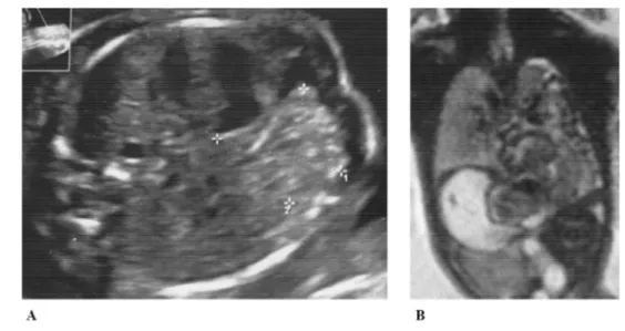
Fig. 1 – Chest images from prenatal ultrasound (A) and magnetic resonance imaging (B)
sided congenital diaphragmatic hernia [2], although the latter has been associated with high incidence of pulmonary com- plications [3].
Here, we report the case of a Bochdalek hernia involving the right side, which was diagnosed in utero.
Case
report
A male newborn baby was delivered by planned caesarean section at 36 weeks and 6 days’ gestational age due to prena- tal diagnosis of right posterolateral congenital diaphragmatic hernia, which was identified by ultrasound during the second trimester of pregnancy ( Fig.1A). At 21 weeks of gestation, fetal magnetic resonance imaging showed intestinal loops, likely to be colic, in the right hemithorax, associated with compression and cranial displacement of the lung, intrathoracic position of the ipsilateral kidney, and anterior and left dislocation of the heart ( Fig.1B). At 28 weeks of gestation, the observed-to- expected lung-head ratio was 78.2%.
At birth, the baby had spontaneous respiratory activity and vigorous crying, with an Apgar score of 7 and 8 at 1- and 5-minute, respectively. He was immediately intubated, and treated with synchronized intermittent positive pressure ven- tilation, with continuous monitoring of vital parameters. A central venous line and a nasogastric tube were placed, seda- tion as well as hemodynamic support with inotropic agents were provided, and total parenteral nutrition was initiated. Chest X-ray, performed soon after birth, revealed multiple bowel loops herniated in the right hemithorax and medi- astinum and, partially, in the left hemithorax. Of note, an ab- normally small right lung volume, suggestive of pulmonary hypoplasia, was observed together with multiple areas of at- electasis of the lungs, likely due to compression ( Fig.2A).
At 3 days of life, after achieving physiologic stabilization, the infant underwent laparotomic. surgical exploration that showed a large diaphragmatic defect of 5 cm × 2cm, with ab- sence of the right posterior diaphragmatic flap. The herniated
abdominal viscera (right colon, right portion of the transverse colon, ileum and distal jejunum, and right kidney) were repo- sitioned into the abdominal cavity, and a patch repair of the right posterolateral diaphragmatic hernia was performed with expanded polytetrafluoroethylene. Notably, the post-surgery chest X-ray did not reveal any morpho-structural alterations of the lungs and the diaphragmatic profile. Namely, the right lung appeared well developed, thus excluding the previous suspect of pulmonary hypoplasia ( Fig.2B).
After surgery, the patient’s conditions progressively im- proved and he was extubated in the tenth day of life with nor- mal respiratory parameters afterwards. At 12 days of life, tho- racic drainage was removed and enteral nutrition was intro- duced. A further chest X-ray was performed at 17 days of life, which confirmed normal findings ( Fig.2C). The infant was dis- charged at twenty days of life in good general condition, with a program of multidisciplinary follow-up.
Discussion
Bochdalek hernia is associated with high morbidity and mor- tality due to the passage of abdominal organs into the tho- racic cavity through a diaphragmatic defect. Intrathoracic herniation of kidneys, such as in our patient, is very rare, with a reported incidence of 0.25%. Of importance, the ab- dominal viscera herniated into the chest compete for areas that would normally accommodate the lungs, thus adversely affecting their growth and development [4]. Indeed, lungs may be severely compromised in newborns, with pulmonary hypoplasia and/or persistent pulmonary hypertension being among the most unfortunate outcomes [5].
Prenatal diagnosis is essential in identifying and manag- ing infants with congenital diaphragmatic hernia, especially those cases at higher risk for the worse outcomes, to optimize their clinical and surgical management [6]. Several prenatal factors may be considered in the evaluation of the defect, such as coexistence of liver herniation, the observed-to-expected
1504
Radiology Ca s e Reports 15 (2020) 1502–1505Fig2– ChestX-ray(A)beforesurgeryshowingherniatedbowelloopsintherighthemithoraxandmediastinumand,partially,inthelefthemithorax,withmultipleareas ofatelectasisofthelungsandasmallrightlungvolume;(B)aftersurgeryshowingtheabsenceofthepreviouslyherniatedvisceraandarightlungnormallydeveloped; and(C)at17daysoflifeconfirmingnormalfindings
R a d i o l o g y C a s e R e p o r t s 1 5 ( 2 0 2 0 ) 1 5 0 2 – 1 5 0 5
1505
lung-head ratio by ultrasound for the estimation of likelihood of pulmonary hypoplasia, and assessment of fetal lung vol- umes by magnetic resonance imaging [6]. An observed-to- expected lung-head ratio <45% has been reported to predict poor outcome in right-sided congenital diaphragmatic hernia, while an observed-to-expected lung-head ratio ≤25%predicts poor outcome in left sided congenital diaphragmatic hernia (25% survival) [6]. In our case, despite an observed-to-expected lung-head ratio of approximately 78%, the chest X-ray per- formed after birth was suggestive of pulmonary hypoplasia and a large diaphragmatic defect that needed patch repair was detected during surgical exploration. However, a normal lung development was observed soon after surgical repair of the defect.
In these patients, surgery should be deferred until clinical conditions are declared stable, thus allowing to relocate vis- cera into their physiological position. Depending on the size, diaphragmatic defects can be repaired primarily or, if large, a permanent patch closure is required [6].
Multidisciplinary approach and post-discharge follow-up of infants with diagnosis of congenital diaphragmatic her- nia are of fundamental importance [7]. Indeed, survivors may suffer of long-term sequelae, including respiratory issues (eg, chronic lung disease). Nevertheless, most infants improve over time [ 6,7], as lung development continues after birth into early childhood [8]. Of note, crucial processes take place up to young adulthood, such as classical and continued alveolariza- tion and microvascular maturation (including the extracellu- lar matrix) [8].
In summary, our clinical case shows that some patients may have a much better lung outcome despite their initial unfavorable picture. Of importance, a close multidisciplinary follow-up is mandatory for congenital diaphragmatic hernia survivors.
R E F E R E N C E S
[1] Kotecha S, Barbato A, Bush A, Claus F, Davenport M, Delacourt C, et al. Congenital diaphragmatic hernia. Eur Respirat J 2012;39(4):820–9 Epub 2011/10/29PubMed PMID: 22034651. doi: 10.1183/09031936.00066511.
[2] Sperling JD, Sparks TN, Berger VK, Farrell JA, Gosnell K, Keller RL, et al. Prenatal diagnosis of congenital diaphragmatic hernia: does laterality predict perinatal outcomes? Am J Perinatol 2018;35(10):919–24 Epub
2018/01/06PubMed PMID: 29304545; PubMed Central PMCID: PMCPMC6033692. doi: 10.1055/s-0037-1617754.
[3] Partridge EA, Peranteau WH, Herkert L, Rendon N, Smith H, Rintoul NE, et al. Right- versus left-sided congenital diaphragmatic hernia: a comparative outcomes analysis. J Pediatric Surg 2016;51(6):900–2 Epub 2016/06/28PubMed PMID: 27342009. doi: 10.1016/j.jpedsurg.2016.02.049.
[4] Tovar JA. Congenital diaphragmatic hernia. Orphanet J Rare Dis 2012;7:1 Epub 2012/01/05PubMed PMID: 22214468; PubMed Central PMCID: PMCPMC3261088. doi: 10.1186/1750-1172-7-1.
[5] Ameis D, Khoshgoo N, Keijzer R. Abnormal lung development in congenital diaphragmatic hernia. Semin Pediatric Surg 2017;26(3):123–8 Epub 2017/06/24PubMed PMID: 28641748. doi: 10.1053/j.sempedsurg.2017.04.011.
[6] Puligandla PS, Skarsgard ED, Offringa M, Adatia I, Baird R, Bailey M, et al. Diagnosis and management of congenital diaphragmatic hernia: a clinical practice guideline. CMAJ: Can Med Assoc J 2018;190(4):E103–Ee12 Epub 2018/01/31PubMed PMID: 29378870; PubMed Central PMCID: PMCPMC5790558. doi: 10.1503/cmaj.170206.
[7] Lally KP, Engle W. Postdischarge follow-up of infants with congenital diaphragmatic hernia. Pediatrics
2008;121(3):627–32 Epub 2008/03/04PubMed PMID: 18310215. doi: 10.1542/peds.2007-3282.
[8] Schittny JC. Development of the lung. Cell Tissue Res 2017;367(3):427–44 Epub 2017/01/31PubMed PMID: 28144783. doi: 10.1007/s00441-016-2545-0.