222
|
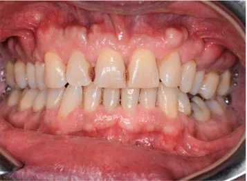
wileyonlinelibrary.com/journal/ccr3 Clin Case Rep. 2019;7:222–223.A 50‐year‐old man was referred at the outpatient clinic for multiple hard masses of the maxilla. The intraoral examination revealed multiple proliferating lesions of hard consistency, pain-less, and covered by normally colored mucosa, in the buccal
aspect of the maxilla above the teeth (Figure 1). A slow but steady dimensional increasing was referred by the patient over the last year. The familial medical history was negative for bowel disease and for similar jaw lesions. On the panoramic radiograph
Received: 11 September 2018
|
Revised: 21 October 2018|
Accepted: 23 October 2018 DOI: 10.1002/ccr3.1918C L I N I C A L I M A G E
Oral maxillary exostosis
Luisa Limongelli
1|
Angela Tempesta
1|
Saverio Capodiferro
1|
Eugenio
Maiorano
2|
Gianfranco Favia
1This is an open access article under the terms of the Creative Commons Attribution License, which permits use, distribution and reproduction in any medium, provided the original work is properly cited.
© 2018 The Authors. Clinical Case Reports published by John Wiley & Sons Ltd. 1Department of Interdisciplinary
Medicine, Aldo Moro University, Bari, Italy 2Department of Emergency and Organ Transplantation, Aldo Moro University, Bari, Italy
Correspondence
Saverio Capodiferro, Department of Interdisciplinary Medicine, Aldo Moro University, Bari, Italy.
Email: [email protected]
Key Clinical Message
Oral maxillary exostoses are proliferating bone lesions with an unknown etiology occurring on the cortical plates both in the maxilla and in the mandible of young in-dividuals, showing a typical slow but continuous enlargement. No treatment is usu-ally required unless they create esthetic or functional limitations during follow‐up; the biopsy is needed only for doubtful lesions. Furthermore, it is mandatory to collect an accurate familiar history of patients affected by exostosis, especially when occur-ring with atypical clinical presentation, in order to exclude or prevent potentially associated systemic diseases.
K E Y W O R D S
benign bone lesions, maxillary exostosis
FIGURE 1 Multiple proliferating lesions of hard consistency in the buccal aspect of the maxilla above the teeth covered by normally colored mucosa
FIGURE 2 Panoramic radiograph showing multiple well‐ defined radiopacities with a round/ovoid appearance all over the upper jaw
|
223LIMONGELLI EtaL.
(Figure 2), multiple well‐defined radiopacities with a round/ ovoid appearance were detectable over the upper jaw. The cone beam computed tomography (Figure 3A‐E) revealed multiple proliferating osseous lesions with irregular appearance emerg-ing from the buccal cortical plate of the maxilla without signs of teeth involvement. The diagnosis was of typical multiple buccal exostosis of the maxilla; a periodical follow‐up was suggested.
Oral exostoses (OEs) are bony protuberances arising from the buccal or lingual cortical plates of the maxilla and/or the mandible, with a variable prevalence ranging from 8% to 51% in the maxilla and 6%‐32% in the mandible.1 Although theories
regarding a possible role of a chronic periosteal inflammation have been suggested, the true etiology of OE remains still un-clear.1,2 Though the appearance reported here is typical of oral
exostosis, benign and malignant bone tumors should be con-sidered in the differential diagnosis when the lesion presents as solitary localized enlargement. In addition, such patients should be investigated for Garden’s syndrome by an accurate familial anamnesis and subsequently by colonoscopy in doubtful cases, considering the high rate of malignant transformation of intes-tinal polyps.
CONFLICT OF INTEREST
Authors declare no conflict of interest.
AUTHOR CONTRIBUTION
LL: performed clinical diagnosis and prepared the manu-script. AT: reviewed the literature. SC: performed clinical diagnosis and prepared the manuscript. EM: contributed to the final revision of the manuscript. GF: performed clinical diagnosis and prepared the manuscript.
ORCID
Saverio Capodiferro http://orcid.org/0000-0002-9819-6229
REFERENCES
1. Kitajima S, Yasui A. Images in clinical medicine. Oral maxillary exostosis. N Engl J Med. 2015;373(15):1457‐1457.
2. Kennedy RA, Thavaraj S, Diaz‐Cano S. An overview of autoso-mal dominant tumour syndromes with prominent features in the oral and maxillofacial region. Head Neck Pathol. 2017;11(3): 364‐376.
How to cite this article: Limongelli L, Tempesta A,
Capodiferro S, Maiorano E, Favia G. Oral maxillary exostosis. Clin Case Rep. 2019;7:222–223. https://doi. org/10.1002/ccr3.1918
FIGURE 3 (A‐E) The cone beam computed tomography showed multiple proliferating osseous lesions with irregular appearance and dimension emerging from the buccal cortical plate of the maxilla without signs of teeth involvement
(A)
(D) (E)