1 23
Current Stem Cell Reports
e-ISSN 2198-7866
Curr Stem Cell Rep
DOI 10.1007/s40778-017-0091-7
Role of PTH in Bone Marrow Niche and
HSC Regulation
Maria Giovanna Sabbieti, Luigi
Marchetti, Roberta Censi, Giovanna
Lacava & Dimitrios Agas
1 23
Your article is protected by copyright and
all rights are held exclusively by Springer
International Publishing AG. This e-offprint
is for personal use only and shall not be
self-archived in electronic repositories. If you wish
to self-archive your article, please use the
accepted manuscript version for posting on
your own website. You may further deposit
the accepted manuscript version in any
repository, provided it is only made publicly
available 12 months after official publication
or later and provided acknowledgement is
given to the original source of publication
and a link is inserted to the published article
on Springer's website. The link must be
accompanied by the following text: "The final
publication is available at link.springer.com”.
CELL:CELL INTERACTIONS IN STEM CELL MAINTENANCE (D BONNET, SECTION EDITOR)
Role of PTH in Bone Marrow Niche and HSC Regulation
Maria Giovanna Sabbieti1&Luigi Marchetti1&Roberta Censi2&Giovanna Lacava1&
Dimitrios Agas1
# Springer International Publishing AG 2017
Abstract
Purpose of Review The bone marrow microenvironment hosts a multicellular complex that is extraordinary in its inter-dependence and function. The composite machinery within the axial and long bones is involved in the homing, mainte-nance, differentiation, and egress of hematopoietic/ progenitors stem cells (HSCS) as well as mesenchymal/ stromal stem cells (MSCs) that dwell in specific anatomical areas inside the marrow space, described as niches. The need for more efficient hematopoietic stem cell transplantation pro-tocols and bone marrow manipulation techniques has motivat-ed scientists to identify effective niche regulators such as the parathyroid hormone (PTH).
Recent Findings PTH treatment is increasingly used with promising outcomes in autologous and allogeneic transplan-tation of HSCs, because PTH operates as a significant medi-ator in HSC engraftment, expansion, and mobilization. In ad-dition to the well-established anti-osteoporotic effect of PTH, there is evidence that it may also coordinate hematopoietic stem cell activities.
Summary This report provides up-to-date information about PTH action within marrow niches and highlights the impor-tance of this hormone in the behavior of hematopoietic ele-ments in the bone marrow.
Keywords PTH . PTHrP . Bone marrow niche . Hematopoiesis . HSCs
Introduction: the Current Understanding of Bone
Marrow Structure and Function
The structure and function of the bone marrow have been the object of scientific research for many decades. Certainly, the study of the elegant equilibrium between the different habitats of the bone marrow reservoir presents a complex challenge. Specialized cellular compartments within the bone cavities are characterized by interdependence and interconnectedness and establish dynamic operational microareas designated as niches. The niche milieu encompasses a panorama of undif-ferentiated and stem elements as well as mature cells. The niche hosts mesenchymal/stromal stem cells (MSCs) many of which can commit to form osteoblasts, chondrocytes, and adipocytes and hematopoietic stem/progenitor components (HSCs) which give rise to blood cells. In particular, the prim-itive murine hematopoietic cells have been identified as a heterogeneous Lin−, Sca1+, and C-kit+ (LSK) population comprising multipotent progenitors (MPP), long-term HSCs (LT-HSCs), and short-term HSCs (ST-HSCs) [1,2].
The niche ontogeny is complemented by various space-specific resident cells, such as perivascular reticular cells, perivascular mesenchyme progenitors, endothelial cells, and neuronal and muscle stem cells, which actively participate in the microenvironmental homeostasis [2]. Current findings in-dicate that some HSCs are located near the endosteal bone surface (endosteal niche) with distinct functions that are de-pendent on the distance from the bone surface while others are in close proximity to the specialized blood vessels within the bone marrow, the sinusoids (vascular niche) [3–5]. Both the endosteal and the sinusoidal regions are strategic bone marrow
This article is part of the Topical Collection on Cell:Cell Interactions in Stem Cell Maintenance
* Dimitrios Agas [email protected]
1
School of Biosciences and Veterinary Medicine, University of Camerino, Camerino, MC, Italy
2 School of Pharmacy, University of Camerino, Camerino, MC, Italy
Curr Stem Cell Rep
DOI 10.1007/s40778-017-0091-7
areas that support HSC maintenance, self-renewal, quies-cence, differentiation, and egression. These features are made possible by the involvement of additional factors including cytokines and hormones, which are critical for the bone mar-row homeostatic tableau.
The Interdependence of Niche Inhabitants
Accumulating evidence indicates that theBbone forming^ os-teoblasts, theBbone feeder^ osteoclasts, and the MSCs are the foremost regulators of the HSC phenotype. The wide array of MSC functional and architectural properties provides the re-quired platform for the assembly and organization of the skel-etal and perivascular hematopoietic niche framework within the bone marrow. For this reason, MSCs and progenies have been considered for about two decades the preeminent niche manufacturers [2]. In point of fact, it is thought that the differ-ent committed MSCs progenies provide signals for HSC dif-ferentiation and osteoclastogenesis [6,7], while the in vivo depletion or impaired function of osteoblasts in mice disrupts hematopoiesis. In accordance, the osteocalcin+ osteoblasts have been identified as HSC-supporting cells because imma-ture hematopoietic cells were found organized in follicle-like structures next to them [3]. Moreover, it has been found that HSCs establish contacts with osteoblasts lining the bone sur-face, named spindle-shaped N-cadherin+CD45−osteoblastic (SNO) cells, which express high levels of the multifunction N-cadherin protein. N-cadherin-mediated interaction between osteoblasts and hematopoietic cells plays a critical role in the survival and homing of HSCs and hematopoietic progenitors [3]. In addition, tunica endothelial cell kinase 2 receptor (Tie2)/angiopoietin-1 (Ang-1) signaling supports tight adhe-sion of HSCs to the niche through an N-cadherin/β1-integrin-dependent mechanism [8]. Taking into consideration the com-plexity of the niche morphology, these precedent studies have deduced that both the osteoblasts and the endosteal niche are key components of HSC maintenance [9–11]. On the contrary, modern concepts argued that osteoblasts create a niche for certain early lymphoid progenitors but not for HSCs [12••].
Specifically, deletion of SCF, CXCL-12, and angiopoietin using Col2.3-Cre ablation of osteoblasts in mice does not affect the HSC function and the overall HSC numbers but impaired LT-self-renewal of HSCs [12,13••,14–17].
In this view, other findings indicated that HSCs depend on a perivascular niche created by endothelial cells and leptin receptor (Lepr)- or Prx1-expressing perivascular stromal cells, whereas osteoblastic cells and endosteal niche support proliferation and differentiation of some early lymphoid progenitors [12••].
Likewise, at least three studies have provided evidence highlighting the contribution of the vascular niche: in one study, human MSCs expressing CD146 proved capable of supporting HSCs at the sinusoidal level [18]. Two other
studies reported that MSC subpopulations within the same compartment, such as CXCL12-abundant reticular (CAR) cells, orchestrate HSC metabolism via stem cell factor (SCF) and CXCL12 production [19,20]. In addition, in vivo deple-tion of the MSC pool expressing the intermediate filament nestin reduces bone marrow homing of HSCs, an indication that = nestin+MSCs are important HSC regulators [21].
The distinct bone marrow anatomical zones and their topo-graphical interactions continue to be the subject of controver-sy. Several authors suggested that the endosteal niche main-tains HSC quiescence [3,5,8], whereas the vascular niche supports stem and progenitor cell homeostasis and regulates megakaryopoiesis [4]. However, current findings have indi-cated quite the opposite, reporting that HSCs reside adjacent to the perivascular niche, whereas early lymphoid progenitors inhabit the endosteal area [12••]. The multifaceted bone
mar-row microarchitecture and the candidate anatomical microareas for the HSCs continue to be the object of intense investigation.
Bone Deposition is Controlled by Niche Elements
Bone formation and osteoblast physiology are influenced by the metabolic features of HSCs [22]. Stem, progenitor, and mature hematopoietic cells coordinate bone cell differentia-tion and matrix deposidifferentia-tion. For instance, osteomacs, a specific macrophage subdivision, provide bone-forming signals dur-ing the deposition phase [2].
The physical interactions between the niches, and conse-quently the fate of the MSCs and HSCs, are broadly influ-enced by a plethora of autocrine, paracrine, and endocrine Bbone-forming^ factors such as bone morphogenetic proteins [23–25], growth factors [26], prostaglandins [27–31], shared cytokines and chemokines [2], and hormones such as the para-thyroid hormone (PTH) [32]. Although all of these molecules appear to be fundamental for the maintenance of bone microarchitecture and stem/progenitor cell homeostatic fea-tures within the bone marrow, PTH has been identified as a key niche element that functionally and spatially links the activities of MSCs and HSCs.
PTH Effects Within the Bone Marrow at a Glance
PTH, a peptide comprised of 84 amino acids, is the fundamen-tal regulatory molecule of calcium and phosphate systemic levels; nonetheless, the PTH metabolic features go beyond this and meet the needs of osteocyte signaling, osteoblast pro-liferation, differentiation and apoptosis, and HSC homing, maintenance, and egression [32,33].
It is well documented that the administration of PTH, its recombinant human analog PTH 1–34, and its nearly
Curr Stem Cell Rep
Author's personal copy
homologous PTH-related peptide (PTHrP) exerts dose-dependent differential effects in the bone and the bone marrow microenvironment [32].
Intermittent PTH (iPTH) injections in experimental an-imals induce a progressive and adaptive response in the cell targets enhancing trabecular bone formation with a concomitant minor loss of the cortical bone [34, 35]. iPTH affects the osteoblastic pool by shifting MSC differ-entiation towards osteoblastogenesis [36] and also in-creases the number of HSCs probably due to the simulta-neous expansion of the osteoblastic cells and, consequent-ly, the fine-tuned interactions between HSCs and osteo-blasts [3, 37,38]. PTH bone marrow anabolic and bone-building effects were observed following defined dose-treatment protocols in rats (80 μg/kg/day for 14 days) [36], mice (40 μg/kg/day for 21 days) [38], and humans (20 μg daily for 24 months) [39]. Nonetheless, hemato-poietic progenitors are preferentially sustained by the early-osteoblastic lineage and PTH administration boosts this anabolic scenery [40]. Certainly, iPTH administration has opened up new avenues for the treatment of bone diseases such as osteoporosis and immune disorders, and iPTH administration could be of use in the future to affect bone marrow transplantation/engraftment outcomes.
In contrast to the anabolic effects of intermittent PTH ad-ministration, chronically elevated PTH levels have catabolic effects. Such supraphysiological PTH levels occur in patho-logical conditions such as primary and secondary hyperpara-thyroidism, chronic renal disease, and chronic inflammation, all of which induce osteopenia. Actually, modern concepts have portrayed osteopenia and osteoporosis as anBaccident of inflammation^ [41]. In this context, PTH levels are in-creased within an acute inflammatory scenario and continu-ous PTH production drives bone resorption and loss of both the cortical and trabecular bones. Moreover, in the context of inflammation, activated immune cells can release PTHrP, which has been shown to be overexpressed in both acute and chronic inflammation [42,43]. Continuous PTH admin-istration and/or release within the bone marrow leads to ab-normal production of factors that affect hematopoietic line-age commitment. For instance, cPTH enhances receptor ac-tivator of nuclear factor-κB ligand (RANKL) and macro-phage colony-stimulating factor (MCSF) release by osteo-blasts and consequently prompts osteoclast differentiation and activation and diverting the fate of HSCs toward the maturation of myeloid progenies. On the other hand, it has been observed that MSCs and osteoblasts release less osteo-protegerin (OPG) (a decoy receptor of RANKL) after cPTH injection [44,45]. Nevertheless, treatment with PTH at high doses (13.6μg/kg/day) for 18–24 months was found to in-crease the risk of osteosarcoma in rats due to the exaggerated bone formation response provided by the PTH receptors on the osteoblastic cells [46].
The Role of PTH in HSC Niche Regulation
PTH Influences HSCs via Specific MSC Subpopulations and Release of Cytokines and Chemokines
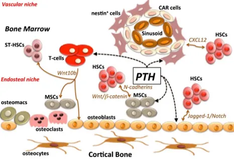
Several studies support the thesis that proper functioning of the bone marrow niche is based on complex interactions be-tween ligands and receptors, as well as physical interactions between the different bone marrow niche residents. In this perspective, PTH provides signals for stem cell differentiation, proliferation, and maintenance, via its specific receptors (PTHR) expressed by the osteoblastic lineage as well as by MSCs within the marrow. An important note is that these receptors are not present in HSCs: this observation indicates that mesenchymal populations and, as recently reported, T-lymphocytes are solely responsible for mediating the effects of PTH within the marrow niche [37,47–49]. The PTH niche targets are summarized in Fig.1.
PTH osteogenic activity and its capacity to stimulate MSC expansion have been correlated with HSC homing and the increase of the HSC pool. PTH orchestrates an operational platform among osteoblasts, MSCs, and HSCs, recruiting me-diators capable ofBsewing the niche patchwork,^ such as N-cadherin, Wnt/β-catenin, and Notch/Jagged1 [33, 37]. In point of fact, PTH stimulates hematopoiesis in mice via up-regulation of cadherin-11 expression in MSCs. Of interest, cadherin-11 is also highly expressed in hematopoietic progen-itors characterized by elevated self-renewal capacity [50,51]. PTH-induced cadherin-11 production in MSCs can facilitate physical interactions between hematopoietic progenitors and MSCs and, as a consequence, hematopoietic progenitor cell expansion. PTH treatment in mice subjected to lethal irradia-tion and bone marrow transplantairradia-tion led to increased cadherin-11 levels in MSCs with concomitant HSCs expan-sion, substantially improving the survival rate of the experi-mental animals [52]. Bearing in mind that cadherin-11 inter-acts withβ-catenin [53] and that Wnt/β-catenin activation in
hematopoietic progenitor cells contributes to their expansion [54], the above findings depict an effective operating system through bone and immune progenitor cells within the niche.
Exogenous PTH increases the bone marrow cell secretion of important niche regulators such as interleukin (IL)-6, IL-11, GM-CSF, and SCF. In a synergistic fashion, these cytokines enhance the number and mobilization of HSCs [55]. A possi-ble explanation for PTH-induced HSC expansion is based on SCF+-secreting cell growth and cytokine release; SCF+ -se-creting cells in combination with IL-6 and IL-11 secreted in-side the niche frames act as signal platform for HSC expan-sion. In point of fact, SCF expressed by osteoblasts, fibro-blasts, CXCL12-expressing perivascular stromal cells, endo-thelial cells, and nestin-expressing MSCs has been identified as a critical mediator of HSC dynamics [15]. Furthermore, considering the fact that IL-6 is a downstream mediator of
Curr Stem Cell Rep
PTH signaling [56], this cytokine can also directly support PTH-mediated HSC expansion and coordinate hematopoiesis, lymphopoiesis, and megakaryopoiesis [57,58]. Of note, the IL-6 soluble receptor sIL-6R has been found upregulated in bone marrow cells after PTH treatment, leading researchers to attribute to this receptor unique orphan homeostatic roles within the niche [59]. Indeed, IL-6 and sIL-6R, in a supportive or independent fashion, enhanced PTH-mediated HSC expan-sion via a STAT3 signaling cascade. In IL-6 null mice, the action of sIL-6R on hematopoietic cells was sufficient to pre-serve PTH-mediated HSC expansion and thus guarantee PTH anabolic effects [59].
Among the various players in bone marrow homeostasis, proteoglycan 4 (PRG4) is thought to have a role as a regulator of the HSC niche, due to its involvement in HSC expansion. PRG4 supports basal expression of both niche moderators, CXCL12 and IL-6. PTH induces upregulation of Prg4 mRNA by osteoblast progenitors within the bone marrow and simultaneous osteoblastic PRG4 secretion, which in turn triggers the release of CXCL12 and IL-6. These events culmi-nate in expansion of HSCs due to PRG4 regulatory and PTH-supporting effects. Though PRG4 has not been considered as aBfront row^ HSC regulator, the fact that in Prg4−/− mice PTH does not significantly augment the marrow Lin−Sca1+ c-kit+pool reveals the significance of PRG4 in PTH outcomes within the niche [60].
As previously mentioned, nestin+MSCs are spatially asso-ciated with HSCs and contribute to HSC maintenance. In line with this observation, PTH administration expands bone mar-row nestin+cells and conducts them toward osteoblastic dif-ferentiation. In addition, PTH-induced nestin+MSC pool ex-pansion is directly correlated with a parallel exex-pansion of HSCs. Thus, PTH seems to amplify the ability of this peculiar nestin-expressed MSC population to support HSC
maintenance within the niche [21], motivating new interest in the structural and functional features of this hormone on the microenvironmental behavior of MSCs and HSCs.
Experiments conducted in humans revealed that long-term teriparatide (PTH 1–34 fragment) administration at FDA-approved doses not only yielded favorable outcomes against post-menopausal osteoporosis, but also increased circulating HSCs in the absence of G-CSF. The dual effects of PTH on bone homeostasis and hematopoiesis seem to follow defined signaling pathways. PTH influences bone growth involving Wnt/β-catenin mechanisms; it also exerts an effect on multi-ple transductional mediators of MSCs, mature osteoblasts, and osteocytes. Concerning hematopoiesis, PTH outcomes were mostly orchestrated by the early-stage osteoblasts and the ac-tivation on their membrane of PTHRs via G-protein (Gsα) signaling cascades. Indeed, mice lacking Gs in cells of the osteoblast lineage present a decrease in pro-B and pre-B cells. Bearing in mind that bone mass may be related to B cell number and, in turn, this process may be regulated by signals downstream of Gs in the osteoblast, it is reasonable to deduce that PTH-PTHR-Gs axis activation may have beneficial ef-fects for immune system maturation [61]. In accordance, teriparatide-activated early osteoblasts within human bone marrow provide a watchdog role in the HSC niche [39].
Of interest, these findings in humans were comparable to results obtained in rodents; in mice treated a short time with PTH, there were increases in circulating HSCs, lymphocytes, and neutrophils, without a reduction in the HSC pool [62]. The effect of PTH on the HSC niche has been studied in mice lacking Bmi 1 (B lymphoma Mo-MLV insertion 1), an impor-tant epigenetic niche regulator. Bmi 1-null mice displayed weakened HSC self-renewal and reduced HSC niche ele-ments. Moreover, Bmi 1 maintains MSC populations and drives mesenchymal stem cell differentiation toward
Fig. 1 PTH targets distinct bone marrow elements within an anabolic scenario. Notably, intermittent administration of PTH affects early osteoblastic cells, MSCs, and T cells, inducing multiple spatiotemporal effects. Due to its many effects and its involvement in MSC and HSC homeostasis, PTH has been identified as one of the major regulators for the maintenance of the bone marrow phenotype. The dashed lines represent PTH target cells. HSCs hematopoietic stem cells; MSCs bone marrow mesenchymal/stromal stem cells
Curr Stem Cell Rep
Author's personal copy
osteoblastic lineage and bone formation via the regulation of alkaline phosphatase, osteocalcin, type I collagen, and Runx2 [63]. Thus, Bmi 1 deficiency affects not only immune cells but also bone stem and progenitor cells, whereas its absence dis-rupts the niche integrity. In this context, PTH 1–34 adminis-tration partially rescued hematopoietic defects in Bmi 1-null mice and reestablished the HSC niche microenvironment. Furthermore, PTH partially reversed the premature osteopo-rosis that occurs in the Bmi 1 knockout mice [64]. These results highlight the many ways PTH maintains niche func-tionality through MSC and HSC interdependency. Extensive investigation is underway on the action of PTH and its bone marrow cell targets.
PTH Targets T Cells that Prompt ST-HSC Expansion and HSC Commitment
Though MSC subpopulations, early-osteoblastic cells, and osteocytes are thought to be the major targets of PTH, some reports have revealed an unexpected role of T lym-phocytes in mediating the osteo-anabolic effects of PTH. In line with these findings, iPTH treatment in mice in-creased T cell-released Wnt10b, a Wnt ligand that drives osteoblastogenesis by activating Wnt receptors on MSCs and osteoblasts. Strong support for this consideration comes from the finding that iPTH administration prompted a reduction in bone anabolic response in mice with T cell deletion [65,66]. Furthermore, since Wnt signaling active-ly participates in hematopoiesis in a dose-dependent man-ner [67], it has been reported that iPTH treatment in mice modulated T cell Wnt10b production and consequently ST-HSC expansion and ameliorated blood cell engraftment after bone marrow transplantation. Interestingly, iPTH-induced ST-HSC expansion did not compromise the quies-cent HSC niche or LT-HSC self-renewal [49]. The fact that iPTH does not increase the number of ST-HSCs at the expense of the LT-HSC pool might break new ground in a therapeutic context, regarding PTH preferential niche targets. Bearing in mind the key role of MSCs and osteo-blasts in HSC maintenance and HSC metabolic features, these findings have added another player, T cells, to the scenario of hematopoietic regulation in the bone marrow. On the other hand, it is well known that PTH anabolic protocols establish molecular pathways for bone remodel-ing and hematopoietic niche maturation via stimulation of bone and blood components. In line with this observation, i P T H i n d u c e s o s t e o b l a s t r e l e a s e o f m o n o c y t e chemoattractant protein-1 (MCP-1), which in turn recruits myeloid precursors and differentiates them into osteoclasts [68]. Nevertheless, it was reported that, after PTH chal-lenge, Th17, a T cell subpopulation, produced IL-17, which participates actively in both bone resorption and control of hematopoietic activities [69].
The Importance of PTH in HSC Bone Marrow Niche Manipulation
Research in bone and bone marrow manipulation has powered the development of heterotopic bone models formed in vivo by transplanted MSCs; these capsular bone-mimicking microenvironments are termed ossicles. PTH treatment plays a key role in bone apposition and HSC engraftment and expansion into these ectopic cortical-like bone assemblies [70]. In mice with PTH-treated ossicles, augmented HSC frequency associated with simultaneous bone growth has been reported. Thus, PTH significantly supports ossicle niche development, probably due to its ability to increase anabolic Jagged-1/ Notch signaling through osteoblasts and HSCs, to modu-late the HSC niche regulator SDF-1 (referred also as CXCL12) and to increase the number of microvessels within this tissue-engineered scenario. In line with this ob-servation, PTH provides ossicle structural and functional sustenance for hematopoietic long-term multilineage re-constitution cells (CD150+CD48−CD41−Lineage− cells) [38]. A recent study also demonstrated that iPTH adminis-tration (40 μg/kg) for 28 days in mice transplanted with human MSC-derived ossicles induced a significant in-crease in the weight of the humanized ossicles, as com-pared to untreated littermate controls [71].
An important challenge is the improvement of HSC trans-plantation techniques and HSC engraftment and egression efficiency. It is well documented that, in patients treated with granulocyte colony-stimulating factor (G-CSF)-based pro-tocols, poor HSC mobilization has been observed. Several authors have noted that targeting the distinct niche popula-tions might improve stem cell-based remedies, since treat-ments with a combination of cytotoxic drugs influence both osteoblasts and HSCs in experimental animals [36]. In this context, a phase I clinical trial established that PTH exerts a prominent pharmacological role in HSC maintenance during G-CSF-induced mobilization treatment [47]. Generally, G-CSF cotreatment used in allogeneic transplantation tech-niques provokes a homeostatic imbalance in the regulation of osteoblasts and osteoclasts. Decrease of osteoblast num-bers leads to reduced levels of HSPC mobilization regulators such as SDF-1, SCF, and OPN. Moreover, it was found that after short-term G-CSF treatment, osteoblast loss and osteo-clast pool expansion altered the fine-tuned signaling be-tween bone remodeling mediators and HSCs [72]. In order to offset bone niche disruption and impaired bone remodel-ing caused by drug treatment, therapy combinremodel-ing PTH and RANKL to enhance HSC egression was tested. PTH and RANKL countered the side effects of cytotoxic chemother-apy in two ways: first, by triggering the anabolic features of osteoblasts and osteoclasts, and second, by protecting the HSC pool during treatment [36].
Curr Stem Cell Rep
Conclusions
Bone marrow homeostasis is related to the specific features of each niche element, and within this complex system, physical interactions and the release of cytokines and hormones govern MSC and HSC homeostasis. PTH plays a leading role in the panorama of interactions inside the bone marrow. Indeed, the administration of an anabolic regimen of PTH supports MSC and osteoblast differentiation and bone deposition, HSC ex-pansion and protection during chemotherapy, HSC post-transplantation engraftment, and ST-HSC pool development and egression. PTH signaling through early osteoblasts and T lymphocytes in the axial and long bones orchestrates hemato-poiesis and coordinates niche microenvironmental dynamics. Given the pharmacological potential of PTH and its important physiological role in the niche apparatus, it is to be expected that this key hormone will be the subject of intense future inquiry.
Compliance with Ethical Standards
Conflict of Interest Maria Giovanna Sabbieti, Luigi Marchetti, Roberta Censi, Giovanna Lacava, and Dimitrios Agas declare that they have no conflict of interest.
Human and Animal Rights and Informed Consent This article does not contain any studies with human or animal subjects performed by any of the authors.
References
Papers of particular interest, published recently, have been highlighted as:
•• Of major importance
1. Hoffman CM, Calvi LM. Minireview: complexity of hematopoietic stem cell regulation in the bone marrow microenvironment. Mol Endocrinol. 2014;28:1592–601.
2. Agas D, Marchetti L, Douni E, Sabbieti MG. The unbearable light-ness of bone marrow homeostasis. Cytokine Growth Factor Rev. 2015;26:347–59.
3. Zhang J, Niu C, Ye L, Huang H, He X, Tong WG, et al. Identification of the haematopoietic stem cell niche and control of the niche size. Nature. 2003;425:836–41.
4. Avecilla ST, Hattori K, Heissig B, Tejada R, Liao F, Shido K, et al. Chemokine mediated interaction of hematopoietic progenitors with the bone marrow vascular niche is required for thrombopoiesis. Nat Med. 2004;10:64–71.
5. Arai F, Yoshihara H, Hosokawa K, Nakamura Y, Gomei Y, Iwasaki H, et al. Niche regulation of hematopoietic stem cells in the endos-teum. Ann N Y Acad Sci. 2009;1176:36–46.
6. Gori F, Hofbauer LC, Dunstan CR, Spelsberg TC, Khosla S, Riggs BL. The expression of osteoprotegerin and RANK ligand and the support of osteoclast formation by stromalosteoblast lineage cells is developmentally regulated. Endocrinology. 2000;141:4768–77.
7. Porter RL, Calvi LM. Communications between bone cells and hematopoietic stem cells. Arch Biochem Biophys. 2008;473:193– 200.
8. Arai F, Hirao A, Ohmura M, Sato H, Matsuoka S, Takubo K, et al. Tie2/angiopoietin-1 signaling regulates hematopoietic stem cell quiescence in the bone marrow niche. Cell. 2004;118:149–61. 9. Visnjic D, Kalajzic Z, Rowe DW, Katavic V, Lorenzo J, Aguila HL.
Hematopoiesis is severely altered in mice with an induced osteo-blast deficiency. Blood. 2004;103:3258–64.
10. Zhu J, Emerson SG. A new bone to pick: osteoblasts and the haematopoietic stem-cell niche. BioEssays. 2004;26:595–9. 11. Whitfield JF. Parathyroid hormone: a novel tool for treating bone
marrow depletion in cancer patients caused by chemotherapeutic drugs and ionizing radiation. Cancer Lett. 2006;244:8–15. 12.•• Ding L, Morrison SJ. Haematopoietic stem cells and early
lym-phoid progenitors occupy distinct bone marrow niches. Nature. 2013;495:231–5. This study illustrates that HSC populations depend on a perivascular niche created by endothelial cells and perivascular stromal cells, whereas only some early lym-phoid progenitors depend on an endosteal niche created by osteoblasts
13.•• Bowers M, Zhang B, Ho Y, Agarwal P, Chen CC, Bhatia R. Osteoblast ablation reduces normal long-term hematopoietic stem cell self-renewal but accelerates leukemia development. Blood. 2015;125:2678–88. This study provides a clear demonstration regarding the role of the osteoblasts and the endosteal niche in maintaining the quiescence and long-term self-renewal poten-tial of HSCs. It highlights also the fact that different HSC sub-populations are accommodated in specialized niches within bone marrow
14. Zhao M, Li L. Osteoblast ablation burns out functional stem cells. Blood. 2015;125:2590–1.
15. Ding L, Saunders TL, Enikolopov G, Morrison SJ. Endothelial and perivascular cells maintain haematopoietic stem cells. Nature. 2012;48:457–62.
16. Greenbaum A, Hsu YM, Day RB, Schuettpelz LG, Christopher MJ, Borgerding JN, et al. CXCL12 in early mesenchymal progenitors is required for haematopoietic stem-cell maintenance. Nature. 2013;495:227–30.
17. Zhou BO, Ding L, Morrison SJ. Hematopoietic stem and progenitor cells regulate the regeneration of their niche by secreting angiopoietin-1. elife. 2015;4:e05521.
18. Sacchetti B, Funari A, Michienzi S, Di Cesare S, Piersanti S, Saggio I, et al. Self-renewing osteoprogenitors in bone marrow sinusoids can organize a hematopoietic microenvironment. Cell. 2007;131:324–36. 19. Omatsu Y, Sugiyama T, Kohara H, Kondoh G, Fujii N, Kohno K, et al. The essential functions of adipo-osteogenic progenitors as the hemato-poietic stem and progenitor cell niche. Immunity. 2010;33:387–99. 20. Danks L, Takayanagi H. Immunology and bone. J Biochem.
2013;154:29–39.
21. Mendez-Ferrer S, Michurina TV, Ferraro F, Mazloom AR, Macarthur BD, Lira SA, et al. Mesenchymal and haematopoietic stem cells form a unique bone marrow niche. Nature. 2010;466: 829–34.
22. Takayanagi H. Osteoimmunology: shared mechanisms and crosstalk between the immune and bone systems. Nat Rev Immunol. 2007;7:292–304.
23. Rosen V, Wozney JM. Bone morphogenetic proteins. In: Belizikian J, Raisz LG, Rodan G, editors. Principles of bone biology. 2nd ed. San Diego, CA: Academic; 2002. p. 919–28.
24. Naganawa T, Xiao L, Coffin JD, Doetschman T, Sabbieti MG, Agas D, et al. Reduced expression and function of bone morphogenetic protein-2 in bones of Fgf2 null mice. J Cell Biochem. 2008;103: 1975–88.
25. Sabbieti MG, Agas D, Marchetti L, Coffin JD, Xiao L, Hurley MM. BMP-2 differentially modulates FGF-2 isoform effects in Curr Stem Cell Rep
Author's personal copy
osteoblasts from newborn transgenic mice. Endocrinology. 2013;154:2723–33.
26. Crane JL, Cao X. Bone marrow mesenchymal stem cells and TGF-β signaling in bone remodeling. J Clin Invest. 2014;124:466–72. 27. Agas D, Marchetti L, Menghi G, Materazzi S, Materazzi G,
Capacchietti M, et al. Anti-apoptotic Bcl-2 enhancing requires FGF-2/FGF receptor 1 binding in mouse osteoblasts. J Cell Physiol. 2008;241:145–52.
28. Sabbieti MG, Agas D, Materazzi S, Capacchietti M, Materazzi G, Hurley MM, et al. Prostaglandin F2alpha involves heparan sulphate sugar chains and FGFRs to modulate osteoblast growth and differ-entiation. J Cell Physiol. 2008;217:48–59.
29. Frisch BJ, Porter RL, Gigliotti BJ, Olm-Shipman AJ, Weber JM, O’Keefe RJ, et al. In vivo prostaglandin E2 treatment alters the bone marrow microenvironment and preferentially expands short-term hematopoietic stem cells. Blood. 2009;114:4054–63.
30. Hoggatt J, Singh P, Sampath J, Pelus LM. Prostaglandin E2 en-hances hematopoietic stem cell homing, survival, and proliferation. Blood. 2009;113:5444–55.
31. Agas D, Marchetti L, Hurley MM, Sabbieti MG. Prostaglandin F2: a bone remodeling mediator. J Cell Physiol. 2013;228:25–9. 32. Agas D, Marchetti L, Capitani M, Sabbieti MG. The dual face of
parathyroid hormone and prostaglandins in the osteoimmune sys-tem. Am J Physiol Endocrinol Metab. 2013;305:E1185–94. 33. Pacifici R. Role of T cells in the modulation of PTH action:
phys-iological and clinical significance. Endocrine. 2013;44:576–82. 34. Neer RM, Arnaud CD, Zanchetta JR, Prince R, Gaich GA,
Reginster JY, et al. Effect of parathyroid hormone (1–34) on frac-tures and bone mineral density in postmenopausal women with osteoporosis. N Engl J Med. 2001;10:1434–41.
35. Jilka RL. Molecular and cellular mechanisms of the anabolic effect of intermittent PTH. Bone. 2007;40:1434–46.
36. Li S, Zou D, Li C, Meng H, Sui W, Feng S, et al. Targeting stem cell niche can protect hematopoietic stem cells from chemotherapy and G-CSF treatment. Stem Cell Res Ther. 2015;15:6–175.
37. Calvi LM, Adams GB, Weibrecht W, Weber JM, Olson DP, Knight MC, et al. Osteoblastic cells regulate the haematopoietic stem cell niche. Nature. 2003;425:841–6.
38. Song J, Kiel MJ, Wang Z, Wang J, Taichman RS, Morrison SJ, et al. An in vivo model to study and manipulate the hematopoietic stem cell niche. Blood. 2010;115:2592–600.
39. Yu EW, Kumbhani R, Siwila-Sackman E, DeLelys M, Preffer FI, Leder BZ, et al. Teriparatide (PTH 1-34) treatment increases periph-eral hematopoietic stem cells in postmenopausal women. J Bone Miner Res. 2014;29:1380–6.
40. Cheng YH, Chitteti BR, Streicher DA, Morgan JA, Rodriguez-Rodriguez S, Carlesso N, et al. Impact of maturational status on the ability of osteoblasts to enhance the hematopoietic function of stem and progenitor cells. J Bone Miner Res. 2011;26:1111–21. 41. Straub RH, Cutolo M, Pacifici R. Evolutionary medicine and bone
loss in chronic inflammatory diseases—a theory of inflammation-related osteopenia. Semin Arthritis Rheum. 2015;45:220–8. 42. Walsh NC, Crotti TN, Goldring SR, Gravallese EM. Rheumatic
diseases: the effects of inflammation on bone. Immunol Rev. 2005;208:228–51.
4 3 . F i e r e r J , B u r t o n D W, H a g h i g h i P, D e f t o s L J . Hypercalcemiaindisseminated coccidioidomycosis: expression of parathyroid hormone-related peptide is characteristic of granulo-matous inflammation. Clin Infect Dis. 2012;55:e61–6.
44. Kearns AE, Khosla S, Kostenuik PJ. Receptor activator of nuclear factor kappaB ligand and osteoprotegerin regulation of bone re-modeling in health and disease. Endocr Rev. 2008;29:155–92. 45. Ohishi M, Schipani E. PTH and stem cells. J Endocrinol Investig.
2011;34:552–6.
46. Watanabe A, Yoneyama S, Nakajima M, Sato N, Takao-Kawabata R, Isogai Y, et al. Osteosarcoma in Sprague-Dawley rats after
long-term treatment with teriparatide (human parathyroid hormone (1– 34)). J Toxicol Sci. 2012;37:617–29.
47. Adams GB, Martin RP, Alley IR, Chabner KT, Cohen KS, Calvi LM, et al. Therapeutic targeting of a stem cell niche. Nat Biotechnol. 2007;25:238–43.
48. Calvi LM, Bromberg O, Rhee Y, Weber JM, Smith JN, Basil MJ, et al. Osteoblastic expansion induced by parathyroid hormone re-ceptor signaling in murine osteocytes is not sufficient to increase hematopoietic stem cells. Blood. 2012;119:2489–99.
49. Li JY, Adams J, Calvi LM, Lane TF, DiPaolo R, Weitzmann MN, et al. PTH expands short-term murine hemopoietic stem cells through T cells. Blood. 2012;120:4352–62.
50. Kiener HP, Brenner MB. Building the synovium: cadherin-11 me-diates fibroblast-like synoviocyte cell-to-cell adhesion. Arthritis Res Ther. 2005;7:49–54.
51. Wagner W, Wein F, Roderburg C, Saffrich R, Faber A, Krause U, et al. Adhesion of hematopoietic progenitor cells to human mesen-chymal stem cells as a model for cell-cell interaction. Exp Hematol. 2007;35:314–25.
52. Yao H, Miura Y, Yoshioka S, Miura M, Hayashi Y, Tamura A, et al. Parathyroid hormone enhances hematopoietic expansion via upreg-ulation of cadherin-11 in bone marrow mesenchymal stromal cells. Stem Cells. 2014;32:2245–55.
53. Kawaguchi J, Takeshita S, Kashima T, Imai T, Machinami R, Kudo A. Expression and function of the splice variant of the human cadherin-11 gene in subordination to intact cadherin-11. J Bone Miner Res. 1999;14:764–75.
54. Reya T, Duncan AW, Ailles L, Domen J, Scherer DC, Willert K, et al. A role for Wnt signalling in self-renewal of haematopoietic stem cells. Nature. 2003;423:409–14.
55. Duarte RF, Franf DA. The synergy between stem cell factor (SCF) and granulocyte colony-stimulating factor (G-CSF): molecular ba-sis and clinical relevance. Leuk Lymphoma. 2002;43:1179–87. 56. Li X, Liu H, Qin L, Tamasi J, Bergenstock M, Shapses S, et al.
Determination of dual effects of parathyroid hormone on skeletal gene expression in vivo by microarray and network analysis. J Biol Chem. 2007;282:33086–97.
57. Imai T, Koike K, Kubo T, Kikuchi T, Amano Y, Takagi M, et al. Interleukin-6 supports human megakaryocytic proliferation and dif-ferentiation in vitro. Blood. 1991;78:1969–74.
58. Pirih FQ, Michalski MN, Cho SW, Koh AJ, Berry JE, Ghaname E, et al. Parathyroid hormone mediates hematopoietic cell expansion through interleukin-6. PLoS One. 2010;5:e13657.
59. Cho SW, Pirih FQ, Koh AJ, Michalski M, Eber MR, Ritchie K, et al. The soluble interleukin-6 receptor is a mediator of hematopoi-etic and skeletal actions of parathyroid hormone. J Biol Chem. 2013;288:6814–25.
60. Novince CM, Koh AJ, Michalski MN, Marchesan JT, Wang J, Jung Y, et al. Proteoglycan 4, a novel immunomodulatory factor, regu-lates parathyroid hormone actions on hematopoietic cells. Am J Pathol. 2011;179:2431–42.
61. Wu JY, Purton LE, Rodda SJ, Chen M, Weinstein LS, McMahon AP, et al. Osteoblastic regulation of B lymphopoiesis is mediated by Gs{alpha}-dependent signaling pathways. Proc Natl Acad Sci U S A. 2008;105:16976–81.
62. Brunner S, Zaruba MM, Huber B, David R, Vallaster M, Assmann G, et al. Parathyroid hormone effectively induces mobilization of progenitor cells without depletion of bone marrow. Exp Hematol. 2008;36:1157–66.
63. Zhang HW, Ding J, Jin JL, Guo J, Liu JN, Karaplis A, et al. Defects in mesenchymal stem cell self-renewal and cell fate determination lead to an osteopenic phenotype in Bmi-1 null mice. J Bone Miner Res. 2010;25:640–52.
64. Lu R, Wang Q, Han Y, Li J, Yang XJ, Miao D. Parathyroid hormone administration improves bone marrow microenvironment and Curr Stem Cell Rep
partially rescues haematopoietic defects in Bmi1-null mice. PLoS One. 2014;9(4):e93864.
65. Terauchi M, Li JY, Bedi B, Baek KH, Tawfeek H, Galley S, et al. T lymphocytes amplify the anabolic activity of parathyroid hormone through Wnt10b signaling. Cell Metab. 2009;10:229–40. 66. Bedi B, Li JY, Tawfeek H, Baek KH, Adams J, Vangara SS, et al.
Silencing of parathyroid hormone (PTH) receptor 1 in T cells blunts the bone anabolic activity of PTH. Proc Natl Acad Sci U S A. 2012;109:E725–33.
67. Luis TC, Naber BA, Roozen PP, Brugman MH, de Haas EF, Ghazvini M, et al. Canonical wnt signaling regulates hematopoiesis in a dosage-dependent fashion. Cell Stem Cell. 2011;9:345–56. 68. Li X, Qin L, Bergenstock M, Bevelock LM, Novack DV, Partridge
NC. Parathyroid hormone stimulates osteoblastic expression of
MCP-1 to recruit and increase the fusion of pre/osteoclasts. J Biol Chem. 2007;282:33098–106.
69. Pacifici R. The role of IL-17 and TH17 cells in the bone catabolic activity of PTH. Front Immunol. 2016;7:57.
70. Schneider A, Taboas JM, McCauley LK, Krebsbach PH. Skeletal ho-meostasis in tissue-engineered bone. J Orthop Res. 2003;21:859–64. 71. Reinisch A, Thomas D, Corces MR, Zhang X, Gratzinger D, Hong
WJ, et al. A humanized bone marrow ossicle xenotransplantation model enables improved engraftment of healthy and leukemic hu-man hematopoietic cells. Nat Med. 2016;22:812–21.
72. Li S, Zhai Q, Zou D, Meng H, Xie Z, Li C, et al. A pivotal role of bone remodeling in granulocyte colony stimulating factor induced hematopoietic stem/progenitor cells mobilization. J Cell Physiol. 2013;228:1002–9.
Curr Stem Cell Rep