Dipartimento di Bioscienze e Territorio
Dottorato in Scienze e Tecnologie biologiche e ambientali
(XXVIII Cycle)
UNIVERSITÀ DEGLI STUDI
DEL MOLISE
S.S.D MED 46
CAMPOBASSO
FROM HEPATOCARCINOMA TO BREAST CANCER:
SELENOPROTEINS AS LINK BETWEEN TWO
DIFFERENT REALITY
Antonella Angiolillo
SUPERVISOR
POSTGRADUATE
Fabiola Rusolo
Matr. 149329
ILLUSTRIOUS Professor
COORDINATOR
ILLUSTRIOUS Professor
Claudio Caprari
CONTENTS
1
ABSTRACT
42
INTRODUCTION
52.1 PREFACE 6
2.2 METABOLISM OF SELENIUM 7
2.2.1 Nutritional source of selenium……….. 7
2.2.2 Selenium uptake and metabolic pathway……… 7
2.2.3 Systemic distribution …….………..………. 9
2.2.4 Selenium ritention and excretion……….. 9
2.2.5 Selenoproteins biosynthesis pathway……….. 10
2.2.6 Selenoproteins in physiology and pathology……….. 11
2.3 OXIDATIVE STRESS AND CANCER - AN INFLAMMATORY NETWORK 16 2.3.1 ROS definition and source………. 16
2.3.2 Physiological functions of ROS……… 18 2.4 HARMFUL EFFECTS OF ROS 19 2.4.1 ROS: common denominator of inflammation and cancer……… 19
2.4.2 Hepatocellular Carcinoma (HCC) and Breast Cancer (BC): two examples of inflammation-related cancer………... 20
3
MATERIALS AND METHODS
233.1 HepG2 and Huh7 Cell Culture……… 23
3.2 Protein Extraction and Western Blot Analyses………... 24
3.3 Bio-Plex Assay for HepG2 and Huh7……….. 25
3.4 Statistical Analysis………... 25
3.5 RNA preparation and Reverse Transcription-qPCR (RT-qPCR) analysis for selenotranscriptoma in HepG2 and Huh7 cells……… 25 3.6 Bioinformatics analysis for HepG2 and Huh7 Selenotranscriptoma……….. 28
3.7 Tissue samples HepG2 and Huh7 cells………... 28
3.8 Tissue immunohistochemistry……….. 28
3.9 Cell culture for two human breast cancer cell lines and human non-cancerous mammary epithelial cell line………... 28 3.10 RNA preparation and quantitative Reverse Transcription Polymerase Chain Reaction (RT-qPCR) analysis in MCF-7, MDA-MB231 and MCF-10A………... 29
3.11 Bioinformatics analysis……….. 30
3.12 Interactomic Studies ……….. 30
4
RESULTS
31 4.1 EVALUATIONS OF THE SELENOTRANSCRIPTOME ON HEPG2, HUH7 AND NORMAL HEPATOCYTE CELLS 31 4.1.1 Network analysis for selenotranscriptome on HCC cells….………. 344.1.2 Validation of the up-expression of SELM, GPX4 and GPX7 by immunohistochemistry……....………. 36
4.2 EVALUATION OF THE SODIUM SELENITE EFFECT ON HCC CELLS 38
4.2.1 SELENBP1, SELK and GPX1 Expression in HepG2 and Huh7 Cells after
stimulation with sodium selenite ….………..………. 38
4.2.2 Evaluation of Selenium Concentrations …….……….. 39
4.2.3 Bio-Plex assay on HepG2 and Huh7 cells ………..……… 40
4.2.4 Interactomic Studies ….………. 41
4.3 EVALUATION OF THE SELENOTRANSCRIPTOME ON MCF-7, MDA-MB231 AND HUMAN NON-CANCEROUS BREAST CELL LINE (MCF-10A) 43 4.3.1 Network Analysis for MCF-7, MDA-MB231 and MCF-10A selenotranscriptome………….. 44
5
DISCUSSIONS
456
CONCLUSIONS AND FUTURE PROSPECTIVES
551
ABSTRACT
Selenium is an essential trace mineral of fundamental importance to human health. It is known primarily for its antioxidant activity, for its chemopreventive, anti-inflammatory and antiviral properties, (Papp et al. 2007) hence its deficiency has been recognized as a contributing factor to pathophysiological
conditions, including heart disease,
neuromuscular disorders, cancer, male infertility and inflammation. Much of its beneficial influence on human health is attributed to its presence within at least 25 proteins (Selenoproteins) (Papp et al. 2007). It is becoming more evident how the cancer and its the behaviour not dependent only on the genetics of tumor cells but also by surrounding milieu [stromal tissue (immune cells, fibroblasts, myofibroblasts, cytokines, and vascular tissue), as well as the surrounding extracellular matrix], necessary for tumor cells survival, growth, proliferation and metastasis. Inflammatory cells and
mediators are present in the
microenvironment of most, if not all, tumors, irrespective of the trigger for development
(Leonardi et al. 2012). Hepatocarcinoma
(HCC) and Breast Cancer (BC) are examples. The liver is a hormone-sensitive organ and a
several lines of evidence suggest that sex hormones and their receptors play a role in liver carcinogenesis (Wang et al. 2006). Continuous oxidative stress, impaired synthesis of antioxidant enzymes, in HCC and in BC, un-regulated synthesis and secretion of sex hormones laid the foundation for research into selenoproteins a great help, not only to mitigate these mechanisms, but also to modulate the hormonal signaling exacerbating the hepatocarcinogenesis. The aim of thesis has been to identify selenoproteins, whose de-regulation was, potentially, associated to hepatocarcinogenesis and to breast cancer. These preliminary investigations direct future studies to understand how BC cells can influence the hepatocarcinogenesis through the secretion of hormones, cytokines, chemokines and growth factors and how the HCC cells can exercise control on breast cancer progression. Furthermore, it will be interesting to investigate how the modulation of selenoproteins, by treatment with selenium
alone or in combination with
chemotherapeutic molecules, might influence the key signaling of these two cancers.
2
INTRODUCTION
2.1 PREFACE 6
2.2 METABOLISM OF SELENIUM 7
2.2.1 Nutritional source of selenium……….. 7
2.2.2 Selenium uptake and metabolic pathway……… 7
2.2.3 Systemic distribution ………..………. 9
2.2.4 Selenium ritention and excretion……….. 9
2.2.5 Selenoproteins biosynthesis pathway……….. 10
2.2.6 Selenoproteins in physiology and pathology……….. 11
2.3 OXIDATIVE STRESS AND CANCER - AN INFLAMMATORY NETWORK 16 2.3.1 ROS definition and source………. 16
2.3.2 Physiological functions of ROS……… 18 2.4 HARMFUL EFFECTS OF ROS 19 2.4.1 ROS: common denominator of inflammation and cancer……… 19
2.4.2 Hepatocellular Carcinoma (HCC) and Breast Cancer (BC): two examples of inflammation-related cancer………... 20
2.1 PREFACE
Initially identified as a new species of sulfur, the selenium (Se) was discovered as new element in 1817 by the Swedish chemist Berzelius (Fig.1), establishing its properties, as well as the properties of the compounds it formed with metals, oxygen, hydrogen, sulfur, phosphorus, and different salts. In the appendix to the third volume of his Textbook in Chemistry, published in 1818, Berzelius gave the formulas of 90 different selenium compounds (58 selenias, 20 selenietum, and 12 hydroselenietum) together with the atomic weight of the element itself. A remarkably high number of compounds (Jan Trofast, 2011). Today, almost 200 years later, selenium is well established as an essential trace mineral of fundamental importance to human health. It is known primarily for its antioxidant activity and, in therapeutic aspects, for its chemopreventive, anti-inflammatory, and antiviral properties (Papp et al. 2007), hence its deficiency has been recognized as a contributing factor to pathophysiological conditions, including heart disease, neuromuscular disorders, cancer, male infertility, and inflammation, as well as numerous other disorders. Selenium has been also implicated in mammalian development, immune function, inhibition of viral expression and delaying the progression of AIDS in HIV positive patients (Labunsky et al. 2014). Much of its beneficial influence on human health is attributed to its presence within at least 25 proteins. Unlike other metal elements that interact with proteins in form of cofactors, selenium becomes co-translationally incorporated into the polypeptide chain as part of the amino acid selenocysteine (SeCys) e through which carries out its functions (Papp et al. 2007). The group of proteins that contain Sec as an integral part of their polypeptide chain are defined as selenoproteins. Selenoproteins are present in all lineages of life; the largest repertoire exists in fish with 30 individual selenoproteins, followed by humans and rodents with 25 and 24 selenoproteins, respectively. Their small size can be explained by a limited selenium supply in nature and a rather energy-expensive synthesis process (Papp et al. 2007). These include glutathione peroxidases (GPx) (five genes), thioredoxin reductases (TrxR) (three genes), iodothyronine deiodinases (DIO; three genes), and selenophosphate synthetases 2 (SPS2). The remaining selenoproteins have been annotated in alphabetic order and include the 15-kDa selenoprotein/ Sep15, SelH, SelI, SelK, SelM, SelN, SelO, SelP/SepP, SelR, SelS, SelT, SelV, and SelW. Only a few of these proteins have been functionally characterized. These Figure 1. Jacob Berzelius (1779-1848)
include the GPxs, the TrxRs, SPS2, and DIOs, which all have oxidoreductase functions. Mapping the selenoproteomes in species across the domains has paved the way for the main challenge within the selenium field: the functional characterization of these proteins and their involvement in the etiology of disease (Papp et al. 2007).
2.2 METABOLISM OF SELENIUM
2.2.1 Nutritional source of selenium
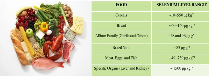
The main route for Se intake is via the diet that varies widely depending on the food type and composition. The major contributors to Se intake is typically provided by bread and cereals, meat, fish, eggs, and milk/dairy products (Roman et al. 2014). An crucial aspect is that food types provide Se in distinct combinations of chemical forms which in turn entail a different bioavailability of the element, depending on the plant/animal species, the environment and the growth conditions (natural or supplemented) (Roman et al. 2014)(Fig.2). The forms of selenium more prevalent in plants are
selenomethionine(SeMet) and inorganic selenate (SeO42) or selenite (SeO32-) which is then used to
produce the organic compound, SeMet, through the sulfur assimilation pathway (Terry et al. 2012). Minor species are selenocysteine (SeCys), major prevalent in animal tissues,
Se-methyl-selenocysteine (SeMCys) and g-glutamyl-Se-methylSe-methyl-selenocysteine (GGSeMCys) (Roman et al. 2014).
Figure 2. Selenium-rich foods
2.2.2 Selenium uptake and metabolic pathway
Once it has been ingested, the distribution of selenium within the body of humans, as well as, of other animals, and also its absorption and excretion, depend on several factors, particularly the chemical form as well as on total amount of the element in the diet (Terry et al. 2012). In addition
FOOD SELENIUM LEVEL RANGIE
Cereals ~10–550 μg kgˉ¹
Bread ~ 60–160 μg kgˉ¹
Allium Family (Garlic and Onion) ~ 68 and 96 μg gˉ¹
Brazil Nuts ~ 83 μg gˉ¹
Meat, Eggs, and Fish ~ 49–739 μg kgˉ¹
intake can be affected by the presence of certain other components of food, including sulfur, heavy metals, and vitamins, as well as, on other factors, such as, sex, age, condition of health and nutritional status (Terry et al. 2012). The absorption of Se-species occurs mainly across intestinal epithelial membrane, by different pathways (Fig.3). Absorption of selenate (SeO42) appears to be by
a sodium-mediated carrier transport mechanism shared with sulfur, while selenite (SeO32-) uses
passive diffusion. Both forms of inorganic selenium compete with inorganic sulfur compounds for absorption (Terry et al. 2012). In contrast, the Se-amino acids SeMet and SeCys are absorbed through transcellular pathways mediated by transporters which are basically shared with their sulphur-containing analogues (Roman et al. 2014). Absorbed selenium is transported in the blood mainly bound to protein, following an initial reduction within the erythrocytes to selenide (Hseˉ) by reduced glutathione and involving the enzyme glutathione reductase (Terry et al. 2012). In humans, almost all the protein-bound selenium in blood is reported to be in the very low-density β-lipoprotein fraction, with smaller amounts bound to other proteins (Terry et al. 2012). However, distribution between these proteins appears to depend on the composition of the diet, in fact Whanger et al. (1993) showed that nearly 50% of the selenium in plasma is associated with albumin in people who consume a diet in which selenomethionine is the main form of the element (Terry et
al. 2012).
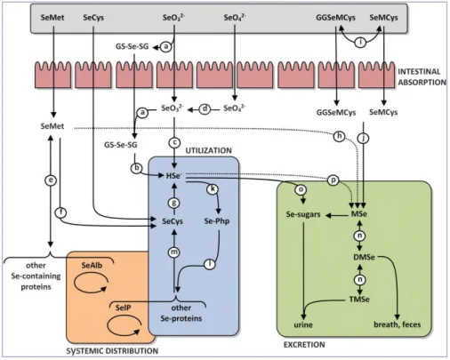
2.2.3 Systemic distribution
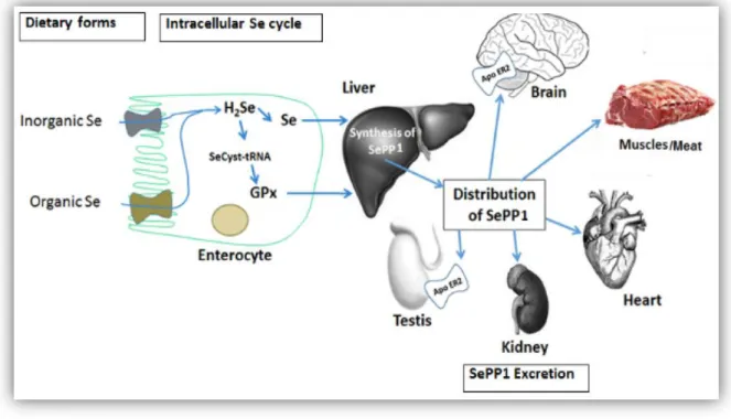
The Se-species absorbed into the gastro-intestinal tract are firstly transported into the liver: SeMet is usually transported in the form of Se-albumin (SeAlb) while selenate and the other organic species may be transported intact or through mechanisms which are still not elucidated. The liver is the foremost organ in Se metabolism, since it synthesizes most of the Se-proteins and regulates the excretion of Se metabolites. The Selenoprotein P (SelP), produced into the liver, is released into the bloodstream and is responsible for the distribution of Se to the other organs, where other Se-proteins can be synthesized. The local uptake of Se from plasma has been shown to occur by endocytosis mediated by receptors of the Apolipoprotein family such as apoER2 in testis and brain and megalin (Lrp2) in kidney. Thus, the liver regulates the whole-body Se distribution by sorting the metabolically available Se between the two pathways of Se-proteins synthesis and the excretory metabolite synthesis. Such regulation might be passive, so that the fraction of Se that cannot be utilized for Se-proteins synthesis enters the excretory pathway. Active regulation of the excretory metabolites has been also hypothesized, but not yet investigated (Roman et al. 2013) (Fig.4).
Figure 4. General pathways of Se absorption and distribution to various organs
2.2.4 Selenium retention and excretion
Actual retention times will depend on a number of factors, including present selenium status, the specific form in which the element is ingested, as well as the state of health of the subject. Selenium is excreted from the body by three distinct routes, in urine via the kidneys, in feces from the
gastrointestinal trace, and in expired air via the lungs. The amounts and proportions of each type of excretion depend on the level and form of the element in the diet (Terry et al. 2012). The urinary pathway is the dominant excretion route for selenium in humans (Yang et al. 1989). The proportion of intake excreted in this way depends on the level of intake in the diet. When this is high, urinary excretion will also be high (Thomson and Robinson, 1986). At low levels of intake, half or less of the dietary selenium will appear in urine (Robinson et al., 1973). These findings point to the importance of renal regulation of selenium levels in the body that are not, apparently, homeostatically controlled by the gut (Burk, 1976). Fecal selenium consists largely of unabsorbed dietary selenium, along with selenium contained in bilary, pancreatic, and intestinal secretions
(Levander and Baumann, 1966). It has been postulated that secretion of selenium in bile and its
enterohepatic reabsorption may provide a mechanism, in addition to renal control, for conserving body stores. This could have major implications for populations with a low dietary intake (Dreosti,
1986). Excretion of selenium via the pulmonary route in expired air and via the dermal route in
sweat are of minor significance at normal levels of dietary intake. Excretion through the lungs occurs principally when intake is unusually high. Excess selenium is detoxified by successive methylation to form the volatile dimethyl selenide and other methylated species. The garlic-like odor of dimethyl selenide on the breath is characteristic of selenium intoxication (Terry at al. 2012)
2.2.5 Selenoproteins biosynthesis pathway
Selenium is the key component of the active site of several Se-proteins having essential biological functions (Roman et al. 2014), which synthesis is an evolutionary conserved process and requires a several components. Co-translational incorporation of Sec into proteins is dictated by in-frame UGA codons present in selenoprotein mRNAs. Sec is introduced into selenoproteins by a complex mechanism that requires special trans-acting protein factors, Sec-tRNA[Ser]Sec (Fig.5a) and a cis-acting Sec insertion sequence element (SECIS). When a ribosome encounters the UGA codon,
which normally signals translation termination, Sec machinery interacts with the canonical translation machinery to augment the coding potential of UGA codons and prevent premature termination. SECIS elements serve as the factors that dictate recoding of UGA as Sec. In response to the SECIS element in selenoprotein mRNA, Sec-tRNA[Ser]Sec, which has an anticodon complimentary to the UGA, translates UGA as Sec. At least two trans-acting factors are required for efficient recoding of UGA as Sec in eukaryotes: SECIS binding protein 2 (SBP2, itself is a selenoprotein in most organisms with selenoproteomes [Papp et al. 2007]) and Sec-specific
translation elongation factor (eEFSec). SBP2 is stably associated with ribosomes and contains a
specificity. Aside from binding to ribosomes and SECIS elements, SBP2 also interacts with eEFSec, which recruits Sec-tRNA[Ser]Sec and facilitates incorporation of Sec into the nascent, growing polypeptide (Fig.5b). Since the discovery of SBP2, additional SECIS-binding proteins were identified and their roles in selenoprotein synthesis were characterized, including ribosomal
protein L30, eukaryotic initiation factor 4a3 (eIF4a3) and nucleolin that together serve as regulatory
proteins that modulate synthesis of selenoproteins and may contribute to the hierarchy of selenoprotein expression (Labunsky et al. 2014).
Figure 5a) Sec biosynthesis pathway in mammalian cells. Sec biosynthesis initiates with the attachment of serine to the Sec tRNA[ser]sec by seryl tRNA synthetase to yield SeryltRNA[ser]sec. The phosphoseryl tRNA kinase phosphorylates the complex. The phosphate is then replaced by the selenium donor selenide (H2Se-P), activated by selenophosphate synthetase. The resulting molecule is selenocysteyl- tRNA[Ser]Sec, which delivers the Sec into the growing polypeptide chain (Papp et al. 2007) Figure 5b)Factors that are required for Sec incorporation into proteins in response to the UGA codon and the factors that may influence the efficiency of the Sec insertion (Nebraska Redox Biology Center Educational Portal).
2.2.6 Selenoproteins in physiology and pathology
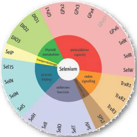
Only a few of the 25 identified mammalian selenoproteins have so far been functionally characterized. The Sec localization in the enzyme active site is responsible of enzymatic redox function, which confers their catalytic or antioxidant activities but many others are cellular
processes in which the selenoproteins are involved: biosynthesis of dNTPs for DNA, removal of damaging or signaling peroxides, reduction of oxidized proteins and membranes, regulation of redox signaling, thyroid hormone metabolism, selenium transport and storage and potentially protein folding (Papp et al. 2007) (Fig.6).
Figure 6. The 25 human selenoproteins classified by their determined or potential function (Benstoem et al. 2015)
Overall, Thioredoxin Reductases (TrxRs), Glutathione Peroxidases (GPxs) and Deidinases (DIOs) are the three best characterized selenoprotein families. TrxRs (TrxR1 in the cytosol/nucleus, TrxR2 in mitochondria and and thioredoxin glutathione reductase in the testis) are the enzyme able to reduce oxidized Trx. Trx provides electrons to ribonucleotide reductase, which is essential for DNA synthesis by converting ribonucleotide to deoxyribonucleotides. In addition, the Trx system participates in many cellular signaling pathways by controlling the activity of transcription factors containing critical cysteines in their DNA-binding domains, such as NF-κB, AP-1, p53 and the glucocorticoid receptor. It is also known that reduced Trx can bind to and inhibit apoptosis signal regulating kinase 1 (ASK1), whereas the oxidization of Trx results in the activation of ASK1 and the induction of ASK1-dependent apoptosis. Therefore, TrxRs are involved in the control of cellular proliferation, viability, and apoptosis through the control of Trx activity and redox state (Lu and
Holmegren 2009). GPxs protect cells against peroxidative damage by reducing hydrogen peroxide,
free fatty acid hydroperoxides, and phospholipid hydroperoxides (Brenneisen et al. 2005). In humans, there are now five Sec-containing GPxs: the ubiquitous cytosolic GPx (GPx1), the gastrointestinal-specific GPx (GPx2), the plasma GPx (GPx3), the ubiquitous phospholipid hydroperoxide GPx (GPx4), and the olfactory epithelium- and embryonic tissue-specific GPx (GPx6). GPx1–3 catalyze the reduction of hydrogen peroxide and organic hydroperoxides, whereas GPx4 can directly reduce phospholipid and cholesterol hydroperoxides. GPx4 is also involved in sperm maturation and male fertility because it has been found to be a main structural component of the sperm mitochondrial capsule in mature spermatozoa as an enzymatically inactive, oxidatively cross-linked, insoluble protein (Lu and Holmegren 2009). Conversely, GPx5, secreted in epididymis, have not the selenocysteine in the active site, and GPx7, recently described as new
phospholipid hydro-peroxide glutathione peroxidase (PHGPX), incorporates cysteine instead of
selenocysteine in the conserved catalytic motif (Rusolo et al. 2015). Three DIOs (DIO1, DIO2 and DIO3) constitute a group of dimeric integral membrane thioredoxin fold–containing proteins that can activate or inactivate the thyroid hormone, depending on their action on the phenolic or the tyrosil ring of the iodothyronines (Rusolo et al. 2015). DIO1 and DIO2 catalyze the deiodination of T4, the major thyroid hormone secreted by the thyroid gland, into the active hormone T3; DIO3 converts T4 into reverse T3 and also T3 into 3,3-diidothyronine. DIO1 and DIO2 can also convert reverse T3 into 3,3-diidothyronine (Lu and Holmgren, 2009). The specific function of several other human selenoproteins are unknown although some details of their biology have been established. Thus, selenoproteins S, and K are Endoplasmatic Reticulum (ER) transmembrane proteins, involved in regulating ER-associated degradation of misfolded proteins and may have a role in immune function. The selenoprotein 15 (Sep15/SEL15) and selenoprotein M (SelM) are also ER-localized and implicated in quality control of protein folding (Brigelius and Sies, 2015). SelN (selenoprotein N) is found in the membrane of the ER and appears to be necessary for proper muscle development
(Bellinger et al. 2009). SelV was the least conserved mammalian selenoprotein that likely arose
from a duplication of SelW in the placental stem. The functions of SelV and SelW are not known, but SelV is expressed exclusively in testes, whereas SelW (Mariotti et al. 2012), expressed in a variety of organs, is similar to the GPxs family in that it shares the redox motif and binds glutathione. The ‘W’ stands for ‘white muscle disease’, a disorder among grazing livestock found in regions with low selenium soil levels. The completion of the human genome project led to the identification of many other selenoproteins though sequence homology and characteristic elements. SelH (selenoprotein H) is a nuclear-localized DNA-binding protein that may act as a transcription factor. SelH increases glutathione levels and GPx activity, and may up-regulate other selenoproteins
in response to stress (Bellinger et al. 2009). Selenoprotein I (SelI) is one of the least studied selenoproteins. It contains a highly conserved CDP-alcohol phosphatidyltransferase domain. This domain is typically encountered in choline phosphotransferases (CHPT1) and choline/ethanolamine
phosphotransferases (CEPT1). CHPT1 catalyzes the transfer of choline to diacylglycerol from
CDPcholine. CEPT1 catalyzes an analogous reaction but accepts both choline and ethanolamine
(Mariotti et al. 2012). Selenoprotein R (SelR)/selenoprotein X (SelX) is a member of the
methionine sulfoxide reductase family, important for reduction of sulfoxymethyl groups (Bellinger
et al. 2009). The physiological and pathological effects of selenoproteins are closely related to
selenium status. Selenium deficiency leads to a dramatic loss of activity of selenoproteins though the their expression exhibits a hierarchical style during selenium deprivation and repletion
(Lu and Holmgren 2009). Furthermore, genetic mutations in selenocodons have been identified like
cause of different human deseases. One form of congenital muscular dystrophy, termed
multiminicore disease, is characterized by a distinct loss of organization of muscle fibres. Mutations
in ryanodine receptors, responsible for calcium-stimulated release of calcium from intracellular stores, and SelN have been identified as causing the disorder. There is evidence that other selenoproteins may also regulate calcium signalling and calcium stores. Overexpression of SelT, another ER localized selenoprotein, led to an increase in calcium levels, but inhibited calcium responses and endocrine release from pituitary adenylate cyclase-activating polypeptide (PACAP). Selenoprotein function in cardiovascular disease has been investigated primarily by analysis of oxidative stress under conditions of selenium supplementation and/or deficiency (Bellinger et al.
2009). Selenium supplementation elevates expression and activity of GPx1, GPx4 and TRxR1 in
vascular endothelial or smooth muscle cells and thus inhibits oxidative stress, cell damage and apoptosis from oxidized low-density lipoprotein (LDL) or triol, a cytotoxic hydroxylated cholesterol derivative found in blood, cells, tissues and atherosclerotic plaques in humans (Bellinger et al.
2009). Low blood Se concentrations have been associated with increased cardiovascular disease
mortality. This may be a reflection of sub-optimal GPx4 activity in the prevention of LDL oxidation, with subsequent uptake by endothelial cells and macrophages in arterial blood vessels
(Brown and Arthur, 2001). Also other selenoproteins are involved in cardiovascular disease. GPx1
has been shown to inhibit ischaemia/ reperfusion-induced apoptosis of cardiac myocytes in mice and its deletion produces heart and vascular dysfunction and tissue irregularities; decreased GPx3 activity, generally abundant in plasma and modulatating redox-dependent aspects of vascular function, results in inadequate nitric oxide (NO) levels for excess ROS, which disrupts platelet inhibitory mechanisms and increases arterial thrombosis (Bellinger et al. 2009). Moreover, the TRxR/TRx system contributes in regulating myocardial remodelling through the reversible
oxidation of signalling molecules. For example, adrenergic receptor activation-induced hypertrophy of adult rat cardiac myocytes is affected by the oxidation of cysteine thiols of Ras that can be reduced by TRxR1 (Bellinger et al. 2009). Levels of ROS influence, also, inflammatory gene expression, thus selenoproteins affect inflammatory responses by regulating the oxidative state of immune cells. GPxs and TRxRs are necessary for optimal function of immune cells by controlling oxidative stress and redox regulation but also other selenoproteins also have ROS-independent roles in modulating inflammatory responses (Bellinger et al. 2009). SelS, for example, is located in the ER membrane and its expression and secretion from liver cells is regulated by inflammatory cytokines as well as extracellular glucose concentrations. SelS has an antiapoptotic role and reduces ER stress in peripheral macrophages and brain astrocytes (Bellinger et al. 2009). Oxidative stress and generation of reactive oxygen species (ROS) are strongly implicated also in a number of neuronal and neuromuscular disorders, including stroke and cerebrovascular disease, Alzheimer’s disease, Parkinson’s disease, familial amyotrophic lateral sclerosis, and Duchenne muscular dystrophy (Chen and Berry, 2003) and selenoproteins, as GPx1 and SelP, are envolved in protection mechanisms against cellular demage - ROS induced. While GPx1 prevents lipid peroxidation on neuronal cell membranes, SelP has a function to chelate heavy metals (mercury, zinc, cadmium and silver), reducing their toxicity (Chen and Berry, 2003) and can also stimulate the survival of cultured central neurons (Almondes et al. 2010). Since the genotoxic damage caused by the accumulation of oxidative modifications in DNA bases and DNA single- or double-strand breaks are outstanding features in the development of many forms of cancer, selenium has been suggested to exert its anticarcinogenic function through ROS detoxification selenoenzymes (Almondes et al.
2010). There is evident correlation between cancer risk and a pattern of altered expression of
selenoproteins: a single nucleotide polymorphism at codon 198 of GPx1 gene is correlated with increased risk of lung cancer (Almondes et al. 2010), while allelic loss of one of two GPx-1 alleles on chromosome 3p is a common event in the development of several types of cancer, including that of the lung, breast and cancers of the head and neck (Zhuo and Diamond, 2010). GPx1, GPx3 and SelP decrease expressions and GPx2 increase are observed in colorectal cancer progression
(Almondes et al. 2010). Furthermore, methylation of CpG islands in the GPx3 promoter regions
typically results in transcriptional silencing, gene down-regulation and to contribute to prostate cancer development (Zhuo and Diamond, 2010). GPx4 is another member of the glutathione peroxidase family of selenoproteins that has received considerable attention. Lower GPx4 protein levels have been observed in cell lines derived from pancreatic cancers compared to normal pancreatic tissue (Zhuo and Diamond, 2010). An examination of the dbEST database identified two polymorphic positions in Sep15 gene (C/T variation at position 811 and a G/A polymorphism at
position 1125), both included in the predicted SECIS element and correlated to Malignant
mesothelioma (MM) (Zhuo and Diamond, 2010). Selenoproteins role in cancer is much more
complex; they appear to have roles in both preventing and promoting cancer. TRx1, for example, activates the p53 tumor suppressor, manifests other tumor suppressor activities, and is specifically targeted by carcinogenic electrophilic compounds, suggesting its major role in cancer prevention. On the other hand, TRx1 overexpression in many cancer cell lines and cancers, targeting by a number of anti-cancer drugs and potent inhibitors that alter cancer-related properties of malignant cells and its deficiency that reverses cell morphology point to a role of TR1 in cancer promotion
(Hatfield et al. 2009). The elucidation TRx roles, as well as, unknown function of other
selenoproteins in cancer biology may yield some promising anti-cancer paradigms for new cancer therapeutic agent development.
2.3 OXIDATIVE STRESS AND CANCER - AN INFLAMMATORY NETWORK
2.3.1 ROS definition and sources
All animals need O2 for efficient production of energy in mitochondria. This need for O2 obscures
the fact that it is a toxic mutagenic gas; aerobes survive only because they have evolved antioxidant defenses (Barry Halliwell, 2006). The O2 molecule is itself a free radical because has two unpaired
electrons. Although this is the most stable, O2 is, thermodynamically, a potent oxidizing agent,
ready to accept a pair of electrons from non-radical molecules, like proteins, DNA and lipids (Barry
Halliwell, 2006). The resulting peroxidation and modification of these molecules can increase the
risk of mutagenesis (Reuter et al. 2010). So, reactive oxygen species (ROS) is a collective term that includes both oxygen radicals and certain non radical derivatives of O2 that are oxidizing agents and/or are easily converted into radicals (HOCl, O3, 1O2, and H2O2). It should be noted that all
oxygen radicals are ROS but not all ROS are oxygen radicals (Barry Halliwell, 2006). The acronym ROS may include also several nitrogen-containing compounds or RNS (Reactive Nitrogen Species), such as nitric oxide (NO), nitroxyl anion (NO−), and peroxynitrite (ONOO−) (Tafani aet al. 2016). The cell is exposed to a large variety of ROS and RNS from both exogenous and endogenous sources. Major exogenous source of ROS is represented by ionizing and nonionizing irradiation and air pollutants such as car exhaust, cigarette smoke, and industrial contaminants that attack and damage the organism either by direct interaction with skin or following inhalation into the lung. Drugs are also a major source of ROS. There are drugs, such as belomycinem and adreamicine, whose mechanism of activity is mediated via production of ROS. A large variety of xenobiotics (eg, toxins, pesticides, and herbicides) and chemicals (eg, mustard gas, alcohol) produce ROS as a by-product of their metabolism in vivo. The invasion of pathogens, bacteria, and viruses might result in
the production of many ROS species by direct release from the invaders or an endogenous response induced by phagocytes and neutrophils. One of the major sources of oxidants is food for a large portion of the food we consume is oxidized to a large degree and contains different kinds of oxidants such as peroxides, aldehydes, oxidized fatty acids, and transition metals (Kohen and
Nyska, 2002) (Fig.7).
Figure 7. Major exogenous sources of ROS
Although the exposure of the organism to ROS is extremely high from exogenous sources, the exposure to endogenous sources is much more important and extensive, because it is a continuous process during the life span of every cell in the organism (Kohen and Nyska, 2002). ROS are, also, products of a normal cellular metabolism and play vital roles in stimulation of signaling pathways in plant and animal cells in response to changes of intra- and extracellular environmental conditions
(Reuter et al. 2010). Five main cellular compartments contain ROS: mitochondria that produce in large quantities through oxidative phosphorylation for ATP synthesis, cytosol that produce ROS from many endogenous (growth factors, cytokines, and metabolisms) or exogenous sources (nutrients, radiation, microbiome, and xenobiotics), single membrane-bound organelles
(peroxisomes, endosomes, and phagosomes) that can produce substantial amount of ROS as typically occurs in the respiratory burst of activated leukocytes (macrophages and eosinophils),
exosomes, released by plasma membranes by shedding and extracellular fluids (such as blood plasma and spermatic, peritoneal, and pleural fluid) in which ROS are released through aquaporins (hydroperoxides) and some anion channels (superoxides) and by secretion (external opening of
phagosomes and granules) as typically occurs in activated degranulating leukocytes (Tafani et al.
2016) (Fig.8).
Figure 8. Subcellular compartmentation of ROS. 1. Mitochondrial ROS; 2. Cytosolic ROS; 3. Redoxosomes (Peroxisomes, Phaosomes); 4. Exosome ROS that include vesicles shedding from damaged plasma membranes; 5. Extracellular ROS in extracellular fluids and plasma (Tafani et al. 2016).
2.3.2 Physiological functions of ROS
ROS has dual functionality in biological systems, with both beneficial and detrimental effects in cells (Ramoutar et al. 2010). Beneficial effects of ROS occur at low or moderate concentrations and their ‘stady state’are determined by the balance between their rates of production and their rates of removal by various antioxidants (Valko et al. 2007). A great number of physiological functions are controlled by redox-responsive signalling pathways. These, for example involve: (i) redox regulated production of NO; (ii) ROS production (oxidative burst) by NAD(P)H oxidase in activated neutrophils and macrophages in an inflammatory environment; (iii) ROS production by NAD(P)H oxidases in non-phagocytic cells, such as, fibroblasts, vascular smooth muscle cells, cardiac myocytes and endothelial cells, in order to regulate intracellular signalling cascades: regulation of cardiac and vascular cell functioning; (iv) modulation of the protein kinases functions and ion channels, as well as, regulation of vascular tone and the inhibition of platelet adhesion mediated by NO•; (v) ROS production as a sensor for changes of oxygen concentration and regulation of certain hormones such as erythropoietin, VEGF (vascular endothelial growth factor) and IGF-II (insulin-like growth factor), controlled by HIF-1 (the transcription hypoxia inducible factor-1); (vi) redox regulation of cell adhesion that plays an important role in embryogenesis, cell growth,
differentiation and wound repair; (vii) activation of T lymphocytes and induction of their function, such as the IL-2 production; (viii) ROS-induced apoptosis, triggered internal signals: the intrinsic or mitochondrial pathway (Valko et al. 2007).
2.4 HARMFUL EFFECTS OF ROS
2.4.1 ROS: common denominator of inflammation and cancer
An imbalance between the continuous efflux of ROS from endogenous and exogenous sources and their elimination by protective mechanisms, referred to as antioxidants (Fig.9), results in continuous and accumulative oxidative damage to cellular components, impairing many cellular functions and inducing somatic mutations (Reuter et al. 2010; Kohen and Nyska, 2002). ROS may be important initiators and mediators in many clinical disorders: heart diseases, endothelial dysfunction, atherosclerosis and other cardiovascular disorders, burns, intestinal tract diseases, brain degenerative impairments, diabetes, eye diseases and ischemic and post-ischemic (eg, damage to skin, heart, brain, kidney, liver, and intestinal tract) pathologies (Kohen and Nyska, 2002). ROS, also, are involved in chronic inflammation and in a wide variety of different cancers, indeed, epidemiological and experimental data have demonstrated how inflammation is strongly associated with an increased risk of several human cancers and how anti-inflammatory therapies show efficacy in cancer prevention and treatment (Reuter et al. 2010). How oxidative stress placed in this context? And how it modulates these different stages of inflammation-induced carcinogenesis? Microbial and viral infections, exposure to allergens, radiation and toxic chemicals, autoimmune and chronic diseases, obesity, consumption of alcohol, tobacco use, and a high-calorie diet are sources both inflammation and ROS. During inflammation, mast cells and leukocytes are recruited to the site of damage, which leads to a ‘respiratory burst’ due to an increased uptake of oxygen, and thus, an increased release and accumulation of ROS at the site of damage. On the other hand, inflammatory cells also produce soluble mediators, such as metabolites of arachidonic acid, cytokines and chemokines, which act by further recruiting inflammatory cells to the site of damage
and producing more reactive species. These key mediators can activate signal transduction cascades, as well as, induce changes in transcription factors, such as nuclear factor kappa B (NF-κB) and activator protein-1 (AP-1) that control transcription of DNA, cytokines production and cell survival. Also aberrant expression of inflammatory cytokines [tumor necrosis factor (TNF),
interleukin-1 (IL-1), IL-6] and chemokines, such as IL-8, CXC chemokine receptor 4 (CXCR4)
sustain oxidative stress-induced inflammation. This inflammatory/oxidative environment leads to a vicious circle, which can damage healthy neighboring epithelial and stromal cells and over a long period of time may lead to carcinogenesis (Reuter et al. 2010).
2.4.2 Hepatocellular Carcinoma (HCC) and Breast Cancer (BC): two examples of
inflammation-related cancer
Hepatitis B and C viruses (HBV and HCV) are the two global causes of hepatitis with 5 to 10 % of HBV infected and more than 70% of HCV infected patients developing chronic infection which alters the state of liver homeostasis inducing inflammatory responses. Chronic inflammatory state, during HBV and HCV infection, is associated with activated inflammatory cells and is mediated by various cytokines (Mohammad Khalid Zakaria et al. 2014). Two are key mediators of HBV associated liver inflammation: IL-8 which maintains an inflammatory environment by activation of cyclooxygenase (COX)-2 gene and IL-29 which inhibits virus replication by PKr production, activated by NK cells subset and enriched in the HBV infected Liver (Mohammad Khalid Zakaria
et al. 2014). A transactivant and multifunctional viral protein, HBx, is responsible to viral genes
expression, the activation of NFκB, AP1 and NF-AT transcription factors (Mohammad Khalid
Zakaria et al. 2014) and upregulates some oncogenes (such as, Rab18 or Yes-associated protein).
Moreover, induces apoptosis by upregulating FasL protein through activating MLK3/MKK7/JNKs signaling and integration of the HBV gene into the house genome (cancer-related genes such as
TERT, MLL4, and CCNE1 are integrated by HBV), an important mechanism that is responsible for
HCC development (Akinobu Takaki et al. 2015). HCV produces, by STAT3 signaling cascade, a several cytokines: RANTES, IL-8, MIP1α, MIP1β, converging on common inhibitor kappa beta (IκB)-kinase dependent kinases, thereby responsible to interferon regulatory factor 3/7 (IRF3/7) and Nuclear Factor Kappa Beta (NFκB) activations. The letter driving the INFα/β and pro-inflammation cytokines synthesis which exacerbate pro-inflammation. The continuous production of ROS, lipid peroxidation and the activation of hepatic stellate cells (HSC) are responsible to progression From Liver chronic desease to steatosis. The organ becomes more sensitive to
apoptosis, injury and inflammation mediated by TNF-α,
extracellular matrix production and degradation, as well as, of complex mechanisms of fibrinogenesis, cell proliferation, contractility, chemotaxis, degradation of matrix, and cytokine release, develop in fibrosis (Mohammad Khalid Zakaria et al. 2014). The release of free radicals, chemokines and cytokines as a result of HBV and HCV infection and inflammation, result in DNA damage, cell proliferation, fibrosis and angiogenesis. Indeed, free radicals, for example, reactive nitrogen or oxygen species, can directly damage DNA and proteins, and indirectly damage these macromolecules via lipid peroxidation. The abrogation of p53-mediated apoptosis, both inhibition by HBx and for its mutation, as in HepG2 and HuH7 cells, infected with HCV, provide a selective clonal advantage for preneoplastic or neoplastic hepatocytes and contribute to hepatocellular carcinogenesis (SP Hussain et al. 2007) (Fig.10).
Figure 10. Stages of Hepatocellular Carcinoma (HCC)
The liver is also a hormone-sensitive organ and a several lines of evidence suggest that sex hormones, like estrogens, and their receptors play a role in liver carcinogenesis (Wang et al. 2006) by the formation of free radical-mediated DNA and RNA adducts. In humans, indeed, the chronic use of estrogens is associated with increased risk of developing liver neoplasms such as benign nodular hyperplasia and hepatic adenoma. Therefore, HCC could therefore be an estrogen-dependent cancer like breast cancer and the use of anti-estrogen drugs should control the growth of this tumor (De Maria et al. 2002). Host microenvironment plays an important role also in breast cancer tumorigenesis. Mutations, like BRCA1/2 are responsible to extent of genomic instability in the malignant breast epithelium while, the letter, activate the tumor-associated stroma to foster
tumor growth by secreting growth factors, increasing angiogenesis or facilitating cell migration. The inflammatory component tends to exacerbate this context. Infiltration of lymphocytes,
tumor-associated macrophages (TAM), mast cells and neutrophils have been shown to exert both direct
and indirect tumoricidal action by their ability to express numerous tumor-promoting characteristics, such as growth factors for breast tumor cells, angiogenic mediators, ECM-degrading enzymes (MMPs), and inflammatory cytokines. For instance, monocyte-derived cytokines, such as TNF-a, may intensify chemokine expression by tumor or stroma cells besides possessing other promalignant properties. As for hepatocarcinogenesis, inflammatory cells can secrete cytokines, growth factors and chemokines, release reactive oxygen intermediates to stimulate proliferation, prevent apoptosis, direct morphogenesis and induce mutagenic changes that may increase tumor cell DNA damage. The overall effect of these activities may be a key factor in breast cancer metastasis and disease progression (Artacho-Cordón et al.2012). Secretion of tumour necrosis factor-α by TAM is known to induce cellular oxidative stress. Oxidative stress trigger mitosis, in vitro, either by the activation of mitogen-activated protein kinases (MAPKs) or via MAPK independent mechanisms. Furthermore, persistent oxidative stress at sublethal levels may cause resistance to apoptosis. The induction of programmed cell death by ROS is dependent on p53 in both mouse and human cell lines. Constitutive oxidative stress within breast carcinoma cells may therefore accelerate the selection of p53 knockout tumour cell clones, which have an apoptosis resistant phenotype (Brown and Bicknell, 2001), fostering to progression from normal mammary gland to ductal carcinoma in situ (DCIS) and finally to invasive ductal carcinoma (IDC)
(Artacho-Cordón et al.2012) (fig.11).
3
MATERIALS AND METHODS
3.1 HepG2 and Huh7 Cell Culture……… 23
3.2 Protein Extraction and Western Blot Analyses………... 24
3.3 Bio-Plex Assay for HepG2 and Huh7……….. 25
3.4 Statistical Analysis………... 25
3.5 RNA preparation and Reverse Transcription-qPCR (RT-qPCR) analysis for selenotranscriptoma in HepG2 and Huh7 cells………
25
3.6 Bioinformatics analysis for HepG2 and Huh7 Selenotranscriptoma……….. 28
3.7 Tissue samples HepG2 and Huh7 cells………... 28
3.8 Tissue immunohistochemistry……….. 28
3.9 Cell culture for two human breast cancer cell lines and human non-cancerous mammary
epithelial cell line………...
28
3.10 RNA preparation and quantitative Reverse Transcription Polymerase Chain Reaction
(RT-qPCR) analysis in MCF-7, MDA-MB231 and
MCF-10A………... 29
3.11 Bioinformatics analysis……….. 30
3.12 Interactomic Studies ..……… 30
3.1 HepG2 and Huh7 Cell Culture
Human hepatoma cell lines (HepG2 and Huh7) were kept in culture and expanded at 37°C in a humidified atmosphere of 5% CO2 in culture medium DMEM (Dulbecco's Modified Eagle’s Medium, Lonza, Verviers, Belgium), supplemented with FBS (Invitrogen, Camarillo, CA, USA) at 10%, Penicillin/Streptomycin 100x (Euroclone, Devon, UK), Glutamax 100x (Invitrogen) and non-essential amino acids 100x (Invitrogen) at 1%. Phosphate buffer (PBS phosphate buffered saline Ca2+ and Mg2+ free) and trypsin (Ca2+ and Mg2+ free) were supplied by Euroclone. The cells (2 × 10⁴) were seeded in 100 mm plates in 8 mL of culture medium and left to grow for 24 h at 37 °C to allow adhesion. Then, the cells were treated with sodium selenite (Na2SeO3), dissolved in H2O, at
were performed in duplicate and repeated for three times. However, the concentrations of sodium selenite used in this study were chosen concerning that human physiological concentration of selenium is less than 3 μM.
3.2 Protein Extraction and Western Blot Analyses
HepG2 and Huh7 cells were washed once in cold phosphate buffered saline (PBS) and lysed in a lysis buffer containing 20 mM Tris HCl pH 7.5, 150 mM NaCl, 1 mM EDTA and NP40 after 24 h of treatment with 0.25 μM, 0.5 μM and 1 μM of sodium selenite. The lysis buffer was complemented with protease inhibitor cocktail tablets (Roche Applied Science, Penzberg, Germany), diluted in H2O to obtain a stock solution 7X concentrated, and with phosphatase inhibitor cocktail tablets (Roche Applied Science), diluted in H2O to obtain a 100X concentrated stock solution. The lysates were clarified by centrifugation at 13,000 rpm for 15 min. Protein concentrations were estimated by a BioRad assay (Bio-Rad Laboratories, Hercules, CA, USA), based on the Method of Bradford, that is a simple and accurate procedure for determining concentration of solubilized proteins. Then the proteins were boiled for 5 min before electrophoresis in Laemmli Sample buffer (Bio-Rad) containing 62.5 mM Tris-HCl pH 6.8, 2% sodium dodecyl sulphate (SDS), 25% glycerol, 0.01% bromophenol blue complemented with 10% β-mercaptoethanol. 60 μg of proteins were subjected to SDS–polyacrylamide gel electrophoresis (SDS-PAGE) using 12% acrylamide concentrated gels under reducing condition. After electrophoresis, proteins were transferred to nitrocellulose membranes (Amersham Hyperfilm MP, High performance autoradiography, GE Healthcare, Hertfordshire, UK); complete transfers were assessed using prestained protein standards (Fermentas, Milano, Italy). After blocking with Tris-buffered saline 5% non fat dry milk (Bio-Rad), membranes were incubated ON at 4°C in shaking with the Goat anti GPX1 antibody (R&D Systems, Minneapolis, MN, USA), with the rabbit anti SELK antibody (ABCAM, Cambridge), and with the rabbit anti SELENBP1 antibody (ABCAM, Cambridge, UK) diluted 1:500, 1:500 and 1:1,000 overnight at 4°C, respectively, and then incubated with the horseradish peroxidase conjugated secondary antibody (1:3,000) for 60 min at room temperature; the reaction was detected with a Western blotting detection system (ECL; Amersham Biosciences, Little Chalfont, UK). To ascertain that equal amounts of protein were loaded, membranes were incubated with antibodies against the γ-tubulin protein (1:500) (Santa Cruz Biotechnology, Santa Cruz, CA, USA).
3.3 Bio-Plex Assay for HepG2 and Huh7
In our approach, the levels of a panel of numerous cytokines, chemokines and growth factors were evaluated at the same time by BioPlex assay. The simultaneous quantitative determination of a large panel of cytokines, able to report the correct ratios and dynamics between highly and poorly represented molecules, has emerged as an accurate, simple, specific, noninvasive, reproducible and less expensive method (Costantini et al. 2010, Capone et al. 2010). The multiplex biometric ELISA-based immunoassay, containing dyed microspheres conjugated with a monoclonal antibody specific for a target protein was used, according to the manufacturer’s instructions (Plex Bio-Rad), to evaluate the concentrations of different cytokines by Human Cytokine 27-Plex Panel after 24 h of incubation with sodium selenite in HepG2 and Huh7 supernatants. In particular, the following cytokines were evaluated: IL-1β, IL-1ra, IL-2, IL-4, IL-5, IL-6, IL-7, IL-8, IL-9, IL-10, IL-12 (p70), IL-13, IL-15, IL-17, eotaxin (CCL11), basic FGF, G-CSF, GM-CSF, IFN-γ, CXCL10, MCP-1, MIP-1α, MIP-1β, PDGF-ββ, RANTES, TNF-α and VEGF. Each experiment was performed in duplicate as previously described (Costantini et al. 2010, Capone et al. 2010). Protein concentrations were determined using a Bio-Plex array reader (Luminex, Austin, TX, USA) that quantitates multiplex immunoassays in a 96-well format with very small fluid volumes. The analyte concentration was calculated using a standard curve, with software provided by the manufacturer (Bio-Plex Manager Software).
3.4 Statistical Analysis
The cytokines concentrations evaluated in HepG2 and Huh7 supernatants after 24 of incubation with sodium selenite were compared by T-test. Values of p < 0.05 were considered to be statistically significant. The statistical program Prism 4 (GraphPad Software, San Diego, CA, USA) was used.
3.5 RNA preparation and Reverse Transcription-qPCR (RT-qPCR) analysis for
seleno-transcriptoma in HepG2 and Huh7 cells
Total RNA from hNHEPS® Human Hepatocytes (Lonza, Basel, Switzerland), HepG2 and Huh7 (Lonza, Basel, Switzerland) was obtained using the TRizol Reagent (Invitrogen, Milan, Italy) following the manufacturer's instructions. Each total RNA sample was treated with the DNa-free kit according to the manufacturer’s instructions (Ambion). RNA samples were quantified using a NanoDrop ND-1000 spectrophotometer (Thermo Scientific, Wilmington, DE). The mRNA levels of the analysed genes were measured by a RT-qPCR amplification procedure that was previously reported. Sequences for mRNAs from the nucleotide data bank (National Center for Biotechnology
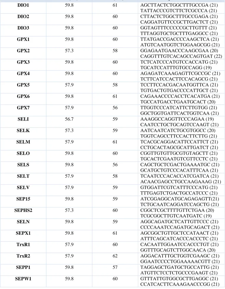
Information, USA) were used to design primer pairs for RT-qPCR (Primer Express, Applied Biosystems, CA, USA). Amplicon sequences were aligned against the human genome by BLAST to check for specificity. Oligonucleotides were obtained from Sigma Aldrich. The primer sequences of 25 selenoprotein mRNAs are provided in Table 1. An appropriate region of 18S rRNA was used as control. Reverse transcriptase reactions (500 ng) were performed according to the manufacturer’s instructions using iScriptTM Reverse Transcriptase Supermix for RT-qPCR (Biorad, Italia) and subsequently diluted with nuclease-free water (Ambion). RT-qPCR assays were run on an Opticon-4 machine (MJ, Research, Waltham, MT, USA). 2 l of cDNA were amplified in a total volume of 20 l containing 1X iQ SYBR Green Supermix (Bio-Rad) and 300 nM of forward and reverse primers. The thermal cycling conditions were as follows: 5 min of denaturation at 95°C followed by 40 cycles of a two-step program (denaturation at 95°C for 15 sec and annealing/extension for 1 min). For each target the primer sequences and the annealing temperature are reported in Table 1. Fluorescence threshold and baseline setting was adjusted with the aid of the Opticon Monitor software (Biorad). The efficiency of each assay was tested with serial dilutions of cDNA to obtain a standard curve. The PCR efficiency was calculated according to the following formula: 10(-1/slope)-1. All PCR efficiencies were above 95%. Dilutions of standards and test samples were run in triplicate. Each reaction was repeated at least three times. Standard deviations and coefficients of variation were calculated for the Cq values of replicated measurements using Microsoft Excel software. Relative quantities were calculated by the ΔΔCq method using the 18S rRNA as housekeeping gene for normalization. Statistical analyses (paired Student's t) were performed using Prism software (Graphpad Software, La Jolla, CA, USA). Significant differences in relative gene expression between hepatocytes and HepG2 or Huh7 are marked by* (p-value<0.05),** (p-value<0.01).
Gene Tm [°C] Ta [°C ] Sequence (5’→ 3’) DIO1 59.8 61 AGCTTACTCTGGCTTTGCCGA (21) TATTACCCGTCTTCTCGCCCA (21) DIO2 59.8 60 CTTACTCTGGCTTTGCCGAGA (21) CAGGATGTTCCGCTTGACTCT (21) DIO3 59.8 60 GGTAGTTTCCCCCGCTTGTTT (21) TTTAGGTGCTGCTTTGAGGCC (21) GPX1 59.8 60 TTATGACCGACCCCAAGCTCA (21) ATGTCAATGGTCTGGAAGCGG (21) GPX2 57.3 58 GGAGAATGAACCCAAGCGAA (20) CAGGTTTGTCACAGCCAGTGAT (22) GPX3 59.8 60 TCTCATCCCATGTCCACCATG (21) TGCATCCATTTGTGCCAGG (19) GPX4 59.8 60 AGAGATCAAAGAGTTCGCCGC (21) TCTTCATCCACTTCCACAGCG (21) GPX5 57.9 58 TCCTTCCACGACAATGGTTCA (21) TGTGACTGTGACCCCATTGCT (21) GPX6 59.8 61 CAGAAACCCCACCTCACATGA (21) TGCCATGACCTGAATGCACT (20) GPX7 57.9 56 TTGGTCCCATCATTCTTGTGG (21) GGCTGGTGATTCACTGGTCAA (21) SELI 56.7 59 AAAGGCCAGGTTCCCAGAA (19) CAATCCTGCTGCAGTCCAAGT (21) SELK 57.3 59 AATCAATCATCTGCGTGGCC (20) TGGTCAGCCTTCCACTTCTTG (21) SELM 57.9 61 TCACGCAGGACATTCCATTCT (21) CCTGCACTAGCGCATTGATCT (21) SELO 59.8 60 CGGTTGTGTTGCGTGTAGCTT (21) TGCACTCGAATGTCGTTCCTC (21) SELS 59.8 56 CAGCTGCTCGACTGAAAATGC (21) GCATGCTGTCCCACATTTCAA (21) SELT 57.9 58 TCAATCCCACACCATCGATCA (21) ACAACGAGCCTGCCAAGAAAG (21) SELV 57.9 59 GTGGATTCGTCATTTCCCATG (21) TTTGAGTCTGACTGCCATCCC (21) SEP15 59.8 59 ATCGGAGGCATGCAGAGAGTT(21) TCTGCAATCAGGATCCAGCTG (21) SEPHS2 57.3 60 CGGCTCGCTTTTGTTCTGAA (20) TCGCGGCTTGTCAATGATC (19) SELN 59.8 59 AGGCAGATGCTCATTGTTCCC (21) CCCCAAATCCAGATGCAGACT (21) SEPX1 59.8 61 AGCGGCTGTTGCTCCATAACT (21) ATTTCAGCATCACCCACCCTC (21) TrxR1 57.9 60 CACAATTGGAATCCACCCTGT (21) GGTTTGCAGTCTTGGCAACA (20) TrxR2 57.9 62 AGGACATTTGCTGGTCGAAGC (21) GGAATCCCCTGGAAAAACGTT (21) SEPP1 59.8 57 TAGGAGCTGATGCTGCCATTG (21) ATGTTCTCCTCTGCCCGAAGT (21) SEPW1 59.8 60 GTTTATTGTGGCGCTTGAGGC (21) CCATCACTTCAAAGAACCCGG (21)
3.6 Bioinformatics analysis for HepG2 and Huh7 Selenotrascrittoma
Biological functional analyses were performed using David and Panther tools whereas human pathway lists and networks were determined by Ingenuity Pathway Analysis (IPA) program setting at 0.001 the significance threshold of t-test. IPA is a system that transforms large data sets into a group of relevant networks containing direct or indirect relationships between genes based on known interactions in the literature and in the experimental studies. In our studies only the “direct relationships” option was selected.
3.7 Tissue samples HepG2 and Huh7 cells
Paraffin-embedded HCC tissues obtained by biopsy of 20 patients were subjected to immunohistochemical staining. All patients in this study provided informed consent, and the study was approved by the Second University of Naples Ethics Committee. All patients had HCV-related cirrhosis with poorly differentiated tumor. No information related to follow-up data of these patients is known.
3.8 Tissue immunohistochemistry
Briefly, xylene dewaxed and alcohol rehydrated paraffin sections were placed in Coplin jars filled with a 0.01M tri-sodium citrate solution and microwaved. After heating, slides were thoroughly rinsed in cool running water for 5 min. Sections were immersed in 3% H2O2 at room temperature
for 30 min to block any endogenous peroxidase activity. They were then washed in Tris-buffered saline (TBS) pH 7.4 before incubating at 4°C O.N with monoclonal rabbit anti-human SELM (LifeSpan BioSciences), polyclonal rabbit anti-human GPX4 (LifeSpan BioSciences) and monoclonal mouse anti-human GPX7 (LifeSpan BioSciences), diluted 1:50, 10µg/mL and 10µg/mL respectively. After incubation with the primary antibody, tissue sections were stained with specie-specific biotinylated secondary antibodies, followed by peroxidase labeled streptavidine (Dako, Denmark); the signal was developed by using diaminobenzidine (DAB) chromogen (Dako) as substrate. Incubations omitting the specific antibody were used as negative controls.
3.9 Cell culture for two human breast cancer cell lines and human non-cancerous mammary
epithelial cell line
Two human breast cancer cell lines, estrogen-receptor (ER)-positive MCF-7 (HTB-22, adenocarcinoma) and ER-negative MDA-MB231 (HTB-26, adenocarcinoma) and the human non-cancerous mammary epithelial cell line, MCF-10A (CRL-10317, fibrocystic disease) were kept in culture. In details, MCF-7 and MCF-10A were expanded at 37°C in a humidified atmosphere of 5%
CO2 in culture medium DMEM (Dulbecco's Modified Eagle's Medium High Glucose with sodium
pyruvate, without L-Glutamine), whereas MDA-MB231 in RPMI 1640 (RPMI 1640 Medium without L-Gluamine with Phenol Red), supplemented with FBS at 10% (Foetal Bovine Serum,
Standard Quality, Australian origin, PAA Cell Culture Company), with 1% of
Penicillin/Streptomycin 100× (Euroclone, Devon, UK) and 1% of Glutamine 200mM (100×) (PAA Cell Culture Company). Moreover, in the case of MCF-10A, the DMEM was supplemented also with human insulin 10 μg/mL (Life Technologies Corporation, Carlsbad CA, 92008 USA), human epidermal growth factor 20 ng/mL (Life Technologies), and hydrocortisone 0.5 g/mL (Sigma-Aldrich 3050 Spruce St. St. Louis, MO 63103) according to the procedure reported in Rothwell et al (Rothwell et al. 2014).
3.10 RNA preparation and quantitative Reverse Transcription Polymerase Chain Reaction
(RT-qPCR) analysis in MCF-7, MDA-MB231 and MCF-10A
Total RNA was extracted from MCF-7, MDA-MB231 and MCF-10A cells using the RNAeasy mini kit (Qiagen Inc., Valencia, CA, USA) according to the manufacturer’s instructions. The extracted RNA was dissolved in diethylpyrocarbonate treated water, and its concentration and purity were assessed by measurement of optical density at 260⁄280 nm, using a NanoDrop 2000 spectrophotometer (Thermo Scientific, Wilmington, DE). 2µg of total RNA of each sample was reverse-transcribed with SuperScript VILO cDNA Synthesis kit (Life Technologies-Invitrogen, Carlsbad, CA, USA) according to the manufacturer’s instructions and subsequently diluted 1:20 with nuclease-free water (Life Technologies-Ambion). Reverse-trascriptions were performed in a Veriti® Thermal Cycler (Thermo Fisher Scientific) by incubating the samples at 25°C for 10 minutes (min), followed by a step at 42°C for 60 min, 85°C for 5 min and the samples were then chilled to 4°C. The reverse-transcribed products were used to perform a qPCR in order to evaluate the expression level of transcripts of seleno proteins. Sequences for mRNAs from the nucleotide data bank (National Center for Biotechnology Information, USA) were used to design primer pairs for RT-qPCR (Primer Express, Applied Biosystems, CA, USA) (Table 1). Oligonucleotides were obtained from Sigma Aldrich. The efficiency of each primer pair was calculated according to standard method curves using the equation E=10−1/slope. Five serial dilutions were set up to determine Ct values and reaction efficiencies for all primer pairs. Standard curves were generated for each oligonucleotide pair using Ct values versus the logarithm of each dilution factor.RT-qPCR assays were run on an 7900HT Fast Real-Time PCR System (Applied Biosystems). Starting with 2 µg of total RNA, we have preparated a 20-fold dilution of the resulting cDNA to achieve the concentration equivalent of starting with 100 ng of RNA (Life Technologies-Invitrogen), according
to the manufacturer’s instructions. 10 ng of cDNA were amplified in a total volume of 25 µL containing 1X SYBR Green PCR Master Mix (Applied Biosystems) and 300 nM of forward and reverse primers. The thermal profile conditions were as follows: 5 min of denaturation at 95°C followed by 44 cycles at 95°C for 30 sec and 60°C for 1 min. We have addicted one cycle for melting curve analysis at 95°C for 15 sec, 60°C for 15 sec and 95°C for 15 sec to verify the presence of a single product. Each assay included a no-template control for each primer pair. To capture intra-assay variability, all qPCR reactions were carried out in triplicate. For all RT-qPCR experiments, the data from each cDNA samples were normalized using β-actin mRNA as endogenous level (Peirce et al. 2001). Sample ΔCt values were calculated as the difference between the means of selenoprotein markers Ct and housekeeping assay Ct from the same sample. We determinated 2∆∆Ct values in order to define the fold change of selenoprotein expression levels in tumor cells compared to the non-cancerous cells, MCF-10A. These data were also confirmed by REST tool (Relative expression software tool, Weihenstephan, Germany) based on the Pfaffi method (Pfaffi, 2001; Pfaffi et al. 2002).
3.11 Bioinformatics analysis
Network analysis was performed by Ingenuity Pathway Analysis (IPA) program and using the same procedure reported in our recent paper (Costantini et al. 2013). In details, IPA builds and explores transcriptional networks to identify regulatory events that lead from signaling events to transcriptional effects.
3.12 Interactomic Studies
Specific undirect or direct physical interactions among the molecules evidenced in this work and, biochemically different in their structure, have been assessed also by an interactomic analysis. In general, interacting molecules form molecular interaction networks that are classified by the nature of the compounds involved.
4
RESULTS
4.1 EVALUATIONS OF THE SELENOTRANSCRIPTOME ON HEPG2, HUH7 AND
NORMAL HEPATOCYTE CELLS 31
4.1.1 Network analysis for selenotranscriptome on HCC cells………. 34
4.1.2 Validation of the up-expression of SELM, GPX4 and GPX7 by
immunohistochemistry …….………. 36
4.2 EVALUATION OF THE SODIUM SELENITE EFFECT ON HCC CELLS 38
4.2.1 SELENBP1, SELK and GPX1 Expression in HepG2 and Huh7 Cells after
stimulation with sodium selenite ………..………. 38
4.2.2 Evaluation of Selenium Concentrations …….……….. 39
4.2.3 Bio-Plex assay on HepG2 and Huh7 cells ………..……… 40
4.2.4 Interactomic Studies ….………. 41
4.3 EVALUATION OF THE SELENOTRANSCRIPTOME ON MCF-7, MDA-MB231 AND HUMAN NON-CANCEROUS BREAST CELL LINE (MCF-10A) 43
4.3.1 Network Analysis for MCF-7, MDA-MB231 and MCF-10A selenotranscriptome………….. 44
4.1 EVALUATIONS OF THE SELENOTRANSCRIPTOME ON HEPG2, HUH7 AND NORMAL HEPATOCYTE CELLS
The gene expression profiles of HepG2, Huh7 and normal hepatocyte cells by means of RT-qPCR have shown that in the two HCC cell lines there were three downregulated genes (DIO1, DIO2, and SELO) (fig.12 Panel A) and ten upregulated genes (GPX4, GPX7, SELK, SELM, SELN, SELT, SELV, SEP15, SEPW1, and TrxR1) (Fig.12 Panel A). In detail, two of the three downregulated genes showed a statistically significant difference between HepG2 and Huh7 versus hepatocytes. On the other hand, in the group of the of the ten upregulated genes five of them (GPX4, GPX7, SELK, SELM, and SEP15) have shown a statistically significant upregulation in HepG2. All the other twelve selenotranscripts have appeared unchanged (Fig.12 Panel A).
![Figure 5a) Sec biosynthesis pathway in mammalian cells. Sec biosynthesis initiates with the attachment of serine to the Sec tRNA[ser]sec by seryl tRNA synthetase to yield SeryltRNA[ser]sec](https://thumb-eu.123doks.com/thumbv2/123dokorg/4793230.49011/12.892.95.829.343.773/figure-biosynthesis-mammalian-biosynthesis-initiates-attachment-synthetase-seryltrna.webp)