1
Alma Mater Studiorum – Università di Bologna
in cotutela con l’Università di Cambridge (UK)
DOTTORATO DI RICERCA IN
SCIENZE FARMACOLOGICHE E TOSSICOLOGICHE,
DELLO SVILUPPO E DEL MOVIMENTO UMANO
Ciclo XXVI
Settore Concorsuale di afferenza: 5/G1
Settore Scientifico disciplinare: Bio/14
TITOLO TESI:
HYPERTENSION, HYPERCHOLESTEROLEMIA,
HYPERALDOSTERONISM: A GENETIC AND PROTEOMIC
PERSPECTIVE FOR PERSONALIZED THERAPY
Presentata da:
Dott. Gianmichele Massimo
Coordinatore Dottorato Relatore
Chiar.mo Chiar.mo
Prof. Giorgio Cantelli Forti Prof. Giorgio Cantelli Forti
2
Contents
Abstract
Introduction
1 Cardiovascular system….………...…..…….pag.10
1.1 Heart, vessels and blood pressure….………...10
1.2 Autonomous nervous system and blood pressure regulation….……….15
1.3 Kidney involvement in the blood pressure regulation….………...17
1.4 Endocrine involvement in the blood pressure regulation….………..21
1.5 Pharmacology of hypertension….………..26
2 Adrenal gland and hormones synthesis….………...31
2.1 Physiological role of the adrenal gland….……….31
2.2 Aldosterone synthesis mechanism….………..34
2.3 Aldosterone activity….………...36
2.4 Resting potential membrane of zona glomerulosa cell and its role in the aldosterone secretion………...……..…...………37
2.5 KCNJ3/KCNJ5 (Kir3.1, Kir3.4 or GIRK1 and GIRK4) implication on resting potential membrane and aldosterone production………..…...…40
2.6 Hyperaldosteronism.……….………...45
3 Lipid metabolism……….………..…...49
3.1 Lipid metabolism. ….………..49
3.2 Disorders of lipid metabolism………...56
3.3 Kinesin like protein 6 role in the lipid pathway………...58
3.4 Pharmacology of lipid metabolism………..….…...60
4 Genetic background……….……….…...64
4.1 Pharmacogenetics, pharmacogenomics, and individualized therapy…….…………....64
4.2 Single nucleotide polymorphisms (SNPs), important genetic variation…….………..………...66
4.3 An overview on the genetic variations…….………...….…...68
5 Aim of the study………...…...70
6 Material and methods………...72
3
6.2 Mutagenesis site specific and X-Gold cells
transformation….………73
6.3 RNA synthesis….……….77
6.4 Oocytes preparation………....78
6.5 Two electrode voltage clamp………..79
6.6 H295R cell culture………...80
6.6.1 Cell counting………...81
6.6.2 H295R transfection………...81
6.6.3 Aldosterone release and radioimmunoassay technique………...82
6.6.4 Cell viability and MTT (3-(4,5-dimethylthiazol-2-yl)-2,5-diphenyltetrazolium bromide) assay………..………...…....84
6.6.5 Statistical analysis...84
6.7 Brisighella population patient recruitment...85
6.7.1 Brisighella blood samples collection……...85
6.7.2 Genotyping analysis, PCR restriction fragment length polymorphism (RFLP) and Real time PCR approaches...86
6.7.3 Brisighella Statistical analysis...90
7 Results...91
7.1 KCNJ5 resequencing...91
7.2 Expression of WT and mutant form of KCNJ5 in Xenopus laevis, how does the resting membrane potential change?...93
7.3 Aldosterone release from transiently transfected H295R cells...96
7.4 KCNJ5 mutations and their effects on cellular viability...97
7.5 Allelic frequency of polymorphisms in the Brisighella population...99
7.5.1 Distribution of our polymorphism in hypertensive and not hypertensive population...100
7.6 Discussion of results……….……...108
7.6.1 KCNJ5 results...108
7.6.2 Brisighella results and conclusion...111
8 Conclusion and future perspectives………...……...114
4 List of Tables
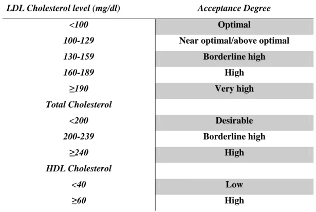
Table n.1 Electrolytes composition of blood plasma and cellular cytoplasm in a normal condition...18 Table n.2 Most important antihypertensive drugs used in therapy...27 Table n.3 Primary Hyperlipidaemia classification based on lipoprotein phenotype...56 Table 4 Classification of lipids level in the blood and acceptance risk according to the international guidelines...61 Table. n.5 PCR primers used to amplify the coding and flanking regions of human KCNJ5...73 Table n.6 Reagent used to make a Real time RFLP master mix...86 Table 7 In this table are summarized the most important informations of our genes of interest: name of gene analysed, singular nucleotide polymorphism variation, type of mutation (synonymous, and non-synonymous, etc…), primers ID, and the technique used to make the analysis...89 Table 8 Clinical phenotype of the PA cohort by mutation and rs7102584 genotype...93 Table 9 Allele frequency of polymorphisms in hypertensive and not hypertensive patients within the Brisighella population; P<0.05 no Hypertension (allele 2) vs Hypertension (allele 0);OR was calculated with a confidence interval of 95%...99 Table 10 Distribution of SNPs within our population, divided in two groups, hypertensive and not hypertensive. Through a statistical approach, a correlation between SNP and the hypertensive condition was evaluated, and results were considered significant for a P<0.05...100 Table 11 Comparison of Hardy-Weinberg P value and X2 probability between hypertensive and not hypertensive patients. P-value of HWE is reported and considered significant if <0.05. Deviation from the HWE can be seen for the only genes AGT and KIF6 rs9471077, and KIF6 rs9462535, whose P-values are 0.03, 0.005 and 0.006 respectively...102 Table 12 In this table is reported the SNP distribution of candidate genes within the hypertensive population undergone to a drug therapy. Significant P value<0.05 was calculated for no responder vs responder ( allele 0 vs 1 and 0 vs
5
2)...103 Table 13 Similarly to the analysis as above, in this table is reported the SNP distribution of candidate genes within the hypertensive population undergone to a drug therapy. Significant P value<0.05 was calculated for no responder vs responder ( allele 0 vs 1 and 0 vs 2)...105 Table 14 Familiarity was considered in the drug-treated subgroup referring to several pathological condition such as hypercholesterolemia, hypertension, acute myocardial infarction, and ictus. A borderline value was seen for the only hypercholesterolemia and not for the other conditions. P-value<0.05 was considered significant...106 Table 15 Familiarity was considered into the untreated subgroup referring to several pathological condition such as hypercholesterolemia, hypertension, acute myocardial infarction, and ictus. Hypertension familiarity was confirmed. P-value<0.05 was considered significant...107
List of Figures
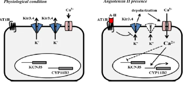
Fig.1 Frontal view of the heart structure and the most important arteries and veins...10 Fig.2 Schematic representation of the volume and pressure values changes during a complete cycle of contraction-relaxation of the heart...12 Fig. 3 Schematic representation of KCNJ5 channel activity across the membrane of H295R cells in a resting condition (figure on the left of the panel) and in a presence of AG-II (figure on the right of the panel). It has been proposed this mechanism: in the absence of AG-II KCNJ5 channels seem to be opened with an outflow of K+ from the inner to the outer of cells and a consequent membrane...39 Fig. 4 KCNJ5 gene homology over several species, from ; both Kir3.1 and Kir3.4 show an amino acid sequence equality ranging from 94 to 99%, demonstrating their importance in the cell functionality and viability...41 Fig. 5 Primary structure of Kir channel; it shows two transmembrane domains TM1 and TM2 linked by a pore-forming H5 loop and cytosolic amino and carboxyl terminals (left image). On the right it is showed the structure of voltage-gated potassium channel which is
6
constituted by six transmembrane domains, two of which, TM5 and TM6 are linked by a H5 loop, similarly to the previous one...42 Fig. 6 Linear correlation existing between LDL-C and Relative Risk for CHD demonstrates the high importance to keep total cholesterol level and LDL within a physiological range suggested from the international guidelines for cholesterol level (Grundy et al. Circulation 2004; 110(2):227-239)...60 Fig. 7 Representation of dose-response curve. Panel A shows the therapeutic window, that is the difference between the efficacy and the toxicity. This window may vary from patient to patient, B and C, requiring dose-adjustment (Qiang Ma and Anthony Y. H. Lu, Pharmacogenetics, Pharmacogenomics, and Individualized Medicine, Pharmacol Rev 63:437459,2011)...65 Fig. 8 Organization of pCMV6-AC-GFP plasmid used as a vector for our KCNJ5 cDNA...74 Fig. 9 Overview of the site-directed mutagenesis protocol: there are 3 different steps through which it is possible to produce a specific mutation, such as insertion/deletion, and point mutation...75 Fig. 10 Colonies of X-Gold ultracompetent cells transformed with site-mutated KCNJ5 cDNA, growth and selected onto LB ampicillin agar...76 Fig.11 pGEM vector structure used to run the cRNA transcription...78 Fig. 12 H295R human adenocarcinoma cell line used for in vitro studies in two different state of confluence; low confluence (left panel) and high confluence (right panel). ATCC company...81 Fig. 13 Fluorescence microscope image of H295R transfected cells with a WT KCNJ5. The same picture was acquired with a fluorescence filter turned off (left panel), and turn on (right panel). Transfected cells are well characterized by the green fluorescent colour...82 Fig. 14 Molecular basis for Taq-man based allelic discrimination processes: a) polymerization, b) strand displacement, c) cleavage, d) completion of polymerization...88 Fig. 15 Sequence chromatograms for the 3 heterozygous missense mutations identified in the population. A) It is showed the missense mutation 155G>A that leads to an Arginine (R) – Histidine (H) substitution at the position 52 of peptide chain; B) it is showed the 736 G>A that leads to a Glutamate (E) – Lysine (K) substitution at the position 247; C) in the last panel is represented the substitution 739 G>A, responsible of Glycine (G) – (Arginine) substitution
7
at the position 247 in the protein structure...91 Fig. 16 KCNJ5 potassium channel structure: all the mutations characterized in our 251 population patient, R52H, E246K, Q282E, are not localized in the selectivity filter but in neighbouring region...92 Fig.17 Different activity of KCNJ5 WT and mutant forms on the resting membrane potential variation in Xenopus laevis oocytes. Great difference has been registered between EK, RH, EQ and WT form (mean±SEM; n=6). It has also been seen that the missense mutation G247R did not exert any effects on this parameter. *Significantly different from WT, P<0.01...94 Fig.18 Current-voltage curves of WT and mutant forms of KCNJ5 potassium channel obtained from clamped Xenopus laevis oocytes in two different bathing solution: high K+ and low Na+( right) and low K+ and high Na+ concentration. The absence or presence of KCNJ5 antagonist, tertiapin-Q (50nM), are indicated respectively by ● and ○ (mean±SEM; n=6-8)...95 Fig.19 Aldosterone release from H295R transiently transfected cells with empty vector or one of the KCNJ5 mutant forms. In the panel A are reported results of aldosterone release under basal condition; panel B shows the AT-II (10nM)-stimulated aldosterone secretion; panel C shows the difference between AT-II stimulated and basal aldosterone release. These results are reported as (mean±SEM; n=6). *P<0.05 and #P<0.01 vs empty vector control...96 Fig. 20 Aldosterone release from H295R cells transfected with empty vector (negative control), del157 (positive control) and the missense mutation G247R. This missense mutation behaves similar to the negative control, empty vector, and do not produce any alteration in terms of membrane depolarization and aldosterone secretion. * P<0.01 versus control...97 Fig. 21 Viability of H295R cells transfected with WT and electrophysiologically active mutant forms of KCNJ5 potassium channel. Cell viability has been evaluated 48h after transfection; results are indicated as fold changes of optical density (OD) versus control (WT or Empty vector). Results are an average of 4 different experiments. * P<0.05 versus WT or empty vector control...98
8
Abstract
Cardiovascular disease (CVD) accounted for 30% of the estimated 58 million deaths globally. This proportion is equal to that due to infectious diseases, nutritional deficiencies, and maternal and perinatal conditions combined. It is noteworthy that a substantial proportion of these deaths (46%) occurred in individuals under 70 years of age, in the most productive period of life. In addition, 79% of the disease burden attributed to cardiovascular disease is in the same age group.
High blood pressure is one of the most important preventable causes of premature illness and death. It is the major risk factor for stroke, heart attack, heart failure, chronic kidney disease and cognitive decline. The risk associated with increasing blood pressure is continuous, with each 2 mmHg rise in systolic blood pressure associated with a 7% and 10% increased risk of mortality from ischaemic heart disease and stroke respectively. According to the international guidelines of the World Health Organization (WHO) and the International Society of Hypertension (ISH) individuals may be divided into different groups according to the blood pressure value: patients with optimum (<120/80 mm Hg), normal (120–129/80–84 mm Hg), high (130/139 - 85/89 mm Hg) blood pressure (BP) and hypertensive (140/90 mm Hg). Periodic screening of BP in adults is recommended to detect the onset of hypertension1, so that appropriate measures can be instituted to prevent morbidity and mortality associated with raised BP2. Due to the absence of definitive data regarding the time course of hypertension evolution from lower BP values, current international guidelines vary widely in their recommendations for the clinical monitoring of individuals without hypertension. For instance, the sixth report of the Joint National Committee on the Prevention, Detection, Evaluation and Treatment of High Blood Pressure in the United States (JNC VI) recommends that people with high-normal BP should undergo yearly monitoring, whereas those with normal or optimum BP should be screened every 2 years. By contrast, the European Task Force on Prevention of Coronary Disease proposes that all individuals without hypertension should be screened at least once every 5 years. The British Hypertension Society advocates an intermediate position: patients with a systolic BP of 135–139 mmHg or a diastolic BP of 85– 89 mmHg should be reassessed yearly, whereas those with lower BP should be assessed at 5– year intervals.3,4
Essential, primary, or idiopathic hypertension is defined as high BP in which secondary causes such as renovascular disease, renal failure, pheochromocytoma, hyperaldosteronism,
9
or other causes of secondary hypertension are not present. Essential hypertension accounts for 80-90% of all cases of hypertension; it is a heterogeneous disorder, with different patients having different causal factors that may lead to high BP. Life-style, diet, race, physical activity, smoke, cultural level, environmental factors, age, sex and genetic characteristics play a key role in the increasing risk.
Conversely to the essential hypertension, secondary hypertension is often associated with the presence of other pathological conditions such as dyslipidaemia, hypercholesterolemia, diabetes mellitus, obesity and primary aldosteronism. Amongst them, primary aldosteronism represents one of the most common cause of secondary hypertension, with a prevalence of 5-15% depending on the severity of blood pressure. Besides high blood pressure values, a principal feature of primary aldosteronism is the hypersecretion of mineralcorticoid hormone, aldosterone, in a manner that is fairly autonomous of the renin-angiotensin system. Primary aldosteronism is a heterogeneous pathology that may be divided essentially in two groups, idiopathic and familial form.
Despite all this knowledge, there are so many hypertensive cases that cannot be explained. These individuals apparently seem to be healthy, but they have a great risk to develop CVD. The lack of known risk factors makes difficult their classification in a scale of risk. Over the last three decades a good help has been given by the pharmacogenetics/pharmacogenomics, a new area of the traditional pharmacology that try to explain and find correlations between genetic variation, (rare variations, SNPs, mutations), and the risk to develop a particular disease.
This study was realized with the aim to add new informations on the hypertensive susceptibility starting from two different populations: an unselected Italian population, the Brisighella population, used for the Brisighella Heart Study, and a selected Australian cohort of 251 patients affected by primary aldosteronism.
10 1.0 Cardiovascular system
1.1 Heart, vessels and blood pressure
Cardiovascular apparatus is one of the most fascinating and intriguing, for complexity and importance, among all systems that regulate our life. It is constituted by an important central organ, the heart, and by a wide number of blood vessels, all differing in function and structure. The heart and blood vessels form a transportation system that delivers to all cell nutrients and oxygen, needed for their proper function, and moves away the products of their metabolism, providing a sort of communication between the cells and the environment.
Fig.1 Frontal view of the heart structure and the most important arteries and veins
The heart is an involuntary muscle, able to contract an relax autonomously with regular rhythm, pumping the blood in the pulmonary and systemic circulations. This organ, located anteriorly to the vertebral column and posteriorly to the sternum, is surrounded by a double wall sac, called pericardium, with a pericardic fluid between them. Outside the parietal pericardium there is a fibrous layer called fibrous pericardium. The human heart can be divided in three layers: the outer layer called epicardium or visceral, the middle layer, called
11
myocardium, constituted prevalently by contractile cardiac muscle, and the inner endocardium, in contact with the blood. The heart (Fig. 1) may be divided in four chambers, two atria left (sx) and right (dx) on top, that constitute the base of the organ and similarly two ventricles dx and sx in the bottom of the heart, named apex. The two sides of the organ dx and sx are separated by a wall, called septum. The two atrium dx and sx communicate with the corresponding ventricle below, allowing the blood to flow downwards. However, the heart needs a set of valves, located across each atrium and ventricle, to keep the fluid flowing in one direction; the dx ventricular valve is called tricuspid valve and the sx atrial-ventricular valve is the bicuspid or mitral. There are other two important semilunar valves, housed between right and left ventricle and pulmonary and aortic artery, respectively with the same function of the others.
Blood circulation is divided in small and big circulation; the first that starts from the dx atrium and receives deoxygenated blood from the systemic circulation through the superior vein cava; once the dx atrium is completely filled, blood starts to flow down in the corresponding ventricle below, from which deoxygenated blood through pulmonary artery is driven in the lug to be oxygenated. From the lug, this blood reach of oxygen and poor of anhydride carbonic flows back in the sx atrium by the pulmonary vein. Thus, the big circulation starts with the oxygenated blood that has completely filled the sx atrium, from which, in the similar way to the right part, it flows down in the ventricle below and get off by aortic artery. In both, sx and dx atrium blood is able to flow down in the ventricle located below for the 90% of the total volume thanks to the force of gravity. The remaining 10% of volume is driven down by the atrial myocytes contraction, atrial systole, reducing the atrial volume and in the same time increasing the pressure inside that overcomes the ventricle pressure, according to the ideal gas law: P= nRT/V.
At the end of atrial systole all the blood volume is located in the ventricle below; in the same time the semilunar valves, located between the ventricle orifice and aortic artery, are still closed, keeping all the volume inside. At the beginning of left ventricle systole, as aforementioned, the two physical parameters, volume and pressure change in an inversely-related manner, volume decreases while the pressure starts to increase overcoming the aortic pressure; this leads to the opening of semilunar valves, allowing the blood to flow out in the aortic artery. The left ventricle is empty now and ready to receive again blood from the overlying atrium. (Fig. 2)
12
Fig.2 Schematic representation of the volume and pressure values changes during a complete cycle of contraction-relaxation of the heart.
The heart is an involuntary muscle able to contract autonomously with a regular rhythm thanks to own electrical system, constituted by three important structures such as sinus atrial node (SAN), atrio-ventricular node (AVN) and His-Purkinjie fibres. SA node is located in the right atrium near the entrance of the superior vein cava; it is the most important of this system because of presence of particular type of cells, named pacemaker cells, responsible of the normal heartbeat, which ranges between 60 and 80 beats for minute. As in all other cells, the resting potential membrane of pacemaker cells range between -60 and -70mV that is kept by a continuous outflow of K+ ions in the extracellular environment. However, this outflow decreases during the time leading to a small depolarization of the resting potential membrane; in the same time there is a slow inward flow of Na+ as well as of Ca2+ ions, increasing the potential membrane until the threshold potential is reached. At a this moment, the true depolarization can start and differently from other exciting cells, in which this phase is characterized by Na+ channel opening, it is caused by a slow influx of Ca2+ ions through L-type calcium channel; for this reason the depolarization is slower than in neurons. At the end of depolarization, Ca2+ and Na+ channels result to be closed, while the K+ ions flow out rapidly repolarizing the resting membrane potential. Under normal conditions the depolarization wave will be propagated throughout the right atrium, and through the Bachmann's bundle to the left atrium, stimulating the atrial myocardium to contract, atrial systole. During the atria relaxation, called diastole, since atria are electrically isolated from the ventricles and the AV node is the only connection between the two areas, the electrical conduction from the AV node to the underlying ventricles is realized with a small delay
13
through the Bundle of His along the septum to the Purkinjie fibres. The conduction delay between atria and ventricles is very important for the heart functionality in order to avoid a simultaneously contraction. This phenomenon gives the possibility to the atria to contract together, allowing the blood flow toward the ventricles. An important characteristic of the myocardium cells is the ability to contract immediately and simultaneously with the depolarization wave passage. Myocytes of the heart can be considered as a functional syncytium because of the electrical impulses propagate freely between cells in every direction, so that the myocardium can contract as a single contractile muscle. The free and rapid propagation of the electrical impulses can happen thanks to the presence of particular junctions, called gap-junctions, between myocytes. Changes of depolarization/repolarization frequency of pacemaker cells and contraction strength of myocytes as well as alterations of the peripheral vascular resistance and alteration of volemia, including water and Na+, K+, Cl -ions levels, have wide effects on the systolic blood pressure values according to the physiological definition of blood pressure:
SBP = Q x TPR SBP = systolic blood pressure
Q = cardiac output
TPR = total peripheral resistance
Therefore, SBP is directly proportional to TPR and Q. The latter is defined as a volume of blood being pumped by the heart, in particular by the right and left ventricle and represents the effective volume that comes back with the vein determining how much blood the heart pumps out.
The principal types of vessels, different in composition and function, include arteries, capillaries and veins. Arteries are characterized by three different layers: the innermost layer, called tunica intima or internal, which show an elastic membrane and a thin layer of endothelial cells directly exposed to the blood; the tunica media, the middle layer, containing concentric sheets of smooth muscle tissue in a framework of loose connective tissue, and tunica external, or adventitia, the outermost layer of a blood vessel that forms a connective tissue sheath. The connective tissue fibres of the tunica external typically blend into those of adjacent tissues, stabilizing and anchoring the blood vessel.
Arteries may be divided into elastic and muscular arteries; the first, which include the pulmonary trunk, aorta, and their major arterial branches, transport large volume of the blood away from the heart; the second group, also known as medium-size arteries, distribute blood to the body's skeletal muscle and internal organ, and it includes the carotid, brachial,
14
mesenteric, and femoral arteries. The tunica media of the muscular arteries contains scattered smooth muscle cells, although they do not form a proper layer, and the contraction or relaxation of this muscle can produce a great changes in the diameters of the lumen. Since resistance to blood flow depends in part on this diameter, the activity of smooth muscle cells is very important on blood pressure regulation.
Capillaries, that show a singular thin layer of endothelial cells, are the only responsible of the exchange between the blood and the surrounding extracellular fluid. There are two major types of capillaries: continuous and fenestrated. Continuous capillaries allow the diffusion of water, small solutes and lipid-soluble material, and avoid the passage of blood cells and peptide. Fenestrated capillaries, as the name implies, are not completely attached each other, but show some fenestration that allow the passage of small peptide. Capillaries do not function as individual units but as part of an interconnected network called capillary bed, or capillary plexus, which is characterized by a lot of connections between capillaries and venules.
Veins transport de-oxygenated blood from periphery to the heart with a blood flows at very low pressure. Veins are classified on the basis of their size in three different types: venules, medium-size veins and large veins. Venules, which collect blood from capillary beds, are the smallest venous vessels and show three layers: an inner endothelium composed by endothelial cells, a middle layer of muscle and elastic tissue and an outer layers of fibrous connective tissue. Medium-size veins, comparable in size to the muscular arteries, characterized by a thin tunica media containing relatively few smooth muscle cells, and a thick layer, the tunica external, which contains longitudinal bundles of elastic and collagen fibres. The blood pressure in venules and medium-size veins is so low that it cannot oppose the force of gravity. Large veins include superior and inferior venae cavae. One of the most important characteristic of the large veins is the presence of valves, at more or less regular intervals, that prevents the retrograde flow of blood. These valves act like the valves of the heart, allowing blood flow in only one direction. In particular, when the individuals is in the standing position, these valves are closed, and only the compression of deeper veins, by contractions of the skeletal muscle, allows to the blood to open these valves, which in turn will be closed by the flowing back of blood itself. This force, called muscle pumping, works through the thoracic force giving to blood the needed energy to overcome the force of gravity.
15
1.2 Nervous system and its role in the blood pressure modulation
It has been well established over the years that modification of one of aforementioned parameters, heart rate, peripheral vascular resistance, volemia, directly modulates the arteriolar blood pressure. For this reason, blood pressure regulation, likes other parameters of our body, is strictly settled by numerous and complex mechanisms such as endocrine and autonomic nervous system (ANS), which often work synergically.
ANS is constituted by sympathetic and parasympathetic pathways, which have opposite activities on the same organs and tissues, and use two different types of neurotransmitter activating several receptors. The most abundant sympathetic fibres use noradrenaline (NA) as neurotransmitter and are named adrenergic or noradrenergic fibres while parasympathetic fibres use acetylcholine (Ach) thus called cholinergic. NA and ACh neurotransmitters are able to exert different activities and evocate different responses thanks to the activation of several receptors. Among the cholinergic receptors it is possible to distinguish two families: muscarinic receptors (M), M1, M2, M3, and nicotinic (N) receptors, Nn, Nm. Noradrenergic
receptors are divided in alpha (α) and beta (β), which in turn are classified in α1 and α2, and
β1, β2, and β3. To date, it is well understood the localization and the mechanism by which
these receptors are activated and the effects that their activation lead to. Muscarinic and β adrenergic receptors are G-protein-coupled-receptor, and their different action depends on the sub-type of G-protein with which they are coupled. It is well known that G-protein can exist as a Gs (stimulatory), Gi (inhibitory) and Gq proteins, that work activating or inhibiting respectively an important effector, adenylate cyclase, that converts ATP in cAMP, which in turn stimulates a kinase cAMP-dependent such as PKC. Activation of Gq protein, leads to an increased activity of phospholipase C, that converts inositol-bisphosphate-membrane-phospholipid (IP2) in inositol-triphosphate (IP3) and dyacilglycerole (DAG) increasing cytoplasmatic calcium (Ca2+) concentration.
All β receptors, β1, β2, and β3 are associated with a Gs protein even if in some case β2 has
been seen coupled with a Gi protein. Alpha receptors, α1 and α2, are coupled with a Gq and a
Gi protein respectively.
M1, M3 receptors are associated with a Gq protein while M2 is associated with an Gi protein.
Nicotinic Nn and Nm receptors are classical potassium and sodium channels, with a
pentameric structure, 2α, β, γ, δ subunits. These receptors are widely distributed, and the most part of organs and tissues receive sympathetic and parasympathetic fibres. Heart, vessels,
16
kidneys, endocrine glands, are the principal organs and tissues, strictly involved in the blood pressure regulation, that undergo sympathetic and parasympathetic modulation.
Heart is modulated by autonomic nervous system; indeed, muscarinic and adrenergic receptors have been isolated from this organ. Pacemakers cells, whose role has been already described, show muscarinic M2 and adrenergic β1 receptors. M2 receptors, coupled to a Gi
protein, once activated by ACh, vary ions flow, increasing the K+ conductance and reducing the Ca2+ inflow in both pacemaker and myocytes cells. The final effect of the M2 receptor
activation is heart rate reduction and decreased electrical conductibility. Furthermore, there is a reduction of the contraction strength. All together these effects wok reducing the blood pressure regulation. Similarly, activation of adrenergic β1 receptors is associated with
electrical and mechanical effects. It has been observed an increased Ca2+ flow across the cell membrane of pacemaker and myocytes, that leads to an increase in the discharge frequency as well as electrical conduction and ventricle strength contraction. These effects have hypertensive implication.
The cardiovascular system maintains an adequate circulation of blood to all part of the body due to the blood vessels regulation, realized through the branches of the autonomic nervous system that reach almost 100% of them. Density and function of this innervation vary widely according to different types of vessels, modulating the vasculature tone. Vascular tone is the result of smooth muscle cells contraction, which may be modulated by different neuronal neurotransmitters, different receptors, cell-to-cell spread excitation, chemical events at the cell membrane. The vascular tone reflects directly the resistance to the blood flow of small vessels, in particular arterioles and pre-capillary arterioles. The increased vascular tone results in reduction of blood vessels diameter and consequently in blood flow, causing inadequate nourishment and oxygenation of the organ or tissue with a decreased activity. For all these reasons vascular tone or vascular resistance regulation is very important in the blood pressure control, and for this reason it is strictly controlled by autonomic nervous system. Vascular tone undergoes influences of adrenergic fibres, through NA release and β2 and α1 adrenergic
receptors activation. β2 receptors, localized on top of the membrane of endothelial cells, are
coupled with a Gs protein; its activation lead to an increased adenylate cyclase activity and cAMP production; raised cAMP concentration activates a cAMP kinase which in turn phosphorylates and activates a myosin light chain kinase, inducing relaxation of the endothelial cells. On the other hand, activation of adrenergic α1 receptors, coupled to a Gq
protein, leads to an increased activity of phospholipase C, that up-regulates the IP3 and DAG production with a resulting increase of Ca2+ concentration. The Ca2+-calmoduline complex
17
activates myosin light chain kinase, which in turn phosphorylates the myosin light chain. It is this phosphorylation that promotes the interaction of myosin ATPase and actin and the cross-bridge formation, leading to the initiation of a transient increase in vascular smooth muscle contraction. These effects mediate vasoconstriction and increase blood pressure. Muscarinic receptors that have been discovered across the membrane of endothelial cells are principally M3. Activation of these Gq-coupled receptors leads to an up-regulation of phospholipase C,
which in turn, releasing DAG and IP3, increase the cytoplasmatic Ca2+ concentration. This
effect seems to be important for activating the endothelial NO synthase (eNOS), that produces NO. This substance is able to spread easily in the smooth muscle cells, where activates the guanylyl cyclase increasing the conversion of GTP to cGMP; furthermore cGMP activates a cGMP-dependent protein kinase that most probably dephosphorylates the MLC, leading to a relaxation of these cells. Even if the relaxation effect of NO is well known, its mechanism to date is not completely clarified.
It is well established that several substances of endothelial cells metabolism, such as CO2
and O2 are involved in the vascular tone and in the blood pressure regulation.
It has been seen that reduction of O2 concentration is associated with diminished
contractility of vascular smooth muscle cells as well as increased CO2 concentration is associated with a vasodilatation. Similarly a wide number of substances with a vasoactive properties have been isolated and characterized, such as kallidin, bradykinin etc.... Some of these have important functions, like angiotensin-II (AG-II): which is an extremely powerful vasoconstrictor; another peptide is the vasopressin also called antidiuretic hormone, that besides the renal effects on Na+ and K+, has been shown to exert a cutaneous vasoconstriction in the renal vascular bed; histamine that is released from cells of the skin in response to injury or antibody-antigen reactions, not only dilates arterioles and venules but also increases their permeability; prostaglandins are a group of biologically active lipids that show a vasodilator effects.
1.3 Kidney involvement in the blood pressure regulation
Kidneys are the principal organs of regulation of the volume and solute composition of the body fluid by reabsorption and excretion of water and electrolytes. The composition of blood plasma varies only slightly from individual to individual in a healthy condition. In the table
18
below are reported the average normal values for the most important ions that come into play in the composition of these two compartments, blood plasma and cytoplasm.
Table n.1 Electrolytes composition of blood plasma and cellular cytoplasm in a normal condition.
Therefore, kidney exerts this important function but is not the only one, because other organs participate in this process, notably the lungs in respect to acid-base regulation, autonomic nervous system and endocrine glands. However, the kidney through its excretory activity, provides the major mechanism for maintaining homeostasis. In order to keep a constant internal and external environment the kidney must respond appropriately to variations in dietary intake and in extra-renal losses of solutes and water. It is well known that ion concentration, measured in terms of osmolarity, is one of the most important factor able to influence the volemia. In case of increased osmolarity our body through autonomic nervous system, endocrine and kidney systems induce a reabsorption of water in order to dilute these ions, increasing the volemia. Otherwise, if there is a wide loss of ions with a consequent decrease of osmolarity our kidneys eliminate the right volume of water to concentrate the solution within physiological range. All of this is possible because our systems always work trying to keep our body in a homeostasis condition. Since the structure and the function of the kidney are so closely related, it is necessary to have some knowledge of its structure before looking at its function. The basic unit of structure and function of the kidney is the nephron, that includes the glomerulus and its attached tubules; in succession there are: proximal convoluted tubule, loop of Henle and distal convoluted tubule and collecting duct. Glomerulus is completed surrounded by a Bowman's capsule, an anastomotic network of freely branching capillaries. These capillaries originate from an afferent arteriole, and leave the glomerulus as a single efferent arteriole. The afferent arteriole in turn breaks up to form peritubular
Ions Blood plasma Cytoplasm
Na+ 145 mM 139 mM K+ 4 mM 12 mM Ca2+ 1-2 mM <0.0002 Mg2+ 1.5 mM 0.8 mM Cl- 116 mM 4 mM HCO3- 29 mM 12 mM Amino acids 9 mM 138 mM
19
capillaries surrounding the cortical tubules reaching also the deepest zone of the nephron, medulla, where they are called vasa recta. The presence of these blood vessels all around the renal tubules is very important because in this way water and electrolytes exchanges between them are possible. Our blood is filtered almost 26.7 times during the day and considering that the blood volume ranges between 5-7 L it results that total volume of 186 L is filtered for day. Of these 186 L of glomerular ultra-filtered liquid the 99% is reabsorbed and only the 1% is eliminated as urine, i.e. 1.8 L per day.
The rate of filtration across the glomerular filtration membrane of the Bowman's space is determined by physical forces. The hydrostatic pressure in the glomerular capillaries (Pgc) is the driving force of the filtration; opposing filtration are the hydrostatic pressure in the Bowman's capsule (Pbs) and the colloid osmotic pressure (COP) exerted by the proteins of the capillary plasma. Because the filtrate in Bowman's space is protein free the COP may be considered negligible. The glomerular filtration rate (GFR) is directly proportional to the net balance of the these forces according to the mathematical equation:
GFR = Kf [Pgc - (Pbs + COP)]
Kf is considered constant and is defined as the product of the hydraulic conductivity and the surface area of glomerular capillaries. According to this definition, it's possible to say that at the Bowman's space the net difference between these forces is almost 8 mmHg, driving the ultra-filtered from the lumen of capillaries to the Bowman's capsule. Along all the distal tubes the difference between these forces becomes positive in the peritubular capillaries with a consequent reabsorption of the fluid that was lost before.
Two thirds of the filtered volume is reabsorbed immediately in the proximal tube, while the remaining part starts to flow down along the descending trait of the Henle's loop. Along all the descending trait of Henle's loop there is a great reabsorption of water and the filtered solution reach a concentration of 1200mOsm, that is comparable with that of the external medulla zone; along the ascending trait of Henle's loop, ions such as Na+, K+ and Cl- but not water, are widely reabsorbed reducing the concentration of filtered liquid. The filtered liquid, that is not yet called urine, continues its trip along the distal tubule, which similarly to the ascending trait is responsible of Na+ reabsorption but in the same time is impermeable to the water. The distal tube is the physiological target of an important hypertensive hormone, aldosterone, released from the glomerulosa cells of the adrenal gland (see chapter 2.0), responsible Na+ and consequently water reabsorption, increasing the volemia and the blood pressure. The last tube of nephron, the collecting duct, exerts an important function of
20
changing the urine concentration, regulating the water reabsorption. In this way, this tube is able to produce hypertonic urine in case of excess of ions that have to be eliminated or hypotonic urine in case of excess of water. This function is realized through specific channels called aquaporin, whose expression is under the control of a particular type of hormone, antidiuretic hormone (ADH) or vasopressin, released from anterior pituitary gland, whose activity is important for maintaining the osmolarity (see parag.1.4). Kidney is able to bring about these adaptive responses because of its interaction with the autonomic nervous system, which constantly receives informations about the ion content, volemia and blood pressure. The major level of regulation lies in a microscopic structure, juxtaglomerular apparatus (JGA), so-called for its proximity to the glomerulus. This apparatus is morphologically a highly specialized structure with both vascular and tubular components. It probably has an important role in a feedback mechanism controlling glomerular filtration rate and/or renal blood flow. It consists of several parts: granulated cells in the afferent and efferent arterioles, the macula densa, and the extra glomerular mesangium. It has been demonstrated only recently the presence of gap-junctions among these cells, whose role seems to be very important in the exchanges of informations. Some of the cells in the media of the afferent and probably the efferent arteriole close to the glomerulus are specialized myoepithelioid cells and contain granules that appear to be composed of the renin enzyme. The macula densa cells position is useful to monitor some aspect of distal tubular function possibly hydrostatic pressure and the concentration of one or more solutes especially Na+ concentration. In case of increased Na+ concentration in the filtered liquid that lies in the distal tubule, this cells start to work, releasing paracrine substances that seem to have double effects: on the one hand act on the afferent arteriole increasing the vascular tone and resistance while on the other hand transfer these informations to the epithelioid cells of juxtaglomerular apparatus, thereby regulating the release of renin.
Renin is a protolithic enzyme that once released and entered in the systemic circulation converts the angiotensinogen, that is produced and released constitutively by the liver, to release the inactive decapeptide angiotensin-I. Angiotensin converting enzyme (ACE) synthesized by several tissues, split off the last two amino acids of the angiotensin I to form the octapeptide AG-II, which has important hypertensive activities. This mechanism represents the key response at the kidney level toward a Na+ reduction. However, other regulation mechanisms, as aforementioned, are involved in this complicated process. Afferent and efferent arterioles show particular type of receptors, called baroceotor, that are able to monitor constantly the pressure with which blood enters and leaves from the kidney, and in
21
case of reduced blood pressure these receptors through afferent fibres induce a sympathetic response. Furthermore, cellular membrane of juxtaglomerular cell has adrenergic β1 receptors
that response to noradrenaline/adrenaline increasing the renin release.
These observations show as the blood pressure regulation is a key mechanism realized in a cooperation manner between cardiovascular, autonomic nervous system, renal and endocrine glands.
1.4 Endocrine involvement in the blood pressure regulation
It is possible to imagine and assume that the connection point between the nervous and the endocrine system is represented by the hypothalamus through the pituitary gland or hypophysis. Hypothalamus is unique in this function and in the same time it is involved in several important processes. The hypothalamus is a relatively small area of the brain located beneath the thalamus nuclei and extends from the mesencephalon to the region of the optic chiasm and the pre-optic area. It has been well described the vascular organization of hypothalamus constituted by a series of capillaries that end in a capillary bed in the hypophysis, from which these vessels flow out with a singular vein into the systemic circulation.
It is well known that hypothalamus receives different types of neuronal fibres, GABAergic, cholinergic and catecholaminergic; indeed, several transmitters, such as acetylcholine, gamma amynobutyric acid, dopamine, serotonin, substance P, adrenaline, histamine have been isolated from this area.
It has become clear that the secretory activities of the cells of the anterior lobe of hypophysis are widely controlled by substances secreted in the hypothalamus and transported in the adenohypophysis by the portal vessel system. As stated previously, the hypothalamus integrates many functions and is involved in a series of reactions essential to the maintenance of homeostasis and to the initiation and control of many behavioural responses, such as reproduction, thirst and control of water balance, control of body weight, temperature regulation, reactions to stress, control of emotional reactions, sleep and arousal, control of somatic reactions.
The hypothalamus exerts its control over body functions through endocrine, hypothalamic-pituitary axis, and somatic neuronal efferent systems. It has a close relationship with the hypophysis and this hypothalamic-pituitary axis regulates the activity of nearly all endocrine
22
organs. Discussion of the role of the hypothalamus in the control of the body economy through the endocrine system can be divided in at least three parts: reproductive functions, water balance, and metabolic processes. The hypothalamus exerts its function through several peptides and biogenic amines, so-called releasing factors, which in turn lead to stimulation or inhibition of hormones secretion of pituitary gland. Among these substances we can recognize gonadotropins-releasing hormone (GnRH), thyrotropin-releasing hormone (TRH), corticotropin-releasing hormone (CRH), growth hormone release-inhibiting hormone or somatotropin (GHRH or STH), somatostatin-releasing hormone (SRIF). Each of these releasing factors secreted by the hypothalamus, reaches the anterior lobe of the pituitary gland and promotes or inhibits pituiry hormones release. Pituitary gland or hypophysis is divided in two parts: anterior lobe or adenohypophysis and posterior lobe or neurohypophysis. Histologically, the adenohypophysis consists of a large cells that contain secretory granules. As it has been already said, adenohypophysis is strictly related with the hypothalamus by a portal system of vessel, through which the releasing factors secreted into the hypothalamus reach this area and can exert their functions. The secretory cells of adenohypophysis show on the cellular membrane specific receptors with which the releasing factor interact leading to activation or inhibition of pituitary hormone secretion. The adenohypophysis secretes at least seven recognized hormones, four of which directly control the functioning of their particular target glands, the adrenals, thyroid, and gonads. These hormones are defined tropic or trophic. Unlike to the adenohypophysis the neurohypophysis contains no large epithelial cells full of secretory granules. Instead, it consists primarily of unmyelinated nerve fibres and modified glial cell, called pituicytes. Although it was thought for many years that the pituicytes were the source of the posterior pituitary hormones, it is now recognized that these hormones are neurosecretory products of the hypothalamus conducted to the neurohypophysis by carrier proteins (neurophysins) via neuronal axons and stored into secretory vesicles in the neuron terminal and released directly in the systemic circulation. While the adenohypophysis release, as aforementioned, several hormones under the hypothalamus stimulating factors, neurohypophysis is responsible of secretion of antidiuretic hormone or vasopressin (ADH) and oxytocin, which escape to the hypothalamus regulation. It seems to be likely that the role of neurohypophysis is just storage and release of these substances; indeed the posterior pituitary gland contains almost 15 unit of ADH, which is enough to maintain a man in maximum antidiuresis for more than 1 week. By contrast, the hypothalamus contains very little hormone less than 5% of the total amount stored in the posterior lobe of pituitary. While
23
the hypothalamus may be capable of meeting some demands for ADH, it may not be able to support intense and sustained antidiuresis.
Pituitary hormones show a feedback mechanism of regulation, and in most cases it is a negative feedback; this means that releasing of these hormones is subjected to an auto-limitation process and the hormone itself inhibits further secretion by the pituitary. Nevertheless, when the plasma concentration of the target gland hormone falls below under critical level, the negative feedback is relieved, and the trophic hormone is again secreted until the target gland produces enough hormone to shut off the pituitary. However, there are particular conditions during which this mechanism may be a positive feedback, leading to a continue releasing of the specific substance. Assumed that each of the pituitary hormones exert an important role in the regulation of several pathways involved in the maintenance of the homeostasis of our body, I want to focus only on the role of those hormones, whose activity is directly correlated with the blood volume, ions absorption or excretion and blood pressure regulation. Among all the hormones aforementioned, ADH, ACTH, TSH, have been demonstrated to work in synergism with nervous system to maintain blood pressure within a physiologic range. Currently it is well know the functional mechanism of TSH, released from the adenohypophysis under the stimulation of specific releasing factor, to stimulate the thyroid gland to produce Thyroxin (T4) and Triiodothyronine (T3) which stimulates the metabolism of almost every tissue in the body. Furthermore, thyroid gland is also involved in the secretion of a polypeptide hormone, calcitonin, also named thyrocalcitonin, whose role seems to be important for the Ca2+ regulation. Its activity is not completely clear but to date we can say that this hormone is involved in the reduction of Ca2+ circulating at a three different levels:
inhibition of Ca2+ absorption by the intestine
inhibition of osteoclast activity in bones
inhibition of renal tubular reabsorption of Ca2+ allowing it to be excreted in the urine
Furthermore, calcitonin is also involved in the inhibition of phosphates reabsorption by the kidney. The role of this hormone is very important considering the key function of Ca2+ ions in the cellular responses.
Similarly, the adrenocorticotropic hormone, ACTH, is secreted by the anterior lobe of the hypophysis in response to the CRH released by hypothalamus and exerts a key role not only in the metabolism but also in the blood pressure regulation. ACTH secretion is increased by reduced level of circulating cortisol and under conditions of stress such as fever, acute
24
hypoglycaemia, major surgery or by noxious stimuli; furthermore, its release resulted to be affected by the diurnal rhythm, increasing during the first few hours of sleep and reaching the peak at the time of awakening while it decrease during the evening. ACTH hormone, once released, by the systemic circulation reach the adrenal gland, that is the major target, where activates the first enzyme involved in the first step of synthesis of glucocorticoids and mineralocorticoid hormones (see chapter 2). Aldosterone is one of these, whose hypertensive role was completely clarified and will be deeper discussed in the next chapter. Furthermore, as an indirect consequence of its actions, ACTH, also maintains the size and blood flow of the adrenal cortex. Indeed, when present in excessive amounts, it causes enlargement of the two zones of the adrenal cortex, fasciculate and reticularis, while in its absence adrenal cortical atrophy occurs. Noteworthy is the aspect that ACTH has a lot of extracortical activities, modulating numerous organs and tissues.
Antidiuretic hormone, ADH, or vasopressin is clearly involved in the urine production and especially in the regulation of the urine osmolarity and consequently is indirectly responsible of blood volemia and blood pressure regulation. As widely explained in the last paragraph, one of the essential functions of the kidney is control of the total body water content so that the total solute concentration of the body fluids in normal humans is maintained within a very narrow limits despite wide variations in the in fluid intake and extrarenal water losses. Thus, urine volume and osmolarity directly reflect changes in the same parameters of blood flow. The osmolarity of human urine can vary widely between 85 and 1400 mOsm/kg of water. These changes in osmolarity are realized mostly in the last part of distal tube and along the collecting duct where ADH exerts its activity. I have already explained the urine flow over the nephron; briefly here I want just to remember how the urine changes in volume and concentration. In the distal tube almost two third of the filtered liquid is reabsorbed in the proximal tube, and the liquid has the same concentration of the plasma of almost 285mOsm/kg. Along the descending trait of the Henle's loop, only water is reabsorbed into the interstitial space, with a consequent increase of filtered liquid osmolarity that riches 1200mOsm; at this point the fluid start to flow up along the ascending trait of Henle's loop, where Na+ and Cl- ions are reabsorbed and the fluid's osmolarity get off again to 285mOsm. In the last two traits, distal tube and collecting duct, urine osmolarity will be changed by ADH hormone, according to the body's needs, producing hypo or hyperosmotic urine. Plasma ADH levels, under normal condition, range approximately between 1 and 3µU/ml and plasma osmolarity is maintained around 285-290mOsm. When this value is changed as little as 2%, ADH output is significantly increased. ADH secretion is influenced by many neural and
25
humoral stimuli that excite or inhibit hypothalamic supraoptic nuclei, from which this hormone is released. Blood volume and pressure are constantly monitored by numerous receptors, osmoreceptor and baroreceptors, which afferent fibres reach the CNS, which in turn integrates the informations and send appropriate responses to the periphery. The location of the volume receptors and their afferents pathways are not known with certain. It seems to be like that atrial and ventricular stretch receptors, receptors located in the great veins of the chest and probably from the pulmonary circulation, may be involved in the blood pressure and osmolarity control. Furthermore, there are particular type of cells of the supraoptic nuclei of hypothalamus that have been recognized to be involved in this mechanism acting as osmoreceptor. ADH releasing undergoes the negative feedback regulation, similarly to others hormones. Thus, increased concentration of solute in the plasma evokes the release of ADH, which travels in the blood to the kidney, where it increases water reabsorption. The water thus reabsorbed dilutes the solutes in the plasma and thereby shuts off the signal for ADH secretion. When the solute concentration of plasma is too low, as might occur after drinking large volumes of water, ADH secretion is inhibited and free water is excreted until the solute concentration in plasma retains some optimal level that again initiates basal secretion of ADH. When the volume of blood decreases following haemorrhage, dehydration, or sodium depletion, signals originating from receptors that monitor either the central venous pressure or the arterial pressure are transmitted to the CNS and initiate the release of ADH. In addition, renin released from kidney may trigger increased ADH secretion through the intermediate AG-II. Increased water reabsorption reduces loss of volume in the urine, and in the presence of water intake increases plasma volume, thereby shutting off the signal for ADH secretion. The mechanism of action of ADH hormone is not completely understood, even if it seems to work activating its receptors V2 located over the basolateral membrane of epithelial cells in
the last part of distal tube and collecting duct. These receptors are Gs-coupled proteins, whose activation lead to an increased level of cAMP, that stimulates the up-regulation of the gene expressing for the aquaporin channels and in the same time triggers their insertion by exocytosis mechanism.5 Thus ADH makes both tubules more permeable to water, that diffuses freely across the collecting duct epithelium into the medullary interstitium, and the urine equilibrates the interstitial concentration of 1200mOsm. The water reabsorbed from the cortical collecting ducts and distal convolution enters the peritubular capillaries of the cortex and is removed from the kidney. In the absence of ADH the epithelium of the distal convolution and collecting duct has a very low permeability to water, the tubular urine flows through the remainder of the tubule without achieving osmotic equilibrium with the
26
surrounding interstitial fluids (1200mOsm), and hypoosmotic urine is produced. It has been seen that ADH hormone exerts its hypertensive action activating also V1 receptors localized
over the membrane of smooth muscle cells and leading to an increased vascular resistance. From these informations results quite clearly that alteration of mechanisms that lead to the ADH release are responsible of pathological conditions. Deficiency of ADH results in a continuous, copious flow of as much as 15L of dilute urine/day. This disease state is known as diabetes insipidus. It is accompanied by a profound thirst to compensate for the water loss in the urine. Otherwise, overproduction of ADH produces a disease state that is characterized by abnormal retention of water and dilution of plasma sodium. Renal compensation for the change in volume results in increased sodium loss, which aggravates the hyponatremia.
1.5 Pharmacology of Hypertension
As already said blood pressure regulation is realized by several integrated mechanism that involve heart functionality, vessels resistance modulation and water and ions reabsorption or excretion. Central and peripheral nervous systems, kidneys and endocrine systems work in a coordinated manner in order to keep blood pressure parameters within physiologic values ranging between 80/120 mmHg. Thus, the pharmacological therapies of hypertension can be realized at a different levels targeting one or more of these physiological mechanisms. Drugs currently available act either by decreasing CO or TPR. However, reduction of blood pressure by any of these mechanisms can trigger compensatory reflex responses that may prevent drug-induced decrease in blood pressure.
Over the years it has been well clarified the complexity of hypertension and it has been described as a multifactorial pathology where others diseases such as dyslipidaemia, diabetes, renal dysfunction and endocrine alterations were included. Sometimes, antihypertensive therapies need an integrated treatment in order to reduce pathological risks and prevent cardiovascular complications too. Thus, the choice of therapy for a patient with hypertension depends on a variety of factors such as age, gender, race, body build, and lifestyle of patient; etiology of disease; other coexisting diseases; presence or absence of risk factors (smoking, alcohol consumption, obesity, etc...).
In some cases patients show borderline blood pressure values for which a no pharmacological approach can be sufficient to restore them. Low sodium chloride diet, weight reduction, cessation of smoking, decrease in alcohol consumption, sports, etc... may be useful
27
to achieve this goal. Unfortunately, the most part of hypertensive patients need a pharmacological intervention because of their higher blood pressure values.
Antihypertensive drugs can be divided into seven classes, based on mechanism of action; ACE inhibitors, α1 antagonists, β-blockers, angiotensin receptor-blockers, Ca2+-antagonists,
central adrenergic inhibitor, diuretics, direct vasodilators and are summarized in the table n.2.
Drug Advantages Disadvantages CO HR TPR
Angiotensin-Converting Enzyme (ACE) Inhibitors Captopril, Zofenopril, Benazepril, Enalapril, Lisinopril, Ramipril, Fosinopril Renoprotective in type 1 diabetics Profoundly antiproteinuric Decreased morbidity and mortality and symptomatic improvement in CHF patients Decreased ventricular remodeling and mortality post-MI Leftward shift of pressure-natriuresis curve Safely combined with other anti-hypertensive, especially diuretics or calcium antagonists Cough (~9%) Rare angioedema (~3/1000) Rare hypokalemia Higher dose requirements in some African-Americans BP lowering effect very sensitive to level of dietary sodium intake Reduced cardiac output by reducing blood volume These drugs have not effects on the heart rate Reduced vascular resistance Alpha1 Antagonists Non-selective: Phenoxybenzaminep hentolamine, tolazoline, ergot-derivates; Selective: prazosine, doxazosine, alfuzosine, terazosine, indoramine, uropidil Positive effect on all lipoprotein fractions Improved insulin sensitivity Unchanged to improved sexual function in men Blunts thiazide-induced rise in cholesterol Improves maximal urine flow in men with BPH Orthostatic hypotension (particularly in the elderly, in combination with other vasodilators, in the setting of autonomic dysfunction, and in volume depleted patients) Slightly higher doses needed in African-Americans Reduced cardiac output by reducing cardiac activity and lowering preload
Not effect Marked reduction of vascular resistance
28 Angiotensin II (AT1) Receptor Antagonists or Sartan: Losartan, Telmisartan, Valsartan, Irbesartan Lesser peak BP lowering compared to ACEs Nearly complete blockade of A-II effect No effect on bradykinin, enkephalins, or substance P Uricosuric (losartan) Profoundly antiproteinuric Metabolically neutral Rare angioedema No cough Relatively flat BP dose-response curve No effect on bradykinin Limited data available in African-Americans Absence of hard clinical end-point (i.e., CHF, MI, renal) data Rare hyperkalemia Reduced cardiac output, by inhibiting sympathetic outflow
Not effect Reduced vascular resistance by inhibiting vascular AT2 receptors β1-Blockers non-selective: Propranolol, Pindolol, Bucindolol, Sotalol Timolol Selective: Atenolol Bisoprolol, Acetobutolol, Nebivolol Esmolol Betaxolol Differential cardiac and hemodynamic effects Proven to lower morbidity and mortality post-MI Selected agents useful in patients with migraine or angina Can worsen CHF; however, at low doses may improve CHF Low cardiac output symptoms Can worsen or precipitate depression Can aggravate PVD symptoms Lowers HDL and raises TGs Delays recovery from and masks symptoms of hypoglycemia Abrupt discontinuation can lead to rebound hypertension Reduced cardiac output by, reducing volemia, inhibiting central sympathetic outflow Inotropic, chronotropic and dromotropic negative effects Increased vascular resistance, mediated by inhibition of β2 receptors