Enzyme Activity Induces Insulin Resistance and
Hepatosteatosis in Mice
Loredana Fiorentino,
1* Alessia Vivanti,
1* Michele Cavalera,
1Valeria Marzano,
1,2Maurizio Ronci,
1,2Marta Fabrizi,
1Stefano Menini,
3Giuseppe Pugliese,
3Rossella Menghini,
1Rama Khokha,
4Renato Lauro,
1Andrea Urbani,
1,2and
Massimo Federici
1Tumor necrosis factor␣–convertingenzyme(TACE,alsoknownasADAM17)wasrecentlyinvolved
in the pathogenesis of insulin resistance. We observed that TACE activity was significantly higher in livers of mice fed a high-fat diet (HFD) for 1 month, and this activity was increased in liver> white adipose tissue> muscle after 5 months compared with chow control. In mouse hepatocytes, C2C12
myocytes, and 3T3F442A adipocytes, TACE activity was triggered by palmitic acid, lipolysaccharide, high glucose, and high insulin. TACE overexpression significantly impaired insulin-dependent phos-phorylation of AKT, GSK3, and FoxO1 in mouse hepatocytes. To test the role of TACE activation in
vivo, we used tissue inhibitor of metalloproteinase 3 (Timp3) null mice, because Timp3 is the specific
inhibitor of TACE and Timp3ⴚ/ⴚmice have higher TACE activity compared with wild-type (WT) mice. Timp3ⴚ/ⴚmice fed a HFD for 5 months are glucose-intolerant and insulin-resistant; they showed macrovesicular steatosis and ballooning degeneration compared with WT mice, which pre-sented only microvesicular steatosis. Shotgun proteomics analysis revealed that Timp3ⴚ/ⴚ liver showed a significant differential expression of 38 proteins, including lower levels of adenosine kinase, methionine adenosysltransferase I/III, and glycine N-methyltransferase and higher levels of liver fatty acid-binding protein 1. These changes in protein levels were also observed in hepatocytes infected with adenovirus encoding TACE. All these proteins play a role in fatty acid uptake, triglyceride synthesis, and methionine metabolism, providing a molecular explanation for the increased hepatosteatosis observed in Timp3ⴚ/ⴚcompared with WT mice. Conclusion: We have identified novel mechanisms, governed by the TACE–Timp3 interaction, involved in the determination of insulin resistance and liver steatosis during overfeeding in mice.(HEPATOLOGY2009;50:000-000.)
P
andemic obesity is now considered the underlying basis for the increasing prevalence of chronic met-abolic-inflammatory diseases including type 2 dia-betes, nonalcoholic fatty liver disease (NAFLD) and atherosclerosis.1Although NAFLD is an emergingmeta-bolic complication of obesity, its pathogenic mechanisms are still unclear.1
The contribution of insulin resistance to the develop-ment of fatty liver occurs in part by deficient control of lipid storage in white adipose tissue and in part by altered
Abbreviations: ADK, adenosine kinase; BSA, bovine serum albumin; FABP1, fatty acid– binding protein 1; GFP, green fluorescent protein; GNMT, glycine N-methyltransferase; HFD, high-fat diet; JNK, c-Jun N-terminal kinase; MAT1A, methionine adenosysltransferase 1A; MATI/III, methionine adenosysltransferase I/III; mRNA, messenger RNA; NAFLD, nonalcoholic fatty liver disease; PA, palmitic acid; SV40, PCR, polymerase chain reaction; Simian virus 40; SD, standard deviation; TACE, tumor necrosis factor ␣–converting enzyme; Timp3, tissue inhibitor of metalloproteinase 3; TNF-␣, tumor necrosis factor ␣; WAT, white adipose tissue; WT, wild-type.
From the1Department of Internal Medicine, University of Rome Tor Vergata, Rome, Italy;2S. Lucia Research Institute, Rome, Italy; the3Department of Clinical
Sciences, Sapienza University of Rome, Rome, Italy; and the4Ontario Cancer Institute, University of Toronto, Toronto, Ontario, Canada.
Received April 1, 2009; accepted August 12, 2009.
Supported in part by grants from Telethon (GGP-08065), Juvenile Diabetes Research Foundation (Research Grant 1-2007-665), Societa` Italiana di Diabetologia (Italian Society of Diabetology) (Research Grant 2007), the Italian Ministry of University (PRIN 2006/2008), Ministry of Health Research Grants (all to M. F.), and Fondazione Roma (to M. F. and A. U.).
*These authors contributed equally to this work.
Address reprint requests to: Massimo Federici, M.D., Department of Internal Medicine, University of Rome Tor Vergata, Via Montpellier 1, 00133 Rome, Italy. E-mail: federicm@uniroma2.it; fax: (39)-0672596890.
Copyright © 2009 by the American Association for the Study of Liver Diseases. Published online in Wiley InterScience (www.interscience.wiley.com). DOI 10.1002/hep.23250
Potential conflict of interest: Nothing to report.
Additional Supporting Information may be found in the online version of this article.
control of hepatic lipogenesis and mitochondrial fatty acid oxidation.2Increased release of inflammatory factors
or diminished secretion of protective adipokines from dysfunctional adipose tissue can predispose hepatocytes to accumulate lipids in obese individuals.3Further
evolu-tion to fibrosis and steatohepatitis may involve activaevolu-tion of hepatic stellate cells and Kupffer cells by insulin resis-tance–related factors.4Tumor necrosis factor␣ (TNF-␣)
is among the cytokines involved in linking nutrient avail-ability to innate immune activation and development of fatty liver disease.5Local/paracrine regulation of TNF-␣
release from plasma membrane through its ectodomain shedding is regulated by TACE.6TACE is naturally
in-hibited by tissue inhibitor of metalloproteinase 3 (Timp3), which has the potential to regulate other ADAM and matrix metalloproteinases during immune responses.6 Activation of TACE is triggered by way of
protein kinase C and extracellular signal-regulated kinase signals upon several stimuli, including metabolic ones such as hyperinsulinemia.7-9We have recently shown that
whereas lack of Timp3 alone has no gross effect on insulin resistance and glucose tolerance in mice fed a regular diet, its deficiency accelerates liver inflammation and steatosis only if coupled to genetic-dependent and nutrient-depen-dent insulin resistance.10-12The TACE/Timp3 system is
therefore emerging as a pivotal mediator between meta-bolic stimuli and innate immunity, although the temporal and spatial regulation of this activation remains un-known. We coupled murine and cellular models to pro-teomic technologies to show that hepatic TACE overactivity is central to the development of fatty liver disease.
Materials and Methods
Reagents. Free fatty acid–free, low endotoxin bovine serum albumin (BSA), palmitic acid, lipolysaccharide, in-sulin, glucose, c-Jun N-terminal kinase (JNK) inhibitor SP600125, and other common chemicals were obtained from Sigma Aldrich (St. Louis, MO). A list of antibodies is available in the Supporting Information.
Cell Culture. 3T3-F442A preadipocytes, C2C12
myocytes, and Simian virus 40 (SV40)-tranformed hepa-tocytes were grown and differentiated as described.13-15
Metabolic Treatments. Palmitic acid was dissolved in methanol by heating at 75°C and mixing, then loaded onto free fatty acid–free low endotoxin BSA by way of sonication and gently shaking overnight at 37°C to yield a 5-mM solution of palmitic acid in 5% BSA. Before treat-ments, all cells were serum-starved in 0.5% BSA over-night and then treated for 2 hours with 0.5 mM palmitic acid (PA) alone or in combination with the JNK inhibitor
SP600125 (20 M). For glucose treatment, cells were either grown in low-glucose medium (C2C12and
hepato-cytes) or were glucose-starved for 4 hours before treat-ment (3T3-F442A).
TACE Activity. TACE activity was determined using the SensoLyte 520 TACE Activity Assay Kit (AnaSpec, San Jose, CA) according to the manufacturer’s protocol. Thirty micrograms tissue proteins or 20g cell proteins were used for the assay. A reaction was started by adding 40M of the fluorophoric QXL520/5FAM FRET substrate. Fluores-cence of the cleavage product was measured in a fluoresFluores-cence microplate reader (FLx800, BIO-TEK Instruments, Wi-nooski, VT) at lex 490 nm and lem 520 nm.
Adenovirus Infection. Adenoviruses expressing green fluorescent protein (GFP) only or GFP and TACE (Vec-tor Biolabs, Philadelphia, PA) were used to infect SV40-tranformed hepatocytes. The infection was carried out at 500 pfu/cells in ␣ minimum essential medium supple-mented with 0.2% BSA for 6 hours at 33°C. The virus-containing medium was then removed, and cells were incubated for 24 hours in␣ minimum essential medium supplemented with 4% fetal bovine serum before being differentiated and treated as described.
Animal Models and Analytical Procedures. Timp3⫺/⫺ mice on a C57/BL6 background have been described,16as
have metabolic testing procedures10-12(see also
Support-ing Information). Animal studies were approved by the University of Tor Vergata Animal Care and Use Commit-tee. All animals received human care according to the criteria outlined in the “Guide for the Care and Use of Laboratory Animals” prepared by the National Academy of Sciences and published by the National Institutes of Health (NIH publication 86-23, revised 1985).
Extraction of Timp3 for Western Blots. For the extraction of matrix-bound Timp3, tissues were treated as described.10
Histology and Quantification of Liver Lesions. Histology was perfomed as described.12(See Supporting
Information for details.)
Gene Expression Analysis. Total RNA was isolated from wild-type (WT) and Timp3⫺/⫺ mice and from SV40-transformed hepatocytes using Trizol reagent (In-vitrogen Corp, Carlsbad, CA). Two micrograms of total RNA were reverse-transcribed into complementary DNA using the High Capacity cDNA Archive kit (Applied Bio-systems, Foster City, CA). Quantitative real-time poly-merase chain reaction was performed using an ABI PRISM 7700 System and TaqMan reagents (Applied Biosystems). Each reaction was performed in triplicate using standard reaction conditions. The Applied Biosys-tems primers used are listed in the Supporting Methods.
Protein Identification and Quantization by LC-MSE and Ingenuity Pathway Analysis. Shotgun
pro-teomics and ingenuity pathways analysis were performed as described17,18and are reported in an extended version
in the Supporting Information.
Liver Methionine Metabolism Assay. Assays for S-adenosylmethionine and S-adenosylhomocysteine in liver and cells—methionine and homocysteine in serum— were performed as described19and are reported in an
ex-tended version in the Supporting Methods.
Statistical Analysis. Results of the experimental stud-ies are expressed as the mean ⫾ standard deviation as indicated. Statistical analysis was performed using one-way analysis of variance, two-one-way analysis of variance, or an unpaired Student t test as appropriate. Values of p⬍ 0.05 were considered statistically significant.
Results
TACE Activation Is Induced by Metabolic Stimuli In Vitro and Impairs Insulin Signaling. We have re-cently described that regulation of TNF-␣ release from plasma membrane through its ectodomain shedding by TACE has a role in accelerating liver inflammation and ste-atosis when coupled with an insulin-resistant environmental and genetic background.10-12 Because TACE
haploinsuffi-ciency protects from lipotoxicity and glucotoxicity in an in vivo model, we analyzed three different in vitro cell culture models—3T3-F442A adipocytes, C2C12 myocytes, and
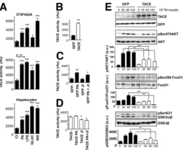
SV40-tranformed hepatocytes—to study mechanisms that link metabolic dysfunction to TACE activation. TACE ac-tivity was significantly increased by treatment with a free fatty acid, palmitic acid (0.5 mM; 2 hours), lipolysaccharide (200 ng/mL; 2 hours), high glucose (15 mM; 2 hours), or high insulin (10⫺7M; 2 hours) (Fig. 1A). To test whether increased TACE activity is a downstream effector of meta-bolic toxicity to impaired insulin action, we overexpressed TACE by way of adenoviral vectors. This resulted in in-creased TACE activity (Fig. 1B). Inhibition of JNK activity by SP600125 partially reversed the effect of palmitic acid and TACE overexpression on TACE activity (Fig. 1C,D). In a preliminary set of results, we observed that TACE overex-pression impairs ligand-dependent phosphorylation of the insulin receptor subunit at different insulin concentrations (10⫺9M and 10⫺7M) and time points (Supporting Fig. 1). Next, we analyzed downstream elements of insulin signaling involved in the control of glucose and lipid metabolism. We found that phosphorylation of AKT on serine 473, FoxO1 on serine 256, and GSK3␣/ on serine 9/21 were all consis-tently reduced by increased TACE activity (Fig. 1E).
TACE Expression and Activity Are Modulated Dur-ing HFD. To identify tissues in which TACE activity
may affect glucose and lipid metabolism, we analyzed its activation in white adipose tissue (WAT), muscle, and liver of C57/BL6 mice fed either a high-fat diet (HFD) or chow for 5, 10, and 20 weeks after weaning. We found that TACE activity was significantly increased by HFD first in liver at 10 weeks and continued to be increased after 20 weeks of HFD compared with chow (Fig. 2A). Both WAT and muscle also displayed increased TACE activity by this time point. Next, we analyzed the expres-sion levels of TACE and its inhibitor Timp3 in all three tissues and found that whereas increased TACE activation associated with a mild increase of TACE expression in WAT and liver, a more significant decrease of Timp3 expression occurs at both messenger RNA (mRNA) and protein levels in all three tissues (Fig. 2B,C).
Overall, these results suggest that prolonged metabolic stress is associated with increased TACE activity and de-creased Timp3 expression.
Impaired Glucose Tolerance and Steatohepatitis in Timp3ⴚ/ⴚMice Fed a HFD for 20 Weeks. Timp3⫺/⫺mice manifest increased TACE activity, especially in the liver.16
Fig. 1. TACE activity is induced by metabolic stimuli in vitro and impairs insulin signaling. (A) 3T3F442A adipocytes (top), C2C12myocytes (middle),
and SV40-transformed hepatocytes (bottom) were treated with 0.5 mM palmitic acid, 200 ng/mL lipolysaccharide, 15 mM glucose or 10⫺7M insulin for 2 hours and then analyzed for TACE activity. (B) SV40-transformed hepatocytes were infected with adeno-GFP or adeno-GFP-TACE and then analyzed for TACE activity. (C,D) SV40-transformed hepatocytes infected with adeno-GFP or adeno-GFP-TACE were treated with 0.5 mM PA in the presence or absence of 20M JNK inhibitor SP600125; data are expressed as the mean⫾ standard deviation (SD) (n ⫽ 3). **P ⬍ 0.01 versus GFP, *P ⬍ 0.05 versus GFP-PA (C). *P⬍ 0.05 versus both TACE and TACE PA (D). (E) SV40-transformed hepatocytes were infected with adeno-GFP or adeno-GFP-TACE and then stimulated with 10⫺7M insulin for different time lengths. Cells were lysed and subjected to western blotting to detect TACE overexpression, and Ser473 AKT, Ser256 FoxO1, and Ser9/21 GSK3␣- phosphorylation, matched against total protein levels. Data are expressed as the mean⫾ SD (n⫽ 3-5). ***P ⬍ 0.001. **P ⬍ 0.005. *P ⬍ 0.05.
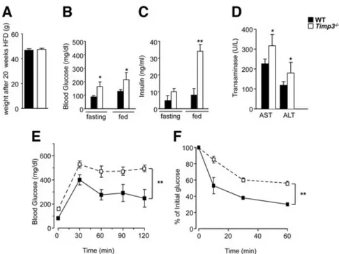
However, we have previously shown that metabolic ho-meostasis in Timp3⫺/⫺mice is similar to that of WT litter-mates at 24 weeks of age, when both are fed chow, offering the ideal scenario to study the interaction between increased TACE activity and the prolonged metabolic stress caused by a diet rich in lipids. Timp3⫺/⫺mice fed a HFD for 20 weeks exhibited a weight similar to that of WT mice (Fig. 3A); however, Timp3⫺/⫺animals showed significantly increased fasting and fed glucose and insulin levels (Fig. 3B,C), in-creased aminotransferases (Fig. 3D), and worsened glucose tolerance (Fig. 3E) and insulin sensitivity (Fig. 3F) compared with WT littermates.
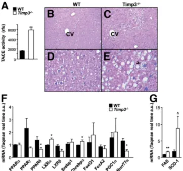
Analysis of liver function and histology revealed that after 20 weeks of HFD, Timp3⫺/⫺ mice manifested in-creased TACE activity (Fig. 4A) and macrovesicular ste-atosis with features of ballooning degeneration as seen in grade 2 human steatohepatitis (Fig. 4C,E) compared with only microvesicular steatosis in WT livers (Fig. 4B,D). Analysis of the expression of several transcription factors known to regulate lipid and carbohydrate metabolism re-vealed that Timp3⫺/⫺livers had significantly higher levels of liver X receptor␣ and carbohydrate response element binding protein 1 along with significantly reduced levels of peroxisome proliferator-activated receptor ␦ and
Fig. 2. TACE expression and activity are mod-ulated during a HFD. C57/BL6 mice were fed either a HFD or chow diet for different periods. WAT, muscle, and livers were homogenated to analyze (A) TACE activity, (B) Timp3, and (C) TACE protein and mRNA levels were quantified by way of western blotting and real-time PCR, respectively, from muscle, WAT, and livers of HFD or chow-fed mice. Data are expressed as the mean ⫾ SD (n ⫽ 3). ***P ⬍ 0.001. **P⬍ 0.005. *P ⬍ 0.05.
Fig. 3. Impaired glucose tolerance in Timp3⫺/⫺mice fed a HFD. WT and Timp3⫺/⫺ mice were fed a HFD for 20 weeks and (A) body weight, (B) blood glucose, (C) insulin, and (D) aminotransferase levels were measured. (E-F) WT and Timp3⫺/⫺mice were fasted overnight and (E) intraperitoneal glucose tolerance test and (F) intraperitoneal insulin tolerance test were performed. Data are expressed as the mean⫾ SD (n ⫽ 6). **P ⬍ 0.005. *P ⬍ 0.05.
Nurr77 (Fig. 4F) compared with WT livers. Expression of targets of liver X receptor␣ and carbohydrate response element binding protein 1 such as fatty acid synthase and stearoyl-coenzyme A desaturase 1 were consequently in-creased in Timp3⫺/⫺ mice compared with WT controls (Fig. 4G).
Shotgun Proteomics Analysis of Steatohepatitis in Timp3ⴚ/ⴚ and WT Mice Fed a HFD for 20 Weeks. Because our data suggested that TACE activation plays a role in the pathogenesis of nonalcoholic steatohepatitis, we were prompted to use a proteomics-based approach to identify TACE targets linked to controlling lipid and glu-cose metabolism in the liver. Shotgun proteomics analysis of hepatic lysates from WT and Timp3⫺/⫺mice revealed 38 differentially expressed proteins in WT versus
Timp3⫺/⫺ mice (Table 1). An unbiased systems biology approach showed that Timp3 knockouts carried signifi-cantly different signals involving liver fibrosis, damage, steatosis, cholestasis, and hyperbilirubinemia (Supporting Table 1). To seek the best candidates to validate our teomic approach, we used bioinformatics to identify pro-teins associated with liver disease and lipid metabolism. Data analysis performed through IPA-Ingenuity software pointed to several proteins in hepatic system disease,
amino acid and lipid metabolism, and highlighted aden-osine kinase (ADK), methionine adenosyltransferase I/III (MATI/III), glycine N-methyltransferase (GNMT), and fatty acid-binding protein 1 (FABP-1) as relevant targets. Supporting Figs. S2 and S3 show representative images of IPA analysis, and proteomic identification data are shown in Supporting Figs. 4 and 5. Interestingly, several of these proteins are involved in the regulation of methionine me-tabolism.20,21Next, liver lysates from WT and Timp3⫺/⫺
mice were immunoblotted to confirm that ADK, MATI/ III, and GNMT protein levels were indeed significantly decreased whereas the FABP-1 level was significantly in-creased in livers of Timp3⫺/⫺ mice compared with WT littermates (Fig. 5A). To control the effect of TACE at the mRNA level, we used quantitative real-time polymerase chain reaction (PCR) to analyze the expression of ADK, methionine adenosysltransferase 1A (MAT1A), GNMT, and fatty acid– binding protein 1 (FABP1) genes and found a pattern comparable with the correspondent pro-tein levels (Fig. 5B). Moreover, we found unchanged expression of methionine adenosysltransferase 2, cystathi-onine-beta-synthase, and 5,10-methylenetetrahydrofo-late reductase—three other enzymes involved in methionine metabolism but not identified by proteom-ics—suggesting that TACE effects are specific (Support-ing Fig. 6A). Analysis of S-adenosylmethionine and
S-adenosylhomocysteine in the liver as well as methionine
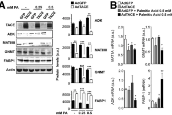
and homocysteine in the blood confirmed that a HFD has to some extent a different effect on methionine metabo-lism in Timp3⫺/⫺ mice compared with their WT litter-mates (Fig. 5C). Because Timp3 controls different families of membrane proteases, we examined whether the proteins identified are linked to TACE activation in synergy with lipotoxicity. Therefore, we adenovirally overexpressed TACE in hepatocytes in the presence or absence of increas-ing concentrations of palmitic acid. Immunoblot analysis confirmed that ADK, MATI/III, GNMT, and FABP-1 ex-pression was modulated in vitro in a manner similar to that observed in vivo (Fig. 6A). Analysis of mRNA levels of the same candidates supported that TACE effects are specific (Fig. 6B) due to lack of effect on methionine adenosysltrans-ferase 2, cystathionine-beta-synthase, and 5,10-methyl-enetetrahydrofolate reductase (Supporting Fig. 6B). Analysis of S-adenosylmethionine and S-adenosylhomocysteine from cell extracts suggested that the TACE effects on the regula-tion of methionine metabolism may depend on several con-ditions, including interaction with lipotoxicity (Supporting Fig. 6C).
Discussion
Epidemiological studies suggest that among the meta-bolic complications of obesity, NAFLD may evolve into
Fig. 4. Steatohepatitis in Timp3⫺/⫺mice fed a HFD. (A) TACE activity was measured in livers from WT and Timp3⫺/⫺mice. (B-E) Livers from WT and Timp3⫺/⫺mice fed a HFD were fixed in formalin, and 5-m-thick sections were stained with hematoxylin-eosin and Masson’s trichrome. Sections were analyzed by way of light microscopy at magnifications of ⫻10 (B,C) and ⫻40 (D,E). (F,G) Expression of transcription factors and enzymes involved in lipid and carbohydrate metabolism was measured using real-time PCR on livers from WT and Timp3⫺/⫺mice fed a HFD. Data are expressed as the mean⫾ SD (n ⫽ 3). **P ⬍ 0.005. *P ⬍ 0.05. P⫽ 0.06.
steatohepatitis, cirrhosis, or hepatocellular carcinoma.1
Experimental models have suggested that direct lipotox-icity (increased circulating free fatty acid) and glucotoxic-ity (aggravating insulin resistance) may interfere with regulation of lipid and carbohydrate metabolism in the liver, resulting in steatosis and consequently progressive liver damage.2,3Although several mediators
accompany-ing the progression from simple steatosis to steatohepati-tis and to more severe degenerative diseases have been identified, the mechanisms explaining how metabolic toxicity initiates the inflammatory burden are still incom-pletely characterized. We recently reported that the TACE/Timp3 dyad, which regulates the bioavailability of cytokines and growth factors such as TNF-␣ and epider-mal growth factor receptor ligands, functions to amplify
the metabolic damage induced by genetic or environmen-tal insulin resistance.10-12
Recent functional genomic and proteomic analysis performed toward dissecting pathways in hepatic steatosis pathogenesis have revealed several ADAM enzymes that are well expressed in the liver, although their functional role has been inadequately studied.22-24TACE is the
pro-totypical alpha secretase, identified as the major enzyme involved in shedding TNF-␣. This cytokine is believed to play a role in the progression of NAFLD due to its ability to increase inflammatory signals by way of nuclear factor B activation and affect insulin action via activation of JNK/IKK kinases. Our data revealed a role for liver-specific TACE activity in the onset of hepatic steatosis and consequent tissue degeneration and showed that liver is
Table 1. List of Proteins Differentially Expressed in WT and Timp3ⴚ/ⴚMice
Accession SwissProt Description (Symbol) Score PLGS WT:Timp3ⴚ/ⴚratio
WT⬎Timp3⫺/⫺
Q91X72 Hemopexin precursor (HPX) 218.67 ⬎5
P97351 40S ribosomal protein S3a (RPS3A) 158.65 ⬎5
P62082 40S ribosomal protein S7 (RPS7) 135.62 ⬎5
P62908 40S ribosomal protein S3 (RPS3) 155.18 ⬎5
P55264 Adenosine kinase (ADK) 193.93 >5
Q61646 Haptoglobin precursor (HPR) 170.37 ⬎5
Q99PG0 Arylacetamide deacetylase (AADAC) 138.27 ⬎5
P48962 ADP/ATP translocase 1 (SLC25A4) 246.33 ⬎5
P68040 Receptor of activated protein kinase C 1 (GNB2L1) 144.83 ⬎5
Q91VS7 Microsomal glutathione S-transferase 1 (MGST1) 142.31 ⬎5
Q99LB7 Sarcosine dehydrogenase, mitochondrial precursor (SARD) 326.9 ⬎5
P19157 Glutathione S-transferase P 1 (GSTP1) 350.31 2.03
Q9QXF8 Glycine N-methyltransferase (GNMT) 299.32 1.73
Q8R0Y6 10-formyltetrahydrofolate dehydrogenase (ALDH1L1) 562.33 1.67
P11725 Ornithine carbamoyltransferase, mitochondrial precursor (OTC) 304.39 1.55
P16460 Argininosuccinate synthase (Citrulline–aspartate ligase) (ASS1) 904.66 1.51
Q61176 Arginase-1 (Liver-type arginase) (ARG1) 582.61 1.51
P15105 Glutamine synthetase (GLUL) 259.51 1.48
P20029 78 kDa glucose-regulated protein precursor (HSPA5) 426.35 1.46
Q8C196 Carbamoyl-phosphate synthase (CPS1) 2003.64 1.43
Q63836 Selenium-binding protein 2 (SELENBP1) 337.87 1.42
P35505 Fumarylacetoacetase (FAH) 291.02 1.4
P62806 Histone H4 283.78 1.39
P50247 Adenosylhomocysteinase (AHCY) 334.52 1.38
P06151 L-lactate dehydrogenase A chain (LDHA) 343.91 1.38
Q91X83 Methionine adenosyltransferase 1 (MATI/III) 362.33 1.38
P17156 Heat shock-related 70 kDa protein 2 (Heat shock protein 70.2) (HSPA2) 284.56 1.34
Q63880 Liver carboxylesterase 31 precursor (Esterase- 31) (CES3) 325.06 1.34
P49429 4-hydroxyphenylpyruvate dioxygenase (HPD) 325.2 1.34
P47738 Aldehyde dehydrogenase, mitochondrial precursor (ALDH2) 599.59 1.31
P27773 Protein disulfide-isomerase A3 precursor (PDIA3) 218.04 1.31
WT⬍Timp3⫺/⫺
Q64442 Sorbitol dehydrogenase (SORD) 348.14 ⬍0.2
P48036 Annexin A5 (ANXA5) 132.75 ⬍0.2
P56395 Cytochrome b5 (CYB5A) 156.26 ⬍0.2
P56391 Cytochrome c oxidase subunit VIb isoform 1 (COX6B1) 88.26 ⬍0.2
Q01853 Transitional endoplasmic reticulum ATPase (VCP) 267.36 ⬍0.2
P12710 Fatty acid-binding protein, liver (FABP1) 330.68 0.64
P16015 Carbonic anhydrase 3 (CA3) 570.14 0.53
Boldface type indicates proteins confirmed by way of western blotting in tissues from WT and Timp3⫺/⫺mice and in hepatocytes infected with adenovirus encoding TACE.
the first tissue to exhibit increased TACE activity upon metabolic stress. TACE activation is consequent to con-comitant actions of intracellular signals mediated by pro-tein kinase C and extracellular signal-regulated kinase as well as reduction of its endogenous inhibitor Timp3. Our data suggest that both fatty acids and stress-activated ki-nases such as JNK may also play a role in TACE activa-tion. We further demonstrate that TACE reduces the ability of insulin to regulate the AKT/FoxO1/GSK3 pathway, the major controller of gluconeogenesis and li-pogenesis.25,26Although increased release of TNF-␣ may
explain TACE effects on insulin signaling and hepatic steatosis, we cannot exclude that other surface proteins shed by TACE may have a part in this process.
To study the in vivo effects of TACE activation, we used the Timp3 knockout model that is characterized by increased TACE activity in the liver. Because it appears that metabolic toxicity induces the activation of this en-zyme, we subjected Timp3⫺/⫺mice to prolonged meta-bolic stress. Our data suggest that prolonged unrestrained TACE activity contributes to liver degeneration following lipid overload. Histological analysis revealed that
Timp3⫺/⫺ mice manifest macrovesicular steatosis and lobular degeneration compared with their WT litter-mates. This phenotype may be explained at least in part by increased expression of transcription factors involved in lipogenesis such as liver X receptor␣ and carbohydrate response element binding protein, supported by the in-creased expression of their substrates fatty acid synthase and stearoyl CoA desaturase 1.2
Because TACE regulates several factors potentially af-fecting inflammation, metabolic homeostasis, fibrosis,
and cell cycle, we used a shotgun proteomic approach to identify proteins linked to the steatosis phenotype in
Timp3⫺/⫺ mice that could be targets of TACE. Recent studies have shown that a proteomic approach linked to bioinformatic analysis is a useful tool to identify novel targets in the pathogenesis of NAFLD. Our analysis clearly identified liver diseases as the most representative for the submitted data, supporting the validity of our observations. Moreover, this unbiased analysis also indi-cated liver fibrosis and steatosis as the top associated dis-ease processes that differentiate Timp3⫺/⫺ from WT mice. Our results led to identify several proteins poten-tially important for the phenotype showed by Timp3⫺/⫺ mice fed a HFD. To substantiate our proteomics findings, we elected to measure those proteins linked to steatosis through both a bioinformatic approach and evidence from the literature. Although we cannot rule out the con-tribution of the other identified proteins— especially those with the highest deviation—we observed that a clus-ter of down-regulated proteins was linked to methionine metabolism, a pathway known to affect steatosis in mouse models.20,21 Among the proteins most significantly
de-creased in Timp3⫺/⫺ mice was ADK, which was impli-cated in protection against hepatic steatosis through the regulation of adenosine levels.27 Both MAT1A and
GNMT knockouts also support our findings.28,29In fact,
deficiency of MATI/III enzyme is characterized by mac-rovesicular steatosis and increased expression of prolifer-ative signals with decreased S-adenosylmethionine and increased methionine.28By contrast, GNMT deficiency
leads to steatosis and hepatocellular carcinoma in mice characterized by increased S-adenosylmethionine but in-creased methionine.29The definition of the role of Timp3
Fig. 6. TACE effects on key elements of methionine metabolism in hepatocyes in culture. SV40-transformed hepatocytes were infected with adeno-GFP or adeno-GFP-TACE and then treated overnight with different concentrations of palmitic acid. Protein and mRNA levels were analyzed by way of (A) western blotting and (B) real-time PCR (n⫽ 3). *P ⬍ 0.05. **P⬍ 0.01. ***P ⬍ 0.001.
Fig. 5. TACE modulates expression of key elements involved in he-patic steatosis. Livers from WT and Timp3⫺/⫺mice fed a HFD were analyzed by way of (A) western blotting and (B) real-time PCR. Data are expressed as the mean⫾ SD (n ⫽ 3). **P ⬍ 0.005. *P ⬍ 0.05. P ⫽ 0.06. (C) S-adenosylmethionine, S-adenosylhomocysteine levels in liver extracts and methionine/homocysteine levels in blood from from WT and Timp3⫺/⫺mice fed a HFD (n⫽ 4 per group). P ⫽ 0.06.
and TACE in the regulation of methionine metabolism will require further studies, although the observation of increased methionine levels in Timp3⫺/⫺mice is a com-mon feature of both MAT1A and GNMT and suggests that these genes play a role in the phenotype described here.21
Among up-regulated signals we found FABP1; mice deficient in FABP1 are protected from liver steatosis in-duced by a HFD, consistent with the hypothesis that increased FABP1 expression, as found in Timp3⫺/⫺mice and in hepatocytes over expressing TACE, may contrib-ute to an opposite phenotype.30
In conclusion, our data support the concept that TACE is a novel regulator of hepatic metabolism that is activated in the course of metabolic toxicity induced by an HFD and contributes to the development of NAFLD through multiple mechanisms.
References
1. Vuppalanchi R, Chalasani N. Nonalcoholic fatty liver disease and nonal-coholic steatohepatitis: selected practical issues in their evaluation and management. HEPATOLOGY2009;49:306-317.
2. Postic C, Girard J. The role of the lipogenic pathway in the development of hepatic steatosis. Diabetes Metab 2008;34:643-648.
3. Postic C, Girard J. Contribution of de novo fatty acid synthesis to hepatic steatosis and insulin resistance: lessons from genetically engineered mice. J Clin Invest 2008;118:829-838.
4. Adachi M, Osawa Y, Uchinami H, Kitamura T, Accili D, Brenner DA. The forkhead transcription factor FoxO1 regulates proliferation and trans-differentiation of hepatic stellate cells. Gastroenterology 2007;132:1434-1446.
5. Feldstein AE, Werneburg NW, Canbay A, Guicciardi ME, Bronk SF, Rydzewski R, et al. Free fatty acids promote hepatic lipotoxicity by stim-ulating TNF-alpha expression via a lysosomal pathway. HEPATOLOGY
2004;40:185-194.
6. Murphy G, Murthy A, Khokha R. Clipping, shedding and RIPping keep immunity on cue. Trends Immunol 2008;29:75-82.
7. Reddy AB, Ramana KV, Srivastava S, Bhatnagar A, Srivastava SK. Aldose reductase regulates high glucose-induced ectodomain shedding of tumor necrosis factor (TNF)-alpha via protein kinase C-delta and TNF-alpha converting enzyme in vascular smooth muscle cells. Endocrinology 2009; 150:63-74.
8. Soond SM, Everson B, Riches DW, Murphy G. ERK-mediated phosphor-ylation of Thr735 in TNFalpha-converting enzyme and its potential role in TACE protein trafficking. J Cell Sci 2005;118:2371-2380.
9. Chen CD, Podvin S, Gillespie E, Leeman SE, Abraham CR. Insulin stim-ulates the cleavage and release of the extracellular domain of Klotho by ADAM10 and TACE. Proc Natl Acad Sci U S A 2007;104:19796-19801. 10. Federici M, Hribal ML, Menghini R, Kanno H, Marchetti V, Porzio O, et al. Timp3 deficiency in insulin receptor-haploinsufficient mice promotes diabetes and vascular inflammation via increased TNF-alpha. J Clin Invest 2005;115:3494-3505.
11. Serino M, Menghini R, Fiorentino L, Amoruso R, Mauriello A, Lauro D, et al. Mice heterozygous for tumor necrosis factor-alpha converting en-zyme are protected from obesity-induced insulin resistance and diabetes. Diabetes 2007;56:2541-2546.
12. Menghini R, Menini S, Amoruso R, Fiorentino L, Casagrande V, Marzano V, et al. Tissue inhibitor of metalloproteinase 3 deficiency causes hepatic
steatosis and adipose tissue inflammation in mice. Gastroenterology 2009; 136:663-672.
13. Menghini R, Marchetti V, Cardellini M, Hribal ML, Mauriello A, Lauro D, et al. Phosphorylation of GATA2 by Akt increases adipose tissue dif-ferentiation and reduces adipose tissue-related inflammation: a novel path-way linking obesity to atherosclerosis. Circulation 2005;111:1946-1953. 14. Hribal ML, Nakae J, Kitamura T, Shutter JR, Accili D. Regulation of
insulin-like growth factor-dependent myoblast differentiation by Foxo forkhead transcription factors. J Cell Biol 2003;162:535-541.
15. Kim JJ, Park BC, Kido Y, Accili D. Mitogenic and metabolic effects of type I IGF receptor overexpression in insulin receptor-deficient hepatocytes. Endocrinology 2001;142:3354-3360.
16. Mohammed FF, Smookler DS, Taylor SE, Fingleton B, Kassiri Z, Sanchez OH, et al. Abnormal TNF activity in Timp3-/- mice leads to chronic hepatic inflammation and failure of liver regeneration. Nat Genet 2004; 36:969-977.
17. Vissers JP, Langridge JI, Aerts JM. Analysis and quantification of diagnos-tic serum markers and protein signatures for Gaucher disease. Mol Cell Proteomics 2007;6:755-766.
18. Ronci M, Bonanno E, Colantoni A, Pieroni L, Di Ilio C, Spagnoli LG, et al. Protein unlocking procedures of formalin-fixed paraffin-embedded tis-sues: application to MALDI-TOF imaging MS investigations. Proteomics 2008;8:3702-3714.
19. Wang W, Kramer PM, Yang S, Pereira MA, Tao L. Reversed-phase high-performance liquid chromatography procedure for the simultaneous de-termination of S-adenosyl-L-methionine and S-adenosyl-L-homocysteine in mouse liver and the effect of methionine on their concentrations. J Chromatogr B Biomed Sci Appl 2001;762:59-65.
20. Kotb M, Mudd SH, Mato JM, Geller AM, Kredich NM, Chou JY, et al. Consensus nomenclature for the mammalian methionine adenosyltrans-ferase genes and gene products. Trends Genet 1997;13:51-52.
21. Mato JM, Martı´nez-Chantar ML, Lu SC. Methionine metabolism and liver disease. Annu Rev Nutr 2008;28:273-293.
22. Calvert VS, Collantes R, Elariny H, Afendy A, Baranova A, Mendoza M, et al. A systems biology approach to the pathogenesis of obesity-related non-alcoholic fatty liver disease using reverse phase protein microarrays for multiplexed cell signaling analysis. HEPATOLOGY2007;46:166-172. 23. Diamond DL, Proll SC, Jacobs JM, Chan EY, Camp DG 2nd, Smith RD,
et al. HepatoProteomics: applying proteomic technologies to the study of liver function and disease. HEPATOLOGY2006;44:299-308.
24. Younossi ZM, Baranova A, Ziegler K, Del Giacco L, Schlauch K, Born TL, et al. A genomic and proteomic study of the spectrum of nonalcoholic fatty liver disease. HEPATOLOGY2005;42:665-674.
25. Matsumoto M, Pocai A, Rossetti L, Depinho RA, Accili D. Impaired regulation of hepatic glucose production in mice lacking the forkhead transcription factor Foxo1 in liver. Cell Metab 2007;6:208-216. 26. Matsumoto M, Han S, Kitamura T, Accili D. Dual role of transcription
factor FoxO1 in controlling hepatic insulin sensitivity and lipid metabo-lism. J Clin Invest 2006;116:2464-2472.
27. Boison D, Scheurer L, Zumsteg V, Ru¨licke T, Litynski P, Fowler B, et al. Neonatal hepatic steatosis by disruption of the adenosine kinase gene. Proc Natl Acad Sci U S A 2002;99:6985-6990.
28. Lu SC, Alvarez L, Huang ZZ, Chen L, An W, Corrales FJ, et al. Methio-nine adenosyltransferase 1A knockout mice are predisposed to liver injury and exhibit increased expression of genes involved in proliferation. Proc Natl Acad Sci U S A 2001;98:5560-5565.
29. Martı´nez-Chantar ML, Va´zquez-Chantada M, Ariz U, Martı´nez N, Varela M, Luka Z, et al. Loss of the glycine N-methyltransferase gene leads to steatosis and hepatocellular carcinoma in mice. HEPATOLOGY2008;47: 1191-1199.
30. Newberry EP, Xie Y, Kennedy SM, Luo J, Davidson NO. Protection against Western diet-induced obesity and hepatic steatosis in liver fatty acid-binding protein knockout mice. HEPATOLOGY2006;44:1191-1205.