1
UNIVERSITÀ DI PISA
FACOLTÀ di SCIENZE MATEMATICHE, FISICHE e NATURALI
Corso di Laurea Specialistica in Neurobiologia
Tesi di Laurea
Characterization of the Astroglia in an animal model of
Alzheimer’s Disease
RELATORI CORRELATORE Chiar.mo Prof. Antonino Cattaneo Prof.ssa Simona Capsoni Chiar.mo Prof. Massimo Pasqualetti
LAUREANDO
Alberto Ferrari
2
Summary
Introduction ... 3
The astroglia: morphology and classification ... 3
Astrocyte functions and physiology ... 6
CNS compartimentalization ... 6
Developmental functions ... 6
Homeostasis during adulthood ... 7
Glial Fibrillary Acidic Protein (GFAP) ... 10
Vimentin ... 12
NGF and its receptors ... 13
Astrocytes and NGF ... 18
Astrocytes and neurodegenerative diseases ... 22
Alzheimer’s Disease ... 23
Astrocytes in Alzheimer Disease ... 24
NGF and Alzheimer’s disease: Rationale for creating an AD mouse model based on NGF deprivation ... 28
NGF and Alzheimer: the emerging complexity of the proNGF/NGF system. ... 28
The AD11 mouse model ... 30
Aim of the thesis ... 40
Materials and methods ... 42
AD11 Mice production ... 42
DNA extraction ... 43
PCR (Polymerase chain reaction) ... 44
Tissue collection and processing ... 45
Immunohistochemistry ... 46
Fluorescence microscopy ... 47
Confocal microscopy and image analysis ... 47
ELISA to dected free NGF ... 48
Statistical Analysis ... 49
Results ... 51
The number of astrocytes does not change in AD11 mice ... 52
Astrocyte atrophy and Aβ deposition do not correlate in AD11 mice. ... 53
Astroglial atrophy is present in AD11 mice since 2 month of age. ... 57
Astrocytic atrophy negatively correlates with NGF levels ... 61
Atrophic astrocytes are not immature cells. ... 64
Discussion ... 65
3
Introduction
The astroglia: morphology and classification
Brain neuroglia comprises non-neuronal cells which support the activity of neurons by maintaining homeostasis, producing myelin, and providing support and protection to neuronal cells. It can be divided in microglia and macroglia.
Microglia constitutes about the 20% of the total number of glial cell, and is composed by the resident macrophages of the Central Nervous System (CNS), thus constituting its most important immune defense (Gehrmann et al., 1995).
On the other side, macroglia comprises (a) oligodendrocytes, which form the myelin sheath that coats axons in the CNS, (b) ependimoglial cells, which constitute the membrane lining the ventricular system of the brain and the spinal cord, and are involved in the production of cerebrospinal fluid, and (c) astrocytes (Somjen, 1988; Reichenbach, 1989; Chan et al., 2007). Astrocytes are the most numerous cell type in the human brain. They are a sub-type of glial cells in the CNS, so called because of the star shaped morphology of the most dominant cell types, which bear peculiar arborized processes (Cajal, 1909).
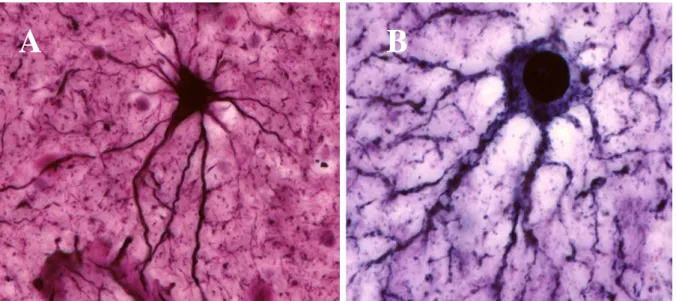
With routine staining and the light microscope, astrocytes can be viewed as pale-staining oval to round or lobate nuclei with little or no discernible cytoplasm. Only after metallic impregnation, other special stains and immunocytochemical examination can the process-bearing nature of these cells be fully appreciated (Montgomery, 1994).
Already in the 19th century, it was recognized that astrocytes are a morphologically heterogeneous population. They can be divided in two major classes termed protoplasmic and fibrous astrocytes.
Fibrous astrocytes (Fig.1A) have many glial filaments, tend to have regular contours and to
4 1994). They are located mainly in white matter, often arranged in rows, and their processes usually develop parallel to neurons, and extend multiple finger-like outgrowths in the perinodal space of adjacent neurons.
Protoplasmic astrocytes (Fig. 1B) have fewer glial filaments and are found mainly in gray matter
(Wolff, 1968; Bushong et al., 2002). They have irregular contours and extend sheet-like processes that extend more or less radially from the soma, and appear to fill most of the space between the other elements in the neuropil. These projection account for about 50% of the cell’s volume, but more than 80% of its surface (Chao et al., 2002).
Figura 1: A, fibrous, and B, protoplasmic astrocytes. Adapted from Garcia-Lopez, 2010 (Garcia-Lopez et al., 2010)
Astrocytes processes are more extensive and branched in mature astrocytes than in younger ones, and often envelope the synapses formed by neurons in the CNS, or surround the blood vessels, such constituting the blood-brain barrier (Peters and Webster, 1991).
Immunocytochemical techniques have improved our knowledge about the expression of antigens that are, within the CNS, restricted to astrocytes. In addition, some of these markers allow distinguishing within the same population between developing, mature ad reactive astrocytes. Some of the biochemical markers expressed in mature astroglia are glial fibrillary acidic
5 protein (GFAP), or Glutamine Synthetase (GS), the high affinity glutamatetransporters GLAST or GLT-1, the calcium binding protein S100β and tenascin-C (Ghandour et al., 1983). The specificity o f GFAP has been a major breakthrough in the study of astrocytes both in situ and in cell culture, being commonly used to label astrocytes in the CNS to study their distribution and morphology, despite the fact that it labels only 15% of the total volume of astrocyte (Bushong et al., 2002).
Table 1
Young astrocytes Mature astrocytes A) Electrophysiology
Varied Vm Vm=Ek
VDCs VDCs not apparent; linear I-V plot
Varied input resistence Very low input resistence Varied degree of cell-cell coupling Extensive cell-cell coupling
B) Markers
Aos Aos
- Glutathione transport
- D-serine racemase
EAA transporters EAA transporters
GFAP sometimes GFAP
Nestin -
Vimentin -
GS GS
- S-100β
C) Morphology
Extesive processes More extensive processes and arborizations
Ionotropic and metabotropic receptors Mainly metabotropic receptors Processes contact blood vessels and
partition the CNS, e.g, glial limitants, or parts thereof.
Also contact mature synapses
Studies investigating the spatial relationships occurring between astrocytes in three-dimensional (3D) arrays in situ concluded that astrocyte somata achieve a nonrandom, region-dependent degree of spacing between themselves throughout the brain. Furthermore, the processes of each astrocyte are extensively intermingled with those of neighboring astrocytes (Dreher et al., 1994; Bushong et al., 2002).
6 The contacts occurring between processes were proposed to impart structural integrity to the nervous tissue while simultaneously separating somata during development, allowing the 3D array of astrocytes to effectively fill the nervous tissue, a phenomenon termed “contact spacing” (Tout et al., 1993).
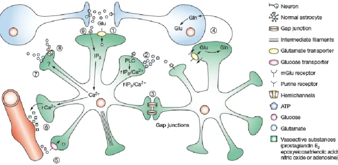
Astrocyte functions and physiology
Astrocytes’ functions range over a wide variety of domains (summarized in Fig. 2).
CNS compartimentalization
On the structural level, protoplasmic astrocytes shape the microarchitecture of the grey matter by dividing it in to relatively independent structural units, creating micro anatomical domains within the limits of the ramification of their processes. Individual astrocytes are further integrated into astroglial syncytia through gap junctions localized on the peripheral processes. The syncytia can also be segregated within defined anatomical structures, such as the individual barrels of the somatosensory cortex (Dreher et al., 1994; Bushong et al., 2002). Disruption of astrocyte domains has been associated with several mouse models for epilepsy (Seifert et al., 2010) and aberrant expression of the astrocyte gap junction protein connexin has also been reported in the autistic brain (Fatemi et al., 2008).
Developmental functions
The well-defined spacing of the astrocyte network, with its capacity for intercellular communication, has the potential to work as a boundary system for neurons, guiding their search for targets during development and ensuring that the activity of mature synapses is appropriately contained. Furthermore, in vitro studies have found that astrocytes exert powerful control over
7 the number of CNS synapses. Astrocytes are essential for postsynaptic function, and are required for synaptic stability and maintenance (Barker and Ullian, 2008). Recent in vivo studies increasingly implicate astrocytes as participants in activity-dependent structural and functional synaptic changes throughout the nervous system (Ullian et al., 2004; Eroglu and Barres, 2010).
Homeostasis during adulthood
Glutamate uptake
High affinity glutamate transporters, GLT1 and GLAST1 subtypes, are enriched in astrocytic processes, and play the predominant role for glutamate clearance in the adult CNS (Rothstein et al., 1996). Glutamate is co-transported with three Na+ (or two Na+ and one H+) in exchange for one K+ and one OH- (or one HCO3-;). The consequence of this stoichiometry is an increase in the Na+ concentration within the astrocytes, accompanied by an intracellular acidification and extracellular alkalization (Robinson and Dowd, 1997). Within the astrocyte, glutamate is converted to glutamine through an ATP-requiring reaction catalyzed by the astrocyte-specific enzyme glutamine synthetase. Glutamine is subsequently released to the extracellular space to fuel neurons and recycled into glutamate for glutamatergic neurotransmission (Loo et al., 1995).
Glutamate signaling
A variety of ionotropic glutamate receptors and metabotropic glutamate (mGlu) receptors have been characterized in glia in situ and in cultured astrocytes (Liao and Chen, 2003).
Astrocytic mGluR are activated during neuronal activity promoting the turnover of ionositol triphosphate (IP3), and triggering Ca2+ release from intracellular stores independently of extracellular Ca2+ (Burnashev et al., 1992).
8
Purinergic signaling
Primary glial cells express several members of the P2Y purinergic receptor family (Fam et al., 2000; Simard et al., 2003).
Purinergic signaling in astrocytes is involved in interactions with synapses and regulation of plasticity and excitability of synaptic networks (Pascual et al., 2005). Release of cytokines (Hide et al., 2000) and growth factors (Ciccarelli et al., 1999a). From astrocytes in response to purinergic signaling implicates astrocytes in developmental and injury-related processes (see “Astrocytes and NGF” paragraph).
Ion and water homeostasis
The extracellular space of the CNS represents the microenvironment of both neurons and glia. Communication of neurons and glial cells is almost exclusively limited to the diffusion of neuroactive substances and ions in extracellular space. The role of astrocytes in maintaining ionic , pH and and volume homeostasis of the extracellular space is crucial for their modulation of synaptic transmission efficacy.
Ion spatial buffering is based on the high permeability of glial membrane and astrocytic coupling, allowing the movement of K+ , Ca2+ and other ions (Coles and Orkand, 1983; Kettenmann et al., 1983).
Homeostasis of extracellular pH is regulated by coordinated expression of alkaline membrane transporters in neurons and of acid extruders in the glial membrane (Jendelova and Sykova, 1991).
In addition, centrally released neuropeptides such as vasopressin (AVP), atriopeptin (ANP), angiotensinogen (AGT) and angiotensin (Ang) II appear to regulate fluid and ionic environment
9 and cell volume in the CNS, possibly via intrinsic osmoregulation of glial cells (Simard and Nedergaard, 2004).
Ca
2+signaling
Astrocytic Ca2+ signaling is expressed as oscillations in cytosolic Ca2+ concentrations ([Ca2+]i) and as slowly propagating waves of [Ca2+]. Astrocytes display regular oscillations when acti-vated by various stimuli including hormones (Berridge, 1994; Smith, 1994). Astrocytes also ac-tively propagate Ca2+ waves. Mechanical stimulation of single astrocytes resulted in expanding waves of [Ca2+] increments, which engaged 20–100 neighboring astrocytes in culture (Smith, 1994). They can modulate the Ca2+ level, and thereby the firing pattern, of neurons in their sur-roundings. In turn, neurons can trigger astrocytic Ca2+ signaling by releasing glutamate.
Cerebrovascular regulation and Blood Brain Barrier control
Astrocytes send processes to synapses (Chao et al., 2002; Volterra et al., 2002) and blood vessels (Reichenbach, 1989), and thus are an integral component of the neurovascular unit.
Astrocytes has been recently shown to be essential participants in the control of cerebral blood flow (CBF) through the control of cerebral vessel diameter. As stated above, astrocytes respond to glutamate with an increase in intracellular Ca2+ through the activation of mGLURs (Burnashev et al., 1992). The application of mGLUR agonists triggers the release of diffusible factors that then act on vascular smooth muscle cells to cause dilation of arterioles (Zonta et al., 2003b). Among this factors, arachidonic acid metabolite lead to a localized increase in calcium at astrocyte end-feet, which results in dilation of nearby arterioles (Zonta et al., 2003a).
A second function of astrocytes in the regulation of cerebral vasculature is the participation to the formation of the the blood-brain barrier (BBB). The BBB is a metabolic diffusion barrier which
10 plays a role of “check point” to regulate influx of most compounds from blood to brain. The BBB consists of endothelial cells, pericytes with smooth muscle-like properties, and astroglial processes that ensheath more than 95% of the abluminal blood vessel surface (Zlokovic, 2008). Although the impact of astrocytes in the maintenance of the BBB is largely unidentified, it can be presumed that a constant astrocytic influence is needed to maintain the BBB. A physiological role could be met by the lactate shuttle hypothesis (Magistretti et al., 1994), control of ingress of compounds such as glucose and aminoacid or egress of waste metabolites (Pellerin and Magistretti, 2004), or control of the concentration of K+ and blood flow.
Figure 2. Summary of astrocyte functions. Adapted from Maragakis and Rothstein, 2006
Glial Fibrillary Acidic Protein (GFAP)
GFAP is a 8-9 nm intermediate filament (IF) protein preeminently expressed in astrocytes. First described in 1971 (Eng et al., 1971), it is a type III IF protein closely related to its non-epithelial family members, vimentin, desmin, and peripherin, which are all involved in the structure and function of the cell cytoskeleton.
Initially, GFAP was purified from large Multiple Sclerosis plaques consisting primarily of fibrous astrocytes and demyelinated axons (Eng et al., 1971). The cloned mouse gene and the
11 cloned human gene were subsequently presented, respectively in 1984 (Lewis et al., 1984) and 1989 (Reeves et al., 1989).
The human GFAP gene maps in chromosome 17q21 (Bongcam-Rudloff et al., 1991) and is composed of nine exons and eight introns, plus four alternative exons and two alternative introns, for about 10 kb of DNA, and yields a mature mRNA of about 3 kb (Kumanishi et al., 1992). The original mRNA transcript can give birth, by alternative splicing, to six different isoforms of the protein, the most abundant one being GFAPα (Reeves et al., 1989; Zelenika et al., 1995; Nielsen et al., 2002; Hol et al., 2003; Roelofs et al., 2005; Blechingberg et al., 2007).
Originally thought to be an astrocyte-specific IF, GFAP has now been detected even in non-glial and non-CNS cells, like chondrocytes (Kepes et al., 1984; Hainfellner et al., 2001), fibroblasts (Hainfellner et al., 2001), myoepithelial cells (Viale et al., 1991; Hainfellner et al., 2001), lymphocytes (Riol et al., 1997) and liver stellate cells (Carotti et al., 2008).
However, possible structural differences between central and peripheral GFAP molecules have been suggested (Feinstein et al., 1992).
GFAP gene expression is regulated by the GFAP promoter (Eng et al., 2000), which it is induced by multiple factors such as brain injury and disease fluctuates under the circadian light-dark cycle (Hajos, 2008). One well known element of the basal promoter is the so called TATA-box which binds the general transcription factor IID (TFIID) (Nakatani et al., 1990). However there are multiple sites of the promoter which are likely to regulate GFAP expression [reviewed in (Middeldorp and Hol, 2011)], and epigenetic mechanisms like phosphorylation and DNA methylation can have significant effects on gene transcription.
In the mouse CNS, GFAP is the major IF protein in the adult brain and is a characteristic protein of mature astrocytes. GFAP expression begins at the end of gestation, while there are not clear data about the age of onset of its production in humans, though it is thought to begin at gesta-tional week (gw) 9–12 (at 6 gw already in some studies) (Antanitus et al., 1976; Stagaard and Mollgard, 1989; Honig et al., 1996; Simonati et al., 1997; Messam et al., 2002; deAzevedo et al.,
12 2003; Middeldorp et al., 2010). Gene transcription increases between birth and day 15 and then decreases until day 55 (Riol et al., 1992). After reaching a plateau lasting into the second year of adult life, GFAP mRNA and protein levels increase again in hippocampus, striatum and cortex, usually corresponding to the increase of reactive astrocytes that during senescence is one of the most generalized markers for brain aging (Morgan et al., 1997; Morgan et al., 1999). In more general terms, the enlargement of astrocytes and the increased expression of GFAP is an indica-tion of reactive gliosis, a process which has shown to be highly related to brain damage and ag-ing and is related to decreased astrocyte proliferation (Eng and Ghirnikar, 1994).
GFAP is thought to help to have mainly mechanical roles, modulating astrocyte motility and shape and providing structural stability to astrocytic processes. Moreover, some data indicate that GFAP might play an important role in the control of neurological disease and response to injury by limiting the lesions (Maragakis and Rothstein, 2006).
Although GFAP function in brain physiology and pathology is still controversial, a large amount of evidence has been accumulating in the past years in favor of an active and relevant role for this structural intermediate filament protein in brain development (Middeldorp and Hol, 2011).
Vimentin
Vimentins are class-III intermediate filaments found in various non-epithelial cells, especially mesenchymal cells. It was found in astrocytes, fibroblasts, endothelial cells, macrophages, neutrophils and lymphocytes (Evans, 1998).
Vimentin biological function is not clear at the present time. We know that in the course of as-trocyte development, a transition in the expression of IF protein genes is observed. Early during development, radial glia and immature astrocytes express mainly vimentin. Towards the end of gestation, a switch occurs whereby vimentin is progressively replaced by GFAP in differentiated astroglial cells, although certain astrocytes also co-express GFAP and vimentin in the adult
ani-13 mal, mainly the Bergmann glia and the astrocytes in the corpus callosum, and the hippocampus, although in a variable manner in the latter one (Stichel et al., 1991; Missler et al., 1994; Kalman et al., 1998).
Data obtained from vimentin knockout mice (-/-) demonstrated that those animals developed and reproduced without presenting an obvious new phenotype, thus heavily calling into question the biological function of vimentin. Several data, however, argue in favor of a relevant function for vimentin. In vimentin (-/-) lineages, cells that normally co-express vimentin and GFAP do not present GFAP filament. Transfection of cultured vimentin -/- astrocytes with a vimentin cDNA restores the vimentin-GFAP filament network, suggesting that in these cells vimentin might be required for co-assembly with GFAP filaments (Goldman et al., 1996).
NGF and its receptors
The neurotrophins are a four-member peptide growth factor family that includes nerve growth factor (NGF) (Levi-Montalcini, 1952), brain-derived neurotrophic factor (BDNF) (Leibrock et al., 1989), neurotrophin 3 (NT-3) (Maisonpierre et al., 1990), and neurotrophin 4/5 (NT-4/5) (Ip et al., 1992), which exhibit well-described actions in the nervous system. Classically defined as target-derived survival factors for developing neuronal populations, the roles of the neurotrophins are now known to include growth cone guidance, synaptic modulation, injury protection, and influence on memory and behavior (Levi-Montalcini, 1987; Barde, 1989).
All neurotrophins are translated from single coding exons as larger precursors (proneurotrophins) of ~30–34 kDa that rapidly associate as noncovalent homodimers. Proneurotrophins can be cleaved in the ER and Golgi to produce C-terminal mature neurotrophins (~13 kDa) (Shooter, 2001).
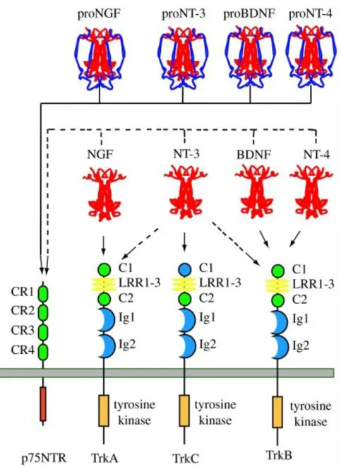
14 At the molecular level, proneurotrophins and mature neurotrophins interact with two distinct type of receptors: p75NTR, one of the tumor necrosis factor superfamily of receptors (Chao et al., 1986; Johnson et al., 1986), and tropomyosine related kinase receptors, Trks (Barbacid, 1993) (Fig. 3).
Figura 3 Neurotrophin–receptor interactions. This illustrates the major interactions of each of the four
mammalian neurotrophins. Each proneurotrophin binds p75NTR, but not the Trk receptors. Following maturation through proteolysis of the proneurotrophins, each mature neurotrophin is able to bind and activate p75NTR, but exhibits more specific interactions with the three Trk receptors. NGF binds specifically TrkA; BDNF and NT4 recognize TrkB; NT3 activates TrkC. In some cellular contexts, NT3 is also able to activate TrkA and TrkB with less efficiency. For simplicity, only the major tyrosine kinase-containing isoforms of the Trk receptors are depicted. Differential splicing generates isoforms of TrkB and TrkC that have truncated cytoplasmic domains lacking a tyrosine kinase motif. Splicing also generates an isoform of TrkC with a small insert in the kinase domain that affects substrate specificity. Splicing of exons that generate the extracellular domain of each Trk receptor results in the expression of receptors with small peptide inserts between the second immunoglobin and transmembrane domains that affect ligand-binding specificity. Ligand-binding specificity is also affected by the presence of p75NTR [adapted from (Reichardt, 2006)].
15 Indeed most neurotrophin actions on neuronal differentiation and survival can be ascribed to this coreceptor system. Trk receptor tyrosine kinase is the specific signaling entity for each neurotrophin while p75NTR serves to restrict and augment ligand recognition specificity (Hempstead et al., 1991).
Nerve growth factor (NGF) is the member of the neurotrophin family that was first to be discovered and purified. It supports neuronal survival during development and influences neuronal function throughout adulthood.
In particular, it influences development of sensory and autonomic neurons in the peripheral nervous system (Levi-Montalcini, 1987), and of basal forebrain cholinergic neurons in the CNS (Mobley et al., 1986). During adulthood plays an essential role in pain transmission (Pezet and McMahon, 2006) and in the maintenance of the cholinergic phenotype of the CNS (Mufson et al., 2008).
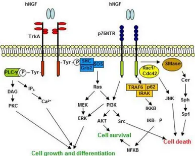
It sends its survival signals through activation of TrkA receptor (Kaplan et al., 1991; Klein et al., 1991) and can induce death by binding to p75(NTR). Neurotrophin engagement of Trk receptors leads to activation of Ras, phosphatidylinositol 3-kinase, phospholipase C-γ1 and signaling pathways controlled through these proteins, including the mitogen-activated protein kinases (Fig. 4).
Figura 24 Summary of the NGF signaling pathways. Adapted from Nicol and Vasco, 2007(Nicol and Vasko, 2007) .
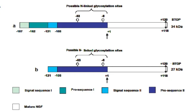
16 NGF, like the other neurotrophins, is translated as a pre-pro-protein (Shooter, 2001). Two splicing variants lead to the formation of a long and a short form of precursor protein (Fig. 5A), both glycosylated in vivo, yielding different molecular weight forms identified (Reinshagen et al., 2000; Lee et al., 2001; Fahnestock et al., 2004; Fasulo et al., 2005; Pagadala et al., 2006; Mouri et al., 2007; Nomoto et al., 2007).
The signal peptide mediating secretion is cleaved upon translocation into the endoplasmic reticulum. In the trans-Golgi network, the pro-peptide is further processed by furin at a conserved dibasic amino acid site, resulting in the release of the 26 kDa mature neurotrophin (Seidah et al., 1996; Mowla et al., 1999; Fahnestock et al., 2004; Mouri et al., 2007; Nomoto et al., 2007).
Figure 5 Schematic representation of the major translation products arising from alternative splicing of the NGF transcript: the long pre-proNGF form of 34 kDa (a) and the short pre-proNGF form of 27 kDa (b).
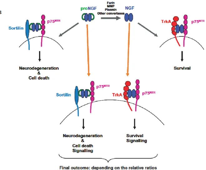
Recently it was found that in cultured neurons, proNGF exhibits a higher affinity for p75NTR than for TrkA, determined by the coexpression of the coreceptor sortilin (Nykjaer et al., 2004), and can induce p75NTR-dependent apoptosis (Lee et al., 2001; Harrington et al., 2004; Pedraza et al.,
17 2005). ProNGF can also bind to TrkA and induce neuronal survival, although to a lesser extent than mature NGF. ProNGF is the predominant form of NGF in normal brain, but its synthesis is up-regulated in various pathological conditions, including AD, and can elicit apoptosis of BF neurons even when Trk receptors are activated (Volosin et al., 2006) (see “NGF and Alzheimer: The emerging complexity of the proNGF/NGF system”).
Thus, the balance between cell death and cell survival might be determined by the ratio of proNGF and mature NGF secreted by cells.
Figure 6 Possible scheme for the signaling of NGF and proNGF, involving Sortilin receptor, besides the “traditional” re-ceptors p75NTR and TrkA.
18
Astrocytes and NGF
Several cell culture studies showed that NGF can be synthesized and secreted from embryonic or neonatal astrocytes (Furukawa et al., 1986; Tarris et al., 1986; Assouline et al., 1987; Furukawa et al., 1987; Yamakuni et al., 1987; Rudge et al., 1992; Moretto et al., 1994; Condorelli et al., 1995).
Rodent cultured astrocytes express both p75 (Marchetti et al., 1987; Hutton et al., 1992) and TrkA NGF receptors (Hutton et al., 1992; Hutton and Perez-Polo, 1995). The expression of both receptors can be increased by NGF itself (Kumar et al., 1993; Hutton and Perez-Polo, 1995). An autocrine regulation mechanism has been recently found according to which Nerve growth factor attenuates proliferation of astrocytes via the p75 neurotrophin receptor (Cragnolini et al., 2009).
No regional differences were detected, in terms of amount of NGF in astrocytes. Among cultures of glial cells of hippocampal, cortical, striatal, hypothalamic or mesencephalic origin, levels of NGF released by the cells were very similar (Houlgatte et al., 1989; Gonzalez et al., 1990). They are closely correlated to the growth rate as shown by the fact that exponentially growing cells produced relatively more factor than did confluent cell .
In these cultures, NGF synthesis/secretion can be differentially regulated by several factors, and this regulation is different according to the state of the cells. NGF production can be increased by glutamate, cathecolamines and dopamine agonists in quiescent astrocytes while the same cate-cholamines and noradrenaline decrease NGF content in growing cells (Furukawa et al., 1987; Lu et al., 1991; Pechan et al., 1993; Inoue et al., 1997; Culmsee et al., 1999; Ohta et al., 2000; Ohta et al., 2003). Cholinergic agonists, such as metacholine and carbamylcholine, nicotine and nicotine agonists slightly increased the NGF content in quiescent cells, but showed no effects on growing astrocytes (Furukawa et al., 1987; Martinez-Rodriguez et al., 2003)
19 All cathecolamines act on NGF synthesis at transcriptional level, by increasing mRNA content in astrocytes (Furukawa et al., 1989). Cyclic AMP (Schwartz and Mishler, 1990). Stimulation of adenosine or serotonin receptors induces the release of NGF (Ciccarelli et al., 1999b; Rathbone et al., 1999; Krzan et al., 2001; Wittendorp et al., 2004). Changes in aminoacid and ionic concen-tration, and particularly an increase in Tryptophan or [K+] content stimulate respectively NGF synthesis (Dong-Ruyl et al., 1998) and secretion from cultured astrocytes (Abiru et al., 1998). Inflammatory cytokines like Interleukin1 α and β (Gadient et al., 1990; Spranger et al., 1990; Carman-Krzan et al., 1991; Yoshida and Gage, 1992; Juric and Carman-Krzan, 2001; Miklic et al., 2004; Jauneau et al., 2006), Interleukin-6 (Marz et al., 1999; Ales et al., 2008) , Tumor ne-crosis factor α (Gadient et al., 1990; Yoshida and Gage, 1992; Miklic et al., 2004; Kuno et al., 2006), transforming growth factor β1 (Lindholm et al., 1990; Yoshida and Gage, 1992; Yu and Fahnestock, 2002), Interleukin-4 (Boutros et al., 1997; Brodie et al., 1998), Interferon- (Boutros et al., 1997), Interleukin-8 (Kossmann et al., 1997), Interleukin-10 (Brodie, 1996), Interleukin 4 and 5 (Awatsuji et al., 1993) and inflammatory/oxidative stress related factors such as Platelet activating factor (PFA) (Brodie, 1995), thrombin (Neveu et al., 1993) histamine and its related receptor H1 (Lipnik-Stangelj and Carman-Krzan, 2004b, a; Miklic et al., 2004; Ales et al., 2008), anaphilatoxins (Jauneau et al., 2006), prostaglandins (Toyomoto et al., 2004) and nitric oxide (Xiong et al., 1999) were showed to have an enhancing effect on the production of NGF in astro-cytes, providing a molecular link between inflammation and astrocytic synthesis of NGF (Lipnik-Stanglj and Carman-Krzan, 2000; Guo et al., 2001).
Different growth factors such as acidic and basic fibroblast growth factors (aFGF and bFGF) and ciliary neurotrophic factor (CNTF) significantly increase NGF secretion by astrocytes (Fukumoto et al., 1991; Ono et al., 1991; Yoshida and Gage, 1991; Semkova and Krieglstein, 1999; Cassina et al., 2005),while epidermal growth factor fails to increase NGF synthesis in cul-tured rat astrocytes (Vige et al., 1991). Synthesis of NGF can be also enhanced by glycerophos-pholipids, sphingolipids, and their related compounds (Galve-Roperh et al., 1997; Furukawa et
20 al., 2007), retinoic acid (Ahlemeyer et al., 2000), 1,25-dihydroxyvitamin D3 (Neveu et al., 1994a; Neveu et al., 1994b) and glucocorticoids hormones (Lindholm et al., 1992).
In cultures of immature astrocytes, NGF induces proliferation of GFAP-positive cells (Yokoyama et al., 1993), possibly by stimulating mitosis as well as enhancing survival or diffe-rentiation. Moreover, increases expression of the neural adhesion molecule L1, leading to a glia-mediated L1-specific increase in neurite outgrowth of dorsal root ganglion neurons on the astro-cyte substrate (Saad et al., 1991).
In pathological conditions, increased NGF synthesis/secretion by astrocytes is found in astrocy-tomas (Saito et al., 2006), gliomas (Fukumoto et al., 1994).
All the data described so far were obtained from astrocytes cultured in vitro. In vivo, conflicting results have been reported. Bacia et al al. did not found localization of NGF in hippocampal as-trocytes (Bacia et al., 1992). Asas-trocytes in brain tissue from human subject do not express, in normal conditions, NGF or TrkA (Calatozzolo et al., 2007). In adult rats, NGF receptors are ex-pressed in small, poorly ramified cells (Soltys et al., 2003) in the hippocampal dendritic fields (McCarthy et al., 2002). On the contrary, spinal cord from matured or aged rats express TrkA (Oderfeld-Nowak et al., 2003).
However, increasing evidence is recently accumulating that, although in intact brain the main source of NGF are target neuronal cells, in some pathological conditions an increase in extra-neuronal pool of NGF takes place. Several lines of evidence indicate that in noxious conditions astrocytes behave just as the astroglia in vitro and thus are an important site of NGF production in damaged brain. The rise of NGF levels in brain in various models of reactive gliosis has indi-rectly shown that brain astrocytes can synthesize the nerve growth factor. Moreover, in several cases NGF immunoreactivity has been directly revealed in activated astrocytes.
In neurotoxic damage a large increase in total NGF content in the hippocampus was observed (Bakhit et al., 1991; Chen et al., 2006; Volosin et al., 2008) but, pyramidal and granular layer neurons, which produce NGF under normal conditions, were lost and a large increase in GFAP
21 immunoreactivity was seen instead. In general, NGF synthesis in astrocytes is upregulated in a number of stress or damage condition, like toxic or mechanic damage (Junier et al., 1994; Strauss et al., 1994; Rudge et al., 1995; Kossmann et al., 1996; Rossner et al., 1997; Goss et al., 1998; Wu et al., 1998; Koczyk and Oderfeld-Nowak, 2000; Yu and Fahnestock, 2002), oxidative stress (Pechan et al., 1992; Naveilhan et al., 1994), apoptosis (Pehar et al., 2004), ischemia (Lee et al., 1996; Himeda et al., 2007; Tonchev et al., 2008; Tonchev, 2011), multiple sclerosis and experimental autoimmune encephalitis (Micera et al., 1998; Biernacki et al., 2005), amyotrophic lateral sclerosis (Barbeito et al., 2004).
Furthermore, hippocampal astrocytes incubated with Aβ up-regulate NGF expression (Schulte-Herbruggen et al., 2007) and release it in the culture medium, causing tau hyperphosphorylation and death in co-incubated p75 expressing neurons (Saez et al., 2006), thus indicating a possible involvement of astrocytes in Alzheimer’s disease.
Reactive astrocytes can induce also neuronal death, being a possible source of pro-NGF (Domeniconi et al., 2007). In stress conditions, like in response to peroxynitrite, an oxidant and producer of free radicals, (Vargas et al., 2004) or to kainic acid (Volosin et al., 2006) astrocytes up-regulate the levels of pro-NGF production. Cultured motor neurons viability is sensitive to conditioned media from cultured astrocytes treated with peroxynitrite, an effect that could be re-versed using a specific antibody against the pro-domain of pro-NGF.
On the other side, many astrocytes not only synthesize NGF and pro-NGF, but also the receptors TrkA and p75. The production of NGF receptors in several human diseases (Aguado et al., 1998). NGF receptors were found to be up-regulated in rats following ischemic damage (Lee et al., 1998; Oderfeld-Nowak et al., 2003; Soltys et al., 2003), multiple sclerosis lesions (Oderfeld-Nowak et al., 2001; Valdo et al., 2002; Oderfeld-(Oderfeld-Nowak et al., 2003), neurotoxic damage (Koczyk and Oderfeld-Nowak, 2000). In p75 expressing cultured astrocytes, NGF fails to in-duce an autoapoptotic reaction, but it attenuates the cellular proliferation (Cragnolini et al., 2009).
22 The presence of p75NTR and TrkA receptors in reactive astrocytes from different human neuro-degenerative diseases and experimentally induced models in rats, and in neoplastic astrocytes suggests that NGF may participate in the astroglial response to different types of injury and neoplastic proliferation (Hutton et al., 1992; Brown et al., 2008). Since astroglial cells are capa-ble of producing NGF, it is plausicapa-ble that this neurotrophin may function as an autocrine or para-crine factor in TrkA-expressing reactive and neoplastic glial cells.
Astrocytes and neurodegenerative diseases
Astrocytes, in accordance with their homoeostatic function, are deeply involved in neural diseases. Acute and chronic brain insults trigger a specific glial reaction, generally known as reactive astrogliosis, represented by a complex morpho-functional remodelling of astrocytes (Sofroniew, 2005, 2009). Astroglia forms the first line of brain defense by controlling the volume and composition of extracellular space. At the very same time astroglial cells can contribute to neuronal damage, when, for example, severe insults compromise astrocyte metabolism (Giaume et al., 2007). Recently, astrocyte dystrophy has been connected also to different kinds of neurodegenerative diseases, including dementia (Maragakis and Rothstein, 2006). In particular, both astrogliosis and astroglial dystrophy are manifest in different types of dementia. Furthermore, both these processes may develop in parallel depending on the disease form and/or stage. The frontotemporal dementia, is, for example, associated with a very early and profound apoptotic death and dystrophy of astrocytes (Martin et al., 2001). The degree of astroglial loss directly correlates with the severity of dementia (Broe et al., 2004). Conversely, other studies, using postmortem tissues from frontotemporal dementia cases, found prominent astrogliosis in the frontal and temporal cortices at the very early stages of the disease, with astrocyte densities increasing by 4–5 times of the control (Kersaitis et al., 2004).
23 Astrogliosis also assumes the leading pathological role in thalamic dementia and HIV-1-Associated Dementia (Vanzani et al., 2006).
Alzheimer’s Disease
AD is a chronic neurodegenerative disease clinically characterized by cognitive loss in two or more domains, including memory, language, calculations, orientation and judgment. In more than 90% of cases, AD develops after the age of 65 years, and doubles its prevalence with every successive decade of life (Aranda-Abreu et al., 2011). The disease eventually leads to the death of affected individuals an average of nine years after diagnosis. These clinical features are the re-sult of neuronal death and dysfunction in the cerebral cortex, entorhinal area, hippocampus and basal forebrain, eventually resulting in severe dementia. Pathologically, the three hallmark find-ings of the disorder are cholinergic deficit (Bartus et al., 1982; Whitehouse, 1992), neurofibril-lary tangles (Grundke-Iqbal et al., 1986) and extracellular senile plaques composed by the β-amyloid protein (Glenner, 1988; Selkoe et al., 1988).
Since in CNS BFCNs provide major projections to the cerebral cortex and the hippocampus and cortical cholinergic mechanisms, they are directly involved in superior cognitive functions such as learning and memory (Hefti et al., 1984; Hefti and Weiner, 1986; Fischer et al., 1987) and for these reason the disease was first attributed to a decrease in cholinergic innervation of the brain (Bartus et al., 1982). More recently, with the discovery of the negative effects of β-amyloid on learning and memory capabilities, an “amyloid cascade hypothesis” has been formulated (Selkoe, 1991; Hardy and Higgins, 1992).
Several chromosomes have been shown to be implicated in the pathology of AD, including chromosomes 1, 14, and 21, associated with the familial early-onset forms of the disease, and chromosomes 12 and 19, linked to late-onset forms. However, most cases cannot be explained genetically, and thus several hypotheses have been raised over the years in an attempt to explain the etiology of this complex disease (Aranda-Abreu et al., 2011).
24 β-amyloid peptide, the principal constituent of neuritic plaques, may have a major role in the neuropathology of AD. It is processed from the Amyloid Precursor Protein (APP) the sequential actions of the enzymes β-secretase (BACE1) (Vassar et al., 2009) and γ-secretase (De Strooper and Woodgett, 2003) in two forms, one 40 amino acids long (Aβ40) and the other 42 amino acids
long (Aβ42), which differ in their terminal carbon structure (Haass and Selkoe, 2007). Of these
two forms, Aβ42 is the most prone to aggregation, promoting the formation of oligomers and
ag-gregation into fibrillar amyloid plaques. Soluble Aβ-oligomers possess profound pro-apoptotic and synaptotoxic effects, and induce learning deficits and neuronal loss to a substantially higher degree than larger assemblies of aggregated fibrillar Aβ. Moreover, high concentrations of Aβ oligomers interfere with long-term potentiation (LTP) (Haass and Selkoe, 2007).
Beside fibrillar Aβ plaques, neurofibrillary tangles (NFTs) represent the second major neuropa-thological hallmark of AD. They consist of hyperphosphorylated forms of tau protein (p-tau) that is assembled into paired helical filaments (PHFs). Tau can be phosphorylated at multiple sites, but because of their high diagnostic relevance for AD, p-tau181and p-tau231 isoforms are gener-ally used in p-tau targeting AD biomarker-studies (Iqbal et al., 2009).
Astrocytes in Alzheimer Disease
The pathological potential of glial cells in progression of dementia (as well as in other types of brain pathology) was originally suggested by Alois Alzheimer himself in 1910 (Alzheimer, 1910), and is now becoming object of several investigations. Many studies of the AD-related pathological potential of astroglia have focused on the investigations of the effects of β-amyloid on astrocytes.
25
Human studies
Astrocytes play significant roles during AD. Senile plaques found in the brain of patients with AD are surrounded by clusters with active astrocytes, which might be activated by human amyloid-β(Aβ), indicating a correlation between this protein and subsequent alterations in astrocyte function. Studies have shown that both aggregated Aβ and the intact cores of Aβ plaques isolated from human AD brain tissue can stimulate activation of astrocytes (DeWitt et al., 1998).
While microglia infiltrate Aβ plaques in AD, astrocytes aggregate around the periphery, walling off the plaque. From this location, astrocytes project thick processes that envelop the Aβ plaque and thinner branches that infiltrate deep into the plaque interior (Schwab and McGeer, 2008). Astrocytes also accumulate neuron derived amyloid material resulting from local neurodegeneration. Once substantial accumulation of this debris occurs, the astrocytes themselves might undergo cell death, resulting in the formation of GFAP+ amyloid plaques. The reason that the astrocytic cytoplasm is accumulated with Aβ42 and neuronal debris lies in the fact that Aβ42 binds with exceptionally high affinity to α7 nAChRs on neuronal surfaces. The cortical molecular layer is densely packed with fine α7 nAChR-rich dendrite branches emanating from underlying neurons. The Aβ42-α7 nAChR complex is present on degenerating dendrites and synaptic surfaces, suggesting that astrocyte-mediated clearing of this debris via phagocytosis and endocytosis and the targeting of this material to the lysosomal system explains both the source of astrocytic Aβ42 as well as its co-localization in astrocytes with other neuron-specific proteins, such as α7nAChR and ChAT (Nagele et al., 2003).
The origin of astrocytic Aβ42 and the mechanism by which it accumulates selectively in activated astrocytes and not in their more quiescent counterparts is unknown. Aβ42 found in astrocytes could be either internally produced or extracellulary taken up. Since expression of the Aβ42 precursor protein (APP) in astrocytes is known to be extremely low, internal production is
26 unlikely to be a major source of accumulated Aβ42. Thus, internalization of extracellular Aβ42 is more apparent and could occur via receptor-mediated endocytosis and/or phagocytosis (Aldskogius et al., 1999).
Animal models
Alzheimer’s disease, as every other dementia, is a sole prerogative of humans. No animal suffers from AD. Hence, efforts were invested in producing relevant animal models of AD.
The simplest transgenic models are based on the expression of mutated forms of proteins found in the familiar form of the disease. Thus, the transgenic mice harbors single-mutated β-amyloid related proteins (amyloid precursor protein (APP) or presenilins, (PS). Some of the most notorious models were the one developed by Games et al. in 1995, called PDAPP (Games et al., 1995), the Tg2576 mouse developed by Hsiao et al. in 1996 (Hsiao et al., 1996), and the APP23 mouse, harboring a double mutated APP protein (Sturchler-Pierrat et al., 1997).
However, mutations in PS proteins account for the majority of cases of familiar forms of AD. Co-expression of PS1dE9 with APP resulted in a viable model that showed accelerated Aβ deposition and memory deficits without tangle formation (Savonenko et al., 2005).
In parallel, pathological tau animals were developed, the first model being created in 1995 (Gotz et al., 1995). These transgenic animals showed somatodendritic localization of hyperphosphorilated tau, but have not developed neurofibrillary tangle pathology.
After identification of the pathogenic mutations of tau in FTDP-17, different transgenic models with clear neuronal NFT were produced (Gotz et al., 1995; Cassel et al., 2008).
One of the most advanced AD animal models is represented by the triple-transgenic mice developed in 2003 by Salvatore Oddo and Frank LaFerla (Oddo et al., 2003). These animals (the 3xTg-AD mouse model) harbor the mutant genes for amyloid precursor protein (APPSwe), for presenilin 1 PS1M146V and for tauP301L, which is a mutation related to frontotemporal
27 dementia, another neurodegenerative disease. As well as progressively developing plaques and tangles, the 3xTg-AD animals also show clear functional and cognitive impairments including LTP, spatial memory and long-term memory deficits.
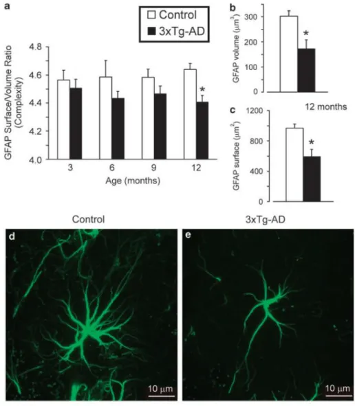
Olabarria et al, examined the astroglial morphology of 3xTg-AD mice (Olabarria et al., 2010), and found a significant reduction in surface area, volume and complexity in mutant mice compared to WT. As can be seen in Fig. 7, this difference was progressive with age and became particularly significant in 12 month old mice. On a cellular level, this decrease in complexity was reflected by reduced number of main processes and their ramification. accumulation.
Figura 7 Bar graphs showing the complexity (a) of glial cytoskeleton by measuring the GFAP area coverage versus vo-lume ratio of within the molecular layer of the dorsal dentate gyrus of both control and 3xTg-AD mice at different ages. (b and c) Bar graphs showing the coverage area (b) and volume (c) of GFAP at 12 months of age in control and 3xTg-AD mice; *Po0.05 compared with age matching controls. (d and e) Fluorescence photomicrographs showing GFAP-positive cells in control (d) and 3xTg-AD mice (e) at 12 months of age. There is an evident decrease in arborisation, surface area and volume in 3xTg-AD mice when compared with matching controls. Adapted from Rodriguez et al., 2009.
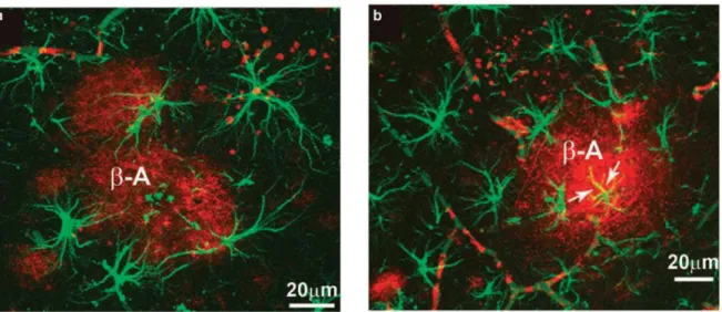
28 The same decrease in astrocytes complexity was found in the postmortem brains of dementia patients, whose astrocytes showed significant decrease in fractal dimension, thus indicating the decreased complexity. Furthermore, the volume of the brain parenchyma occupied by the processes of single astrocyte was smaller in demented than in healthy brains (Senitz et al., 1995). Confocal images revealed that the specific population of astrocytes surrounding amyloid plaques display the typical reactive characteristics (Fig. 8), showing thick processes and enlarged cell bodies. Some of these astrocytes closely associated with amyloid plaques showed β-amyloid accumulation.
Figura 8 Confocal image showing GFAP-positive (green) reactive astrocytes surrounding b-amyloid plaques (red; a). (b) Reactive astrocytes (green) and an astrocyte showing cytoplasmic b-amyloid accumulation (indicated by ar-rows; colocalisation, yellow) near a neuritic plaque (red). Adapted from Rodriguez et al., 2009.
NGF and Alzheimer’s disease: Rationale for creating an AD mouse model
based on NGF deprivation
NGF and Alzheimer: the emerging complexity of the proNGF/NGF system.
In the central nervous system (CNS), mature basal forebrain cholinergic neurons depends on the availability of NGF for the maintenance of their phenotype and for survival after lesions or CNS insults (Hefti et al., 1984; Hefti and Weiner, 1986; Fischer et al., 1987).
29 BFCNs have been identified as the most significant NGF-sensitive target population: they express both TrkA and p75NTR NGF (Sobreviela et al., 1994) and are able to retrogradely transport NGF from their cortical projections up to their cell (Seiler and Schwab, 1984). They respond to administration of exogenous NGF with an increase of cholinergic phenotypic markers (Li et al., 1995; Pongrac and Rylett, 1998), and an extensive evidence now establishes that NGF prevents neuronal death or atrophy in BFCNs from the adult brain.
In AD brains, the level of NGF mRNA are unchanged (Goedert et al., 1986; Jette et al., 1994), whereas increased levels of NGF protein can be detected in the cortex and hippocampus, associated to a decreased amount of NGF in the basal forebrain (Crutcher et al., 1993; Mufson et al., 1995; Scott et al., 1995; Fahnestock et al., 1996). Furthermore, a reduced expression of the TrkA receptor has been demonstrated in BFCNs and neocortex from AD brains (Mufson et al., 1996; Boissiere et al., 1997), and loss of TrkA expressing BFCNs has been described in mild cognitive impairment (MCI) and in early AD neuropathological stages (Counts et al., 2004; Ginsberg et al., 2006).
Remarkably, the AD-linked decline in TrkA cortical contents does not occur for the p75NTR receptor (Ginsberg et al., 2006), determining disequilibrium in the TrkA/p75NTR ratio that might have functional consequences (Cattaneo et al., 2008). Therefore, the putative “off Trk” cycle of deficient NGF signaling may contribute to the selective degeneration of cholinergic neurons observed in AD.
There are interesting pieces of evidence possibly linking NGF to cholinergic dysfunction and to AD.
NGF is axonally retrotransoported to the somata of the BFCNs, and cytoskeletal transport dysfunctions are a common hallmark of AD (Mufson et al., 1995), due to hyperphosphorylation of tau and the ensuing microtubule destabilization (Cattaneo et al., 2008). Furthermore, amyloid-β protein precursor has been shown to have a role in axonal transport (Gunawardena and Goldstein, 2001). Thus, a deregulation of intracellular transport mechanisms, by different causes
30 linked to AβPP, tau, and NGF signaling and/or processing abnormalities, might be one crucial aspect for initiating a negative neurodegeneration loop in AD, leading to a further reduced neurotrophic support to BFCNs (Mufson et al., 1995).
Beside cholinergic dysfunction and neuronal transport defects, other mechanisms could provide a link between an imbalance of NGF signaling and AD.
ProNGF is the predominant form of NGF in the brain, and its content is increased in AD and MCI (Fahnestock et al., 2001; Peng et al., 2004). It can trigger cell death, as shown in cellular models using the purified protein (Lee et al., 2001) or AD brain extracts (Pedraza et al., 2005; Podlesniy et al., 2006). In addition, evidence was provided linking NGF deprivation to the activation of Aβ aberrant processing of APP and tau in vitro (Tan et al., 1999; Matrone et al., 2008a; Matrone et al., 2008b) and in vivo, from the studies in the anti-NGF AD11 mouse model (see below).
The AD11 mouse model
The AD11 model was developed in the group of Antonino Cattaneo at the International School for
Advanced Studies, Trieste, Italy (Ruberti et al., 2000). This model expresses a recombinant monoclonal antibody called αD11 which neutralizes mature NGF (Cattaneo et al., 1988), producing a sequestration of the protein that prevents it from exerting its biological functions. This unique mouse model permits study of NGF deficiency and its connection to neurodegenerative disorders. The phenotype of the mouse model resembles that of human Alzheimer’s disease.
31
The αD11 antibody
αD11 is a monoclonal antibody mAb obtained after long term immunization of rats with mouse NGF (Cattaneo et al., 1988) and it neutralizes the biological action of NGF, in vitro (Molnar et al., 1998) and in vivo (Berardi et al., 1994; Molnar et al., 1998).
The epitope of mAb αD11 on NGF includes the loop region from residues 41–49, which participates in the interaction surface between NGF and its high-affinity receptor TrkA and distinguishes NGF from other members of the neurotrophin family. A comprehensive functional and structural study of MAb αD11 showed that it not only prevents NGF from binding to TrkA, but also to p75NTR receptors (Covaceuszach et al., 2008).
MAb αD11 is characterized by specificity for NGF neutralization. Indeed, the crossreactivity of the antibody with other neurotrophins has been analyzed with a bioassay based on embryonic chick dorsal root ganglions. MAb αD11 blocks the activity of NGF, but it does not affect BDNF, NT3 and NT4/5 functions (Molnar et al., 1998; Capsoni et al., 2000). The implantation of hybridoma cells secretin MAb αD11 in the third ventricle transiently decreases the number of cholinergic neurons in the basal forebrain, thus demonstrating MAb AD11 activity in vivo (Molnar et al., 1998).
AD11 mice express a recombinant version of MAb αD11. It was obtained by reassembling variable regions of the light and heavy chains of mAb aD11 with the human K and g1 constant regions, respectively, to facilitate the detection of transgenic antibodies against the background of mouse Igs. The chains of the antibody are each placed under the transcriptional control of the early region promoter of the human CMV in separate plasmids (Ruberti et al., 1993).
Time dependent expression of anti-NGF antibody in AD11
As expected for the ubiquitous CMV promoter (Baskar et al., 1996a; Baskar et al., 1996b; Ruberti et al., 2000), the VH and VK mRNAs are expressed in different adult organs, including
32 brain, heart, kidney, testis, liver. For both chains the mRNA levels in adult tissues were higher than those in neonatal tissues (Ruberti et al., 2000).
Both antibody chains can be detected in non-neuronal tissues and in many areas of the brain, such as cortex, hippocampus, thalamus, olfactory bulb, spinal cord, retina, and cerebellum. The association of the heavy and light chains requires that the two chains be coexpressed within the same cell. Double-labeling immunohistochemistry demonstrated the coexpression of the two chains in a high percentage of neuronal and non-neuronal cells. The number of cells coexpressing both antibody chains is much greater in the adult than in the neonatal tissues (Ruberti et al., 2000).
Interesting, while mice in which NGF has been knocked out by targeted mutation die soon after birth, the AD11 mice develop normally as anti-NGF mAb levels are low at birth and rise in adulthood. Anti-NGF antibody levels are undetectable between E15 and P45, but rise at high levels in adult mice (P90) (Capsoni et al., 2000; Ruberti et al., 2000). Free NGF in the brain is more than 50% lower in AD11 mice than in control mice (Ruberti et al., 2000).
Neurodegenerative phenotype in the AD11 model
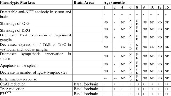
AD11 mice show a progressive and complex phenotype summarized in Table 2.
Cholinergic deficit
The primary target of NGF in the CNS are BCNFs. These neurons express both TrkA and p75NTR receptors (Sobreviela et al., 1994). the analysis in Ad11 mice was performed using the evaluation of the number of neurons expressing the enzyme choline acetyltransferase (CHAT). As expected from the time course of the transgenic antibody expression, the number of cholinergic neurons is unchanged between 0 and P45, while an initial 10-20% decrease can be found at 2 months of age (Ruberti et al., 2000; Capsoni et al., 2002c). The cholinergic deficit
33 increases with age, reaching a 40-50% of decrease in choline acetyltransferase - positive neurons at 6 months of age, and being stable thereafter (Capsoni et al., 2002b).
Hyperphopshorylated tau
The temporal progression of tau hyperphosphorylation in AD11 mice has been studied using antibodies directed against differente epitopes of phosphorylated tau.
The antibody that was most useful in characterizing the progression of fibrillar neurodegeneration was the antibody directed against Ser202/205, clone At8.
This antibody showed that at 2 months of age, similarly to what found in AD human brains, intracellular phosphorylated tau is localize mainly in the enthorinal cortex (Capsoni et al., 2002a). With aging, phospho-tau positive neurons are found in the other cortical areas and in the hippocampus (Capsoni et al., 2002c, a). In AD brains , phosphorylated tau accumulates in soluble and insoluble forms. By western blot analysis and the use of MAb AT8 and PHF1 antibody both states of phosphotau were found in brain extracts of aged AD11 mice (Capsoni et al., 2000).
The use of MAb AT8 combined with immunoelectron microscopy on insoluble fractions of brain extracts showed that phosphotau accumulates in PHF like structures (Capsoni et al., 2002a).
β amyloid
Amyloidogenic forms of APP, containing Aβ-region epitopes, are produced in the brain of aged AD11 mice. Large numbers of amyloid plaques are detectable in the cortex, thalamus, hippocampus and other regions. Diffuse nonneuritic plaques (Fig. 9C), compact neuritic plaques (Fig 9E) and dense cored nonneuritic plaques are all observed in the brains of AD11 mice, but absent from the neostriatum, cerebellum and locus coeruleus (Capsoni et al., 2002c).
34 Extracellular deposits of amyloid beta, but not plaques, are observed as early as six months of age (Capsoni et al., 2002a).
Cerebrovascular Aβ deposition (Fig. 9G) is found consistently throughout cerebral cortex, hippocampus, and thalamus of aged AD11 mice with antibodies against Aβ1–40, Aβ1–42 and Aβ17– 24 and is completely absent in nontransgenic mice. Leptomeningeal vessels are also heavily
affected. Activated microglia is present surrounding plaques and in the parenchyma (Capsoni et al., 2002c). ELISA performed on formic acid brain extracts showed a ratio between Aβ1–40 and
Aβ1–42 equal to 0.55 (Capsoni et al., 2002c). AD11 Mice display an increased expression of
presenilin 1 and β-secretase (BACE) (Capsoni et al., 2004). It is important to underline that unlike FAD mice, and similarly to sporadic AD, in AD11 mice Aβ pathology derives from the endogenous APP in the absence of mutations in APP or AP processing genes.
35
Figure 9 Distribution of β-amyloid plaques in AD11 mice. Immunohistochemistry with antibodies against Aβ17–24 on brain sections of aged AD11 mice (A) and nontransgenic control (B) reveals the presence of β-amyloid plaques in the hippocampus. (C) A central core surrounded and overlayed by Aβ-positive cells (arrows) and dystrophic neurites (arrowheads) forms the most abundant form of plaques. (D) Antibodies against Aβ17–40 label Aβ plaques in human AD brain. Nonneuritic plaques are detected by antibodies against Aβ1–42 and NH2-terminus of Aβ in sections from AD11 mice (E) and human AD brains (F). Cerebral amyloid angiopathy is detected in AD11 mice (G) and not in nontrasgenic control mice (H). Arrows point to cerebral vessels. Bars in A and B, 360 μm; in C–H, 25µm. Adapted from Capsoni et al., 2002.
Imbalance of inflammatory markers
It has been recently proposed that neuroinflammation might be an early event in the pathogenesis of AD (Eikelenboom et al., 2010).
36 mRNAs involved in inflammation and immune response are upregulated in young AD11 mices’ brain. Differential expression of mRNA for various proteins of the complement system is particularly noteworthy compared to WT. Complement C3 mRNA is highly up-regulated at P30 in cortex (5.10 ratio) and hippocampus (5.16 ratio) and at P90 in all brain areas (9.67 and 11.62 ratio in cortex and basal forebrain, respectively), C1qa mRNA is up-regulated in cortex and hippocampus at P90 and the C1qb is up-regulated in the basal forebrain and the hippocampus at P90, while C1qc expression is unchanged in the different brain areas and C2 mRNA is down-regulated in the BF and cortex at P30 and in the hippocampus at P90 (D'Onofrio et al., 2011). The expression of mRNAs coding for Class I Major Histocompatibility Complex (MHC) is also altered, with a significant decrease in some areas at P30 and increased at P90 (H2-T23).
The remarkable, early activation of the complement cascade in the AD11model is noteworthy, not only for the role played by complementin in immune response, but also because of the recently demonstrated role of the complement system in mediating CNS synapse elimination, during normal brain developing (Stevens et al., 2007). The relevant over-expression of some complement cascade factors in the AD11 mouse model could mediate the synaptic loss and remodeling occurring early in this mouse model.
These data show an involvement of central components of the innate and the adaptive immune response in the early neurodegeneration phases in AD11 mice.
Synaptic deficit
Synaptic failure and subtle alterations of hippocampal synaptic efficacy have been suggested to anticipate frank neuronal degeneration (Selkoe, 2002).
In the cortex, Ad11 show a progressive impairment of long term potentiation (LTP). At 2 months of age, LTP can be induced but not maintained (Origlia et al., 2006). This failure can be reverted by acute administration of acetylcholine or of the cholinesterase inhibitor galantamine (Origlia et
37 al., 2006). However, the cholinergic treatment did not rescue the impairment of LTP in 9-10 months old AD11 mice characterized by a severe cortical neurodegeneration, i.e. when LTP is not even induced, thus indicating that the impairment is progressive.
.In the hippocampus of Ad11 mice, synaptic plasticity deficits are subtler. In AD11 nicotine fails to modify synaptic strength and to increase the frequency of spontaneous miniature glutamatergic currents in hyppocampal CA1 (Rosato-Siri et al., 2006; Sola et al., 2006). This occurs via loss of cholinergic function at CA3-CA1 synapses that might be compensated by a rearrangement of the γ-aminobutiric acid (GABA)ergic circuit (Sola et al., 2006). Indeed, in adult AD11 mice, GABAergic signaling shifts from a hyperpolarizing to an immature depolarizing state (Lagostena et al., 2010), which may contribute to or protect from neurodegeneration. In general, the excitatory imbalance in AD11 mice is a major event in early neurodegeneration and it could contribute to alter hippocampal circuits.
A recent study has also enlighten that, while CA1 plasticity remains relatively spared, as in FAD-based transgenic mice (Palop et al., 2007), also in AD11 short and long term plasticity in the dentate gyrus (Houeland et al., 2010).Thus, AD11 mice share with FAD-related mouse models several aspects of hippocampal synaptic plasticity and synaptic network deficits.
Memory impairment
Ad11 mice show progressive memory deficits that well correlate with the expression of the antibody and the onset of the neurodegenerative phenotypes.
The first mild deficit appear at 1 month of age, and is revealed by the object recognition test, which takes advantage of the natural curiosity of rodents for new objects and consequently of their capacity to discriminate old from new objects (Ennaceur and Delacour, 1988). In this test, AD11 mice don’t discriminate between new and old objects, and thus they do not remember the old object.
38 The early appearance of the working memory deficit in 1 month old AD11 mice might correlate with the upregulation of gene and protein related to neurodegeneration. In particular, it might correlate with the role played by complement related protein in regulating synaptic functions. The deficits in working memory progress with age and becomes severe at 8 months of age, when AD11 mice fail to remember objects explored one hour before the last test (De Rosa et al., 2005). Spatial memory deficits, involving the hippocampal complex, were analyzed using the Morris Water Maze Test. The analysis revealed that AD11 show learning deficits not before 7 months, i.e. not before the appearance of Aβ deposits (Capsoni et al., 2002a). Spatial memory deficits appear at 10 months of age (De Rosa et al., 2005). The late onset of special memory deficits was confirmed using the object localization test (De Rosa et al., 2005).
In summary, the AD11 mice display a comprehensive neurodegeneration that is reminiscent of the sporadic forms of AD.
Table 2. Summary of phenotypic characterization in AD11 mice
Phenotypic Markers Brain Areas Age (months)
1 2 4 6 8 9 10 12 15 Detectable anti-NGF antibody in serum and
brain - + + + + + + + + Shrinkage of SCG ND + ND ND N D ND ND ND ND Shrinkage of DRG ND + ND ND N D ND ND ND ND
Decreased TrkA expression in trigeminal
ganglia ND + ND N D N D ND ND ND ND Decreased expression of TrkB or TrkC in
vestibular and nodose ganglia ND - ND
N D
N
D ND ND ND ND
Decreased sympathetic innervation in
spleen ND + ND
N D
N
D ND ND ND ND
Apoptosis in the spleen ND + ND ND
N
D ND ND ND ND
Decrease in number of IgG+ lymphocytes ND + ND ND N D ND ND ND ND Inflammatory response ++ ++ ND N D N D ND ND ND ND
ChAT reduction Basal forebrain - + + ++ ++ ++ ++ ++ ++
TrkA reduction Basal forebrain - + + ++ ++ ++ ++ ++ ++
39
Table 2 (continued). Summary of phenotypic characterization in AD11 mice
Phenotypic Markers Brain Areas Age (months)
1 2 4 6 8 9 10 12 15 Hyperphosphorylated tau* Entorhinal cortex - + + ++ ++ ++ ++ ++ + ++ + Occipital cortex - - + + + + ++ ++ +++ Hyperphosphorylated tau* Entorhinal cortex - + + ++ ++ ++ ++ ++ + ++ + Occipital cortex - - + + + + ++ ++ +++ Parietal cortex - - + + + ++ ++ +++ +++ Hippocampus - - + + + ++ ++ ++ +++
Dystrophic neurites (tau-positive) Cerebral cortex - - - - + + ++ ++ +++
Hippocampus - - - + + ++ ++
ALZ50-positive neurons Cerebral cortex - - - + ++ ++
Neurofibrillary tangles (NFT200 antibody) Cerebral cortex - - - + +++
PHF-1-positive neurons Cerebral cortex - - - ++
Insoluble tau (western blot) Cerebral cortex - - - ++
Hippocampus - - - ++
PHFs Cerebral cortex - - ND ND
N
D ND ND ND ND
MAP-2 altered distribution Cerebral cortex - + + ++ ++ ++ ++ +++ ++ +
Hippocampus - - + ++ ++ ++ ++ +++ ++
Accumulation of Aβ Hippocampus - - - + + ++ ++ +++ ++ +
β-amyloid plaques Hippocampus - - - ++ ++
+
Gallyas staining Hippocampus ND - - ND
N
D ND ND ND +
Bielschowsky staining Hippocampus ND - - ND ND ND ND ND +
Campbell-Switzer staining Hippocampus ND - + ND N
D ND ND ND +
Neuronal loss Cerebral cortex - - - - + + + ++ ++
DNA fragmentation Cerebral cortex - - - +
Cortical LTP deficit Cerebral cortex - + ND ++ ND ++
+ ND ND ND
Nicotine enhancement failure Hippocampus ND ND ND + ND
ND ND ND ND
Abnormal GABA excitatory action Hippocampus ND ND ND + N D
ND ND ND ND
Object recognition test deficit +/- +/- + + ++ ++ +++ ++ +
++ +
Object location deficit ND ND ND - ND
ND ND ND ND
Object context deficit ND ND ND + ND
ND ND ND ND
Radial Maze (retention) ND - + - ++ ND ND ND ND
Morris Water Maze deficit (acquisition) ND - - - + + ++ ++ ++
Morris Water Maze deficit (retention) ND - - - - - ++ ++ ++