Università degli studi Roma Tre
Scuola Dottorale in Biologia
Sezione Biologia applicata alla salute dell’Uomo
XXIV CICLO
(A.A. 2010/2011)
"Role of the MUTYH protein in the response to oxidative damage to DNA"
“Ruolo della proteina MUTYH nella risposta al danno ossidativo al DNA”
Dottoranda
Vitalba Ruggieri
Docente guida: Dr.ssa Margherita Bignami
INDEX
ABBREVIATIONS 5
RIASSUNTO 7
SUMMARY 13
I.INTRODUCTION 17
1. Damage to DNA and human disease 17
1.1. The response to DNA damage: the genome-maintenance network 19
1.2. DNA-damage repair and checkpoint pathways 22
2. The oxidative damage to DNA 24
2.1. Response to oxidative damage: the special problem of 8-oxo-dG 25
2.2. Sources of cellular 8-oxo-dG: potassium bromate and combination of 6-thioguanine and UVA 26
2.3. Base excision repair: structural and functional aspects 28
2.4. The problem of 8-oxo-dG: the special function of MUTYH 30
2.5. Defective MUTYH: MUTYH-associated polyposis 32
II. RESULTS 35
III. DISCUSSION 53
IV. CONCLUSIONS 59
V. REFERENCES 61
ABBREVIATIONS
AP sites: Apurinic/Apyrimidinic sites
APE1: AP endonuclease
Aza: Azathioprine
BER: Base Excision Repair
DDR: DNA-damage response
dNTPs: deoxynucleoside triphosphates
DSBs: double strand breaks
FAP: Familial adenomatous polyposis
HR:homologous recombination
IR: ionizing radiation
LCLs: lymphoblastoid cell lines
LP-BER: Long-Patch BER
MAP: MUTYH-associated polyposis
MEFs: Mouse embryo fibroblasts
me6-TG: methyl-6-TG
MMR: Mismatch Repair
NER: Nucleotide Excision Repair
NHEJ: nonhomologous end-joining
Pol: polymerase
RNS: reactive nitrogen species
SSBs: single-strand breaks
SP-BER: Short-Patch BER
ssDNA: single-strand DNA
UV: ultraviolet radiation
5’dRp: 5’-deoxyribose-5’-phosphate 6-TG: 6-thioguanine
8-oxo-dG: 7,8-dihydro-8-hydroxyguanine
8-oxo-dGTP: 8-oxo-2’-deoxyguanosine triphosphate
8-oxo-dGMP: 8-oxo-2’-deoxyguanosine monophosphate
RIASSUNTO
Da un numero sempre più crescente di evidenze sperimentali emerge che il danno al DNA è uno dei principali fattori causali nell’insorgenza del cancro e di molte patologie legate all’invecchiamento. Il DNA, al pari di tutte le molecole biologiche, rappresenta un target preferenziale di potenziali agenti di danno, sia esogeni che endogeni, che ne mettono a repentaglio l’integrità.
Le radiazioni ultraviolette, i raggi X, gli agenti chimici genotossici come anche i prodotti del metabolismo endogeno o di reazioni chimiche spontanee possono indurre lesioni permanenti nel genoma con gravi conseguenze a livello cellulare. Le rotture a singolo (SSBs) o doppio filamento (DSBs) come anche gli appaiamenti errati di basi azotate, i foto-prodotti indotti dai raggi ultravioletti e le modificazioni chimiche a livello delle basi sono solo alcune delle potenziali modificazioni indotte da tali agenti di danno (Hoeijmakers, 2009), (figura 1, pagina 18). L’importanza del rischio associato a queste lesioni si riflette nell’esistenza di numerosi sistemi di riparazione del DNA (tabella 1, pagina 21), veri e propri meccanismi di salvaguardia del genoma dai quali dipende il destino della cellula in termini di sopravvivenza, senescenza o morte, in caso di danno. Nei mammiferi, alcuni fra questi sono rappresentati dal sistema di riparazione per escissione di basi (BER), dal sistema di riparazione per escissione di nucleotidi (NER), la riparazione degli appaiamenti errati (MMR) e la ricombinazione omologa (HR) e non omologa (NHEJ), (Maynard et al., 2009). Il processo di riparazione del danno, così come la replicazione del DNA, è coordinato in maniera fine e strettamente regolata con la progressione del ciclo cellulare attraverso dei complessi meccanismi di sorveglianza, noti come checkpoint del ciclo cellulare. Questi sono composti da una rete di sensori, trasduttori ed effettori (Bartek et al., 2004; Sancar et al., 2004), e vengono considerati essenziali per la sopravvivenza cellulare e dell’intero organismo (Brown et al., 2000; de Klein et al., 2000), (figura 2, pagina 23).
Alcuni fra i principali fattori endogeni di danno al DNA includono le specie reattive dell’ossigeno (ROS) generate dal normale metabolismo cellulare che, pur esercitando importanti funzioni in numerosi processi fisiologici, svolgono un ruolo causale in fenomeni quali la mutagenesi, la carcinogenesi e l’invecchiamento (Valko et al., 2006).
Le basi azotate sono fra i bersagli più sensibili all’ossidazione da parte dei ROS, e tra di esse la guanina risulta essere particolarmente suscettibile a causa del suo basso potenziale redox. Non sorprende dunque che la 8-oxo-7,8-diidroguanina (8-oxo-dG) sia una delle lesioni più abbondanti e meglio caratterizzate, spesso usata come bio-marcatore cellulare di stress ossidativo a carico del DNA. A causa della sua capacità di mimare funzionalmente la timina nella sua conformazione syn, essa si appaia stabilmente con l’adenina, introducendo nel genoma trasversioni di tipo G>T (David et al., 2007), identificate come mutazioni somatiche predominanti in diverse tipologie di cancro. Alla rimozione di tale lesione sono preposti molteplici sistemi di riparazione del DNA e tra
questi il BER è in prima linea. Il processo di eliminazione della 8-oxo-dG catalizzato dalle proteine del BER consiste in una sequenza di reazioni finemente coordinate in cui l’evento chiave è l’idrolisi del legame N-glicosidico tra la base modificata e il deossiribosio, catalizzata da DNA glicosilasi aventi ciascuna una diversa specificità di substrato (Sharma and Dianov 2007), (figura 3, pagina 29). Tra queste, la proteina MUTYH (mutY homolog (E.Coli)) rimuove l’adenina erroneamente appaiata con la 8-oxo-dG e contribuisce in tal modo, in cooperazione con la DNA glicosilasi OGG1 (8-Oxoguanine glycosylase), ad eliminare tale lesione dal genoma (figura 4, pagina 32). Diversi domini funzionali sono stati identificati nella struttura proteica di MUTYH, deputati a processi quali il legame al DNA, la escissione della 8-oxo-dG e l’interazione con fattori della replicazione (PCNA, RPA), della segnalazione del danno al DNA (il complesso 9-1-1), o proteine di altri sistemi di riparazione come il MMR (MSH6) (Oka and Nakabeppu, 2011).
La capacità di MUTYH di interagire con proteine appartenenti a sistemi diversi dalla semplice riparazione del DNA suggerisce il suo possibile coinvolgimento in un network complesso di intercomunicazione che lascia supporre un suo ruolo più ampio e rilevante nel mantenimento dell’integrità del genoma.
L’importanza funzionale di questa proteina è inoltre sottolineata dall’associazione fra la presenza di mutazioni germinali nel gene MUTYH ed una malattia autosomica recessiva, la poliposi associata a MUTYH (MAP), caratterizzata da poliposi colorettale e da predisposizione al cancro. In tale sindrome ereditaria le mutazioni bialleliche di MUTYH sono tipicamente associate alla presenza di trasversioni G>T nel gene oncosoppressore APC (Adenomatous Polyposis Coli), oltre che, in un’alta percentuale di tumori derivati da pazienti MAP, nell’oncogene K-RAS.
Partendo da tali osservazioni, nel presente studio sono stati analizzati gli effetti prodotti dalla mutazione o dalla inattivazione del gene che codifica per questa proteina nella risposta biologica al danno ossidativo al DNA. In particolare, nella prima parte della tesi si è esaminato l’impatto prodotto a livello cellulare dalla presenza di mutazioni diverse di MUTYH in linee linfoblastoidi di pazienti affetti da MAP, in termini di ossidazione basale del DNA, riparazione del danno ossidativo, mutagenesi spontanea ed indotta.
Diverse varianti geniche di MUTYH sono state finora caratterizzate mediante l’uso di proteine purificate (Bai et al., 2005; Bai et al., 2007; Ali et al., 2008; Yanaru-Fujisawa et al., 2008; D’Agostino et al., 2010) sebbene gli studi biochimici siano stati limitati ad un numero relativamente ridotto di esse. Inoltre, mancano informazioni riguardanti il fenotipo mutatore associato all’inattivazione di MUTYH nell’uomo. Da uno studio condotto in precedenza nel nostro laboratorio era emerso che tutte le varianti di MUTYH associate a MAP erano accomunate dall’accumulo di 8-oxo-dG nel DNA e dall’ipersensibilità agli effetti citotossici del bromato di potassio (KBrO3), un
noto agente ossidante. Tale studio era stato condotto utilizzando un saggio in cui le singole proteine mutanti venivano espresse in fibroblasti embrionali di topo
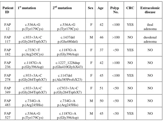
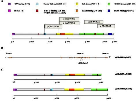
(MEFs) derivate da topi Mutyh difettivi (Molatore et al., 2010). Tale tipo di approccio, tuttavia, non può essere usato per analizzare mutazioni eterozigoti composte di MUTYH, che si riscontrano piuttosto comunemente tra i pazienti italiani affetti da MAP. Allo scopo di studiare tali varianti complesse abbiamo utilizzato linee linfoblastoidi derivate da pazienti MAP con mutazioni missenso e troncanti in condizioni di omozigosi o di eterozigosi composta (Tabella 2, pagina 35 e figura 5, pagina 36).
La quantificazione dei livelli di espressione di MUTYH mediante RT-PCR e Western blotting ha rivelato che, mentre in alcune linee cellulari esiste una buona correlazione fra i livelli di trascritto e quelli di proteina, in altri casi invece, per lo più in linee caratterizzate da mutazioni di tipo frameshift, la presenza del trascritto non corrisponde a quella della proteina (Figura 6, pagina 37), lasciando ipotizzare che alcune mutazioni siano associate ad instabilità dei relativi trascritti.
In seguito alla messa a punto di un saggio in cui viene valutata l’attività glicosilasica sia di MUTYH che di OGG1 (figura 7, pagina 38) è stato possibile dimostrare che tutte le linee mutanti sono difettive nella rimozione dell’adenina da un substrato 8-oxoG:A, pur presentando una normale attività della glicosilasi OGG1 (Figura 8, pagina 39). In accordo con quanto dimostrato da altri autori (Wooden et al., 2004; Parker et al., 2005; Ali et al., 2008; Kundu et al., 2009) si è identificata una residua benché minima attività glicosilasica nelle due varianti p.Tyr179Cys/Tyr179Cys e p.Tyr179Cys/Gly396Asp caratterizzate dalla presenza di livelli di proteina simili alle linee wild-type di donatori sani.
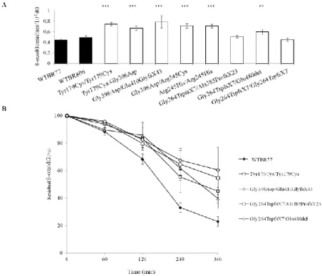
E’stato inoltre interessante osservare che mentre la quasi totalità delle linee esaminate mostra livelli basali di 8-oxo-dG maggiori rispetto alle linee wild-type, le linee caratterizzate dalle mutazioni frameshift p.Gly264TrpfsX7 e p.Ala385ProfsX23, che non esprimono livelli rilevabili di proteina, fanno eccezione. Tale osservazione potrebbe suggerire la presenza di un effetto dominante negativo associato alla proteina mutante, probabilmente connesso all’interferenza con altri meccanismi di riparazione della 8-oxo-dG (Figura 9A, pagina 40).
Con l’obiettivo di verificare se l’incremento nei livelli basali della base ossidata fosse dovuto ad una sua difettiva rimozione nelle linee mutanti, si è eseguito un saggio di cinetica di riparazione dopo trattamento con KBrO3, un agente
ossidante che introduce un alto livello di 8-oxo-dG nel DNA. Tutte le linee con mutazioni di MUTYH hanno mostrato in effetti una rimozione più lenta della lesione quando confrontate con una linea di donatore sano (Figura 9B, pagina 40). La presenza di specifiche trasversioni G>T nei geni APC o K-RAS nei tumori di pazienti MAP può essere plausibilmente ritenuta un’evidenza indiretta della esistenza di un fenotipo mutatore associato alla presenza di una proteina MUTYH non funzionale (Al-Tassan et al., 2002; Jones et al., 2002, Lipton et al., 2003; van Pujienbroek et al., 2008). Partendo da tale osservazione, si è misurata, per la prima volta in linee di pazienti MAP, la frequenza di mutazioni spontanea, utilizzando una metodologia innovativa messa a punto dal gruppo di L. Luzzatto
(Araten et al., 2005) basata sulla determinazione per via citofluorimetrica della frequenza di mutazione di un gene sentinella, PIG-A, già applicata allo studio del fenotipo mutatore associato ad altri difetti della riparazione del DNA. Dai risultati ottenuti si è identificato un aumento medio di quattro volte nel valore della frequenza di mutazione di PIG-A nelle linee di pazienti rispetto al valore medio di tre linee wild-type (Figura 10, pagina 42). Inoltre, la ricomparsa di cellule mutanti dopo previa eliminazione tramite “sorting” dalla popolazione cellulare iniziale, in una delle linee con più alta frequenza di mutazione, ha dimostrato che il fenotipo mutatore identificato è una caratteristica intrinseca della linea cellulare. Nella stessa linea mutante trattata con KBrO3, si è per di
più osservata sia ipersensibilità agli effetti citotossici dell’agente ossidante che un fenotipo di iper-mutabilità rispetto ad una linea di donatore sano (Figura 11, pagina 43), confermando la presenza di un forte impatto biologico associato alla presenza di mutazioni di MUTYH nella risposta al danno ossidativo.
Nella seconda parte della tesi sono invece riportati e discussi i risultati ottenuti in un progetto condotto in parallelo in cui si sono studiati gli effetti dovuti all’assenza di MUTYH nella risposta ad un interessante modello di danno ossidativo prodotto dalla interazione del farmaco immunosoppressore Azatioprina (Aza) con i raggi ultravioletti di tipo A (UVA).
Come suggerito da diversi studi, il danno al DNA indotto dalla combinazione di tali agenti è stato riconosciuto come uno dei possibili fattori responsabili dell’aumentata insorgenza di cancro della pelle in pazienti immunosoppressi con Aza dopo trapianto d’organo (Brem et al., 2009). Il trattamento sistemico con Aza causa l’incorporazione nel DNA di 6-tioguanina (6-TG) (Relling and Dervieux, 2001) che, a differenza delle basi azotate canoniche assorbe le radiazioni UVA, agendo da cromoforo e generando ROS.
La stessa 6-TG è fortemente esposta al processo di ossidazione. In particolare, la sua interazione con i raggi UVA genera una lesione nota come guanina 6-sulfonato che costituisce un blocco alla replicazione del DNA ed è potenzialmente mutagena.
Studi realizzati su cellule umane hanno dimostrato che la 6-TG e i raggi UVA sono sinergicamente citotossici e mutageni inducendo pericolose lesioni a livello del DNA e delle proteine (O' Donovan et al., 2005).
Sulla base di tali osservazioni, e considerando l’importante funzione di MUTYH nella risposta al danno ossidativo, abbiamo esaminato il ruolo di questa proteina nella risposta al danno cellulare indotto dalla combinazione di 6-TG ed UVA. Dato il suo coinvolgimento nella regolazione della risposta cellulare alla 6-TG come anche nella rimozione dell’8-oxo-dG abbiamo esteso parte degli studi anche alla proteina MSH2 facente parte del sistema MMR.
Dai nostri saggi di sopravvivenza in vitro sulle MEFs è risultato che la combinazione di 6-TG e raggi UVA produce effetti tossici in cellule wild-type (WT), mentre il trattamento singolo con 6-TG o con UVA non ha effetti rilevanti sulla sopravvivenza cellulare. Sorprendentemente, sia l’assenza di Mutyh che di Msh2 conferisce resistenza all’effetto tossico prodotto dal
trattamento combinato, (Figura 12, pagina 44) sebbene tutte le linee, indipendentemente dal genotipo, mostrano aumenti simili dei livelli di 8-oxo-dG nel DNA (Figura 13, pagina 45).
Poichè numerosi dati sperimentali indicano che, in cellule trattate con 6-TG, tale analogo di base risulta essere presente nel “pool” cellulare di deossinucleosidi trifosfato (dNTPs) dove può agire da fonte di ROS per esposizione ai raggi UVA (Cooke et al., 2008), si è pensato di determinare il potenziale contributo di questa “riserva” di 6-TG agli effetti biologici osservati. Esperimenti svolti a tale scopo (Figura 14A, pagina 46) hanno rivelato che anche nel sistema da noi analizzato la 6-TG del “pool” contribuisce notevolmente sia agli effetti di citotossicità (Figura 14B, pagina 47) che di ossidazione (Figura 15 pagina 47) osservati dopo il trattamento combinato.
Con l’obiettivo di studiare il meccanismo alla base della tossicità riportata nelle cellule WT ed il ruolo esercitato da MUTYH in questo processo abbiamo analizzato la progressione del ciclo cellulare mediante citometria a flusso. Dai profili ottenuti è emerso che l’assenza MUTYH è associata al mancato arresto delle cellule in fase S dopo esposizione a 6-TG ed UVA, un arresto che si osserva invece in maniera prominente nelle cellule WT (Figura 16, pagina 48). Tale fenomeno si riflette in una diversa cinetica di attivazione del checkpoint: l’attivazione per fosforilazione della proteina Chk1, coinvolta nell’attivazione del checkpoint di fase S e G2/M, si osserva a tempi brevi dal trattamento combinato nelle cellule WT, mentre nelle cellule Mutyh difettive il segnale della proteina fosforilata compare in tempi molto più lunghi e con un’intensità notevolmente ridotta (Figura 17, pagina 49). Tali osservazioni sembrano supportare il coinvolgimento di MUTYH nell’attivazione precoce come anche nella regolazione del checkpoint di fase S in risposta a tale tipo di danno. E’ noto che la fosforilazione di Chk1 da parte della chinasi ATR è coinvolta nel controllo della riparazione dei DSBs mediante il sistema di riparazione HR (Sørensen et al.; 2005). La quantificazione dei DSBs misurati come numero di foci dell’istone fosforilato, H2AX, ha rivelato che, anche nel nostro modello murino, come nelle cellule umane (Brem et al.; 2010), il trattamento combinato con 6-TG e raggi UVA produce questo tipo di danno al DNA, e che i livelli sono paragonabili fra i due genotipi. Sorprende tuttavia che le due linee si distinguano nella modalità di processamento di tale danno. La determinazione numerica dei foci della proteina RAD51, facente parte del sistema HR, ha infatti rivelato che se da un lato nelle cellule WT vi è un’induzione più elevata di tale proteina nonché una sua persistenza a tempi lunghi dopo il trattamento con 6-TG ed UVA, dall’altro invece l’assenza di MUTYH è associata a livelli decisamente più bassi di foci e ad un graduale decremento degli stessi nel tempo (Figura 18, pagina 50). Se si considera che RAD51 esercita una funzione chiave nella sopravvivenza cellulare contro i potenziali effetti letali associati ai DSBs, l’ipersensibilità delle cellule WT alle conseguenze del trattamento combinato è in apparente contraddizione con l’induzione di una risposta chiaramente protettiva. Una possibile spiegazione di questo fenomeno consiste nell’ipotizzare
che il tentativo di riparazione del danno prodotto dall’interazione della 6-TG con i raggi UVA mediante attivazione del sistema HR induce la formazione di una quantità considerevole di intermedi riparativi estremamente tossici incompatibili con la sopravvivenza cellulare. In quest’ottica, l’assenza MUTYH e quindi di una modalità efficiente e/o canonica di riparazione del danno comporterebbe un vantaggio selettivo per la cellula stessa in termini di vitalità, associato tuttavia ad un possibile aumento dell’instabilità genomica.
Lo dimostrano infatti i dati derivati da uno studio parallelo condotto in vivo su animali wild-type e Mutyh-difettivi, sottoposti a trattamenti singoli o combinati (Aza somministrata per via intraperitoneale seguita o meno da esposizione ad UVA) per 12 mesi consecutivi. Come nel modello in vitro, il trattamento con entrambi gli agenti ha determinato una notevole differenza nella tossicità: l’80% dei topi difettivi è sopravvissuto al trattamento contro il 20% dei topi wild-type. Il trattamento singolo con i raggi UVA non ha invece prodotto effetti sulla sopravvivenza in nessuno dei due genotipi, mentre l’immunosoppressione prodotta dall’Aza ha determinato livelli di mortalità simile nei due genotipi. E’ stato inoltre osservato un incremento notevole nei livelli di 8-oxo-dG nella pelle di entrambi i gruppi di animali, sebbene non siano state riscontrate differenze significative nei livelli di ossidazione del DNA tra i due genotipi (Figura 19A, pagina 52). E’ degno di nota, invece, che l’analisi istologica della pelle ha identificato la presenza di due carcinomi a cellule squamose in due topi Mutyh-/- (Figura 19B, pagina 52) dopo trattamento combinato suggerendo che l’assenza di una proteina fondamentale nella salvaguardia del genoma è associata ad una possibile predisposizione all’insorgenza di tumori della pelle. Considerati nel loro complesso i dati ottenuti da tali studi mettono in luce l’importanza dell’effetto protettivo esercitato dalla proteina MUTYH nella risposta al danno ossidativo al DNA. Le osservazioni condotte sulle linee di pazienti affetti da MAP, oltre a sottolineare l’aspetto di patogenicità associato alla presenza della proteina mutante, consentono anche di individuare nell’accumulo di danno ossidativo nel DNA e nella presenza di un fenotipo mutatore sia spontaneo che indotto da stress ossidativo, fattori di estrema rilevanza per una migliore valutazione clinica della patogenesi associata alle varianti di MUTYH. Infine, i risultati ottenuti dallo studio condotto nel modello murino di inattivazione di MUTYH confermano la rilevanza biologica di tale proteina nella salvaguarda della stabilità genomica, indicando tuttavia un suo ruolo più complesso nella risposta al danno al DNA che va oltre il semplice meccanismo di riparazione del danno ossidativo.
SUMMARY
The oxidized base 7,8-dihydro-8-hydroxyguanine (8-oxo-dG) is one of the most deleterious injuries induced by oxidative stress. Multiple DNA repair proteins have evolved to protect the genome against the detrimental effects of this promutagenic lesion. One of the major ones is the Base Excision Repair (BER) MUTYH DNA glycosylase that removes adenine from 8:oxoG-containing mispairs originated by DNA polymerases and via the OGG1 DNA glycosylase contributes to 8-oxo-dG repair. Germline mutations in the MUTYH gene lead to MUTYH-associated polyposis (MAP), an autosomal recessive syndrome characterized by colorectal polyposis and cancer predisposition. Although several reports characterized MUTYH variants using purified proteins, relatively few mutations have been investigated from the biochemical point of view. In addition no information is available on the mutator phenotype associated with MUTYH inactivation in humans. In our previous study accumulation of 8-oxo-dG and hypersensitivity to killing by the oxidizing agent KBrO3 were identified
as a common phenotype among the investigated MAP-associated variants. These results were based on an assay in which single mutant MUTYH proteins were expressed in mouse embryonic fibroblasts (MEFs) derived from Mutyh-null mice (Molatore et al., 2010). This approach however cannot be exploited to analyze compound heterozygous MUTYH mutations, a very common situation among Italian MAP patients. With the aim of studying these particularly complex variants, we derived human lymphoblastoid cell lines (LCLs) from MAP patients harbouring missense and truncating mutations. RT-PCR and Western blotting analyses revealed that while in some cell lines there is a good correlation between transcripts and protein levels, in other instances no MUTYH protein is detectable.
When basal levels of 8-oxo-dG were measured in these cell lines, increases were detected in DNA of six LCLs expressing MUTYH variants when compared to two wild-type cell lines. Interestingly the only two exceptions were cells in which no detectable expression of the MUTYH protein could be identified. To determine whether this increase in steady-state levels was due to a defective repair of 8-oxo-dG in these mutants, repair kinetics of this oxidized base were determined following exposure to KBrO3. All the LCLs harbouring MUTYH
mutations showed a defective repair of 8-oxo-dG when compared to a cell line from a healthy donor.
Results of a novel assay where both MUTYH and OGG1 activity could be evaluated indicate that all these variants were defective in removing adenine from an 8-oxoG:A DNA substrate, but retained wild-type OGG1 activity. Mutation frequency measurements at the PIG-A gene identified a four-fold increase in spontaneous mutagenesis in six LCLs from MAP patients when compared to three LCLs from healthy donors. Finally KBrO3 hypersensitivity
was accompanied by a hyper-mutable phenotype in a MUTYH mutant cell line. These observations support the pathogenic role of these MUTYH mutations and
identify accumulation of 8-oxo-dG and a mutator phenotype as relevant factors for a better clinical assessment of MUTYH variant pathogenesis.
In the second part of this thesis the results of a parallel study on the effects of MUTYH loss in response to a different type of oxidative damage are reported. This study addresses the toxicity and carcinogenicity of the anti-cancer immunosuppressant drug Azathioprine (Aza) combined with UVA radiation. Systemic treatment with Aza causes the incorporation of 6-thioguanine (6-TG) into DNA. 6-TG is a chromophore which generates reactive oxygen species
(ROS) on exposure to UVA and is itself highly susceptible to oxidation. DNA
damage induced by the Aza/UVA combination is thought to contribute to the huge incidence of skin cancer in immunosuppressed organ transplant patients. Several studies have shown indeed that Aza combined with low doses of UVA may cause mutagenic damage in human cells. In particular, 6-TG/UVA generates a novel DNA lesion, the guanine-6-sulfonate that blocks DNA replication and is potentially mutagenic. Here we examined the role of the MUTYH DNA glycosylase and the mismatch repair (MMR) MSH2 protein in the cellular response to 6-TG/UVA-induced DNA damage.
6-TG and UVA were synergistically toxic to wild-type mouse embryo fibroblasts (MEFs) while neither 6-TG or UVA alone detectably affected survival. Mutyh- or Msh2-defective cells were more resistant than wild-type MEFs to killing by 6-TG/UVA. Nevertheless, the combined treatment significantly increased the levels of DNA 8-oxo-dG irrespectively of the genotype. Interestingly, we also found in wild-type cells that the deoxynucleoside triphosphates (dNTP) pool contributed to both the increased levels of DNA 8-oxo-dG and the enhanced toxicity of a combined 6-TG/UVA treatment.
To better understand the mechanism of 6-TG/UVA toxicity and the relative role of the MUTYH protein we also analysed cell cycle progression by flow cytometry. The data suggest that the MUTYH protein is involved in the S phase arrest induced by the combined 6-TG/UVA treatment
We also reported a difference between the two cell lines in the timing of the checkpoint activation: phosphorylation of the Chk1 protein occurred immediately after 6-TG/UVA treatment in wild-type cells, while a later and weaker appearance of phosphorylated Chk1 occurred in Mutyh-/- cells. These data suggest that MUTYH might be involved in the activation of an S phase checkpoint following this type of oxidative damage. We also confirmed that in this mouse model, as in human cells, treatment with 6-TG/UVA produces an increase in double strand breaks (DSBs). These DSBs, as measured by γH2AX foci, were formed immediately and maintained up to 48 hr from the end of the treatment. The comparable increase in DSBs observed in Mutyh-defective cells indicates that the initial level of DNA damage is similar in the two genotypes. However analysis of the homologous recombination (HR) protein RAD51 foci following the combined treatment indicates that the DSBs resolution differs between the two genotypes. Mutyh-/- MEFs showed a significantly lower
induction of RAD51 foci in comparison to wild-type cells with a gradual decrease in the levels of foci at late post-treatment times. This contrasts with the persistence in wild-type cells of high levels of these foci indicating the presence of unresolved DSBs.
Finally we report the results of in vivo experiments in which the long-term toxicity of single or combined Aza/UVA exposures was analysed in wild-type and Mutyh-defective animals. Mice were treated with Aza given by intraperitoneal injection and/or UVA for 12 months. A differential toxicity between WT and Mutyh-defective animals was observed as consequence of the Aza plus UVA exposure. In fact, a high level of toxicity was identified in the group of wild-type mice (only 20% of the animals survived this exposure), while the 80% of Mutyh-/- animals survived this treatment. Survival was 100% in both UVA treated groups, while immunosuppression in Aza-treated groups was associated with some mortality, which was unaffected by the genotype.
A significant increase of DNA oxidation in the skin of animals exposed to the combined treatment was also observed, irrespectively of the genotype. Intriguingly when histopathological examination of the skin was performed two squamous cell carcinomas were identified only in Mutyh-/- mice exposed to Aza plus UVA, revealing a possible skin cancer proneness conferred by loss of this protein.
I. INTRODUCTION
1. Damage to DNA and human diseases
The genome integrity is pivotal in the maintenance of life. A variety of both exogenous and endogenous factors may compromise the architecture of the DNA double helix. Differently from proteins, lipids and RNA that, if damaged, are usually subjected to degradation, DNA lesions should be repaired. If unrepaired they may alter normal cell physiology and cell viability or even result in genomic instability (Radak and Boldogh, 2010).
If we bear in mind that the genome is a critical target for time-dependent deterioration, it is not surprising that DNA damage has been considered a critical factor for many diseases associated with aging.
There is a multitude of potential damage sources that may challenge DNA integrity and compromise its function. Hydrolysis, the principal spontaneous reaction occurring in a cellular environment, is intrinsic to the chemical nature of DNA in aqueous solution and it is responsible for creation of abasic sites and deamination events.
DNA is also continuously exposed to metabolism products, such as reactive oxygen and nitrogen species (ROS and RNS), endogenous alkylating agents, estrogen and cholesterol metabolites, reactive carbonyl species and lipid peroxidation products. Finally, some sources of carcinogenic DNA lesions originate in the environment: various exogenous physical and chemical agents, could represent a risk for DNA stability. Such chemicals can attack DNA, leading to adducts that impair base-pairing and/or block DNA replication and transcription, causing base loss and DNA single-strand breaks (SSBs). Ultraviolet light (UV) and ionizing radiation (IR) also generate various forms of DNA damage. The most toxic lesions caused by IR are double-strand breaks (DSBs). The biological consequences of both endogenous and exogenous injury factors generally depend on the location and number of lesions, the cell type, as well as the stage in the cell cycle and during differentiation (Figure 1) (Hoeijmakers, 2009).
If not properly removed, DNA damage can lead to DNA mutations and/or cell death, especially in the case of cytotoxic lesions that block the progression of DNA/RNA polymerases (Maynard et al., 2009). If not repaired, DNA mutations and large genomic alterations such as deletions, translocations, loss of heterozygosity, and amplifications may lead to cancer onset, while the occurrence of cell death or senescence may cause the acceleration of the aging process. Cell senescence and apoptosis are in fact suspected causes of aging under biological conditions associated to stem-cell exhaustion. Interestingly, whereas p53-induced cell death protects against tumorigenesis, pro-apoptotic p53 activity is harmful in settings such as stroke or heart attack. Induction of p53
by different sources of DNA damage can also affect the development of atherosclerosis (Jackson and Bartek, 2009).
1.1. The response to DNA damage: the genome-maintenance network
“We totally missed the possible role of.. [DNA] repair although I later came to realize that DNA is so precious that probably many distinct repair mechanisms would exist” Francis Crick, Nature, 26 April 1976
Genome instability caused by the continual attack to DNA from endogenous and environmental agents, would be an overwhelming problem for cells and organisms in the absence of DNA repair. In consideration of the great variety of DNA-damaging factors, it is not surprising that cells have evolved mechanisms – collectively termed the DNA-damage response (DDR) to detect DNA lesions, signal their presence and promote their repair (Wood et al., 2001).
Highly conserved pathways are tailored to deal with different classes of lesions, although some share many components and they usually occur by a common general program (Table 1). For example, three types of excision repair have been described: mismatch repair (MMR), nucleotide excision repair (NER) and base excision repair (BER) (Risinger and Groden, 2004). The principal aspects of the first two systems have been comprehensively summarized below, while the BER system will be analysed in full detail later on.
MMR is a central system in the repair of DNA replication errors and in the inhibition of recombination between non-identical DNA sequences. The key proteins in MMR are highly conserved from bacteria to mammals: in eukaryotes there are multiple homologs of the key bacterial MutS and MutL MMR proteins. (Harfe and Robertson, 2000). Six human mismatch repair genes (MSH2, MSH3, MSH6, MLH1, PMS1 and PMS2) have been identified as components of this repair system that efficiently corrects single base mismatches and loops of one to three extrahelical nucleotides. The MSH2 and MSH6 proteins compose the heterodimer hMutS that recognizes single base-base mismatches and small insertion/deletion mispairs. The MSH2 and MSH3 gene products form a heterodimer recognizing larger insertion/deletion mispairs, while the MLH1 protein together with PMS2 forms the human homolog of MutL, the hMutL complex (Gu L et al., 1998).
The binding to DNA by one of the two mismatch recognition complexes (MutS , MutS ) is the first step in the error correction process. Complete excision and replacement of the mismatched section of DNA involves the MutL complex (or another heterodimeric complex formed by MLH1 and hMLH3) as well as PCNA, RPA, DNA polymerase pol and EXO1 (Kunkel and Erie, 2005; Jiricny, 2006). MMR is also one of the alternative pathways involved in minimizing the toxic and mutagenic effects of the DNA 7,8-dihydro-hydroxyguanine (8-oxo-dG), a harmful DNA oxidation product causing G>T transversions and implicated in frameshift formation. The microsatellite instability and the mutator phenotype of MMR-defective cells strongly support a crucial role of MMR in the DDR (Macpherson et al, 2005). Moreover, inherited
MMR defects are associated with the hereditary nonpolyposis colorectal cancer (HNPCC), an autosomal dominant disorder.
NER is a highly versatile and sophisticated DNA damage removal pathway mainly defending mammalian cells against UV-induced DNA damage. Besides cyclobutane pyrimidine dimers and (6-4) photoproducts produced by UV rays, it also deals with bulky adducts resulting from exposure to various agents like cisplatin. The first step in the NER pathway is the lesion recognition process involving the ERCC1, XPA and XPF gene products followed by the interaction with the TFIIH transcription factor. A dual incision event is accomplished by the ERCC1 and XPG gene products, and this is followed by exonuclease activity. DNA is then synthesized to fill the gap using the undamaged strand as a template, and the ends are ligated (Bohr , 1995).Defects in NER are reflected in the severe photosensitivity and predisposition to skin cancer associated to the prototype repair syndrome Xeroderma pigmentosum (de Laat et al., 1999) . DSBs represent one of the most severe type of DNA damage and two additional types of DNA repair, homologous recombination (HR) and non-homologous end-joining (NHEJ), are tailored to deal with this type of lesion. Using a copy (usually the sister chromatid available during the S and G2 phases of the cell cycle) of the damaged segment as a template to mediate a faithful repair, HR is considered an error-free pathway. NHEJ, on the contrary, is an error-prone pathway, since free ends are joined without the use of a template via very small microhomologous repeats. As a result, repair may be associated to loss of nucleotides or translocations (Risinger and Groden, 2004). In NHEJ, DSBs are recognized by the Ku proteins that then bind and activate the protein kinase DNA-PKcs, leading to recruitment and activation of end-processing enzymes, polymerases and DNA ligase IV (Jackson and Bartek, 2009).
HR is always initiated by single-strand DNA (ssDNA) generation, which is promoted by various proteins including the MRE11-RAD50-NBS1 (MRN) complex. In events catalyzed by RAD51 and the breast-cancer susceptibility proteins BRCA1 and BRCA2, the ssDNA invades the undamaged template and, following the actions of polymerases, nucleases, and helicases, DNA ligation and substrate resolution occur. HR is also used to restart stalled replication forks and to repair interstrand DNA crosslinks.
Table 1. DDR mechanisms and components
DDR mechanism Prime lesions acted upon Key protein components
Direct DNA-lesion reversal
UV photo-products O6 alkylguanine
Photolyase O6-methylguanine methyltransferase (MGMT)
Mismatch repair (MMR) DNA mismatches and insertion/deletion loops arising from DNA replication
Sensors MSH2-MSH6 and MSH2-MSH3 plus MLH1 PMS2, MLH1-PMS1, MLH1-MLH3, EXO1, polymerases δ and ε, PCNA, RFC, RPA, ligase I Base excision repair (BER)
and single-strand break repair (SSBR)
Abnormal DNA bases, simple base- adducts, SSBs generated as BER intermediates by oxidative damage or by abortive topoisomerase I activity
DNA glycosylases (sensors), APE1 endonuclease, DNA polymerases (β, δ, ε) and associated factors, flap endonuclease FEN1, ligase I or ligase III. SSBR can also involve polymerase β lyase activity, XRCC1, PARP-1, PARP-2, polynucleotide kinase (PNK) and aprataxin (APTX)
Nucleotide excision repair (NER)
Lesions that disrupt the DNA double-helix, such as bulky base adducts and UV photo products
Sensors elongating RNA polymerase, XPC-HR23B and DDB1/2, plus XPA, XPE, XPF/ERCC1, XPG, CSA, CSB, TFIIH (containing helicases XPB and XPD), DNA polymerases and associated factors, RPA, ligase I
Trans-lesion bypass mechanisms
Base damage blocking replication-fork progression
“Error-prone” DNA polymerases, including polymerases eta, iota, kappa, REV3 and REV1; plus associated factors
Non-homologous end- joining (NHEJ)
Radiation- or chemically-induced DSBs plus V(D)J and CSR intermediates
Sensors Ku and DNA-PKcs plus XRCC4, XLF/Cernunnos and ligase IV. Can also employ the MRE11-RAD50-NBS1 (MRN) complex, Artemis nuclease, PNK, Aprataxin and polymerases μ and λ Homologous
recombination (HR)
DSBs, stalled replication forks, inter-strand DNA cross-links and sites of meiotic recombination and abortive Topoisomerase II action
RAD51, RAD51-related proteins (XRCC2, XRCC3, RAD51B, RAD51C, RAD51D, DMC1), RAD52, RAD54, BRCA2, RPA, FEN1, DNA polymerase and associated factors. Promoted by MRN, CtIP, BRCA1, and the ATM signalling pathway Fanconi anaemia
(FANC) pathway
Inter-strand DNA cross-links FA-A, C, D1/BRCA2, D2, E, F, G, I, J, L, M, N plus factors including PALB2 and HR factors
ATM-mediated DDR signalling
DSBs ATM, MRN and CHK2. Promoted by mediator proteins such as MDC1, 53BP1 MCPH1/BRIT1, and by ubiquitin ligases RNF8, RNF168/RIDDLIN and BRCA1
ATR-mediated DDR signalling
ssDNA, resected DSBs Sensors ATR ATRIP and RPA plus the RAD9-RAD1-HUS1 (9-1-1) complex, RAD17 (RFC1-like) and CHK1. Promoted by MRN, CtIP and mediator proteins such as TOPBP1, Claspin, MCPH1/BRIT1 and BRCA1
1.2. DNA-damage repair and checkpoint pathways
DNA repair, as well as DNA replication, is strictly coordinated with cell cycle progression. DNA damage and replication blocks activate signals that arrest cell cycle progression providing time for repair or trigger cell apoptosis when repair cannot be completed. These complex surveillance mechanisms, known as cell-cycle checkpoints, are controlled by a network of DNA damage sensors, transducers and effectors (Bartek et al., 2004; Sancar et al., 2004)(Figure 2). Highly conserved DNA repair and cell cycle checkpoint pathways allow cells to deal with both endogenous and exogenous sources of DNA damage (Kastan and Bartek, 2004). Originally defined as dispensable regulatory pathways, DNA damage checkpoints are now considered as integrated components of the larger DDR. Recent lines of evidence demonstrated, in fact, the existence of an intimate connection between checkpoint components and DNA repair proteins (Zhou and Elledge, 2000).
Several checkpoint genes are also essential for cell and organism survival (Brown et al., 2000; de Klein et al., 2000) suggesting that their function is not restricted to DNA surveillance but is essential for cellular physiology.
Partly overlapping or redundant checkpoint pathways operate in various cell-cycle phases. Cells in G1 or G2 phases can counteract genotoxic stress by promoting checkpoints that provoke an arrest in G1 or G2 before re-entry into S phase or M phase of cell cycle, respectively. During DNA replication, on the contrary, the cellular response to potential DNA damage factors leads to a delay of cells progression through the S phase in a transient manner. If damage is not repaired, cells exit S phase and arrest at the G2 checkpoint (Bartek J et al., 2004). Despite the recent explosion of information regarding the molecular components of cell cycle checkpoints in eukaryotic cells, there is not a wide understanding of the identity of the DNA damage sensors or the mechanisms through which they initiate and terminate the activation of checkpoints. However, members of the Rad group of checkpoint proteins, which include Rad17, Rad1, Rad9, Rad26, and Hus1 are widely expressed in all eukaryotic cells where act as damage sensors (Green et al. 2000; O’Connell et al. 2000). Three of these Rad proteins, Rad1, Rad9, and Hus1 form a heterotrimeric DNA damage responsive complex (the 9-1-1 complex) that exhibits structural similarity with the homotrimeric clamp proliferating cell nuclear antigen, PCNA, that has the ability to slide across double-strand DNA (Majka et al., 2003). The 9-1-1 complex physically and functionally interacts with several DNA repair proteins, including factors involved in the BER pathway suggesting the presence of a close connection between checkpoint activation by 9-1-1 complex and recruitment of repair machinery (Meister et al., 2003). The 9-1-1 complex is loaded onto DNA by the Rad17/RFC2-5 complex and subsequently serves as a recruitment platform for several proteins, such as the Chk1 and Chk2 serine/threonine kinases. These effectors kinases promote cell cycle arrest,
transcriptional activation and apoptosis by phosphorylating critical targets (Guan et al., 2007). Chk1 and Chk2 are targets of regulation by two signal transducers proteins, ATM (ataxia telangiectasia mutated) and ATR (ATM and Rad3-related protein) kinases, respectively. ATM and ATR are PIKK (phosphoinositide three-kinase-related kinases) that share a number of phosphorylation substrates, even if they respond to different type of DNA damage. ATR plays a central role in the response to certain types of genotoxic agents, including hydroxyurea and UV and seems to have a central function in the S and G2 checkpoints. On the contrary, ATM has a major function for management of the G1 checkpoint and in contrast to ATR, provides a rapid protective response to an extremely lethal form of DNA damage, the DSBs. (Abraham, 2001). The main relevance for cell and organism life of this “network of genome surveillance” is reflected in the evidence that a major mechanism whereby tumor cells acquire genetic instability is through the acquisition of mutations that modify checkpoints. This feature renders them more dependent on the remaining intact pathways to promote repair and arrest the cell cycle, representing an important approach for the development of therapeutic strategies in the personalized cancer treatment (Medema and Macurek, 2011).
2. The oxidative damage to DNA
The variety of reactions in which oxygen is implicated in the cellular environment leads to the formation of chemical intermediates known as ROS, that account for the background levels of oxidative DNA damage detected in normal tissue (Cooke et al., 2003).
During oxidative metabolism in mitochondria, oxygen is mainly converted to water and only a small percentage (0.2–2%) leads to ROS, because of the leakage of electrons directly to oxygen leading to formation of superoxide anions (•O2
-). Either spontaneously or through catalysis by superoxide dismutases, superoxide anions can be further converted to hydrogen peroxide (H2O2), that can be next reduced to H2O or partially reduced to the hydroxyl
radical (•OH), a very powerful oxidant (van Loon et al., 2010).
ROS may also be generated by IR or UV radiation, chemotherapeutic drugs and environmental exposures to transition metals and chemical oxidants (Maynard et al., 2009). Although ROS have a physiological role in numerous signaling pathways, in inflammatory processes and in preventing infections, they can also be genotoxic and oxidize various cellular components such as lipids, proteins and nucleic acids (Mitra et al., 2001). These “oxidative damage” events are thought to be involved in mutagenesis, carcinogenesis and aging. Thus, oxidative stress is accepted as a critical pathophysiological mechanism in different frequent pathologies, including cardiovascular diseases, cancer, diabetes, rheumatoid arthritis, or neurological disorders such as Alzheimer or Parkinson disease (Mena et al., 2009).
ROS-induced DNA damage involves SSBs and DSBs, purine, pyrimidine, or deoxyribose modifications, and DNA cross-links, that can lead to either arrest or induction of transcription, activation of signal transduction pathways, replication errors and genomic instability (Valko et al., 2006).
More than 100 products have been identified as generated by the oxidation of DNA, with bases in DNA being particularly sensitive to ROS oxidation. The low redox potential of guanine makes this base particularly susceptible to oxidation resulting in the formation of a great variety of potential oxidized products. Thus, not surprisingly, 8-oxo-dG is one of the most abundant and well-characterized DNA lesions generated by ROS, often used as a biomarker in cells to indicate the extent of DNA oxidative stress. It arises by the introduction of an oxo group on the carbon at position 8 (C8) and a hydrogen atom to the nitrogen at the position 7 (N7). 8-oxo-dG in syn conformation is particularly mutagenic because of its strong ability to functionally mimic T and to form a stable base pair with adenine. In contrast to many other types of DNA damage, these structural features provide for efficient, though inaccurate, bypass of the lesion by replicative DNA polymerases representing a direct source of C:G to A:T transversion mutations (David et al., 2007). The estimated steady-state level of
8-oxo-dG lesions is about 103 per cell/per day in normal tissues and up to 105 lesions per cell/per day in cancer tissues.
Recent extensive studies of the patterns of somatic mutations in genomes of different cancer types shed light on the abundance of C:G to A:T transversion mutations and identified them among the most predominant somatic mutations in lung, breast, ovarian, gastric and colorectal cancers (van Loon et al., 2010).
2.1. Response to oxidative damage: the special problem of 8-oxo-dG
The removal of oxidative DNA lesions has a crucial role in the limitation of mutagenesis, cytostasis, and cytotoxicity. The existence of multiple overlapping repair processes of oxidative DNA damage introduces a fail-safe element to DNA repair, such that attenuation or elimination of one repair process does not preclude removal of a particular lesion (Cooke et al., 2003).
The deleterious effect of 8-oxo-dG, in particular, is emphasized by the evolution, from bacteria to humans, of several mechanisms to neutralize it. The main protective device that many organisms have developed is a three-component enzyme error-preventing system (termed the “GO system” after dG). In humans this system consists of three repair proteins: the 8-oxo-dGTPase (MTH1), the 8-oxo-dG DNA glycosylase (OGG1) and the MutY glycosylase homologue (MUTYH), that will be described in details below. The MTH1 protein, the homolog of E.Coli MutT, acts by hydrolyzing 8-oxo-dGTP to 8-oxo-dGMP, thereby eliminating it from the pool of DNA synthesis precursors so that it cannot be incorporated into DNA by DNA polymerases (Tsuzuki et al., 2007).
OGG1 targets the C:8-oxo-dG mispair, removing the lesion. Upon DNA binding, the C:8-oxo-dG base pair is disrupted and the 8-oxo-dG flipped out of the double helix. Several studies have indicated that OGG1 initiated repair follows the SP-BER pathway (Dianov et al., 1998), in which DNA pol seems to be responsible for the re-synthesis step (Fortini et al., 1999).
MUTYH acts instead by removing the misincorporated adenine opposite an 8-oxo-dG localized in the template strand. The direct association of MUTYH with both PCNA and RPA is fundamental to direct the MUTYH activity to the newly synthesized strand. This is of great importance in order to prevent a possible fixation of an A:T to C:G mutation. Interestingly, MUTYH was also found to excise another type of oxidative lesion, the 2- hydroxyadenine from DNA, (Ohtsubo T et al., 2000), suggesting that its function may be broader than the removal of A from 8- oxoG:A mispairs.
There are several lines of the evidence indicating that even though the “GO system” is the most important repair mechanism of 8-oxo-dG from genomic DNA, cells possess alternative repair pathways that can handle 8-oxo-dG damage (Klungland et al., 1999). Besides OGG1 and MUTYH, the two bifunctional DNA glycosylases NEIL1 and NEIL2 might be involved in BER of
8-oxo-dG. NEIL1 can in fact excise an 8-oxo-dG from a duplex DNA containing C:8-oxo-dG base pair (Hazra et al.,2002). NEIL2 can also excise 8-oxo-dG, but only when it is present inside a bubble structure suggesting that it may function during BER linked to transcription or replication (Dou et al., 2003). The MMR proteins also contribute to 8-oxo-dG repair. The human MutS complex efficiently recognizes DNA 8-oxo-dG as well as another oxidation product, 2-hydroxyadenine, in some contexts that resemble frameshift intermediates (Macpherson et al., 2005; Barone et al., 2007). It has been shown that MMR also provides supplementary protection by excising incorporated 8-oxo-dGMP (Colussi et al., 2002) and this 8-oxo-dG derived from an oxidized pool of deoxynucleoside triphosphates (dNTPs) is an important cofactor in the genetic instability that characterizes MMR-deficient cells. Indeed hMTH1 overexpression in MMR-defective Msh2-/- mouse embryonic fibroblasts (MEFs) drastically attenuates their mutator phenotype (Russo et al., 2004).
There is also evidence that NER might act as a backup for the repair of this lesion (Reardon et al., 1997). Several data suggest a role of both the Cockayne syndrome B (CSB) and A (CSA) proteins, in this process. The similar impairment of 8-oxo-dG repair observed in the absence of the CSB (Licht et al., 2003) or CSA (D’Errico et al., 2007) proteins suggests a role in the same pathway of response to oxidative stress.
Furthermore, CSB interacts with BER proteins such as APE1 (Wong HK et al., 2007) and there are some evidence of a possible interplay between CSB and OGG1 (Khobta et al., 2009). Another member of the NER pathway, the Xeroderma Pigmentosum complementation group C (XPC) protein, might also be involved in 8-oxo-dG repair. Mice deficient in XPC display an elevated sensitivity to oxidative damage, susceptibility to lung carcinogenesis (Melis et al., 2008) and increased levels of C:G to A:T transversion mutations in lympohocytes (Wijnhoven et al., 2000). Some data indicate also a role for XPC as a cofactor of BER by stimulating the activity of the DNA glycosylase OGG1 in the removal of 8-oxo-dG from DNA (D'Errico et al., 2006).
2.2. Sources of cellular 8-oxo-dG: potassium bromate and combination of 6-thioguanine and UVA
Potassium bromate (KBrO3) is an oxidising agent that has been used as a food
additive and can also be produced from bromide during the disinfection of water by ozonation (Parsons and Chipman, 2000). Although its activity is only very weak in some microbial assays, there is no doubt that KBrO3 can act as a
mutagen as indicated by chromosome aberration and micronucleus tests, (Ishidate et al.,1984). Furthermore, KBrO3 acts as a tumor promoter in renal cell
tumours, mesotheliomas of the peritoneum and follicular cell tumours of the thyroid in F344 rats (Kurokawa et al., 1990).
Several observations, in particular the detection of increased levels of 8-oxo-dG in the kidney DNA of treated rats and the inhibition of this DNA oxidation by various antioxidants (Sai et al., 1992) led to a correlation between KBrO3
induced-carcinogenicity in rodents with its ability to oxidize DNA.
The mechanism whereby KBrO3 can oxidize DNA is however not clear. It is
well known that glutathione, which is protective against most oxidants and alkylating agents, mediates KBrO3 metabolic activation, while bromate itself
does not react directly with DNA, despite its high oxidation potential. Even though the ultimate DNA damaging species has not yet been established, experiments under cell-free conditions suggest that ROS such as superoxide, hydrogen peroxide or singlet oxygen are not involved. Rather bromine radicals or oxides might be responsible (Ballmaier and Epe, 2006).
KBrO3-induced DNA damage was found to consist mostly of base modifications
sensitive to the Fpg protein, a glycosylase recognizing particularly 8-oxo-dG, while lesions such as SSBs and sites of base loss (apurinic/apyrimidinic sites, AP sites) were formed only in low amounts. A similar “damage profile” was also found in mammalian cells (Ballmaier and Epe, 1995).
As well as being produced as a result of simple oxidizing agents such as KBrO3,
DNA 8-oxo-dG might also be generated by more complex interactions among different chemical and/or physical agents. One of these is the combination between the thiopurine Azathioprine (Aza) and UVA radiation.
Aza is an anti-cancer immunosuppressant drug, often used in the treatment of inflammatory conditions. Thiopurines are all prodrugs and their metabolism results in the formation of thioguanine (TG) nucleotides and, finally, in 6-TG incorporation into DNA (Relling and Dervieux, 2001). Long-term treatment with Aza results in detectable DNA 6-TG in patients’ lymphocytes and in cells of the skin (Warren et al., 1995). In addition continuous immunosuppression in organ transplant patients is associated with an incidence of skin cancer that is up to 200-fold higher than that of non-immunosuppressed individuals (Brem et al., 2009). Although several factors may be involved in this phenomenon, some epidemiological evidences suggest that sunlight exposure is an important cofactor (Euvrard et al., 2003). Differently from the canonical DNA bases, thiopurines do absorb UVA (wavelengths 320–400 nm) and incorporated 6-TG therefore introduces into DNA a strong UVA photosensitizer. This suggests a possible mechanism by which sunlight and Aza might interact to promote the development of skin cancer (Brem et al., 2009). The interaction between DNA 6-TG and UVA is an important source of oxidative stress, generating ROS. The oxidative damage caused to DNA by ROS might therefore contribute to the development of transplant-related skin cancer (Zhang et al., 2007). ROS can inflict collateral modifications on surrounding normal DNA bases, sugars and DNA-associated proteins. As well as being a source of ROS, 6-TG itself is also a target for oxidation and one oxidized form of the thiobase is guanine-6-sulfonate (GSO3), a bulky DNA adduct, incapable of stably pairing with any of
polymerases and can be bypassed, in a potentially mutagenic strategy, only by DNA polymerases with a relatively low fidelity (O' Donovan et al., 2005). A further outcome of the enhanced reactivity of the thiol group is the major susceptibility of 6-TG to other chemical modifications such as methylation. Similarly to 6-TG, methyl-6-TG (me6-TG) codes ambiguosly during replication and directs C and T insertion approximately equally. DNA me6-TG base pairs are recognized by MMR in a futile processing effort that causes cell death. One important consequence of this phenomenon is the possible selective proliferation of rare MMR-defective cells following long exposures to this base analog, and since MMR defects are associated to a marked mutator phenotype, this might favour the development of malignancy (Karran and Attard 2008).
2.3. The Base-excision repair: structural and functional aspects
The BER pathway is the primary mechanism for repair of oxidized base lesions. Besides 8-oxo-dG, other base modifications repaired by BER include formamidopyrimidines (4,6-diamino-5-formamidopyrimidine, FapyG) uracil and 3-methyladenine, resulting from cytosine deamination and alkylating agents, respectively (Krokan et al., 2002, Sedgwick et al., 2007). An overlap of the other DNA repair systems with BER has also been described. MMR and recombinational repair, in fact, provide protection against base pair mismatches and strand breaks (Russo et al., 2007), while NER can overlap with BER in repairing a variety of single-base DNA modifications (Memisoglu and Samson L, 2000). Although the understanding of the mammalian BER system is not yet complete, the fundamental enzymatic steps operating in this pathway have been extensively studied. The sequence of reactions consists of base removal, strand cleavage at the AP site, processing of the incised strand, gap filling DNA synthesis and DNA ligation (Wilson et al., 2010). Since unattended BER intermediates are cytotoxic and highly mutagenic (Loeb, 1985), a sequential step-to-step coordination is probably operating to sequester these dangerous repair intermediates, as described in the suggestive “passing the baton” model elaborated by Wilson and Kunkel (Wilson et al., 2000).
DNA glycosylases initiate the BER process by catalyzing hydrolysis of the N-glycosylic bond between the sugar and the base, thus releasing the damaged base to form an AP site. Eleven DNA glycosylases, with various specificities have been described so far in mammalian BER and classified in two distinct mechanistic classes: mono-functional such as UDG, and bi-functional such as OGG1. The first type DNA glycosylases create AP sites through cleavage of the N glycosylic bond using an activated water molecule as an active site nucleophile (Sharma and Dianov, 2007). The major 5’-AP endonuclease, APE1, utilizes the AP site and generates a DNA repair intermediate that contains a SSB with 3’-hydroxyl and 5’-deoxyribose-5’ phosphate (5’dRp) termini (Demple et al., 1991). The 5’dRp terminus is next excised by the dRp lyase activity of pol
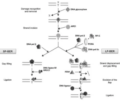
and a one-nucleotide gap is created. Bi-functional glycosylases have an associated AP lyase activity which can further process the AP site by incising the DNA backbone 3’ to the AP site. The next step in the BER repair process consists of two distinct sub-pathways (Figure 3): short-patch BER (SP-BER) and long-patch BER (LP-BER), depending on the damage and the responsive enzymes. They are differentiated by the size of the repair patch synthesized by the repair DNA polymerases: one nucleotide in the case of SP-BER (Dianov et al., 1992) and two to 7-8 nucleotides in the case of LP-BER (Frosina et al., 1996). DNA pol is the major repair DNA polymerase involved in SP-BER. In the LP-BER DNA pol has been described to most likely incorporate the first nucleotide (Podlutsky et al., 2001), while the subsequent elongation step is carried out by the replicative DNA pols or LP-BER also involves flap endonuclease 1 (FEN1), proliferating cell nuclear antigen PCNA, replication factor C (RFC), DNA ligase I in addition to DNA glycosylase and AP endonuclease (Klungland and Lindahl, 1997). The final ligation step in SP-BER is coordinated by DNA ligase III/X-ray repair cross complementing 1 protein (XRCC1) complex and in LP-BER by DNA ligase I. (Tomkinson et al., 2001).
Figure 3. Short-patch (SP) and long-patch (LP) base excision repair (BER) pathways (van Loon et
The importance of BER as a critical process for genomic maintenance, is highlighted by the severe phenotypes observed in animals deficient in BER function, including cancer, premature aging and metabolic defects. Mouse knockouts of genes coding for core BER proteins, including XRCC1, POL , APE, FEN1 and DNA ligase I are embryonic lethal. On the contrary, Mutyh and Ogg1 knockout mice show a much more moderate phenotype (Maynard et al., 2009): they are generally characterized by increases in DNA 8-oxo-dG levels in an age- and tissue-specific fashion accompanied by moderate increases in mutation rates (Russo et al, 2004 and 2009). Nevertheless recent reports showed an increased cancer susceptibility of Ogg1-/- and Mutyh-/- mice affecting respectively the lung and the gastrointestinal tract and occurring late in life (Sakumi et al, 2003; Sakamoto et al, 2007).
Genetic diseases caused by mutations in the BER pathway genes are rare. Up to now the only known example is the MUTYH associated polyposis (MAP) caused by inherited biallelic mutations in the MUTYH gene.
2.4. The problem of 8-oxo-dG: the special function of MUTYH
MUTYH, the human E. coli mutY homolog, is a 59-kDa protein encoded by a gene located on the short arm of chromosome 1 (1p32.1-p34.3) that spans 11.2 kb and contains 16 exons (Out et al., 2010). Transcription of MUTYH is initiated from three distinct exon 1 sequences and results in the production of three different primary transcripts: and with different 5’-untranslated regions. These transcripts are furthermore subjected to alternative splicing in exon 1 and exon 3 with the production of 15 additional transcripts encoding at least nine different isoforms of the MUTYH protein (Oka and Nakabeppu, 2011). The functional significance of these isoforms is not entirely clear: they might have different glycosylase activity levels and/or different expression levels in various tissues (Ma et al., 2004).
The most abundantly expressed is the isoform 2, which has been found localized in mitochondria, while the isoform 4 is the most abundant nuclear isoform (Ohtsubo et al., 2000; Takao et al.,1999). For what concerns the structure, MUTYH is characterized by the presence of 15 functional domains involved in DNA binding, base flipping, catalysis, excision, 8-oxo-dG and adenine detection/recognition, and interaction with other DNA replication and repair proteins (Out et al., 2010). In particular, MUTYH consists of a catalytic core domain with an [4-Fe–4S] iron sulfur cluster at the N-terminus (Guan et al., 1998) and a C-terminal “MutT-like” domain (Shibutani et al., 1991). The N-terminal domain also contains a mitochondrial localization signal and the interacting motif with RPA, while the C-terminal domain is involved in the nuclear localization sequence and in the interaction with PCNA (Takao et al., 1998; Parker et al., 2001).
As previously mentioned, the MUTYH protein protects the cells from the mutagenic effects of 8-oxo-dG. In particular its function, as part of the BER pathway, consists in the removal of adenines misincorporated opposite 8-oxo-dG. The structural analysis of the bacterial protein MutY revealed the biochemical basis for recognizing both bases in the A:8oxoG pair and for catalysing the removal of adenine. Through the catalytic core and the MutT-like domains, MutY encircles the DNA making close contacts to the appropriate DNA strand. In a second step, MutY completely extrudes the substrate adenine nucleoside from the DNA helix and inserts it into a deep extrahelical active site pocket on its N-terminal domain. The oxo-dG lesion is, on the other hand, fully intrahelical and establishes extensive contacts with the MutT-like domain (Lee and Verdine, 2009). After the lesion recognition, the following step is the glycosidic bond cleavage through acid catalyzed protonation of the nucleobase (Figure 4).
The catalytic activity of MUTYH is probably subjected to an accurate modulation through the interaction with other proteins. Several MUTYH interactors have been hitherto identified, such as APE1, PCNA, RPA, Hus 1 (9-1-1 complex), MSH6 as well as other proteins involved in BER (Oka and Nakabeppu, 2011).
The interaction with APE1, PCNA, and RPA suggests that MUTYH catalyzes the base excision repair via a PCNA-dependent LP-BER route. Moreover, the docking of MUTYH onto PCNA and RPA couples BER to DNA replication: in this way the MUTYH activity can be directed to repair of the misincorporated adenines on the newly synthesized strand, but not on the parental strand (Parker et al., 2001). On the other side, the physical and functional interaction of MUTYH with the 9-1-1 complex, mainly via Hus1, might promote its catalytic activity in a stress-inducible way and supports a model in which MUTHY might act as an adaptor for sensor checkpoint proteins (Shi et al., 2006). MutS through a direct interaction of MUTYH with MSH6 has been proposed to promote MUTYH activity, enhancing the binding affinity of the enzyme for A:8oxo-G containing DNA substrates. Thus MMR enzymes might efficiently assist the repair activity of a BER component. Taken together, these evidences suggest that the MUTYH protein is engaged in a network of molecular interactions, which is suggestive of a complex role of this protein outside the repair of oxidative DNA damage. Thus the role of this protein might be more complex than previously thought, reflecting its fundamental role in genome integrity maintenance (Parker and Eshleman, 2003).
Figure 4. Proposed mechanism of repair by MUTYH : step 1, recognition; step 2, Excision; step 3,
processing of the AP site; step 4, DNA synthesis; step 5, processing of flap structure; step 6, MUTYH dissociation (Parker and Eshleman, 2003)
2.5. Defective MUTYH: MUTYH-associated polyposis
The significance of preventing mutations caused by 8-oxo-dG is emphasized by the functional consequences of germline biallelic mutations in the MUTYH gene. MAP is a recessively heritable sindrome, characterized by the development of multiple colorectal adenomas, resulting in an increased risk of colorectal cancer (Al-Tassan et al., 2002; Jones et al., 2002; Sieber et al., 2003; Poulsen and Bisgaard, 2008; Jasperson et al., 2010).
The colorectal phenotype of MAP closely resembles Familial adenomatous polyposis (FAP), an autosomal dominantly inherited syndrome, caused by mutations in the adenomatous polyposis coli (APC) gene. However, in contrast to FAP, defective MUTYH in MAP results in a typical pattern of somatic G:C to T:A transversions in the APC gene (Pope et al., 2005). This is a novel mechanism by which inherited defects in a gene for a BER enzyme leads to somatic mutations in another cancer predisposing gene (APC). In addition to APC, also the K-RAS oncogene harbors G to T transversions in the first G of codon 12 in a high proportion of tumors from patients with biallelic MUTYH mutations. This is consistent with the typical spectrum of somatic mutations in MAP tumors reflecting both selection and hypermutation to which certain guanine residues are particularly prone (Lipton et al., 2003). The same type of K-RAS mutation was also observed in mice deficient in both Mutyh and Ogg1