UNIVERSITÀ DEGLI STUDI DI SASSARI SCUOLA DI DOTTORATO DI RICERCA
Scienze e Biotecnologie dei Sistemi Agrari e Forestali e delle Produzioni Alimentari
Biotecnologie microbiche agrolimentari Ciclo XXVIII
Study of microbial consortium for table olives production
Dr.ssa Cinzia PorruDirettore della Scuola Prof. Antonello Cannas Referente di Indirizzo Dott. Severino Zara
Docente Guida Prof.ssa Marilena Budroni Correlatore Prof.ssa Ilaria Mannazzu
UNIVERSITÀ DEGLI STUDI DI SASSARI SCUOLA DI DOTTORATO DI RICERCA
Scienze e Biotecnologie dei Sistemi Agrari e Forestali e delle Produzioni Alimentari
Biotecnologie microbiche agroalimentari Ciclo XXVIII
Cinzia Porru gratefully acknowledges Sardinia Regional Government for the financial support of her PhD scholarship (P.O.R. Sardegna F.S.E. Operational Programme of the Autonomous Region of Sardinia, European Social Fund 2007-2013 – Axis IV Human Resources, Objective l.3, Line of Activity l.3.1.) and the Sardinia Foundation (Prat.2013.1364) for financial support.
SUMMARY
Directly brined black table olives of Bosana variety are a traditional food produced and consumed in Sardinia. Bosana table olives are produced by “natural” fermentation, an empirical process where yeasts involved. To date, the yeast community of Bosana table olives has not been investigated. In this work yeast isolates were identified by random amplified polymorphic DNA with primer M13 and sequencing of D1/D2 domains of rDNA 26S gene. Technological and probiotic properties were evaluated by biochemical and spectrophotometric methods. Biofilm formation between yeasts and Lactobacillus pentosus was investigated by crystal violet staining. Moreover, ability to form biofilm in vitro and genome sequencing of eight strains of Candida boidinii were investigated. Data of multivariate analysis revealed that Wickeramomyces anomalus Wa1 exhibited better technological characteristics while Saccharomyces cerevisiae Sc24 and Candida boidinii Cb18 showed a probiotic potential. Results about biofilm formation showed that presence of Lactobacillus pentosus stimulates biofilm formation by Candida boidinii. Information about probiotic and technological features of these yeasts can be used to design a potential multifunctional starter in order to improve and optimize natural process and nutritional characteristics of commercialized product of Sardinia.
In addition genomic study of Candida boidinii offers a helpful starting point to understand the mechanism of biofilm formation.
RIASSUNTO
Le olive da tavola ottenute mediante fermentazione al naturale della varietà Bosana sono un prodotto tradizionale prodotto e consumato in Sardegna. Fino ad oggi, non sono stati effettuati studi sulla popolazione di lieviti coinvolta nella fermentazione delle olive della varietà Bosana. In questo lavoro gli isolati sono stati identificati mediante analisi RAPD con il primer M13 e tramite sequenziamento dei domini D1/D2 del gene 26S dell’rDNA. Le caratteristiche tecnologiche e probiotiche sono state studiate tramite metodi biochimici e spettrofotometrici; la produzione di biofilm è stata esaminata mediante la colorazione con il cristal violetto. Inoltre è stata esaminata la capacità di produzione di biofilm dei lieviti ed è stato effettuato il sequenziamento del genoma di otto ceppi di Candida boidinii. I risultati dell’identificazione molecolare rivelano che le specie dominanti sono Wickerhamomyces anomalus and Nakazawaea molendini-olei. I dati ottenuti dall’analisi multivariata mostrano che Wickerhamomyces anomalus Wa1 possiede le migliori caratteristiche tecnologiche mentre Saccharomyces cerevisiae Sc24 e Candida boidinii Cb18 mostrano un potenziale probiotico. I risultati sulla produzione di biofilm mostrano che la presenza di Lactobacillus pentosus stimola la formazione di biofilm da parte di Candida boidinii. Le informazioni acquisite sulle proprietà probiotiche e tecnologiche potranno essere utilizzate per progettare uno starter multifunzionale per migliorare ed ottimizzare il processo di fermentazione al naturale e per migliorare le caratteristiche nutrizionali del prodotto.
Inoltre, lo studio del genoma di Candida boidinii offre un punto di partenza per la comprensione dei meccanismi della formazione di biofilm.
CONTENTS
SUMMARY... RIASSUNTO... LIST OF ABBREVIATIONS...
1. INTRODUCTION...1
1.1. Olive and historical background...1
1.2 Table olives...2
1.3 Italian table olives processing...4
1.3.1 Itrana style...4
1.3.2 Cracked olives...4
1.3.3 Castelvetrano style...5
1.3.4 Ferrandina style...5
1.3.5 Salt dried olives...6
1.3.6 Ascolana style...6
1.3.7 Sardinian preparations...6
1.4 Production of table olives...7
1.5 Microbiology of table olives...8
1.5.1 Role of lactic acid bacteria in table olives fermentation...8
1.5.2 Role of yeasts in table olives production...9
1.5.3 Molecular methods to study table olives microbiota...15
1.6 Biofilm...16
1.7 Aim of thesis...18
2. MATERIALS AND METHODS...20
2.1 Strains...20
2.2 Culture media...22
2.3 Isolates identification...26
2.3.1 Getyping and identification of yeast isolates...26
2.3.2.1 Random amplified polymorphic DNA analysis...26
2.3.2.2 PCR products electrophoresis...27
2.3.2.3 Sequencing of D1/D2 region of 26S rDNA gene...27
2.3.5 DNA purification and sequencing...28
2.3.6 Sequences analysis...28
2.4 Qualitative technological characterization...28
2.4.1 Qualitative salt resistance assay...28
2.4.2 β-glucosidase assay...29
2.4.3 Esterase activity...29
2.4.4 Catalase activity...29
2.4.5 Production of biogenic amines...29
2.5 Quantitative technological characterization...30
2.5.1 Quantitative salt resistance assay...30
2.5.2 Quantitative enzymatic assays...31
2.5.2.1 β-glucosidase activity...33
2.5.2.2 Esterase and lipase activity...33
2.5.3 Growth in presence of oleuropein...33
2.6 Probiotic characteristics...34
2.6.1 In vitro simulated gastric and pancreatic digestion...34
2.6.2 In vitro cholesterol removal...34
2.7 Interaction between yeasts and lactic acid bacteria...36
2.7.1 In vitro biofilm production...36
2.7.2 Biofilm assay of Candida boidinii-Lactobacillus pentosus mixed cultures...37
2.7.3 Biofilm production in mixed cultures of Candida boidinii and lactic acid bacteria...37
2.8 Statistical analysis...39
2.9 Candida boidinii genome sequencing...40
2.9.1 Genomic DNA extraction of Candida boidinii strains...40
3. RESULTS...42
3.1 Study of the yeast biodiversity in Bosana brines...42
3.2 Qualitative technological characterization...47
3.3 Quantitative technological characterization...49
3.3.1 Salt resistance assay...49
3.3.2 Enzymatic activities...52
3.3.3 Growth in presence of oleuropein...53
3.4 In vitro simulated gastric and pancreatic digestion...55
3.5 In vitro cholesterol removal...57
3.6 Interaction between yeasts and lactic acid bacteria...60
3.7 Statistical analysis...63
3.8 Genotyping of Candida boidinii strains...65
3.9 Biofilm production in vitro in mixed culture with different combinations of strains of Candida boidinii and acid lactic bacteria...66
4. DISCUSSION...68
4.1 Yeast biodiversity in Bosana olive fermentations...68
4.2 Multifunctional features of the yeast biotypes...70
4.2.1 Enzymatic activities and assimilation of oleuropein...71
4.3 Probiotic characteristics...74
4.4 Interaction between yeasts and lactic acid bacteria...76
4.4.1 In vitro biofilm production...76
4.2.1 Biofilm production in mixed culture of Candida boidinii and lactic acid bacteria...77
5. CONCLUSIONS...78
LIST OF FIGURES
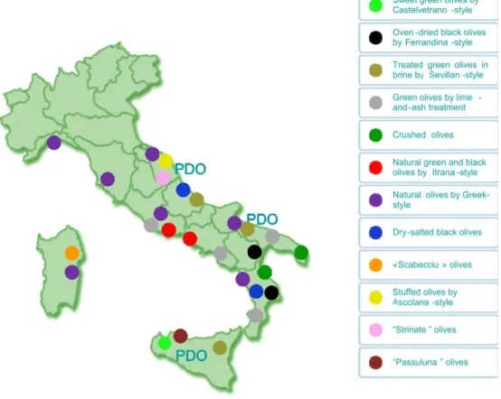
Figure 1. Distribution of table olives production in Italy. PDO: Protected Designation of Origin (Lanza, 2012). ...5
Figure 2. Bosana fruiting...8 Figure 3. Potential mechanism of action of S. boulardii on intestinal tract. On the left, effects of different pathogenic microbes are showed. On the right, seven different protective effects of S. boulardii are showed. Within the lumen of the intestine, S. boulardii may degrade toxins of pathogens, interfere with pathogenic adherence, modulate normal microbiota and preserve normal intestinal physiology (McFarland, 2010)...14
Figure 4. Scanning electron microscopy (SEM) image of mixed biofilm of LAB and yeast on olive surface. (Olifilm)...18
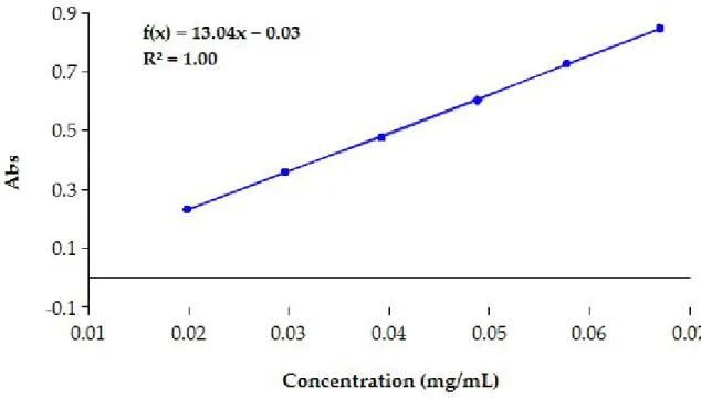
Figure 5. Calibration curve of nitrophenol for enzymatic assay...34 Figure 6. Calibration curve for spectrophotometric analysis of cholesterol. Abs, absorbance...38
Figure 7. Dendrogram obtained by RAPD-PCR pattern of 72 isolates of Bosana brines. The dendrogram was built using UPGMA algorithm. The red asterisks indicate representative strain. The right column shows Roman numerals (I- VIII) of identified clusters. Y45: control strains used to assess reproducibility of RAPD-PCR. S.b: S. boulardii. ...46
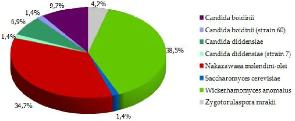
Figure 8. Distribution of genotypes of yeasts isolated from Bosana brines...48 Figure 9. β-glucosidase activity. Brown halo indicated β-glucosidase production...49 Figure 10. Example of reparametrized curve of Gompertz used to calculate MIC and NIC of C. boidinii Cb60...51
Figure 11. MIC values of selected strains isolated from Bosana brines. Standard error is indicate by vertical bars...53
Figure 12. NIC values of selected strains isolated from Bosana brines. Vertical bars indicate standard error...53
Figure 13. Oleuropein assimilation. Vertical bars indicate standard error...56 Figure 14. Percentage of survival of selected strains, L. rhamnosus GG and S. boulardii during in vitro gastric digestion. Vertical bars indicate standard error...58
Figure 15. Percentage of survival of selected strains, L. rhamnosus GG and S. boulardii during in vitro pancreatic digestion. Vertical bars indicate standard error...58
Figure16. Spectrophotometric assay for cholesterol degradation. Tube 1, sample without cholesterol, tube 2, 3 and 4 tubes with cholesterol...59
Figure 17. Percentage of cholesterol removal. Vertical bars indicate standard error....61 Figure 18. Biofilm formation (expressed as absorbance of CV) of selected yeast strains and S. boulardii in single and mixed cultures with L. pentosus TOMC 2. Vertical bars indicate standard error...64
Figure 19. Biofilm production in microtiter plate of C. boidinii Cb18 and mixed culture C. boidinii-L. pentosus TOMC 2. Control wells contained uninoculated medium...64
Figure 20. Scanning Electronic Microscopic images of biofilms formed by C. boidinii. A and B single culture of C. boidinii 18 and 60; C and D, combined cultures of C. boidinii strain 18 and 60 with L. pentosus TOMC 2...65
Figure 21. PCA analysis of yeasts variables for evaluation of technological and probiotic characteristics. A, Projection of the variables onto the plane of the first two Factors. B, Projection of the two major factors as function of yeast strains...66
Figure 22. Dendrogram generated by comparison of RAPD-PCR patterns of eight strains of C. boidinii. Y45, W. anomalus strain Y45...68
Figure 23. ANOVA representations of formed biofilm in mono and combined culture of strains of C. boidinii and LAB. Vertical bars indicated standard error...69
Figure 24. Scheme for enzymatic hydrolysis of oleuropein and products formed according to the reaction type: (1) oleuropein, (2) hydroxytyrosol, (3) glucosyl derivate, (4) aglycone, and (5) elenolic acid. (Ozdemir et al., 2014)...76
LIST OF TABLES
Tab 1. Reference strains used in this study. CSIC, Consejo Superior de Investigaciones Científicas; GD, gastric digestion; PD, pancreatic digestion...21
Tab 2. Media formulations for study ability of selected yeast strains to remove cholesterol...34
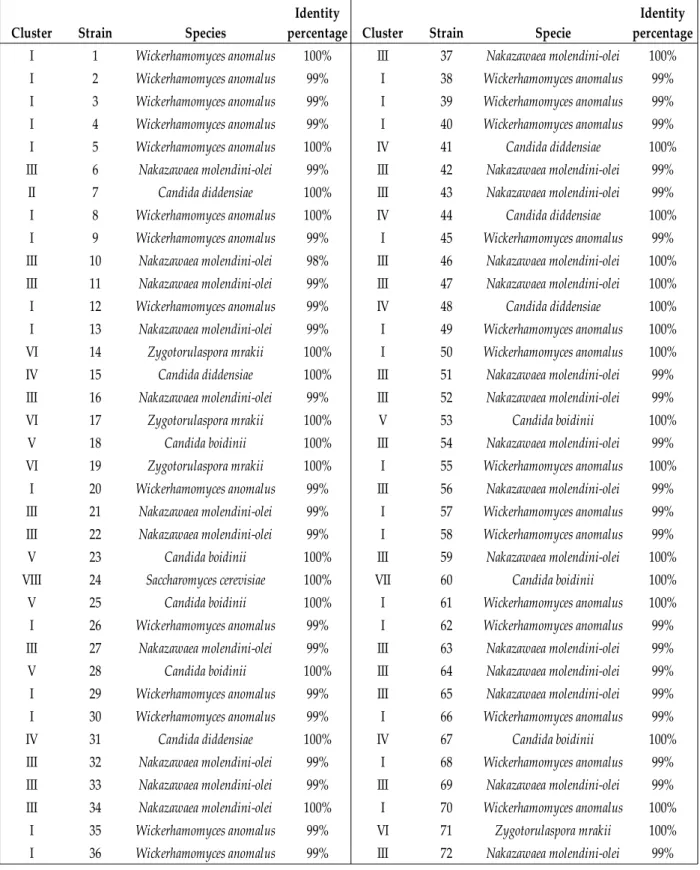
Tab 3. Identification of isolates by sequencing D1/D2 domain. The homology obtained by comparison of strains sequence with reference sequence of GenBank is reported...44
Tab 4. Enzymatic activities and biogenic production of yeast isolated from Bosana brines. Number indicates number of strains that exhibited each activity...46
Tab 5. Growth of yeast strains on YPD supplemented with 6%, 8%, 10% and 12% of NaCl. Data are expressed as number of strains growth on medium...47
Tab 6. MIC (minimum inhibitory concentration) and NIC (non inhibitory concentration) values obtained using growth model. Data was reported as mean and standard deviation in parenthesis. Different letters after each value indicate significant differences according to Scheffé test. Sbo, S. boulardii; Wa1, W. anomalus; Nm6, N. molendini-olei; Cd7 and Cd15, C. diddensiae; Zm14, Z. mrakii; Cb18 and Cb60, C. boidinii; Sc24, S. cerevisiae...49
Tab 7. Data of cellular and extracellular enzymatic activities. Data was reported as mean and standard deviation in parenthesis and are expressed in nmol mL-1 h-1. Each value followed by a letter, are significantly different in accord to Scheffé test. B-C: β-glucosidase cellular, B-E β-β-glucosidase extracellular, E-C esterase cellular, E-E esterase extracellular, L-C lipase cellular, L-E lipase extracellular...51
Tab 8. Values of maximum specific growth rate (μmax) of selected yeasts grown in
presence of oleuropein. Data are reported as mean of three replicates followed by standard deviation in parenthesis. Values followed by letter denote significant differences according to Scheffé test...52
Tab 9. Survival (%) of yeast strains to gastric and pancreatic digestion in vitro. Results followed by letter indicate significant differences according to Scheffé post-hoc test. LGG refer to strain of L. rhamnosus GG used as probiotic control...55
Tab 10. Cholesterol removal (%) of assayed yeast strains. Data were expressed as mean of three replicates followed by standard deviation in parenthesis. Each values followed by letters indicate significant differences in accordance with Scheffé test. YNB-CG, Yeast Nitrogen Base supplemented with cholesterol and glucose; YNB-CGO Yeast Nitrogen Base supplemented with cholesterol, glucose and Oxgall; YNB-CO Yeast Nitrogen Base supplemented with cholesterol and Oxgall...59
Tab 11. Biofilm formation of tested yeast strains in microtiter plate. Data are expressed as mean of six replicates followed by standard deviation in parenthesis. Letter after each value indicate significant differences according to Scheffé test...61
Tab 12. Biofilm production, measured as absorbance of CV released. Standard deviation in parenthesis. Letters after each value indicate significant differences according to Scheffé test...68
LIST OF ABBREVIATIONS
Abs – AbsorbanceAGRIS – Regional agency for agricultural research of Sardinia ANOVA – Analysis of Variance
BLAST – Basic Local Alignment Search Tool CFU – colony forming unit
CSIC – Consejo Superior de Investigaciones Científicas CV – Crystal violet
DNA – Deoxyribonucleic acid
EFTAS – Authority for Land and Agrarian Transformation of Sardinia EU – European Union
Fa – fractional area
FAO – Food and Agriculture Organization of the United Nations GC – gastric digestion
GI – gastrointestinal digestion
ISTAT – National Institute of Statistics ITS – Internal transcribed spacer LAB – Lactic acid bacteria
LSU – Large Subunit
MIC – minimum inhibitory concentration MRS – De Man, Rogosa and Shape
mQ – Milli-Q
NCBI – National Center for Biotechnology Information NIC – non inhibitory concentration
OD – Optical density
PBS – Phosphate – buffered saline PCA -Principal Component Analysis PCR – Polymerase Chain Reaction PD – pancreatic digestion
PDO – Protected Designation of Origin
RAPD-PCR – Random Amplification of Polymorphic DNA rep-PCR – Repetitive extragenic palindromic PCR
RNA – Ribonucleic acid rpm – revolution per minute
SEM – Scanning Electron Microscope
TOMC – table olive microorganism collection
UNAPROL – National Union of Associations of Producers of Olives UPGMA – Unweighted Pair Group Method
YNB – Yeast Nitrogen Base YPD – Yeast Peptone Dextrose
1. INTRODUCTION
1.1. Olive and historical background
The olive belongs to Oleacee family, genus Olea, species Olea europaea (L.). The origin of olive tree cultivation is lost in legend and tradition. Olive tree had origin approximately 6,000 to 7,000 years ago in the region corresponding to ancient Persia and Mesopotamia. The first historical mentions of olive’s presence were observed in Ebla tablets found in ancient city of Ebla, in Syria. The tablets provide information about olive oil production in III millennium BC indicating the value of olive oil was five times that of wine and two and a half times that of seed oils (Vossen, 2007). Records indicate the introduction of olive trees into Greece, Egypt, and western Turkey. Olive cultivation was reported in the Bible books such as Genesis, where the flight of the dove with an olive branch announce the end of the flood (Garrido-Fernández et al, 1997). Olives continued to move westward into Sicily, Sardinia, Italy, France, Spain, Portugal, Algeria, Tunisia, and Morocco. In these areas, there are many archaeological sites with olive-related findings (Vossen, 2007).
In Sardinia, Neolithic evidences of Cave of Refuges in Oliena show presence of olive tree. The archaeologist Giovanni Lilliu hypothesized that a little room found in nuragic village of Barumini was used as laboratory for olive oil preparations used to lighting. The expansion and prosperity of Roman Empire contributed to spread olive plantings and olive processing. Many archaeological findings and toponyms of Latin origin in Sardinia testify presence of olive, such as regions of Parteolla and Ogliastra or villages of Dolianova, Oliena and Ollastra Simaxis (Arca et al., 2010). During the Middle Age olive tree cultivation declined. The greatest expansion of olive tree cultivation in Sardinia came in XIII century under Spanish rule, which contributed significantly to spread olive and to introduce Iberian varieties. The House of Savoy in Sardinia continued the work of protection and diffusion of olive granting title of nobility to people that planted olive trees. Recently, agrarian reform performed by EFTAS (Authority for Land and Agrarian
Transformation of Sardinia) increased cultivation of olive and introduced new varieties such us Leccino, Frantoio and Biancolilla (Arca et al., 2010).
1.2 Table olives
Table olives are defined as “product prepared from the sound fruits of varieties of the cultivated olive tree which are chosen for their production of olives particularly suited to curing, and which are suitably treated or processed and offered for trade and for final consumption” (Olive International Council, 2015).
Olive fruit is a drupe consisting of epicarp, or olive skin, a mesocarp, or flesh, and an endocarp. The epicarp accounts for 1-3% of fruit, the mesocarp accounts for 84-90% while the endocarp contains the seed. The seed contains 2–4 g oil /100 g. The drupe is mainly made up of water (50%), oil (22%), carbohydrates (2%), phenolic compounds (1-3%) and inorganic substances (1.5%) (Ghanbari et al., 2012).
Fats present in table olives include oleic (C18:1; 75-80%), followed by palmitic (C16:0; 10-12%), linoleic (C18:2; 5-7%), stearic (C18:0; 2-3%), linolenic (C18:3; 0.5-1%) and palmitoleic (C16:1; 0.5-1%) acids (Lanza, 2012). Glucose is the most abundant simple sugar, followed by fructose and sucrose. Amount of these components depends from olive variety and maturity degree (Garrido-Fernández et al., 1997).
Olive fruits contain bitter components (oleuropein and phenolic compounds) that make it unpalatable. The main goal of table olives processing is to remove the bitter taste of freshly harvested fruits by oleuropein hydrolysis, obtaining preservation and improvement of organoleptic characteristics of final product (Corsetti et al., 2012). The most common table olives preparations are the Spanish or Sevillan style with about 60% of production, the Greek style and the Californian style. Spanish-style method consist of lye treatment of green olives (1.8–2.5 NaOH, w/v) followed by washes with tap water in order to remove excess of NaOH solution. After washes olive are immersed in brine (10–11% NaCl, w/v) where occurs partial or complete lactic acid fermentation (Garrido-Fernández et al., 1997). After fermentation, olives are packaged in new brine and sold. This process is
conducted by lactic acid bacteria, which degrade carbohydrate and produce lactic acid and bacteriocins, contributing to safety and quality of table olives.
In Greek style method, drupe are immersed directly in brine after harvesting. Usually, brine has a salt concentration of 6-10% (Corsetti et al., 2012). This process is slow and partial, oleuropein hydrolysis is due to enzymatic activities of esterase and β-glucosidase produced by indigenous microbiota (Garrido-Fernández et al., 1997; Tassou et al., 2002). In this olives processing, debittering is also produced by diffusion of oleuropein from fruits to the surrounding brine. The fermentation process can last 8–12 months and it is mainly conducted by mixed population of LAB and yeasts (Botta and Cocolin, 2012). The fermentation time depends on the physic-chemical conditions, the cultivar, the salt content, and the temperature (Tassou et al., 2002). Olive fruits are subjected to spontaneous fermentation performed by natural microbiota that consist of different microorganisms such as Gram negative bacteria, LAB and yeasts (Nisiotou et al., 2010). After initial phase of vigorous fermentation, prevailing microorganisms are LAB and yeasts that compete for nutrients (Tassou et al., 2002; Nisiotou et al., 2010). Presence of microorganisms depends on different factors such as salt concentration, oxygen availability, initial pH, temperature of fermentation and the presence of microorganisms contaminating the fruit (Tassou et al., 2002). Concentration of NaCl used in natural processing affects growth of LAB and improves development of fermentative yeasts (Tassou et al., 2002).
Olives prepared with Californian style method are obtained from ripe olives and are treated with NaOH solution and then darkened by oxidation with iron salts (Garrido-Fernández et al., 1997; Arroyo-López et al., 2008).
The Mediterranean diet pyramid recommends the consumption of table olives on a daily basis due to nutritional benefits associated with this fruit (Bach-Faig et al., 2011). Among vegetable foods, table olives are the most important and well-known with an estimate world production of 2,660.5 million of tons per year (Olive International Council, 2015). In the world, the main producing area is the European Union (EU), which produces
794.0 million of tons per year. Spain is the leading country for table olives production in EU (793.9 tons), followed by Greece (130.0 tons) and Italy (69.3 tons) (Olive International
Council, 2015).
1.3 Italian table olives processing
In Italy, table olives production is concentrated in central and southern regions (Figure 1). The Italian table olives labelled with Protected Designation of Origin are “Nocellara del Belice” (Sicilia), “La Bella della Daunia” (Apulia) and “Oliva Ascolana del Piceno” (Marche and Abruzzo) (Lanza, 2012).
The main Italian table olive preparations are the following. 1.3.1 Itrana style
The Itrana variety is grown mainly in Lazio region, in Latina district. This variety is used to produce the famous “Oliva nera di Gaeta“ by natural method. Harvested olives are immersed in water for about 1 month to stimulate the growth of specific microflora that contributes to the debittering of the fruits. After 10-30 days salt is added to the liquid, in quantities not exceeding 7 kg per 100 kg of fresh olives. After 4-6 months of storage in brine the olive flesh shows a typical red-wine colour and acidic taste probably due to the contribution of hetero fermentative bacteria and yeasts (Lanza, 2012).
1.3.2 Cracked olives
This type of processing is typical of the Calabria, Apulia and Campania regions. The olives fruits harvested at the green stage of ripening, are crushed with a wooden hammer. Then the olives are put in an earthenware pot with water for at least 2 weeks changing the water at least twice a day. The finished product is seasoned with garlic, pepper, oregano and other spices (Lanza, 2012).
1.3.3 Castelvetrano style
This method is used in Sicilia, almost exclusively in the Castelvetrano district using the Nocellara del Belice cultivar. Selected olives are put into plastic vessels and covered with NaOH solution (1.7-2.4% w/v). Eight hours after the lye treatment begins, 6-7 kg of salt is added. The olives are kept in this “alkaline brine” for 8- 10 days (Lanza, 2012).
1.3.4 Ferrandina style
Olives of Majatica cultivar are treated with an ancient method, Ferrandina method, whose name derives from the town Ferrandina, in Basilicata region. Initially the olives are blanched in water at 90°C for 3 min to make the skin more permeable and facilitate the osmotic processes. After blanching, the olives are salted with grinded NaCl (10:1 w/w) for 3 days and finally they are oven-dried in an air-oven at 50 °C. Blanching and salting steps
Figure 1. Distribution of table olives production in Italy. PDO: Protected
cause drupe debittering while oven drying contribute to preservation of final product (Lanza, 2012).
1.3.5 Salt dried olives
Black ripe olives are put in alternating layers with coarse salt (equivalent to 10–20% w/w of the weight of olives), orange peel and spices like oregano, fennec and garlic. The resulting olives, or “date olives”, are shrivelled in appearance and have a salty bitter-sweet taste.
1.3.6 Ascolana style
The stuffed olives by Ascolana-style are prepared from treated green olives in brine from Ascolana tenera cultivar. (PDO “Oliva Ascolana del Piceno). The meat (beef 40-70%; pork 30-50%; chicken/turkey max 10%) is triturated and browned with onion, carrot and celery in olive oil and cooked on low heat with the addition of dry white wine and salt. When cooked, meat and add-ingredients are shredded and combined with beaten egg, grated cheese and ground nutmeg. The pre-pitted olives are filled with the mixture, are dipped in flour, then beaten egg and finally in breadcrumbs (Lanza, 2012).
1.3.7 Sardinian preparations
Natural olives placed in the brine is the most important preparation of Sardinian table olives. After harvesting, sorting and washing, drupes are immersed in brine (8-14% w/v of NaCl) (Piga et al., 2002). Another traditional preparation of table olives is “Scabecciu olives”. The ripe olives Tonda di Cagliari and Pizz'e Carroga varieties are harvested and are engraved in three points. Then the olives are immersed in brine for three days, washed with water, blanched in vinegar-water and dried in the sun. Finally, they are fried with garlic and parsley and placed in oil. After about 1 month of preparation they can be consumed as appetizer.
1.4 Production of table olives
In Italy, table olives production is concentrated in the southern regions, led by Sicily (43%) and Apulia (25%), and followed by Calabria (18%), Sardinia (4.1%), Basilicata (4.1%), Lazio (3.6%) and Campania (1.9%). In Italy there are several varieties of olive trees (Olea europaea). The most widespread variety is “Coratina”, followed by minor varieties such as “Ogliarola Salentina”, “Cellina di Nardò”, “Ogliarola Barese”, “Moraiolo”, “Bosana” and “Cima di Mola” (Source UNAPROL).
Olive tree cultivation plays an important role in economy, landscape and tradition of Sardinia. Cultivation is widespread in the island, with a patchy distribution and concentrated in the area of Sassari, Parteolla, Oliena, Montiferru and Linas (Arca et al., 2010). The data reported by the Census of Agriculture (ISTAT, 2010) showed that about 39,075 hectares are planted with olive trees, of which 1,660 hectares for the production of table olives. The province of Sassari is the major cultivating area, accounting for 12,000 hectares followed by the provinces of Cagliari and Nuoro (Bandino and Dettori, 2001).
According to data released by the Regional agency for agricultural research of Sardinia (AGRIS), Sardinia produced about 10,000 quintals of table olives, contributing approximately for 1.5% to Italian production. This data is far below the average for the producer regions and shows that, despite being a major consumer, the table olive sector is somewhat of a sideline for Sardinia. In the island, production of naturally table olives is very common homemade. Most of producers are occasional producers that do not able to provide a constant quality and amount. Only ten medium companies are specialized in olives processing of drupes harvested in Sardinia. These companies use the natural method to produce table olives and are concentrated in South Sardinia.
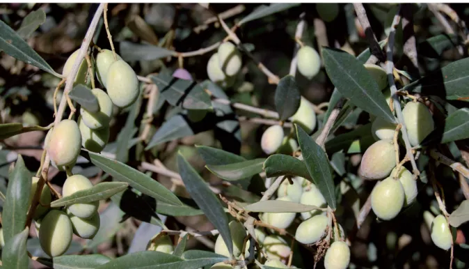
Sardinian varieties (Bosana, Nera di Villacidro, Tonda di Cagliari, Nera di Gonnos, Pizz’e Carroga, and Semidana) include six cultivar. Bosana is the most widespread cultivar in Sardinia, particularly in northern and central Sardinia.
Presumably it has a Spanish origin and it also called Palma, Tondo Sassarese and Olieddu. The areas of greatest Bosana olive concentration are Nurra and Sassarese areas, with an estimated number of tree approximately of 3 million (Bandino and Dettori, 2001).
Fruits with elliptical shape has an average weight of approximately 3 g, which have maturated take on a very bright black colour. This variety is appreciated for the quality of olive oil; big size drupes are used to make black naturally table olives (Bandino and Dettori, 2001). Bosana table olives are a typical product of North Sardinia. After harvesting, sorting and washing, drupes are immersed in brine (8-14% of NaCl) (Piga et al., 2002).
1.5 Microbiology of table olives
1.5.1 Role of lactic acid bacteria in table olives fermentation
Lactic acid bacteria play an important role in Spanish-style table olive fermentation (Garrido-Fernández et al., 1997), in fact they produced antimicrobial substances, bacteriocins and lactic acid from fermentable substrates resulting in pH decrease that
enhance table olives preservation (Ruiz-Barba and Jimenez-Diaz, 1995). LAB and yeasts were able to form mixed biofilm on olive surface during fermentation (Domínguez-Manzano et al., 2012; Arroyo-López et al., 2012). In addition, they also improve the aroma and flavour of final product (Corsetti et al., 2012).
Lactic acid bacteria species commonly found in table olives are Lactobacillus plantarum, Lactobacillus pentosus, Lactobacillus paraplantarum and Lactobacillus brevis both in Spanish style fermentation and in directly brined olives (Campaniello et al., 2005; Hurtado et al., 2008. In natural olive fermentation these species are usually observed after at least 10 days from the beginning of technological process (Tassou et al., 2002).
The two most representative species of LAB ecology in table olives are L. pentosus and L. plantarum. Hurtado et al., (2010) reported that in case of the co-inoculation of strains of L. pentosus and L. plantarum in Aberquina fermentation only the first dominated. Panagou et al., (2008) observed better performance of L. pentosus starter than L. plantarum inoculated separately as starter cultures in black olive fermentations.
1.5.2 Role of yeasts in table olives production
Sometimes, yeasts can be dominant microorganisms during table olive fermentation, causing a milder taste and less preservation of final product. This fact was reported by (Tassou et al., 2002).
In the first phase of fermentation, yeasts could produce gas pocket as CO2 causing
formation of blisters, a spoilage known as “Alambrado” (Lamzira et al., 2005; Hernández et
al., 2007). During phase of packing yeasts could cause clouding of brines, swollen
containers, off flavours, odours and resistance to preservatives (Turantaş et al., 1999).
Recently, role of yeasts table olives production has been re-considered by different researchers. The presence of yeasts during table olives processing is linked to raw materials and related to type of fermentation process (Botta and Cocolin, 2012). Concerning fermentation processing, the role of yeasts is very important in directly brined
olive processing (Garrido-Fernández et al., 1997). Bautista-Gallego et al., (2011) reported high amount of yeast in directly table olive than in Spanish style olives. The most important yeast species found in table olive belong to Aureobasidium, Candida, Cryptococcus, Issatchenkia, Pichia, Rhodotorula, Saccharomyces, and Zygotorulaspora genera (Bevilacqua et al., 2013).
In relation to growth matrices (brine and drupe), Hernández et al., (2007) found higher count of yeasts in brine than fresh fruit. The dominant species detected in the brine belong to Wickerhamomyces anomalus, Kluyveromyces marxianus and Saccharomyces cerevisiae species while mains species detected in the drupe was Cryptococcus laurentii.
During fermentation, yeasts produce volatile compounds and organic acids such as such as ethanol, glycerol, higher alcohols and acetaldehyde that define organoleptic characteristics and maintenance of texture during table olive processing (Fernández et al., 1995; Arroyo-López et al., 2008). Aponte et al., (2010) detected several aromatic compound synthesized by catabolism of fatty acids during green Sicilian table olive fermentations dominated by yeasts. Yeast improve the growth of lactic acid bacteria. Tsapatsaris and Kotzekidou, (2004) reported improvement of growth of L. plantarum after inoculation of Debaryomyces hansenii in same olive juice.
Regarding organoleptic characteristics of table olives, yeasts exhibit important enzymatic activities such as catalase, esterase, lipase, and β-glucosidase which are strains-specific (Botta and Cocolin, 2012). Catalase positive yeasts contribute to preserving olives against unsaturated fatty acid oxidation and peroxide formation (Hernández et al., 2007). Esterase and lipolytic activities are desirable because they can enhance the flavour of olives through the formation of volatile compounds that can be generated by the catabolism of free fatty acids (Bautista-Gallego et al., 2011). Candida boidinii, D. hansenii, and Torulaspora delbrueckii showed high lipase activity in assays in vitro (Psani and Kotzekidou, 2006; Rodríguez-Gómez et al., 2010).
Enzymatic activities are also important for polyphenols degradation. Spanish style processing produced a large amount of wastewater and water with high concentrations of phenols. β-glucosidase produced by yeasts may lower the level of phenols in wastewater produced by lye treatment of olives. So, β-glucosidase can be used to design an eco-friendly olive process to replace lye treatment.
Yeasts produce toxic proteins or glycoproteins, called killer toxins, which are able to decrease or inhibit the growth of bacteria, fungi and non-desirable yeasts (Bevilacqua et al., 2015). Therefore, yeast can be used as biocontrol agents in olive fermentation in order to inhibit development of spoilage and pathogen microorganisms. For example, Hernández et al., (2008) studied killer activity of strains isolated from green seasoned table olives against spoilage microorganisms. Moreover, Psani and Kotzekidou, (2006) found strains of D. hansenii and Torulaspora delbrueckii with action against food-borne pathogen.
Moreover, yeasts can synthesize vitamins (Ruiz-Barba and Jimenez-Diaz, 1995), compounds with killer activity (Psani and Kotzekidou, 2006). The vitamins synthesized or accumulated by yeasts are thiamin, pantothenic acid, nicotinic acid and pyridoxine (Abbas, 2006).
Table olives can be considered itself as a functional food, because of their high content in dietary fiber, antioxidant compounds, vitamins, anticancer substances (Rodriguez-Gomez et al., 2014). The functional value of table olives can be improved by turning this fermented vegetable in a carrier of beneficial microorganisms to the human body. In this sense, probiotics are defined as “live microorganisms which when administered in adequate amounts confer a health benefit on the host” (FAO, 2001). So addition of probiotic microorganisms is useful to produce functional foods.
Probiotic characteristics of LAB was extensively reviewed in the past. Lavermicocca et al., (2005) used table olives as a vehicle to incorporate probiotic bacteria species into the human gastrointestinal tract. Many LAB belong to L. pentosus, L. plantarum, Lactobacillus
paracasei isolated from table olives exhibited probiotic characteristics in vitro (Argyri et al., 2013; Bautista-Gallego et al., 2013). Moreover, LAB with probiotic properties can be used in the packing stage for table olive fortification. In fact, L. pentosus TOMC 2 survived under packing conditions for long time (Rodríguez-Gómez et al., 2014). Recent study showed that use of probiotic L. pentosus as starter for fermentation of table olives by Spanish style method led to decrease growth of Enterobacteriaceae (Rodríguez-Gómez et al., 2013).
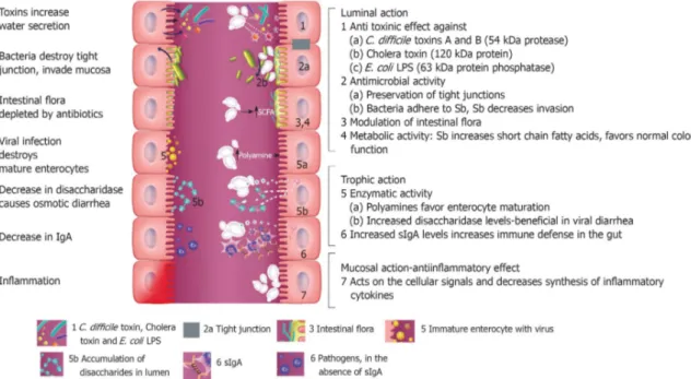
In addition to LAB, many authors focused on probiotic traits of yeast such as adhesion to intestinal mucosa, survival and/or persistence into the gut, antimycogenic and antibacterial activity, production of vitamin B-complex, biodegradation of phytate complexes and reduction of cholesterol levels. Saccharomyces boulardii is a probiotic yeast isolated for the first time from litchi fruit in Indochina in 1923 (Moslehi-Jenabian et al., 2010). It is the only yeast whose use has been recommended for its clinical effects (Hatoum et al., 2012). In fact, several authors described the effect of S. boulardii on antibiotic-associated diarrhoea (D’Souza et al., 2002; Erdeve et al., 2004). Moreover, administration of S. boulardii with antibiotics showed beneficial effect on Clostridium difficile associated diarrhoea. Several case works reported that some patients with recurrent C. difficile diarrhoea treated with S. boulardii showed improvement. This effect is due to production of a 54 kDa protease that degrade toxin A and toxin B secreted from C. difficile, leading to a reduction on effects of C. difficile infection (Castagliuolo et al., 1996, 1999). Finally, the effect of S. boulardii in prevention of traveller's diarrhoea has been demonstrated (McFarland, 2010).
Numerous studied showed benefits of S. boulardii to patients affected with inflammatory bowel disorders (Hatoum et al., 2012). Mechanism of action of S. boulardii is showed in Figure 3.
Regarding yeasts associated to table olives, only Psani and Kotzekidou, (2006), Silva et al., (2011) and Bonatsou, et al., (2015). Psani and Kotzekidou, 2006 and Silva et al., (2011) reported that D. hansenii, T. delbrueckii, Candida oleophila and S. cerevisiae tolerated 0.3% (w/v) bile salts and low pH. Finally, Bonatsou et al., (2015) in a study about selection a multifunctional starter recorded high resistance to simulated digestion of Pichia guilliermondii.
Phytase is an enzyme that catalyses the phytic acid hydrolysis, an indigestible form of phosphorus present in vegetables and cereals. Therefore, phytase activity enhances adsorption of iron, zinc, magnesium and phosphorus. These enzyme has been common detected in several microorganisms. Among yeasts, Candida krusei (Issatchenkia orientalis), Schwanniomyces castellii, Debaryomyces castellii, W. anomalus, Pichia rhodanensis, Pichia spartinae, Cryptococcus laurentii, Rhodotorula gracilis, S. cerevisiae, Saccharomyces kluyveri, T.
Figure 3. Potential mechanism of action of S. boulardii on intestinal tract. On the left, effects of
different pathogenic microbes are showed. On the right, seven different protective effects of
S. boulardii are showed. Within the lumin of the intestine, S. boulardii may degrade toxins of
pathogens, interfere with pathogenic adherence, modulate normal microbiota and preserve normal intestinal physiology (McFarland, 2010).
delbrueckii, Candida spp. and Kluyveromyces lactis have been identified as phytase producers (van der Aa Kühle et al., 2005). This activity was detected for the first time in P. guilliermondii, Pichia kluyveri, and Metschnikowia pulcherrima isolated from Greek natural table olives (Bonatsou et al., 2015).
Yeasts synthesize folic acid (vitamin B9) but mammals lack the ability to synthesize them and therefore are dependent on their intake in the diet. Folates are involved in the synthesis of nucleic acids and metabolise amino acids necessary to cell division, therefore folate producing yeasts can be used in table olives fortification (Arroyo-López et al., 2012).
High level of cholesterol in blood causes hypercholesterolemia, and it is related to risk factor in coronary heart disease. Low cholesterol and saturated fatty acid diet is suggested to people who have high serum cholesterol level. Decrease of cholesterol is a trait exhibited by several yeast strains such as K. lactis, S. cerevisiae, and I. orientalis, usually found in table olives (Arroyo-López et al., 2012).
Candida and Saccharomyces species produce natural antioxidants such us carotenoids,
citric acid, D-erythro ascorbic acid, tocopherols and glutathione with interesting antioxidant properties (Abbas, 2006). The synthesis of bioactive antioxidants can reduce the oxidative degeneration of fatty substances improving human health (Arroyo-López et
al., 2012).
Mycotoxins are toxic secondary metabolites produced by fungi belonging mainly to the
Aspergillus, Penicillium and Fusarium genera. The presence ochratoxins A, citrinin and
aflatoxin B has been recently reported in table olives by El Adlouni et al., (2006). Some yeasts exhibited detoxification ability of mycotoxins. For example, S. cerevisiae showed ability to inhibit ochratoxin A production in Aspergillus carbonarius and Aspergillus
ochraceus (Cubaiu et al., 2012). Therefore, S. cerevisiae could be used for mycotoxins
1.5.3 Molecular methods to study table olives microbiota
In the past, characterization of microorganisms associated to table olives processing was performed by biochemical and morphological methods. Recently, molecular methods have revolutionized approaches used to study microorganisms of fermented products. In particular, two strategies were used. In culture-dependent approach, in which microorganisms are isolated from food matrix by traditional microbiological techniques and are subsequently studied by molecular method. In culture independent method, DNA or RNA are extracted and analysed directly from food matrix. This method allows to investigate complexity of table olives microbiota, detecting the not cultivable microorganisms. On the contrary, culture independent methods are not adapt to select new starter culture (Botta and Cocolin, 2012).
As far as yeasts, the restriction analysis of the ITSs (ITS1 and ITS2) and the 5.8S rRNA gene described by Esteve-Zarzoso et al., (1999) has been used to identify a total of 132 species of yeasts (Botta and Cocolin, 2012). Restriction analysis of 5.8 rRNA gene and internal transcribed spacers ITS1 and ITS2 and sequence analysis of 26S rRNA gene (Kurtzman and Robnett, 1998) allowed the identification of yeast species such as I. occidentalis, Geotrichum candidum, Hanseniaspora guilliermondi, S. cerevisiae and C. boidinii (Arroyo-López et al., 2006). W. anomalus, C. boidinii and Debaryomyces etchellsii,
predominant species in French naturally black olives, were identified by rDNA ITS method and sequencing of the D1/D2 region of the 26S rRNA gene (Coton et al., 2006). Hurtado et al., (2008) identified the species C. boidinii, Candida sorbosa, Candida diddensiae,
Candida membranifaciens, K. lactis, Pichia membranifaciens, W. anomalus, P. kluyveri, and Rhodotorula glutinis from of Arbequina table olives. However, in 2011, sequencing of
domains 1 and 2 (D1/D2) of the LSU rRNA gene and/or the ITS1-5.8SITS2 region were proposed as universal barcode for fungi (Schoch et al., 2012).
With reference to molecular typing, molecular method commonly used both for LAB and yeasts in fermented food are rep- PCR and RAPD-PCR (Andrighetto et al., 2000; Tofalo
et al., 2013; Mari et al., 2016). Molecular typing was used to group the isolates from olive
fermentations in relation to their similarity and subsequently to choose the representative strains, which are identified using the techniques described above (Torriani et al., 2001; Gardini et al., 2006).
1.6 Biofilm
Biofilms are defined as structured communities of microbial cells enclosed in a self-produced polymeric matrix adherent to abiotic or biotic surfaces (Steenackers et al., 2012). Presence of biofilms consists of yeast or LAB or both microorganisms in fermented food such as wine and beer is well documented. Regarding yeasts, biofilm formation of S. cerevisiae and its mechanism was described from several authors (Budroni et al., 2005; Zara et al., 2009; Legras et al., 2016).
An essential requirement for potential probiotic/human-healthy olive yeasts is that they must be able to adhere to olive skin to survive during storage/packaging and to be finally ingested by consumers at elevated numbers (Arroyo-López et al., 2012). For this reason, recently attention of researchers focus on biofilm formed on olive skin during the fermentation (Domínguez-Manzano et al., 2012; Arroyo-López et al., 2012; Grounta and Panagou, 2014; Grounta et al., 2015; León-Romero et al., 2016). Imagines obtained by Scanning Electronic Microscope of biofilm revealed presence of yeasts and LAB embedded in an extracellular matrix. Arroyo-López et al.,(2012) and Domínguez-Manzano et al., (2012) reported presence of mixed species biofilm formed by yeasts and LAB on the epidermis of Gordal and Manzanilla fruits processed according to Spanish style. Pichia galeiformis, C. sorbosa and G. candidum for the yeast species, and L- pentosus for LAB on olive skin. In a scanning electron microscopy study, Grounta and Panagou, (2014) showed the formation of biofilms between L. pentosus and P. membranifaciens on oxidized Greek black olives, while Benítez-Cabello et al., (2015) described the formation of microbial biofilms on the epidermis of directly brined “natural” green olives. Recently, Grounta et al., (2015) have investigated the formation of biofilm on abiotic surface of fermentation
vessels and observed that the most abundant species were W. anomalus, D. hansenii and P. guilliermondii.
Probably, the formation of this mixed biofilms improves their viability during the passage through the gastrointestinal tract (Arroyo-López et al., 2012). However, biofilm formation have an impact on table olive processing.
In a recent work about in vitro ability of L. pentosus and yeast isolated from table olives to form biofilm, was reported strong biofilm production in Candida boidinii- L. pentosus mixed cultures (León-Romero et al., 2016).
Figure 4. Scanning electron microscopy (SEM) image of mixed biofilm of
1.7 Aim of thesis
The aim of this PhD thesis were:
● to design an autochthonous multifunctional starter for table olive processing in order to improve and optimize the fermentation process and the quality and safety of the final product.
● to identify genes related to biofilm formation by genome sequencing of different strains of C. boidinii.
In order to obtain the first objective, characterization of the yeast biodiversity of Bosana brines using RAPD-PCR analysis with primer M13 and sequencing of D1/D2 domain of 26S gene was carried out. Moreover, qualitative and quantitative technological properties and probiotic features were investigated. Subsequent bioinformatic analysis was applied for obtaining dendrogram of yeast population and selecting representative strains to evaluate:
● enzymatic activities (esterase, β-glucosidase and lipase) and assimilation of oleuropein by quantitative spectrophotometric assays;
● quantitative resistance to NaCl by modelling techniques;
● probiotic properties in vitro: resistance to simulated gastric and pancreatic digestion and cholesterol removal;
● in vitro biofilm formation in mono-culture and mixed culture with Lactobacillus
pentosus.
Data were analysed by ANOVA and multivariate analysis to select yeast strains with the best probiotic and technological characteristics.
To achieve the second objective, genotyping of eight strains of C. boidinii and biofilm-forming ability in vitro were assessed. Finally, sequencing of whole genomes of all C.
2. MATERIALS AND METHODS
2.1 Strains
This study was conducted on 72 isolates of yeasts obtained from Bosana table olives brines, a widespread traditional variety of Northern Sardinia, collected during the 2003/2004 season (Pinna, 2005). Drupes of Bosana cultivar were harvested during the 2003/2004 season at black ripening stage. After sorting, sizing, and washing, the olive fruits were dried and were put into barrels. Each barrel had 20 L of total capacity, and was filled with 10.5 kg of olive fruits and 9.5 L of brine (6% NaCl w/v), with a ratio/brine drupe of 1:1.1. Spontaneous fermentation started when olives were placed in the brine. Yeasts were isolated as described by Pinna (2005) and stored for long term in yeast extract, peptone, glucose and glycerol (YEPGly) at -80°C. Reference strains are listed in Table 1.
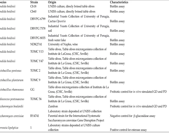
Saccharomyces cerevisiae strain BY4741 was used in β-glucosidase assay and Yarrowia lipolytica 1 in esterase assay. Yarrowia lipolytica 1 belong to UNISS collection of University of Sassari. Wickerhamomyces anomalus strain Y45 was used to assess reproducibility of RAPD-PCR. Strains of Saccharomyces boulardii and Lactobacillus rhamnosus GG were used as reference strain for probiotic test. Lactobacillus pentosus TOMC 2 was used in biofilm assay because it is capable to produce biofilm. Strain Y45, TOMC 2 and L. rhamnosus GG belongs to Table Olives Microorganisms Collection (TOMC) of Institute of la Grasa (CSIC, Seville). Eight strains of C. boidinii in single culture and combined culture with the LAB L. pentosus TOMC 2, L. plantarum TOMC 9 and Pediococcus pentosacesus TOMC 56, were studied for their capability to form biofilm in vitro. Strains DBVPG 6799, DBVPG 7578 and DBVPG 8035 were purchased from Industrial Yeasts Collection of University of Perugia. Strain NDK27A1 was provided by Giuseppe Blaiotta (University of Naples). All isolates and the reference stains were stored at -80 ° C in a Yeast extract Peptone Dextrose medium (YPD) or YM containing 30% (v / v) glycerol until use. Lactobacillus pentosus TOMC 2, L. plantarum TOMC 9 and Pediococcus pentosaceus TOMC 56 were maintained in MRS broth supplemented with 25% (v/v) of glycerol.
Tab 1. Reference strains used in this study. CSIC, Consejo Superior de Investigaciones Cientificas;
GD, gastric digestion; PD, pancreatic digestion.
Species Strain Origin Characteristics
Candida boidinii Cb18 UNISS culture, direcly brined table olives Biofilm assay
Candida boidinii Cb60 UNISS culture, directly brined table olives Biofilm assay
Candida boidinii DBVPG 6799
Biofilm assay
Candida boidinii DBVPG 7578 Biofilm assay
Candida boidinii DBVPG 8035 Biofilm assay
Candida boidinii NDK27A1 University of Naples, wine Biofilm assay
Candida boidinii TOMC Y13 Biofilm assay
Candida boidinii TOMC Y47 Biofilm assay
Lactobacillus pentosus TOMC 2 Biofilm assay
Lactobacillus plantarum TOMC 9 Biofilm assay
Lactobacillus rhamnosus GG
Pediococcus pentosaoceus TOMC 56 Biofilm assay
Saccharomyces boulardii Zambon
Saccharomyces cerevisiae BY4741 Negative control for β-glucosidase assay
1 Positive control for esterase assay
Industrial Yeasts Collection of University of Perugia, Cactus Opuntia
Industrial Yeasts Collection of University of Perugia, soil
Industrial Yeasts Collection of University of Perugia, fresh water lake
Table olives, Table olives microrganisms collection of Institute de LaGrasa, (CSIC, Seville)
Table olives, Table olives microrganisms collection of Institute de La Grasa, (CSIC, Seville)
Table olives, Table olives microrganisms collection of Institute de La Grasa, (CSIC, Seville)
Table olives, Table olives microrganisms collection of Institute de La Grasa, (CSIC, Seville)
Table olives microrganisms collection of Institute de La
Grasa, (CSIC, Seville) Probiotic control for in vitro simulated GD and PD Table olives, Table olives microrganisms collection of
Institute de La Grasa, (CSIC, Seville)
Probiotic control for in vitro simulated GD and PD Laboratory strain deposited at UNISS collection
Parental strain for the International Systematic Saccharomyces cerevisiae Gene Disruption Project
2.2 Culture media
Media used for culture maintenance and development are shown below.
YEP medium supplemented with glycerol (YEPGly)
Yeast extract 10.0 g/L
Glucose 20.0 g/L
Peptone 20.0 g/L
Glycerol 20%
Yeast Mold Agar (YM, Difco)
Yeast Extract 3.0 g/L Malt Extract 3.0 g/L Peptone 5.0 g/L Dextrose 10.0 g/L Agar 20.0 g/L YM Broth (Difco) Yeast Extract 3.0 g/L Malt Extract 3.0 g/L Peptone 5.0 g/L Dextrose 10.0 g/L YPD agar Yeast extract 10 g/L Dextrose 20 g/L Peptone 20 g/L Agar 20 g/L
YPD Broth
Yeast extract 10.0 g/L
Dextrose 20.0 g/L
Peptone 20.0 g/L
Man, Rogosa and Sharpe broth (MRS, Oxoid)
Magnesium sulphate 0.2 g/L
Dipotassium hydrogen phosphate 2.0 g/L
Triammonium citrate 2.0 g/L Yeast Extract 4.0 g/L Sodium acetate 3 H2O 5.0 g/L Lab-Lemco Powder 8.0 g/L Peptone 10.0 g/L Sorbitan mono-oleate 1 mL Dextrose 20.0 g/L
Yeast Nitrogen Base (YNB, Difco)
Ammonium sulphate 5.0 g/L Monopotassium Phosphate 1.0 g/L Magnesium Sulphate 0.5 g/L Sodium Chloride 0.1 g/L Calcium Chloride 0.1 g/L L-Histidine Monohydrochloride 0.01 g/L Arbutin agar (β-glucosidase activity)
Arbutin
(hydroquinone-β-D-glucopyranoside)
5 g/l
Yeast extract 10 g/l
Agar 15g/L
Tributyrin Agar (esterase activity)
Special peptone 5 g/L
Yeast extract 3 g/L
Tributyrin 10 g/L
Agar 12 g/L
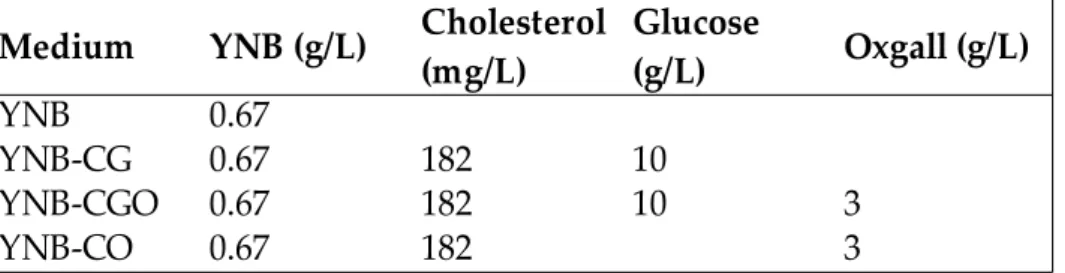
YNB-CG (cholesterol removal)
Yeast Nitrogen Base 0.67 g/L
Glucose 10 g/L
Cholesterol 182 mg/L
YNB-CGO (cholesterol removal)
Yeast Nitrogen Base 0.67 g/L
Glucose 10 g/L
Oxgall 3 g/L
Cholesterol 182 mg/L
YNB-CO (cholesterol removal)
Oxgall 3 g/L
2.3 Isolates identification
2.3.1 Getyping and identification of yeast isolates
Genomic DNA extraction was performed in accordance with the protocol of Senses-Ergul et al. (2012).
Yeasts were cultured overnight in YPD broth at 28°C in an orbital shaker at 150 rpm. Cultures were centrifuged at 14,000 rpm for 5 minutes. After removing of the supernatant, the pellet obtained was washed and re-suspended in 1 mL of sterile ultrapure water. The cells were lysed with 200 µL of breaking buffer (2% Triton X v / v, 1% SDS v / v, 100 mM NaCl, 10 mM Tris-HCl, 1 mM Na2EDTA) and treated with 200 µL solution of
phenol-chloroform-isoamyl alcohol (25: 24: 1, v / v) and 0.3 g of glass beads of diameter <106 µm (Sigma-Aldrich, USA). Samples were vortexed for 3 minutes and centrifuged for 5 minutes at 14,000 rpm. After centrifugation, the DNA contained in the supernatant was precipitated with the addition of 3 volumes of 96% ethanol, washed with 70% ethanol, dried and re-suspended in sterile water. Extracted DNA was quantified using the Spectrostar Nano spectrophotometer (BMG LABTECH, Germany) through the absorbance reading at 260 nm.
2.3.2 Molecular identification of yeast species
In the present work, to identify yeast isolates at species level, two methods were applied:
i. random amplified polymorphic DNA polymerase chain reaction (RAPD-PCR) with primer M13;
ii. sequencing of D1/D2 region of 26S rDNA gene. 2.3.2.1 Random amplified polymorphic DNA analysis
Random amplified polymorphic DNA polymerase chain reaction (RAPD-PCR) with primer M13 was used for discrimination at strain level. Reaction mixture was carried out
in 25 µl containing 80-100 ng/µL of genomic DNA, 5 µl of 5X buffer MyTaqTM (Bioline,
USA), 1 µL (25 pmol/ µL) of M13 primer (5 'GAGGAGGGTGGCGGTTCT 3', (Huey and Hall, 1989), 0.1 µL (0.5 U) of MyTaqTM DNA polymerase (Bioline, USA).
The PCR was conducted in a MasterCycler Pro (Eppendorf, Germany) and the program consisted of the following steps: 1) initial denaturation step at 95 ° C for 5 minutes; 2) 45 cycles of: 93 ° C for 45 seconds, 44.5 ° C for one minute, 72 ° C for one minute; 3) final extension was performed at 72 ° C for 6 minutes (Andrighetto et al., 2000). Negative control PCR mixture without DNA was included in all experiments.
2.3.2.2 PCR products electrophoresis
RAPD-PCR products were separated by electrophoresis on 2% (w/v) agarose gel. Electrophoresis was carried out at 70 V for 90 minutes in TAE buffer (Promega, USA). Ladder 1 kb (Invitrogen, USA) was used as size marker of DNA fragments. After electrophoresis, gel was stained with ethidium bromide solution (10 mg/L). Gel was visualized with a transilluminator EnduroTM GDS (Labnet International, Inc., USA). The
RAPD profiles were captured and analysed using BioNumerics 6.6 software (Applied Maths, Belgium). Dendrogram was built with UPGMA (Unweighted Pair Group Method) method and Pearson correlation (optimization 0.5%, curve smoothing 0%). Reproducibility and sensibility of RAPD-PCR were evaluated by comparing the profiles of strain Y45 obtained from different DNA extractions, different PCR and different gels performed in different days.
2.3.2.3 Sequencing of D1/D2 region of 26S rDNA gene
To corroborate the clustering analysis and for identification of strains, the 26S rDNA gene of all isolates was further sequenced. The genomic DNA was amplified by using the primers NL1 and NL4 (Kurtzman and Robnett, 1998). Sequence of primers were:
NL1 5’-GCATATCAATAAGCGGAGGAAAAG-3’ NL4 5’-GGTCCGTGTTTCAAGACGG-3’
PCR reactions were carried out in a volume of 25 µL containing 1 µL of DNA, 1.25 µl (25 pmol/ µl) of each primer, 5 µl of 5X buffer (Bioline, USA) and 0.1 µL (0.5 U) of Taq polymerase (Bioline, USA). All amplification reactions were carried out in a thermocycler MasterCycler Pro (Eppendorf, Germany) programmed as follows: initial denaturation at 94 °C for 5 minutes, followed by 35 cycles at 94 ° C for 1 minute, 55.5 ° C for 2 minutes, 72 ° C for 2 minutes followed by a final extension at 72 ° C for 10 minutes.
2.3.5 DNA purification and sequencing
Amplification products were analysed by electrophoresis on agarose gel (1% w/v) stained with ethidium bromide (10 mg/L). Molecular weight of amplified DNA fragments was evaluated by comparison with a DNA ladder 1 kb ladder plus (Invitrogen, USA). Purification of the PCR products was carried out using Isolate DNA kit (Bioline, USA) according to the manufacturer’s instructions. Quality and amount of purified PCR product was evaluated by agarose gel electrophoresis (1% w/v). An amount of 10 µL of purified product with forward primer NL1 was used for sequencing by Stab Vida (Lisbon, Portugal).
2.3.6 Sequences analysis
The alignment of sequences was performed with software MEGA 5.0 (Molecular Evolution Genetic Analysis). Sequences were compared with sequences deposited in data bank Gene Bank (http://www.ncbi.nlm.nih.gov/) using software BLAST (Basic Local Alignment Search Tool) of NCBI (National Center for Biotechnology Information, USA).
2.4 Qualitative technological characterization
For qualitative technological characterization, yeast were cultured in YPD agar and incubated at 28°C for two days. Each experiment was carried out in duplicate.
2.4.1 Qualitative salt resistance assay
Growth in presence of NaCl was evaluated on YPD agar supplemented with different concentration of NaCl (Microbiol, Italy): 6%, 8%, 10% and 12% (w/v). Strains were streaked
on plates and incubated at 25°C for 5 days. Results were expressed with the follows: “-” no growth, “+” growth.
2.4.2 β-glucosidase assay
Evaluation of the β-glucosidase activity was assayed on Arbutin agar (Caridi et al., 2005). Yeasts were streaked on the surface of the plates and incubated at 25 ° C for 7 days. The strain Saccharomyces cerevisiae BY4741 was used as negative control. The β-glucosidase activity was indicated by a darkening of the medium and was expressed according as follows: 0 = colourless; 1 = light brown; 2 = brown; 3 = dark brown.
2.4.3 Esterase activity
The esterase activity was evaluated on Tributyrin Agar (Sigma-Aldrich, Milan) (Hernández et al., 2007) supplemented with an emulsion of Tributyrin (Sigma-Aldrich, Milan) and Tween 80 (Acros Organics, Belgium). The yeasts were seeded on TAT and incubated at 25 ° C for five days. Esterase activity is highlighted by a clear halo around the colonies and expressed as follows: "-" (no halo), "+" (alone defined). Strain 1 of Yarrowia lipolytica was used as positive control.
2.4.4 Catalase activity
The catalase activity of yeasts was assayed by the method of Whittenbury (1964), by adding 3% (v / v) of hydrogen peroxide (H2O2) on the colonies grown on YPD agar.
Catalase activity was indicated by the development of oxygen bubbles due to H2O2
decomposition indicated the presence of catalase activity. The results were expressed as "-" (no activity) or "+" (activities).
2.4.5 Production of biogenic amines
The production of biogenic amines was evaluated as decarboxylase activity by the method described by Gardini et al. (2006). Briefly, was used YNB containing 1 g / l of amino acids precursors of biogenic amines (L-lysine, L-proline, glycine, alanine, L phenylalanine, L-tyrosine, L-histidine, ornithine and arginine hydrochloride) with violet
purple as indicator. Strains were grown on medium containing 0.67 g of YNB, 1 g / l of amino acids precursors of biogenic amines (L-lysine, L-proline, glycine, alanine, L phenylalanine, L-tyrosine, L-histidine, ornithine and arginine hydrochloride) and 0.06 g of bromocresol purple (Sigma-Aldrich, Milan). Medium was sterilized by filtration. Plates were incubated at 25 ° C for 72 hours. Medium without amino acids inoculated with yeast was used as a control. If yeasts have a decarboxylase activity, pH increases and a colour change of medium from yellow to purple occurs.
2.5 Quantitative technological characterization
Assays performed on representative strains chosen from each cluster are described in this section.
2.5.1 Quantitative salt resistance assay
Resistance to NaCl of yeast strains was monitored by Bioscreen C automated spectrophotometer (Labsystem, Finland) with a wideband filter (420-580 nm) (Bonatsou et al., 2015). Briefly, overnight culture were centrifuged at 10,000 rpm and collected cells were re-suspended in sterile saline solution reaching an initial OD approximately 0.2. An amount of 20 µL of yeast inoculum was dispensed into wells of Honeycomb plate with 330 µL of sterilized YM broth (pH 4.5) supplemented with 0, 10, 30, 50, 70, 80, 90, 100, 110, 120,130, 150 and 180 g/L of NaCl. Uninoculated wells were included in Honeycomb plate to subtract noise signal. Microplates were incubated at 28°C with shaking of 3 seconds before each OD measurements. All experiments were performed in triplicate. Measures of OD were used to obtain 351 growth curve (9 yeast strains x 13 NaCl concentrations x 3 replicates). The curves were used to calculate the fractional area (fa) by comparison of area of OD/time curve of positive control (strain growth in absence of salt) with area of OD/time curve of the samples:
where areatest is the area of yeast strains and areacont is the area of positive control. Integral
of areas under OD curves were calculated with OriginPro software 7.5 (OriginLab Corporation, USA).
Results in form of fractional area vs logarithm of concentration of NaCl were analysed using the modified Gompertz equation (Bonatsou et al., 2015):
y= exp(- (x/ln(MIC)/exp( - (ln(ln(NIC)/ln(MIC))/2.71828))))(-2.71828/ln(ln(NIC)/ln(MIC)))))
where y is the fa, x is logarithm of NaCl concentration, MIC is the minimum NaCl concentration (g/L) that inhibits yeast growth and NIC is NaCl concentration where a negative effect on yeast growth begin to be detected.
2.5.2 Quantitative enzymatic assays
Quantitative enzymatic assays of β-glucosidase, esterase and lipase were determined in accordance with the protocol described by Bonatsou et al. (2015).
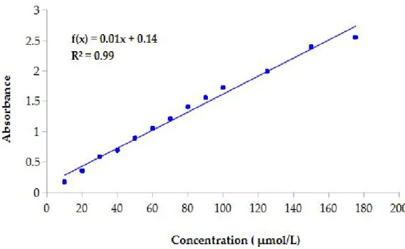
Enzymatic activities were measured using a spectrophotometric assay based on the ability of β-glucosidase, esterase and lipase to catalyse hydrolysis of 4-nitrophenyl-β-D glucoside and 4-nitrophenyl palmitate and p-nitrophenyl stearate to 4-nitrophenol, respectively. The concentration of 4-nitrophenol released from each chromogenic substrate was determined by reading the absorbance at 410 nm. A calibration curve was plotted by measuring the absorbance of 12 solution containing different concentration (10-200 µmol/L) of nitrophenol (Figure 5).
The coefficient obtained from the slope of the calibration curve was used to calculate the molar extinction coefficient (ε).
Molar extinction coefficient allows to determine concentration of nitrophenol in according to Beer-Lambert law:
A= εcl
where A is the absorbance of sample, ε is the molar extinction coefficient, c is the 4-nitrophenol concentration and l is the path length. The equation of calibration curve was A = 0.01c + 0.14. The correlation coefficient (R2) was 0.99 and indicated a good linearity
between concentration and absorbance.
Before each quantitative enzymatic assays, yeasts were cultured in YM broth and incubated at 28°C for 72 hours at 150 rpm. Yeasts culture were centrifuged at 10,000 rpm for 15 minutes to collect cells. Then, supernatant was sterilised by filtration with 0.2 µm filter (Millipore Co., USA) and used as extracellular fraction. Harvested cells were washed