Dottorato di Ricerca in Scienze Farmaceutiche IX-Ciclo NS
2007-2010
Design and synthesis of “small molecules” as
antiviral and radiotracer agents
Tutor PhD Student
Prof. Paolo de Caprariis Grazia Sellitto
Coordinator
“Those who live in freedom has a good reason to live, fight and die”.
“Chi vive nella libertà ha un buon motivo per vivere, combattere e
morire”.
Table of Content
Abstract... PageChapter 1
...1-55 Introduction 1.1 Viruses 21.2 Viruses and human disease 26 1.3 Antiviral strategies 34
Chapter 2
...56-91 Results and discussion2 Aim of project 58 2.1 Synthesis of Arbidol analogs using a convenient five-step scheme 58 2.2 Alternative synthetic scheme 67 2.3 Biological assays 69 2.4 Evaluation of mechanism of Arbidol
anti-influenza action 79
Chapter 3
...92-115 Synthesis of a P.E.T. radiotracer for tumour hypoxia: 18F-FAZA3 Introduction 94 3.1 Tumour hypoxia 94 3.2 Synthesis of 18FAZA 109
Conclusions
...116-119Chapter 4
...120-145 Experimental part4.1 Chemistry 122 4.2 Biological assays 142 4.3 Circular dichroism studies 145
References
...146-154I) Design, synthesis and evaluation of antiviral activity of Arbidol analogs. II) Evaluation of mechanism of Arbidol anti-influenza action.
III) Synthesis and characterization of a P.E.T. radiotracer for tumor hypoxia:
1- (5- [18F] Fluoro-5-deoxy-α-D-arabinofuranosyl) -2-nitroimidazole or
18
F-FAZA.
The initial research activity concerned the design and synthesis of indole derivatives using as a lead compound Arbidol (ARB), a compound that exerts immunomodulatory, antioxidant, antiviral and antimetastatic effects1. ARB is a Russian-made potent broad-spectrum antiviral with demonstrated activity against a number of enveloped and non-enveloped viruses, and pH-dependent and pH-independent viruses. It exhibits antiviral activity against a number of viruses including influenza A (H1N1, H2N2 and H3N2), B and C viruses,
respiratory syncytial virus (RSV), adenovirus type 7, coxsackie B3 virus, parainfluenza type 5 and rhinovirus type 14, avian coronavirus, infectious bronchitis virus and Marek disease virus, hepatitis B virus and hepatitis C virus. The wide spectrum of ARB‘s activity suggests that ARB targets
common critical step(s) in virus – cell interaction. Several studies have shown that the affinity of ARB for lipid membranes could account for its antiviral actions, together with a differential level of interaction with key motifs in glycoproteins of different viruses. Its antiviral activity toward viruses is due probably to a direct effect of ARB on virus-cell membrane interactions where ARB intercalates into membranes and induces membrane alterations. This leads to excessive stabilization of cell membranes, which become resistant to virus fusion and in some cases (HCV) to virus replication2-4. The known biological properties of Arbidol led us to focus on its derivatives as potential antiviral agents. In order to maintain antiviral activity we preserved the groups
positions of the indole ring). Moreover we introduced different substituents at the 2- and 5-position of the indole ring to investigate the influence of these variations on antiviral activity. The synthesis of Arbidol derivatives has been established through the validation of two synthetic schemes. Then, to evaluate anti-HCV and anti-HSV activity of synthesized compounds, biological assays were made. ARB derivatives showed antiviral activity comparable and, in some cases, even better than those of lead Arbidol, on both systems. In particular, it was shown that synthesized compounds are fusion inhibitors on both viruses and also non-selective inhibitors of HCV replication.
The second part of the present research project concerned the study of mechanism of Arbidol anti-influenza action. There are experimental evidences that ARB does not affect viral neuraminidase (NA, a surface protein of influenza virus) activity. It affects early post-adsorption stages of virus replication with possible involvement of the second surface viral protein, the haemagglutinin (HA). Arbidol could act increasing influenza virus HA stability and preventing low pH induced HA transition to its fusogenic state, thus blocking infection at the viral fusion stage5. To support this hypothesis, the interaction of Arbidol with the N-terminal hydrophobic fusion domain of haemagglutinin (HA) was evaluated. Therefore, the peptide host-guest (P20H6) was synthesized using techniques of Solid Phase Peptide Synthesis (SPPS) and
Circular dichroism studies were made6. From these studies we demonstrated that Arbidol interacts with the haemagglutinin fusion domain at pH 5 and 7, through changes in the secondary structure of peptide.
At the end of present Ph.D. project, I spent six months at the University of Aberdeen where I worked to the synthesis of a PET (Positron Emission
characterized by hypoxia (solid tumours, ischemia, stroke)7, but it is not routinely used and synthesized in Scotland. The work done is an important starting point for the introduction of 18F-FAZA in Scotland with the aim of using it in clinical imaging and research. In particular, following a detailed bibliography research on this compound and its synthesis, that is not fully reported, and a subsequent optimization of the synthetic scheme used, the 18 F-FAZA precursor was obtained:
1-α-D-[5’-O-Toluenesulfonyl-2’,3’-Di-Oacetylarabinofuranosyl]-2-nitroimidazole (DAcTs-AZA).
[1] Surinova B. N., Karpova N. A., Kumin Yu. C. (1995). Khim. Farm. Zh.;Chem.-Pharm. J., 3, 14-15.
[2] Eve-Isabelle Pe´cheur, Dimitri Lavillette, Fanny Alcaras, Jennifer Molle, Yury S. Boriskin, Michael Roberts, Francüois-Loı¨c Cosset, and Stephen J. Polyak; Biochemistry 2007, 46, 6050-6059.
[3] Yury S Boriskin1, Eve-Isabelle Pécheur and Stephen J Polyak; Virology
Journal 2006, 3:56:1-9.
[4] Irina A. Leneva, Rupert J. Russell, Yury S. Boriskin, Alan J. Hay; Antiviral Research 2009; 81:132–140.
[5] Irina A. Leneva, Rupert J. Russell, Yury S. Boriskin, Alan J. Hay; Antiviral Research; 81 (2009) 132–140.
[6] Xing Han, John H. Bushweller, David S. Cafiso and Lukas K. Tamm;
Nature Structural Biology; 2001, Volume 8 N.8.
[7] Ernst J. Postema, Alexander J. B. McEwan, Terence A. Riauka, Piyush Kumar, Dacia A. Richmond, Douglas N. Abrams, Leonard I. Wiebe; Eur J
CHAPTER 1
INTRODUCTION
- 2 - 1.1 Viruses
The word virus is from the Latin virus referring to poison and other noxious substances, first used in English in 13921. A virus (Fig.1.1) is a small infectious agent that can replicate only inside the living cells of organisms. Most viruses are too small to be seen directly with a light microscope. Viruses infect all types of organisms, from animals and plants to bacteria and archaea2. They are found in almost every ecosystem on Earth and are the most abundant type of biological entity3,4.
Figure 1.1: Virus.
Despite continuing advances, established and emerging viruses remain major causes of human disease, with dramatic costs in mortality, morbidity and economic terms. In addition to acute diseases, viruses cause at least 15–20% of human cancers and are implicated in neurological and other chronic disorders. One of many challenges in controlling viruses and virus-mediated diseases is that viruses show an amazing diversity in basic characteristics and life cycles, including differences in virion structure, replication strategies, genetic organization, gene expression and many other fundamental processes. Therefore, even the very processes against which antivirals are targeted often differ radically among virus classes5.
- 3 -
1.1.1 Discovery
Viral diseases such as rabies have affected humans for many centuries. There is hieroglyphical evidence of Polio in the ancient Egyptian empire. However, the cause of these diseases was discovered relatively recently. In 1717, Mary Montagu, the wife of an English ambassador to the Ottoman Empire, observed local women inoculating their children against Smallpox. In the late 18th century, Edward Jenner observed and studied Miss Sarah Nelmes, a milkmaid who had previously caught Cowpox and was subsequently found to be immune to Smallpox, a similar, but devastating virus. Jenner developed the first vaccine, based on these findings, and smallpox is currently all but wiped out.
In the late 19th century Charles Chamberland (known today as the Chamberland filter or Chamberland-Pasteur filter) developed a porcelain filterwith pores smaller than bacteria. Thus, he could pass a solution containing bacteria through the filter and completely remove them from the solution (Fig. 1.2)6. This filter was used to study the first documented virus,
tobacco mosaic virus. Shortly afterwards, Dimitri Ivanovski published
experiments showing that crushed leaf extracts of infected tobacco plants were still infectious even after filtering the bacteria from the solution7.
Figure 1.2: Filtration of a mixture of bacteria and viruses.
At about the same time, several others documented filterable disease-causing agents, with several independent experiments showing that viruses were
- 4 -
different from bacteria, yet they could also cause disease in living organisms. These experiments showed that viruses are orders of magnitudes smaller than bacteria. The term virus was coined by the Dutch microbiologist Martinus Beijerinck. He observed that the agent multiplied only in cells that were dividing, but as his experiments did not show that it was made of particles, he called it a contagium vivum fluidum (soluble living germ) and re-introduced the word virus8-10. Beijerinck maintained that viruses were liquid in nature, a theory later discredited by Wendell Stanley.
Since the initial discovery of tobacco mosaic virus by Martinus Beijerinck in 1898,[2] about 5,000 viruses have been described in detail11, although there are millions of different types12.
In 1957, equine arterivirus and the cause of bovine virus diarrhea (a
pestivirus) were discovered. In 1963, the hepatitis B virus was discovered by
Baruch Blumberg13, and in 1965, Howard Temin described the first retrovirus. Reverse transcriptase, the key enzyme that retroviruses use to translate their RNA into DNA, was first described in 1970, independently by Howard Martin Temin and David Baltimore14. In 1983 Luc Montagnier's team at the Pasteur Institute in France, first isolated the retrovirus now called HIV15.
1.1.2 Origins
The origins of modern viruses are not entirely clear, and there may not be a single mechanism of origin that can account for all viruses. As viruses do not form fossils, molecular techniques have been the most useful means of hypothesising how they arose16. These techniques rely on the availability of ancient viral DNA or RNA, but, unfortunately, most of the viruses that have been preserved and stored in laboratories are less than 90 years old. There are three main hypotheses that try to explain the origins of viruses17,18:
Regressive hypothesis: Viruses may have once been small cells that parasitised larger cells. Over time, genes not required by their parasitism
- 5 -
were lost. The bacteria rickettsia and chlamydia are living cells that, like viruses, can reproduce only inside host cells. They lend support to this hypothesis, as their dependence on parasitism is likely to have caused the loss of genes that enabled them to survive outside a cell. This is also called the degeneracy hypothesis.
Cellular origin hypothesis: Some viruses may have evolved from bits of DNA or RNA that "escaped" from the genes of a larger organism. The escaped DNA could have come from plasmids (pieces of naked DNA that can move between cells) or transposons (molecules of DNA that replicate and move around to different positions within the genes of the cell)19. Once called "jumping genes", transposons are examples of mobile genetic elements and could be the origin of some viruses. They were discovered in maize by Barbara McClintock in 1950. This is sometimes called the
vagrancy hypothesis20.
Coevolution hypothesis: Viruses may have evolved from complex molecules of protein and nucleic acid at the same time as cells first appeared on earth and would have been dependent on cellular life for billions of years. Viroids are molecules of RNA that are not classified as viruses because they lack a protein coat. However, they have characteristics that are common to several viruses and are often called subviral agents21. Viroids are important pathogens of plants22. They do not code for proteins but interact with the host cell and use the host machinery for their replication23. The hepatitis delta virus of humans has an RNA genome similar to viroids but has protein coat derived from hepatitis B
virus (HBV) and cannot produce one of its own. It is therefore a defective
virus and cannot replicate without the help of HBV24. Similarly, the virophage 'sputnik' is dependent on mimivirus, which infects the protozoan
- 6 -
presence of other virus species in the host cell are called satellites and may represent evolutionary intermediates of viroids and viruses.
Computer analysis of viral and host DNA sequences is giving a better understanding of the evolutionary relationships between different viruses and may help identify the ancestors of modern viruses. To date, such analyses have not helped to decide on which of these hypotheses are correct. However, it seems unlikely that all currently known viruses have a common ancestor and viruses have probably arisen numerous times in the past by one or more mechanisms20.
1.1.3 Structure
Viruses display a wide diversity of shapes and sizes, called morphologies. Generally viruses are much smaller than bacteria. Most viruses that have been studied have a diameter between 10 and 300 nanometres. Some filoviruses have a total length of up to 1400 nm; their diameters are only about 80 nm26. Most viruses cannot be seen with a light microscope so scanning and transmission electron microscopes are used to visualise virions.[59] To increase the contrast between viruses and the background, electron-dense "stains" are used.
A complete virus particle, known as a virion, consists of nucleic acid surrounded by a protective coat of protein called a capsid. These are formed from identical protein subunits called capsomers. Viruses can have a lipid "envelope" derived from the host cell membrane. The capsid is made from proteins encoded by the viral genome and its shape serves as the basis for morphological distinction27. Virally coded protein subunits will self-assemble to form a capsid, generally requiring the presence of the virus genome. Complex viruses code for proteins that assist in the construction of their
- 7 -
the association of viral capsid proteins with viral nucleic acid is called a
nucleocapsid (Fig. 1.3).
Figure 1.3: Virion structure.
The capsid and entire virus structure can be mechanically (physically) probed through atomic force microscopy28-30.
In general, there are four main morphological virus types:
Helical: These viruses are composed of a single type of protomer stacked around a central axis to form a helical structure, which may have a central cavity, or hollow tube (Fig. 1.4). This arrangement results in rod-shaped or filamentous virions: these can be short and highly rigid, or long and very flexible. Long helical particles must be flexible in order to prevent forces snapping the structure. The genetic material, generally single-stranded RNA, but ssDNA in some cases, is housed on the inside of the tube, protected from the outside, through a bond into the protein helix by interactions between the negatively charged nucleic acid and positive charges on the protein. Overall, the length of a helical capsid is related to the length of the nucleic acid contained within it and the diameter is dependent on the size and arrangement of capsomers. The well-studied Tobacco mosaic virus is an example of a helical virus31.
- 8 -
Figure 1.4: Diagram of a helical capsid.
Icosahedral: Cubic (=Icosahedral) capsid symmetry results in a spherical appearance of viruses at low magnification but actually consists of capsomers arranged in a regular geometrical pattern, similar to a soccer ball, hence they are not truly ―spherical‖. Capsomers are ring shaped structures constructed from five to six copies of protomers. These associate via non-covalent bonding to enclose the viral nucleic acid, though generally less intimately than helical capsids, and may involve one or more protomers.
Most, if not all, of the polygonal viruses are icosahedral; like a geodesic dome, they are formed by equilateral triangles. Each triangle is composed of protein subunits (capsomeres), often in the form of hexons (multiples of six) that are the building blocks of the capsid. There are 12 vertices (the apical junctions of these triangles), each comprising a penton (five subunits). In order to construct a capsid from repeated subunits, a virus must 'know the rules' which dictate how these are arranged. For an icosahedron, the rules are based on the rotational symmetry of the solid, which is known as 2-3-5 symmetry (Fig. 1.5)32:
An axis of two-fold rotational symmetry through the centre of each edge.
An axis of three-fold rotational symmetry through the centre of each face.
- 9 -
Figure 1.5: Icosahedral symmetry33.
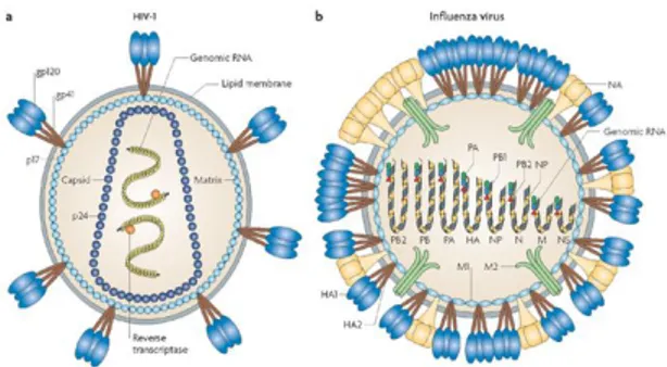
Envelope: Some species of virus envelop themselves in a modified form of one of the cell membranes, either the outer membrane surrounding an infected host cell, or internal membranes such as nuclear membrane or endoplasmic reticulum, thus gaining an outer lipid bilayer known as a viral envelope. This membrane is studded with proteins coded for by the viral genome and host genome; the lipid membrane itself and any carbohydrates present originate entirely from the host. The HIV and influenza virus use this strategy (Fig. 1.6). Most enveloped viruses are dependent on the envelope for their infectivity.
Figure 1.6: Examples of two enveloped viruses. (a) HIV has an icosahedral capsid
which contains two positive-strand RNA copies and (b) Influenza A virus has an helical capsid which contains eight negative-strand RNA segments34.
Complex: These viruses possess a capsid that is neither purely helical, nor purely icosahedral, and that may possess extra structures such as protein tails or a complex outer wall. Some bacteriophages, such as Enterobacteria phage
- 10 -
bound to a helical tail, which may have a hexagonal base plate with protruding protein tail fibres. This tail structure acts like a molecular syringe, attaching to the bacterial host and then injecting the viral genome into the cell27.
Figure 1.7: Structural overview of the T4 phage.
The poxviruses are large, complex viruses that have an unusual morphology. The viral genome is associated with proteins within a central disk structure known as a nucleoid. The nucleoid is surrounded by a membrane and two lateral bodies of unknown function. The virus has an outer envelope with a thick layer of protein studded over its surface. The whole virion is slightly pleiomorphic, ranging from ovoid to brick shape35. Mimivirus is the largest known virus, with a capsid diameter of 400 nm. Protein filaments measuring 100 nm project from the surface. The capsid appears hexagonal under an electron microscope, therefore the capsid is probably icosahedral.
Some viruses that infect Archaea have complex structures that are unrelated to any other form of virus, with a wide variety of unusual shapes, ranging from spindle-shaped structures, to viruses that resemble hooked rods, teardrops or even bottles. Other archaeal viruses resemble the tailed bacteriophages, and can have multiple tail structures36.
- 11 -
1.1.4 Genome
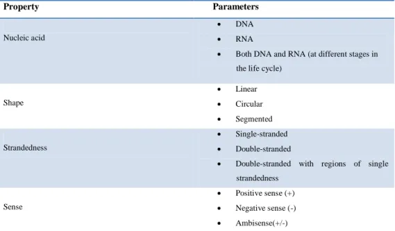
An enormous variety of genomic structures can be seen among viral species; as a group they contain more structural genomic diversity (Table 1.1) than plants, animals, archaea, or bacteria. There are millions of different types of viruses; though only about 5,000 of them have been described in detail. A virus has either DNA or RNA genes and is called a DNA virus or a RNA virus respectively. The vast majority of viruses have RNA genomes. Plant viruses tend to have single-stranded RNA genomes and bacteriophages tend to have double-stranded DNA genomes11,12.
Property Parameters
Nucleic acid
DNA
RNA
Both DNA and RNA (at different stages in the life cycle)
Shape Linear Circular Segmented Strandedness Single-stranded Double-stranded
Double-stranded with regions of single strandedness
Sense
Positive sense (+)
Negative sense (-)
Ambisense(+/-)
Table 1.1: Genomic diversity among viruses.
Viral genomes are circular, as in the polyomaviruses, or linear, as in the
adenoviruses. The type of nucleic acid is irrelevant to the shape of the
genome. Among RNA viruses, the genome is often divided up into separate parts within the virion, in which case it is called segmented. Each segment often codes for one protein and they are usually found together in one capsid.
- 12 -
Every segment is not required to be in the same virion for the overall virus to be infectious, as demonstrated by the brome mosaic virus27.
A viral genome, irrespective of nucleic acid type, is either single-stranded or double-stranded. Single-stranded genomes consist of an unpaired nucleic acid, analogous to one-half of a ladder split down the middle. Double-stranded genomes consist of two complementary paired nucleic acids, analogous to a ladder. Some viruses, such as those belonging to the Hepadnaviridae, contain a genome that is partially double-stranded and partially single-stranded.
For viruses with RNA or single-stranded DNA, the strands are said to be ether
positive-sense (called the plus-strand) or negative-sense (called the
minus-strand), depending on whether it is complementary to the viral messenger RNA (mRNA). Positive-sense viral RNA is identical to viral mRNA and thus can be immediately translated by the host cell. Negative-sense viral RNA is complementary to mRNA and thus must be converted to positive-sense RNA by an RNA polymerase before translation. DNA nomenclature is similar to RNA nomenclature, in that the coding strand for the viral mRNA is complementary to it (−), and the non-coding strand is a copy of it (+).
Genome size varies greatly between species. The smallest viral genomes code for only four proteins and have a mass of about 106 Daltons; the largest have a mass of about 108 Daltons and code for over one hundred proteins37. RNA viruses generally have smaller genome sizes than DNA viruses because of a higher error-rate when replicating, and have a maximum upper size limit. Beyond this limit, errors in the genome when replicating render the virus useless or uncompetitive. To compensate for this, RNA viruses often have segmented genomes where the genome is split into smaller molecules, thus reducing the chance of error. In contrast, DNA viruses generally have larger genomes because of the high fidelity of their replication enzymes.
- 13 -
Viruses undergo genetic change by several mechanisms. These include a process called genetic drift where individual bases in the DNA or RNA mutate to other bases. Most of these point mutations are "silent", they do not change the protein that the gene encodes, but others can confer evolutionary advantages such as resistance to antiviral drugs. Antigenic shift occurs when there is a major change in the genome of the virus. This can be a result of recombination or reassortment.
Viruses with segmented genomes, such as influenza virus, have another mechanism for generating diversity: reassortment38-39.
When an influenza virus infects a cell, the individual RNA segments enter the nucleus. There they are copied many times to form RNA genomes for new infectious virions. The new RNA segments are exported to the cytoplasm, and then are incorporated into new virus particles which bud from the cell. If a cell is infected with two different influenza viruses, the RNAs of both viruses are copied in the nucleus.
When new virus particles are assembled at the plasma membrane, each of the 8 RNA segments may originate from either infecting virus.
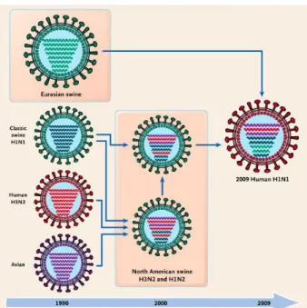
The progeny that inherit RNAs from both parents are called reassortants. One example of the evolutionary importance of reassortment is the exchange of RNA segments between mammalian and avian influenza viruses that give rise to pandemic influenza. For example, the 2009 H1N1 pandemic strain is a reassortant of avian, human, and swine influenza viruses (Fig. 1.8).
- 14 -
Figure 1.8: History of Reassortment Events in the Evolution of the 2009 Influenza A
(H1N1) Virus.
Reassortment can only occur between influenza viruses of the same type. Why
influenza A viruses never exchange RNA segments with influenza B or C viruses is not understood. However, the reason is probably linked to the
packaging mechanism that ensures that each influenza virion contains at least one copy of each RNA segment38-40.
Genetic recombination is the process by which a strand of DNA is broken and then joined to the end of a different DNA molecule. This can occur when viruses infect cells simultaneously and studies of viral evolution have shown that recombination has been rampant in the species studied. Recombination is common to both RNA and DNA viruses41.
1.1.5 Replication cycle
Viral populations do not grow through cell division, because they are acellular. Instead, they use the machinery and metabolism of a host cell to produce multiple copies of themselves, and they assemble in the cell.
- 15 -
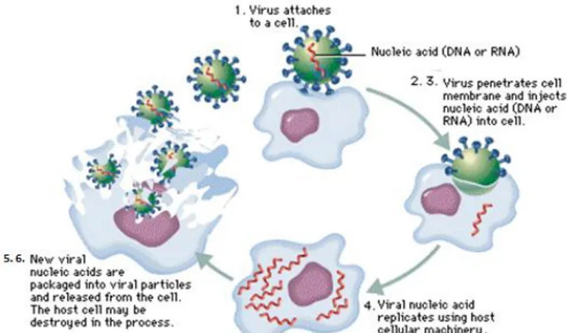
The life cycle of viruses (Fig. 1.9) differs greatly between species but there are six basic stages in the life cycle of viruses:
Figure 1.9: Life cycle of viruses.
1. Attachment is a specific binding between viral capsid proteins and specific receptors on the host cellular surface. This specificity determines the host range of a virus. For example, HIV infects only human T cells, because its surface protein, gp120, can interact with CD4 and chemokine receptors on the T cell's surface. This mechanism has evolved to favour those viruses that only infect cells in which they are capable of replication. Attachment to the receptor can induce the viral-envelope protein to undergo changes that results in the fusion of viral and cellular membranes.
2. Penetration follows attachment; viruses enter the host cell through receptor mediated membrane fusion or endocytosis (Fig. 1.10A). This is often called viral entry. In the case of fusion the viral membrane fuses with the cell membrane and the protein capsid is released. Once in the cytoplasm the protein capsid is removed and the viral genome is freed. Depending on the nature of the virus the viral genome may stay in the cytoplasm or it may enter the nucleus before beginning the next stage of
- 16 -
the life cycle. In the case of engulfment, o endocytosis, the initial adsorption process is the same, but rather than directly fusing with the cell membrane, the cell is stimulated to take-up or engulf the entire eukaryotic virus. Once inside the cytoplasm the enveloped eukaryotic virus is said to be in a vescicle. Subsequently, the two lipid membranes of the vesicle fuse, releasing the capsid covered virus. The capsid is then removed and the viral genome is freed for the next stage of viral replication4,7. Some of the non-enveloped viruses like picornaviruses and phages are capable of directly injecting their genome into the host cell (Fig. 1.10B). The infection of plant cells is different from that of animal cells. Plants have a rigid cell wall made of cellulose, so most viruses can only get inside the cells after trauma to the cell wall. However, viruses such as tobacco
mosaic virus can also move directly in plants, from cell to cell, through
pores called plasmodesmata42.
Figure 1.10: A. Mechanisms of viral penetration: fusion and endocytosis; B.
Genome injection in the host cell by phage.
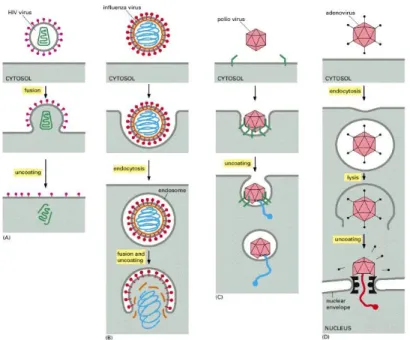
3. Uncoating is a process in which the viral capsid is degraded by viral
enzymes or host enzymes thus releasing the viral genomic nucleic acid. A key step in uncoating is the acidification of the content of the endosome to a pH of about 5, owing to the activity of a proton pump present in the membrane. The low pH causes rearrangement of coat components, which then expose normally hidden hydrophobic sites. They bind to the lipid bilayer of the membrane, causing the extrusion of the viral core into the
- 17 -
cytosol. For influenza virus, the acid-sensitive component is the core HA2
unit of the haemagglutinin, for adenoviruses, it is the penton base. There are four virus uncoating strategies: some enveloped viruses, such as HIV, fuse directly with the host cell plasma membrane to release their capsid
(green) into the cytosol (Fig. 1.11A). Other enveloped viruses, such as influenza virus, first bind to cell-surface receptors, triggering
receptor-mediated endocytosis. When the endosome acidifies, the virus envelope fuses with the endosomal membrane, releasing the nucleocapsid (blue) into the cytosol (Fig. 1.11B). Poliovirus, a nonenveloped virus, binds to a receptor (green) on the host cell surface and then forms a pore in the host cell membrane to extrude its RNA genome (blue) (Fig. 1.11C).
Adenovirus, another nonenveloped virus, uses a more complicated
strategy. It induces receptor-mediated endocytosis and then disrupts the endosomal membrane, releasing part of the capsid into the cytosol. The capsid eventually docks onto a nuclear pore and releases its DNA genome
(red) directly into the nucleus (Fig. 1.11D)43.
- 18 -
4 Replication involves synthesis of viral messenger RNA (mRNA) for
viruses except positive sense RNA viruses, viral protein synthesis and assembly of viral proteins and viral genome replication.
5 Assembly of the virus particles, in which post-translational modification of
the viral proteins often occurs. In viruses such as HIV, this modification (sometimes called maturation) occurs after the virus has been released from the host cell.
6 Viruses are released from the host cell by lysis (a process that kills the cell
by bursting its membrane) or budding (a process that involves taking a portion of the cell membrane) (Fig. 1.12A). Enveloped viruses (e.g., HIV) typically are released from the host cell by budding. During this process the virus acquires its envelope, which is a modified piece of the host's plasma membrane. Some viruses undergo a lysogenic cycle (Fig. 1.12B) where the viral genome is incorporated by genetic recombination into a specific place in the host's chromosome. The viral genome is then known as a "provirus" or, in the case of bacteriophages a "prophage". Whenever the host divides, the viral genome is also replicated. The viral genome is mostly silent within the host; however, at some point, the provirus or prophage may give rise to active viruses, which lyse their host cells44.
- 19 -
The genetic material within viruses, and the method by which the material is replicated, vary between different types of viruses:
DNA viruses: The genome replication of most DNA viruses takes place in the cell's nucleus. If the cell has the appropriate receptor on its surface, these viruses enter the cell by fusion with the cell membrane or by endocytosis. Most DNA viruses are entirely dependent on the host cell's DNA and RNA synthesising machinery, and RNA processing machinery. The viral genome must cross the cell's nuclear membrane to access this machinery45.
RNA viruses: Replication usually takes place in the cytoplasm. RNA viruses can be placed into about four different groups depending on their modes of replication. The polarity (whether or not it can be used directly to make proteins) of the RNA largely determines the replicative mechanism, and whether the genetic material is single-stranded or double-stranded. RNA viruses use their own RNA replicase enzymes to create copies of their genomes.
Reverse transcribing viruses: These replicate using reverse transcription, which is the formation of DNA from an RNA template. Reverse transcribing viruses containing RNA genomes use a DNA intermediate to replicate, whereas those containing DNA genomes use an RNA intermediate during genome replication. Both types use the reverse transcriptase enzyme to carry out the nucleic acid conversion. Retroviruses often integrate the DNA produced by reverse transcription into the host genome. They are susceptible to antiviral drugs that inhibit the reverse transcriptase enzyme, e.g. zidovudine and lamivudine. An example of the first type is HIV, which is a retrovirus. Examples of the second type are the Hepadnaviridae, which includes Hepatitis B virus46.
- 20 -
1.1.6 Classification
Classification seeks to describe the diversity of viruses by naming and grouping them on the basis of similarities. In 1962, André Lwoff, Robert Horne, and Paul Tournier were the first to develop a means of virus classification, based on the Linnaean hierarchical system. This system bases classification on phylum, class, order, family, genus, and species. Viruses were grouped according to their shared properties (not those of their hosts) and the type of nucleic acid forming their genomes. Viruses are mainly classified by phenotypic characteristics, such as morphology, nucleic acid type, mode of replication, host organisms, and the type of disease they cause47-48.
Currently there are two main schemes used for the classification of viruses: the
ICTV system and Baltimore classification system. Accompanying this broad
method of classification are specific naming conventions and further classification guidelines set out by the International Committee on Taxonomy
of Viruses.
a. ICTV classification
The International Committee on Taxonomy of Viruses (ICTV) developed the current classification system and wrote guidelines that put a greater weight on certain virus properties to maintain family uniformity. A unified taxonomy (a universal system for classifying viruses) has been established. The 7th lCTV Report formalised for the first time the concept of the virus species as the lowest taxon (group) in a branching hierarchy of viral taxa49. However, at present only a small part of the total diversity of viruses has been studied, with analyses of samples from humans finding that about 20% of the virus sequences recovered have not been seen before, and samples from the environment, such as from seawater and ocean sediments, finding that the large majority of sequences are completely novel.
- 21 - The general taxonomic structure is as follows:
Order (-virales) Family (-viridae) Subfamily (-virinae) Genus (-virus) Species (-virus)
So far, six orders have been established by the ICTV: the Caudovirales,
Herpesvirales, Mononegavirales, Nidovirales, Picornavirales, and Tymovirales. Most notably, species names generally take the form of virus.
The establishment of an order is based on the inference that the virus families contained within a single order have most likely evolved from a common ancestor. The majority of virus families remain unplaced. Currently (2009) 6 orders, 87 families, 19 subfamilies, 348 genera, and 2,288 species of virus have been defined50.
b. Baltimore classification
The Baltimore classification of viruses is based on the method of viral mRNA synthesis. The Nobel Prize-winning biologist David Baltimore devised the Baltimore classification system. The ICTV classification system is used in conjunction with the Baltimore classification system in modern virus classification.
Baltimore classification is a classification system that places viruses into one of seven groups depending on a combination of their nucleic acid (DNA or RNA), strandedness (single-stranded (ss) or double-stranded (ds)), Sense, and method of replication (may or may not use reverse transcriptase, RT). Additionally, ssRNA viruses may be either sense (+) or antisense (−). These groups are designated by Roman numerals and discriminate viruses depending on their mode of replication, and genome type.
- 22 -
The replication strategy of the virus depends on the nature of its genome. Viruses can be placed in one of the seven following groups (Fig. 1.13)51:
Figure 1.13: Baltimore classification.
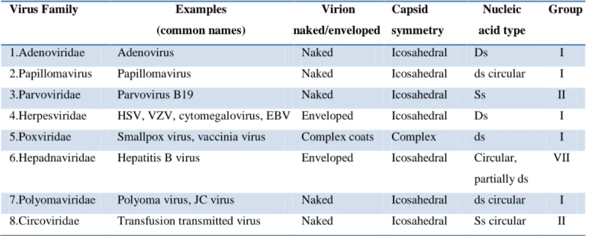
Viruses belonging to group I and II are DNA viruses (Table 1.2).
Group I: Double-stranded DNA (dsDNA) (Adenoviruses; Herpesviruses; Poxviruses; etc).
Viruses having a dsDNA genome that make mRNA by asymmetric transcription. Some replicate in the nucleus e.g adenoviruses using cellular proteins. Poxviruses replicate in the cytoplasm and make their own enzymes for nucleic acid replication.
Group II: Single-stranded (+)sense DNA (ssDNA) (Parvoviruses; circoviruses)
Replication occurs in the nucleus, involving the formation of a (-)sense strand, which serves as a template for (+)strand RNA and DNA synthesis.
- 23 -
Virus Family Examples (common names) Virion naked/enveloped Capsid symmetry Nucleic acid type Group
1.Adenoviridae Adenovirus Naked Icosahedral Ds I
2.Papillomavirus Papillomavirus Naked Icosahedral ds circular I
3.Parvoviridae Parvovirus B19 Naked Icosahedral Ss II
4.Herpesviridae HSV, VZV, cytomegalovirus, EBV Enveloped Icosahedral Ds I 5.Poxviridae Smallpox virus, vaccinia virus Complex coats Complex ds I 6.Hepadnaviridae Hepatitis B virus Enveloped Icosahedral Circular,
partially ds VII
7.Polyomaviridae Polyoma virus, JC virus Naked Icosahedral ds circular I 8.Circoviridae Transfusion transmitted virus Naked Icosahedral Ss circular II
Table 1.2: DNA viruses.
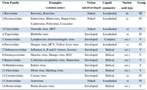
Viruses belonging to group III, IV and V are RNA viruses (Table 1.3).
Group III: Double-stranded RNA (dsRNA) (Reoviruses; Birnaviruses)
Viruses having a dsRNA genome that make mRNA by asymmetric transcription (Fig. 1.14A). Most have multiple pieces of dsRNA, each of which apparently contains the information for the synthesis of a single protein.
Group IV: Single-stranded (+)sense RNA ((+)ssRNA) (Picornaviruses; Togaviruses; Flaviviruses; Coronaviruses; etc)
Viruses having an ssRNA genome that make mRNA of base sequence identical to the genomic RNA. Infect, small amounts of (–)RNA are produced and used as templates to greatly amplify viral (+)RNA, which is encapsidated into new progeny virions (Fig. 1.14B).
- 24 -
Figure 1.14: Distinct life cycle of dsRNA (A), (+)RNA viruses(B).
Group V: Single-stranded (-)sense RNA ((−)ssRNA) (Orthomyxoviruses; Rhabdoviruses; etc)
Viruses having an ssRNA genome that make mRNA with a base sequence complementary to the genomic RNA. Must have a virion particle RNA directed RNA polymerase.
Virus Family Examples
(common names) Virion naked/enveloped Capsid symmetry Nucleic acid type Group
1.Reoviridae Reovirus, Rotavirus Naked Icosahedral ds III 2.Picornaviridae Enterovirus, Rhinovirus, Hepatovirus,
Cardiovirus, Poliovirus, Coxsackie
Naked Icosahedral ss IV
3.Calciviridae Norwalk virus, HEV Naked Icosahedral ss IV 4.Togaviridae Rbubella virus Enveloped Icosahedral ss IV 5.Arenaviridae Lymphocytic choriomeningitis virus Enveloped Complex ss(-) V 6.Flaviviridae Dengue virus, HCV, Yellow fever virus Enveloped Icosahedral ss IV 7.Orthomyxoviridae Influenza A, B and C viruses, Isavirus Enveloped Helical ss(-) V 8.Paramyxoviridae Measles virus, Mumps virus, RSV Enveloped Helical ss(-) V 9.Bunyaviridae California encephalitis virus, Hantavirus Enveloped Helical ss(-) V 10.Rhabdoviridae Rabies virus Enveloped Helical ss(-) V 11.Filoviridae Ebola virus, Marburg virus Enveloped Helical ss(-) V 12.Coronaviridae Corona virus Enveloped Helical ss IV 13.Astroviridae Astrovirus Naked Icosahedral ss IV 14.Bornaviridae Borna disease virus Enveloped Helical ss(-) V
- 25 -
Viruses belonging to group VII are DNA viruses (Table 1.2).
VI: Single-stranded (+)sense RNA with DNA intermediate in life-cycle (ssRNA-RT ) (Retroviruses)
Genome is (+)sense but unique among viruses in that it is diploid, and does not serve as mRNA, but as a template for reverse transcription. After cDNA synthesis by reverse transcription, proviral cDNA is integrated into the host chromosome and transcribed to produce (+)RNA (Fig. 1.15) that is translated (1) and then packaged (2) into new virions that are released by budding. HIV is a member of this group.
Figure 1.15: Life cycle of retrovirus.
VII: Double-stranded DNA with RNA intermediate (dsDNA-RT) (Hepadnaviruses)
This group of viruses also relies on reverse transcription, but unlike the Retroviruses, this occurs inside the virus particle on maturation. Hepadnaviruses replicate through an RNA intermediate which they transcribe back into cDNA using reverse transcriptase. The hepatitis B virus can be found in this group.
- 26 -
As an example of viral classification, the chicken pox virus, varicella zoster (VZV), belongs to the order Herpesvirales, family Herpesviridae, subfamily
Alphaherpesvirinae, and genus Varicellovirus. VZV is in Group I of the
Baltimore Classification because it is a dsDNA virus that does not use reverse transcriptase5.
1.2 Viruses and human disease
Viruses are of intense interest because many cause serious illness in human or domestic animals and others damage crop plants. During the last century, progress in the control of infectious diseases through improved sanitation, safer water supplies, the development of antibiotics and vaccines, and better medical care have dramatically reduced the threat to human health from these agents, especially in developed countries. The first step in treating viral infection is preventing its occurrence and spread. The use of vaccines has led to effective control of the most dangerous of the viruses. Smallpox virus has been eradicated worldwide by means of an ambitious and concerted effort, sponsored by the World Health Organization, to vaccinate all people at risk for the disease. Poliovirus and measles virus have been eliminated from the Americas by intensive vaccination programs. There is hope that these two viruses will also be eradicated worldwide in the near future. Vaccines exist for the control of many other viral diseases including, among others, mumps,
rabies, rubella, chickenpox, shingles, influenza, hepatitis B, hepatitis A, yellow fever, Japanese encephalitis, rotaviral gastroenteritis and, very recently, papillomaviral disease that is the primary cause of cervical cancer. The
dramatic decline in the death rate from infectious disease has led to a certain amount of complacency. However, viral diseases continue to plague humans, as do infectious diseases caused by bacteria, protozoa, fungi, and multicellular parasites52.
- 27 -
There are thousands of viruses, and in humans they cause a wide range of diseases (Fig. 1.16).
Figure 1.16: Simplified classification scheme of the main viruses commonly
associated with human diseases53.
For instance, rhinoviruses (Group: IV-(+)ssRNA, family: Picornaviridae, genus: enterovirus, species: Human rhinovirus A,B,C) cause colds; influenza
viruses (Group: V-(-)ssRNA, family: Orthomyxoviridae, genus: Influenza virus A,B,C) cause flu; adenoviruses (HAdV, Group: I-dsDNA, family:
Adenoviridae, species: Human adenovirus A to G) cause various respiratory problems (mainly species HAdV-B and C), conjunctivitis (HAdV-B and D) and gastroenteritis (HAdV-F serotypes 40 and 41); rotaviruses (Group: III-dsRNA
Family: Reoviridae, genus: Rotavirus, species: Rotavirus A-E) cause gastroenteritis. Poliovirus (Group: IV-(+)ssRNA, family: Picornaviridae, genus: enterovirus, species: human enterovirus C) can make their way to the spinal cord and cause paralysis, while coxsackieviruses (Group: IV-(+)ssRNA, family: Picornaviridae, genus: enterovirus, species: Human enterovirus A,B) and echoviruses (Group: IV-(+)ssRNA, family: Picornaviridae, genus: enterovirus, species: Human enterovirus B) sometimes infect the heart or the membranes surrounding the spinal cord or lungs52.
- 28 -
Although viruses cause disruption of healthy homeostasis, resulting in disease, they may exist relatively harmlessly within an organism. An example would include the ability of the herpes simplex virus, which causes cold sores, to remain in a dormant state within the human body. This is called latency54 and is a characteristic of the herpesviruses. Herpesviruses belong to a large virus family, Herpesviridae (Group: I-dsDNA) and there are eight distinct viruses in this family known to cause disease in humans (Table 1.4)55.
Type Synonym Subfamil
y
Primary target cell
pathophysiology Site of latency
HHV1 Herpes simplex virus-1 (HSV-1) Α Mucoepithelial Oral and genital herpes Neuron HHV2 Herpes simplex virus-2 (HSV-2) Α Mucoepithelial Oral and genital herpes Neuron HHV3 Varicella Zoster virus (VZV) Α Mucoepithelial Chickenpox and shingles Neuron HHV4 Epstein-Barr virus (EBV),
lymphocryptovirus Γ B cells and epithelial cells Infectious mononucleosis, lymphoma, leukoplakia B cell HHV5 Cytomegalovirus (CMV) Β Monocyte, lymphocyte and epithelial cells Infectious mononucleosis, retinitis Monocyte Lymphocyte HHV6 Roseolovirus, Herpes lymphotropic virus
Β T cells Sixth disease T cells
HHV7 Roseolovirus Β T cells Sixth disease T cells
HHV8 Kaposi‘s sarcoma-associated herpesvirus (KSHSV), a type of rhadinovirus Γ Lymphocyte and other cells Kaposi‘s sarcoma, lymphoma, B cell
Table 1.4: Human Herpesvirus (HHV) classification.
For example Herpes simplex viruses (HSV-1,2) cause cold sores and genital herpes (a sexually transmitted disease); the Epstein-Barr virus (EBV) and the
cytomegalovirus (CMV) cause infectious mononucleosis and the varicella zoster (VZV) virus causes shingles and chickenpox.
Most people have been infected with at least one of these types of herpes virus56. However, these latent viruses might sometimes be beneficial, as the presence of the virus can increase immunity against bacterial pathogens, such as Yersinia pestis57.
- 29 -
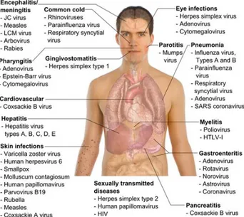
Other viruses cause a variety of conditions from measles and mumps to hepatitis and AIDS. Viral infections primarily affect respiratory and gastrointestinal system; epithelium, mucous membranes and endothelium of skin, mouth and genitals; lymphoid tissue, liver and other organs and central nervous system (SNC)(Fig. 1.17)58.
Figure 1.17: Main types of viral infection and the most notable species involved.
1.2.1 Infection of oropharynx and respiratory tract
Oropharynx and respiratory tract are the most common sites of infection. Viruses spread through droplets and aerosols, food, water and saliva; through close contact and hands. Similar respiratory symptoms may be caused by many viruses; in contrast, a single virus can cause different symptoms in different people. For example, in some individuals the virus can cause mild upper respiratory infection, in others life-threatening pneumonia.
Symptoms and severity of a viral respiratory illness depend on: virus nature, site of infection (upper or lower respiratory tract), immune status and age of
- 30 -
individual. Pharyngitis and other oropharynx diseases are common manifestations of viral infection. For example, symptoms such as acute pharyngitis, fever and oral vesicular lesions are characteristic of coxsackie A
virus (herpangina, hand-foot and mouth disease) and coxsackie B virus and echovirus. Herpesviruses cause local primary infection on oral mucous
membrane and face. Latent infection in neurons is followed by a gingivostomatitis caused by herpes virus and occurs in the form of herpes labialis. Epstein-Barr virus (EBV) and human herpes virus 6 (HHV6) can cause pharyngitis. Rhinovirus and coronavirus are the predominant cause of the common cold, upper respiratory tract infection.
Both pharingitis and colds can be caused by specific serotypes of echovirus and coxsackievirus, adenovirus, influenza virus, parainfluenza virus and
respiratory syncytial virus. Respiratory tract‘s infections may also degenerate
into more serious illnesses. Symptoms of these infections are bronchitis (inflammation of bronchioles), pneumonia and related conditionats.
Parainfluenza virus and respiratory syncytial virus create more damage to
infants and young children, but only cause asymptomatic infections or symptoms of the common cold in adults.
Influenza virus is probably the best known and most feared virus among the
common respiratory viruses, because annually you can check the spread of new strains of viruses. The viral agents that cause pneumonia include
adenovirus, paramyxovirus and varicella-zoster virus primary infections.
1.2.2 Gastrointestinal infections
Gastrointestinal infections can cause gastroenteritis, diarrhea, or be asymptomatic. Norovirus (Norwalk virus), adenovirus, astrovirus, rotavirus,
coronavirus infect small intestine but not colon, causing damage to the
- 31 -
and electrolyte imbalance. Rotavirus and adenovirus 40-41 are the leading cause of infantile gastroenteritis.
1.2.3 Skin infections
Skin viral diseases may be the effect of infections of the mucous membrane or small cuts or skin abrasions (HSV), as in the case of a secondary infection after the onset of viremia (VZV or smallpox) or be the result of an inflammatory response against viral agents (measles rash). The classic childhood exanthems, accompanied by fever, include exanthema subitum (HHV6), erythema infectiosum (parvovirus B19) and in unvaccinated children varicella, measles and rubella. Yellow fever virus (YFV), dengue virus and other viruses, causing hemorrhagic fevers, infect epithelial cells lining the blood vessels and can affect the structure. This produces a hemorrhagic rash with petechiae and ecchymoses.
1.2.4 Eye infections
Conjunctivitis (pink eye) is a typical feature of many childhood infections, and is caused by specific serotypes of adenovirus (3, 4a and 7), measles virus and rubella virus. Keratoconjunctivitis, caused by adenovirus (8, 19a and
37), HSV o VZV, affects the cornea and can cause serious damage. Enterovirus 70 and coxsackie virus A24 can cause acute hemorrhagic conjunctivitis.
Chorioretinitis is associated with congenital and in immunocompromised persons CMV infection.
1.2.5 Organs and tessues infection
Main organs infection can cause significant disease or determine subsequent virus spreading. Symptoms can be the effect of tissues damage or of inflammatory responses. The liver is a major target of many viruses. For example, the hepatitis symptoms may be caused by hepatitis viruses A, B, C,
- 32 -
mononucleosis by EBV and CMV infections. The liver is also the biggest target in disseminated HSV infections in infants and children. The heart and other muscles are susceptible to viral infections and the resulting damage.
Coxsackievirus can cause myocarditis or pericarditis in infants, children and
adults. The coxsackie B virus can infect and cause the muscles pleurodynia (Bornholm disease). Other viruses (eg influenza virus and CMV) can infect the heart. Infection of the secretory glands, accessory sex organs and mammary glands causes a contagious spread of the virus (CMV, mumps virus). For example, mumps and epidemic parotitis is a viral disease of the human species, caused by the mumps virus, whose most typical onset is the painful swelling of the salivary glands (classically the parotid gland). Kidney CMV infection and reactivation cause problems in immunocompromised people and are a major cause of failure of kidney transplantation.
1.2.6 Central Nervous System (CNS) infections
Viral infections of the brain and CNS can cause the most serious viral diseases due to CNS importance and to its very limited capacity to repair damage. A virus can spread to the CNS from the blood (arbovirus) or macrophages (acquired human immunodeficiency virus HIV); from peripheral neurons infection, or it may first infect the skin (HSV) or a muscle (rabies
virus) and then progress to the neurons that innervate the infected area. Virus
then infects neurons that express a viral receptor. For example, the temporal lobe is a target in the encephalitis by HSV, the horn of Ammon in rabies, anterior horn of the spinal cord and motor neurons in paralytic poliomyelitis (Enteroviruses). Aseptic meningitis is caused by inflammation and swelling of the meninges that involve the brain and the spinal cord in response to infection by enteroviruses (particularly coxsackievirus and echovirus), HSV-2,
mumps virus or lymphocytic choriomeningitis virus (LCM virus). The disease
- 33 -
sequelae unless the virus does not access and infect neurons or brain (meningoencephalitis).
Encephalitis and myelitis are fatal or cause significant damage and permanent neurological sequelae. HSV, VZV, rabies virus, California encephalitis virus,
St. Louis encephalitis virus (SLEV) and measles virus are potential causes of
encephalitis. Many polioviruses and other enteroviruses cause a paralytic disease (myelitis). HSV and VZV are ubiquitous and usually cause asymptomatic latent infections of the CNS, but can also cause encephalitis. Other neurological syndromes caused by viruses include HIV dementia, Tropical spastic paraparesis or human T-lymphotropic virus type I
(HTLV-I)-associated myelopathy, progressive multifocal leukoencephalopathy
caused by the papovavirus JC in immunocompromised people, and spongiform encephalopathy (kuru, Creutzfeldt-Jakob syndrome, Gerstmann-Straussler-Scheinker syndrome) that are believed to be caused by prions.
1.2.7 Sexually transmitted diseases
Although sexual transmission is not a very effective way to virus spread, it is the major spread route of HPV, HSV, CMV, HIV, HTLV-1, HBV, hepatitis C
virus (HCV) and hepatitis D (HDV). Papillomavirus and HSV-2 establish local
primary infection causing recurrent disease at the same site. CMV and HIV enter the bloodstream and infect lymphoid cells, while HBV and HCV are transported to the liver. These viruses are present in blood, semen and vaginal secretions, through which virus can be transmitted to sexual partners and infants (neonatal or perinatal transmission).
1.2.8 Syndromes with a possible viral etiology
Many diseases cause symptoms or have the epidemiological features or other similar to those of viral infections, or may be the sequelae of viral infections (eg, inflammatory responses to persistent viral infection). These
- 34 -
include multiple sclerosis, Kawasaki disease, arthritis, diabetes and chronic fatigue syndrome52
1.2.9 Chronic and potentially oncogenic infections
Some viruses can cause life-long or chronic infections, where the viruses continue to replicate in the body despite the host's defence mechanisms59. Chronic infections are established when the immune system has difficulty in resolving the infection. People chronically infected are known as carriers, as they serve as reservoirs of infectious virus60. In populations with a high proportion of carriers, the disease is said to be endemic61. In contrast to acute lytic viral infections this persistence implies compatible interactions with the host organism. Persistent viruses may even broaden the evolutionary potential of host species. CMV and other herpesviruses, hepatitis B, C and D viruses and retroviruses cause chronic productive infections. HBV, HCV, EBV,
papilloma virus, and HTLV-1 are associated with human tumors.
Cofactors, chromosomal aberrations, or both, convert a clone of cells, containing a virus, in cancer. EBV usually causes infectious mononucleosis, but is also associated with African Burkitt's lymphoma and nasopharyngeal carcinoma, HTLV-1 is associated with adult human T cell leukemia. Many
papillomaviruses (HPV) cause a simple hyperplasia, characterized by the
development of warts; however, many other strains of HPV have been linked to cancer in humans (eg, types 16 and 18 are associated with cervical cancer). Chronic infection with HBV and HCV has been associated with hepatocellular carcinoma. HSV-2 has been associated with human cervical carcinoma, most likely as a cofactor52.
1.3 Antiviral strategies
Effective vaccines have led, or might lead, to the eradication of important viral pathogens, such as smallpox, polio, measles, mumps and rubella. But
- 35 -
other viral diseases, particularly human immunodeficiency virus (HIV) and
hepatitis C virus (HCV), have so far proved to be intractable to the vaccine
approach. The need for effective antiviral drugs is further emphasized by the lack of vaccines for most respiratory-tract virus infections (adenovirus,
rhinovirus, parainfluenza virus and respiratory syncytial virus (RSV)), the
widely occurring human papilloma viruses (HPV) and herpesviruses (herpes
simplex virus types 1 and 2 (HSV-1,-2), varicellazoster virus (VZV), Epstein– Barr virus (EBV), cytomegalovirus (CMV), human herpesviruses types 6, 7 and 8 (HHV-6, -7, -8)), and the vast array of haemorrhagic fever viruses. And
although vaccines have been developed for hepatitis B virus (HBV) and
influenza virus types A and B, their use has not eliminated the need for
effective chemotherapeutic agents. Many new antiviral drugs have been licensed in recent years, most of which are used for the treatment of HIV infections; of the current repertoire of more than 30 drugs, 16 are anti-HIV, 5 are anti-CMV, 5 are anti-HSV and anti-VZV, 1 is anti-RSV, 3 are anti-hepatitis and 4 are anti-influenza. But there is considerable room for improvement, as these compounds are not always efficacious or well tolerated.
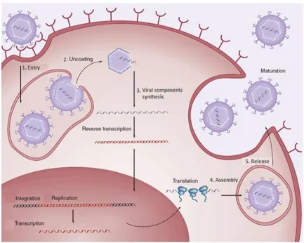
The emergence of viral resistance to drugs and drug-related side effects are among the main reasons for further refinement of antiviral drug design and development62. The basic strategies that are used to design antiviral drugs include to attack viruses at every stage of their life cycles (Fig. 1.18):
1 Entry; 2 Uncoating;
3 Viral components synthesis (through the inhibition of viral enzymes);
4 Assembly; 5 Release phase.
- 36 -
Figure 1.18: Antiviral strategies63.
1.3.1 Entry inhibitors
One anti-viral strategy is to interfere with the ability of a virus to infiltrate a target cell. The virus must go through a sequence of steps to do this, beginning with binding to a specific "receptor" molecule on the surface of the host cell and ending with the virus "uncoating" inside the cell and releasing its contents. Viruses that have a lipid envelope must also fuse their envelope with the target cell, or with a vesicle that transports them into the cell, before they can uncoat. The entry stage of viral replication cycle can be inhibited in two ways:
1. Using agents which act on viral-enveloped proteins. This includes antibodies and synthetic ligands.
2. Using agents which bind to the host cell receptors. This may include natural ligands of the receptor and anti-receptor antibodies.
Agents which act on viral-enveloped proteins
Enveloped proteins or glycoproteins contain oligosaccharide chains (glycans) covalently attached to polypeptide side-chains. Glycoproteins are
- 37 -
often important integral membrane proteins, where they play a role in cell-cell interactions. The N-glycosylation of viral enveloped protein is required for the folding, assembly and infectivity of several viruses.
Belong to this class:
1. Polyanionic compounds: For example, polysulphates (such as
polyvinylalcohol sulphate, Fig. 1.19A), polysulphonates (such as polyvinylsulphonate, Fig. 1.19B), polycarboxylates, polynucleotides (such as zintevir, polyoxometalates and negatively charged albumins) have been shown to inhibit HIV replication by preventing virus attachment (adsorption) to the surface of the host cell. All of these negatively charged polymers might be expected to interact with the positively charged amino acids in the V3 loop of the HIV glycoprotein, gp120, which is rich in arginine (R) and lysine (K) residues. In doing so, the polyanions shield the V3 loop and therefore hamper the binding of the HIV virions to heparin sulphate, the primary binding site at the cell surface, before more specific binding occurs with the CD4 receptor on CD4+ cells. Heparan sulphate is widely expressed on animal cells, and is involved in virus–cell binding for a broad array of enveloped viruses, including HSV and dengue virus. Moreover, these polyanions are not only active against HIV, but also against HSV and other sexually transmitted disease (STD) pathogens, such as Neisseria gonorrhoeae and Chlamydia trachomatis62.
Figure 1.19: Viral adsorption inhibitors: polyvinylalcohol sulphate (A) and
- 38 -
2. Glycans inhibitors: Glycans on the viral envelope often have a crucial
role in enabling an efficient transmission of the pathogen and/or entry into its susceptible target cells. Some agents interfere with the glycosylation enzymes from the cell, others act by directly binding to the intact glycans on the viral envelope. For example, MBI-3253 or
Celgosivir (Fig. 1.20A) is an alpha-glucosidase I inhibitor with a
potent antiviral activity in vitro and in vivo against several viruses, including HIV-1, herpes simplex virus (HSV), bovine viral diarrhea virus (BVDV) and HCV. It is currently undergoing phase II development for the treatment of HCV infection. Among agents that interact with the viral-envelope glycans there are carbohydrate-
binding agents (CBAs)(Fig. 1.20B). These compounds have a dual
mechanism of antiviral action: first, through direct antiviral activity, by binding to the glycans of the viral envelope and subsequently blocking virus entry, and second, through indirect (additional) antiviral action resulting from the progressive creation of deletions in the envelope glycan shield, thereby triggering the immune system to act against previously hidden immunogenic epitopes of the viral envelope64-67.
Celgosivir
A B