Hystrix, the Italian Journal of Mammalogy
Available online at:
http://www.italian-journal-of-mammalogy.it doi:10.4404/hystrix–00100-2018
Research Article
Health survey on the wolf population in Tuscany, Italy
Cecilia Ambrogi
1,∗, Charlotte Ragagli
1, Nicola Decaro
2, Ezio Ferroglio
3, Marco Mencucci
1, Marco Apollonio
4, Alessandro Mannelli
5 1Comando Unità Tutela Forestale Ambientale Agroalimentare Carabinieri2Dipartimento di Medicina Veterinaria, Strada Provinciale per Casamassima 3, 70010 Valenzano (Ba) 3Dipartimento di Scienze Veterinarie, Largo Paolo Braccini 2, 10095 Grugliasco (TO)
4Department of Veterinary Medicine, University of Sassari, Sassari, Sardinia, Italy 5Dipartimento di Scienze Veterinarie, Largo Paolo Braccini 2, 10095 Grugliasco (TO)
Keywords: wolf dog monitoring parasites parvovirus Article history: Received: 26/05/2018 Accepted: 29/04/2019 Acknowledgements
We wish to thank all the staff of RCB Lucca Orecchiella Natural Re-serve, especially Michela Adami for her invaluable help in collecting wolf samples and monitoring wolf-howling and reproduction. The pro-ject was funded by Comando Unità Tutela Forestale Ambientale Agroali-mentare Carabinieri and by Regione Toscana.
We also thank the editor in chief and the anonymous reviewers who greatly contributed to the improvement of the original manuscript.
Abstract
The objective of our study was to survey the occurence of transmissible agents in wolf (Canis lupus) population living in the northern Apennines. A total of 703 wolf fecal samples were collected in the Appennino Tosco-Emiliano National Park (ATENP) and the Foreste Casentinesi National Park (FCNP) in Tuscany, Italy. Parasitic forms (eggs or oocists) were detected in 74.3% of fecal samples, mainly infested by Trichuroidae (60.4%) and Coccidia (27.3%); heavy Trichuroidea and Coccidia infestation were found in 8.5% and 17.4% of samples (the intensity of infestation measured as EPG >1000, OPG >10000). Taking into consideration the main canine viruses, we evaluated the presence of Parvovirus in feces: 54 specimens from the study area in the ATENP and 71 from the study area in the FCNP were negative by PCR for the detection of Parvovirus. Tissue samples from nine wolves found dead were negative for Canine Distemper Virus (CDV), Canine Coronavirus (CCoV), Canine Adenovirus-1 (CAdV-1) and Canine Adenovirus-2 (CAdV-2). Tissue samples of two dead wolves in the FCNP were positive for Canine Parvovirus (CPV) and the virus was characterized as the antigenic variant 2a. Wild boar is the main component of the wolf’s diet in the study areas and 57 out of 135 serum samples (42.2%), collected from wild boars in the surroundings of the FCNP, yielded positive results for the detection of antibodies against Pseudorabies Virus (PrV). Wolves, especially in mountain areas, share the same habitat with dogs: this suggests how useful dog vaccination is for wolf protection.
Introduction
In the last decades, wolves (Canis lupus) have expanded their
geo-graphic range in Italy on account of their legal protection and the
in-crease in the number of ungulates their diet depends upon. Their
num-ber is currently estimated to be between 1260 and 1800 (Galaverni
et al., 2015). Furthermore, in mountain areas, where such traditional
practices as animal husbandry has been abandoned, a natural
reforest-ation has favored wolf populreforest-ations (Ciancio et al., 2006). Health
mon-itoring is considered a priority in the Italian wolf conservation plan
(Genovesi, 2002), since small pack size and high mortality rates may
significantly affect the composition and the stability of wolf
popula-tions (Mech and Goyal, 1993).
Among transmissible agents, canine parvovirus (CPV;
Protopar-vovirus, Parvoviridae), and canine distemper virus (CDV;
Morbilli-virus, Paramyxoviridae) are the most frequently assessed in health
sur-veys on wolf populations in Europe and North America (Millán et al.,
2016; Allison et al., 2013; Almberg et al., 2009; Sobrino et al., 2008;
Fico et al., 1996). A wide range of directly and indirectly transmitted
gastrointestinal parasites were found in wolves and their role in
conser-vation remains to be clarified (Craig and Craig, 2005; Guberti et al.,
1993).
In this study, such non-invasive procedures as the analysis of fecal
samples and carcasses were used to survey the presence of
transmiss-ible agents in wolf populations in two National Parks located in the
Northern Apennines, in Tuscany, Italy. A search for antibodies against
the Pseudorabies Virus (PRV) was carried out among wild boars (Sus
∗Corresponding author
Email address: [email protected](Cecilia Ambrogi)
scrofa
), which are epidemiological reservoir of the virus and the main
prey of the wolves in the study areas at this location (Lari et al., 2006;
Capua et al., 1997; Hahn et al., 1997). Microbial agents can be
trans-mitted between wolves and domestic dogs (Canis lupus familiaris).
Therefore, information from dog owners was collected to estimate
vac-cination coverage in dogs sharing wolves’ habitat.
Materials and methods
Study areas
The study was carried out in two National Parks in the Northern
Apennines, in Tuscany, Italy: the Foreste Casentinesi National Park
(FCNP) and the nearby Alpe di Catenaia area (43°47
026.64
00N,
11°43
024.06
00E) at an altitude ranging between 300–1700 m above
the sea level (a.s.l.); the Orecchiella, Lamarossa, Pania di Corfino
Nat-ural Reserves (44°12
015.78
00N, 10°21
030.83
00E) at an altitude ranging
between 1000–2054 m a.s.l. in the Appennino Tosco-Emiliano
Na-tional Park (ATENP) and the nearby Orrido di Botri Natural Reserve
(44°4
056.77
00N, 10°36
037.76
00E) at an altitude between 900–1300 m
a.s.l.
Wolf populations were monitored for several years in both study
areas (Mattioli et al., 2011; Reggioni unpublished data 2006;
Apollo-nio et al., 2004) by using indirect procedures (transects, snow tracking
and wolf howling). Wolf population was estimated at 2–3 per 100 km
2in the ATENP and 8 wolves per 100 km
2in the FCNP. Genetic
exam-ination in the FCNP identified 35 different genotypes (Caniglia et al.,
2014).
Hystrix, the Italian Journal of Mammalogy ISSN 1825-5272 3rd June 2019
©cbe2019 Associazione Teriologica Italiana doi:10.4404/hystrix–00100-2018
Field data collection
Wolf fecal samples
Wolf fecal samples (n=703) were collected during wolf population
monitoring activities, on transects selected by convenience sampling
criteria, in either one or two monthly sessions in 2006 and 2007 (CFS
— State Forestry Police). Fecal samples were attributed to wolves on
the basis of physical criteria (Caniglia et al., 2014; Darimont et al.,
2008) in the ATENP (n=439) and by means of genetic analysis in the
FCNP (n=264). Feces were stored at −80
◦C for 10 days to neutralize
Echinococcus
spp. eggs and then at −20
◦C prior to laboratory analysis
(Hildreth et al., 2004). Data obtained from the analysis of fecal samples
from the ATENP and the Orrido di Botri Natural Reserve were
com-bined as ATEPN data, since the two areas are relatively close (≈20 km
distance).
Wolf tissue samples
Nine wolves (one from the ATENP, eight from the FCNP), which were
found dead in the study areas between 2005 and 2007, underwent
nec-roscopy. Causes of death included car accidents and poaching.
Tis-sue samples were taken from kidneys, bladder, liver, lungs, mesenteric
lymph nodes, spleen, small intestine, and brain. Samples were frozen
and submitted to the Department of Veterinary Medicine, Aldo Moro
University of Bari, for laboratory analysis. Owing to the poor
conser-vation status of the carcasses, no sample for histopathology was taken.
Wild boar blood samples
Blood samples were collected from the cardiac clot of 135 wild boars
during the 2005 hunting season in the surroundings of the FCNP. Blood
serum was stored at −20
◦C prior to laboratory analysis.
Information on domestic dogs
One of the authors interviewed 81 dog owners living in remote areas
where dogs share part of wolves’ habitat. Data were collected on the
vaccination status of dogs against the major viral diseases (parvovirus,
paramyxovirus; vaccination against rabies is generally not carried out,
since Italy is rabies-free).
Laboratory analyses
Genetic analysis of wolf fecal samples
To identify individual wolves and characterize their circulating
gen-otypes, DNA was extracted from 264 fecal samples collected in the
FCNP and submitted for genetic analysis by using a panel of 12
can-ine microsatellite PCR primers (Galaverni et al., 2012). All of the 35
genotypes identified were matched with the database which is
compre-hensive of all Italian wolves (Caniglia et al., 2014). No hybrid was
found.
Parasitological analysis
Fecal samples (2 g) were examined for parasite eggs or oocysts by
flot-ation in a saturated solution of zinc sulphate (1200 density).
Identi-fication was based on morphology and size (Sloss and Kemp, 1978).
Protozoa and Nematoda eggs/oocysts were classified by genus, whereas
Ascaridae by species (Stronen et al., 2011; Kloch et al., 2005; Gompper
et al., 2003). Fecal eggs/oocyst count was carried out by using
Mac-Master’s technique (Whitlock, 1948) and expressed as eggs/oocysts per
gram (EPG/OPG).
Virological analysis
Wolf fecal samples (including 54 specimens from the ATENP and 71
from the FCNP) were analyzed in pools of five to detect CPV by
us-ing laboratory techniques, as described in Decaro et al. (2006, 2005a).
DNA was extracted from tissue samples by employing the DNeasy
Tissue Kit (QIAGEN S.p.A. Milan, Italy), whereas RNA purification
was obtained by using the QIAamp® Viral RNA Mini Kit (QIAGEN
S.p.A.).
Wolf tissues samples were analyzed by using molecular methods to
detect the viral agents CPV, CDV, CCoV, and CAdV: a real-time PCR
(Decaro et al., 2005b, 2006); TaqMan-based real-time RT-PCR (Elia et
al., 2006); duplex real-time PCR assay (Dowgier et al., 2016).
Virological analysis
Wild boar serum samples were tested for antibodies against PRV
by employing IgB antibody ELISA test (IDEXX antiPRV gB). The
ELISA colorimetric reaction was measured by using a
spectrophoto-meter (BIO-RAD 680 Microplate Reader).
Statistical analysis
For each parasite group, the prevalence of positive fecal samples (in
which at least one parasitic form was detected) was obtained with the
FREQ procedure in the SAS System. The median and third quartiles
(Q1, Q3) of the number of EPG/OPG were calculated (PROC MEANS,
SAS, 2011). Furthermore, the prevalence of samples with high burden
(EPG>1000; OPG>10000) were calculated to assess the frequency of
wolf scats which were highly infested. We chose the cut-off of 1000
EPG (Urquarth et al., 1998) and 10000 OPG for Coccidia infection, to
define the parasitic infection as serious. Confidence intervals of
preval-ence were not calculated, since multiple samples might have belonged
to the same individual. Thus, no statistical analysis was carried out to
test differences in prevalence and in the number of EPG/OPG.
There-fore, data analysis was limited to descriptive statistics. Prevalence of
each viral agent in wolf tissue samples and antibodies against PRV in
wild boars’ serum sample was also obtained.
Results
In both study areas, eggs of Tricuridae and Strongylidae were the most
prevalent Nematode parasitic forms. Although the prevalence of
As-carididae was relatively low, presence of AsAs-carididae eggs was highest
in positive fecal samples (Tab. 1). Infections with EPG >1000 were
detected in 8.5% of specimens for Trichuridae. The intensity of
infec-tion was also detected (UPG>1000, OPG>10000): in particular, 8.5%
of samples showed high levels of Trichuridae, 1.4% of Strongylidae,
22.2% of Ascarididae eggs, and oocysts were found in 17.4% of fecal
samples.
Virological analysis
Twenty-five pools, each composed of five fecal samples (for a total of
125 samples) showed negative PCR for Parvovirus. Tissue samples
from the nine wolves which were found dead were negative for CDV,
CCoV, CAdV-1, and CAdV-2. Samples from intestine, spleen and
mes-enteric lymph nodes of two wolves (of an approximate age of 6 and 18
months) found dead in the FCNP were positive for parvovirus (CPV).
Viral DNA titers were generally low, ranging from 7.19×10³
(intest-ine of one individual) to 2.71×10⁴ (intest(intest-ine of the other wolf) per
mg of feces. Pooled spleen and lymph nodes of the same individuals
were positive, though they displayed lower CPV DNA loads (2.84×10³
and 6.12×10³ per mg of feces, respectively). Both strains were
char-acterized as type 2a, but viral DNA loads were too low to allow any
sequencing of informative regions for subsequent evolutionary studies.
ELISA for the detection of antibodies against PRV yielded positive
results on 57 out of 135 serum samples (42.2%), which were collected
from wild boars in the surroundings of the FCNP.
Table 1– Prevalence of fecal samples with at least one parasitic form, and median (Q1, Q3) numbers of parasite eggs or oocysts per g in positive wolf fecal samples in the study areas in the Northern Apennines, Italy.
Study area (n=703)
FCNP (n=264) ATENP (n=439)
Parasite group % Median Q1, Q3 % Median Q1, Q3
Ascaridia 2.3 180 30, 6570 0.68 120 90, 1350
Tricuridae 67.8 150 45, 480 56 90 45, 255
Strongil 12.5 30 15, 60 6.8 30 30, 120
Cestoda 7.2 105 30, 540 9.6 60 30, 180
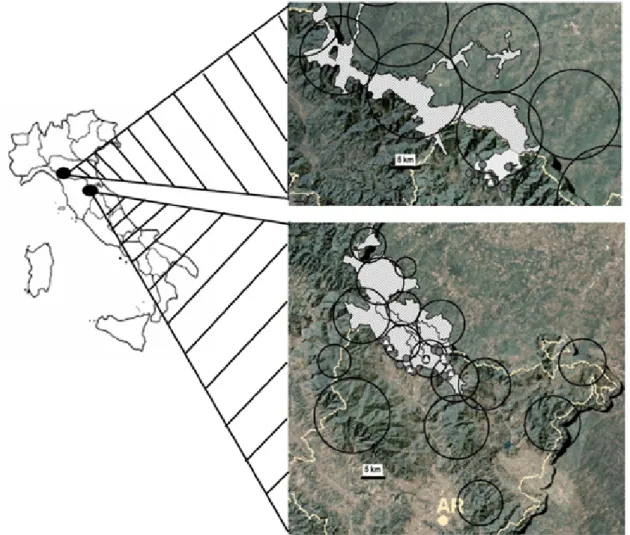
Figure 1– Dogs and wolves share the same territory: the circles represent the home range of different wolf packs obtained from snow tracking, wolf howling and genetic analysis data (LIFE07NAT/IT/000502 “Improving the conditions for large carnivore conservation — a transfer of best practices” LIFE EX-TRA). The white and grey dots represent the location/position of dogs. The grey dots represent unvaccinated dogs.
Dog owner survey
Based upon the analysis of questionnaires administered to dog owners,
in the ATENP, 39% of dogs were correctly vaccinated (three
vaccin-ations administered at four, eight, and sixteen weeks of age), 37%
re-ceived only the first two vaccinations, and 24% were unvaccinated. In
the PNFC, 45% of dogs were correctly vaccinated, 35% were
vaccin-ated only twice, whereas 20% were unvaccinvaccin-ated.
Discussion
Parasitological examination of wolf feces in the two study areas showed
that Trichuridae eggs were the most common Nematode parasitic
forms, followed by Strongylidae eggs. Such finding might be
accoun-ted for by the resistance of eggs of these parasites in the environment,
favoring the transmission of infection within wolf populations (Capelli
et al., 2003). Conversely, eggs of Ascaridae were relatively rare in the
samples examined; in fact, despite being common in carnivores and
previously reported in wolves (Bryan et al., 2012; Byman et al., 1977;
Holmes et al., 1968), Ascaridae are not considered dominant in this
species (Segovia et al., 2003, 2001; Guberti et al., 1993). Moreover, in
our study, the frequency of Ascaridae might have been underestimated
owing to a relatively low likelihood of collecting feces from juvenile
wolves, which are more liable to be infected. In fact, scats found on
transects tend to be part of scent-marking by adult wolves (Mech and
Boitani, 2003; Vilà et al., 1994). With respect to the detection of
Cest-oda (Gori et al., 2015; Guerra et al., 2013; Guberti et al., 1993), the
re-latively low prevalence obtained in our study might be due to the poor
sensitivity of copromicroscopical analysis by flotation in the diagnosis
of these parasites (Poglayen et al., 2017; Villeneuve et al., 2015). We
observed relatively high burdens of Coccidia oocysts; however, none
of the parasitic forms identified belonged to I. canis, which was
previ-ously shown as a pathogen associated with the death of wolf puppies in
North America (Mech and Kurtz, 1999).
The relatively low proportions of wolf feces characterized by great
parasitic counts (EPG>1000 and OPG>10000) suggest an aggregate
distribution of parasites among hosts (Tompkins et al., 2002; Wilson et
al., 2002; Anderson and May, 1978). Under these circumstances, the
few hosts harboring the majority of parasites might be weakened and
particularly vulnerable to secondary pathogens (Stronen et al., 2011;
Scott, 1988).
The high prevalence of seropositivity to PRV in wild boars, which
is consistent with the findings in other areas of Italy (Lari et al., 2006),
suggests that wolves are exposed to the agent of Aujeszky’s disease,
given that wild boar is the main component of a wolf’s diet (Davis et
al., 2012; Mattioli et al., 2011, 1995). Although PRV was identified in
the brain of a wolf which showed nervous signs after being fed with
wild boar offal in a wildlife park in Belgium (Verpoest et al., 2014),
consequences of exposure to PRV for free living wolf populations
re-main to be clarified and are likely scarce.
The detection of CPV in tissues from two wolves confirmed the
cir-culation of such agent within the wolf population of the Northern
Apen-nines, where it was previously found in fecal samples by Martinello et
al. (1997). The presence of CPV had never been investigated before
in the ATENP, where all fecal and tissue samples yielded negative
res-ults. The viruses detected in the present study belong to the variant
CPV-2a. It is unlikely that CPV contributed to the death of the two
positive wolves, since viral DNA loads were very low and the wolves
were found dead with lesions attributable to car accidents. The most
recent report on CPV in wolves in Italy dates back to 2001 and
de-scribes the detection of four CPV-2b strains in 1995 (Battilani et al.,
3
2001). More recently, a CPV-2a strain was reported in a wolf in
Bul-garia (Filipov et al., 2016). Based upon genetic characterization of the
viruses we identified, both individuals harbored one of the antigenic
variants circulating within the dog population. A CPV-2b strain was
recently detected in a wolf found dead in the Campania region, Italy,
which was co-infected by pantropic CCoV and CAdV-2 (Alfano et al.,
2019).
All of the fecal samples examined were negative for parvovirus. The
virus was seldom found in feces, in spite of the frequent detection of
antibodies against CPV and the endemic CPV presence in a number of
wolf populations in North America and Europe (Alfano et al., 2019;
Millán et al., 2016; Molnar et al., 2014; Almberg et al., 2009; Sobrino
et al., 2008). This is due to the viral excretion, which may occur only
during the first phase of the infection. Furthermore, viral excretion in
a population with endemic parvovirosis is likely reduced by immunity,
including the protection of puppies by passive immunization (Mech et
al., 2012). Serological analysis on live trapped individuals may reveal
valuable information regarding the health status of wolves (Zarnke and
Ballard, 1987; Goyal et al., 1985; Choquette and Kuyt, 1974), though
it sparks a debate on the considerable financial outlays required as well
as on ethical considerations (Darimont et al., 2008).
Dogs vaccinated against the old strain CPV-2 may a new infection
by new viral reservoirs, which may emerge following be affected by
a mutation, as shown by the viral lineages identified in the last years
(CPV-2a, b, c). This might explain the occurrence of outbreaks of
in-fection in breeding kennels, where dogs were regularly vaccinated with
the old-type vaccines, based on the classical lineage CPV-2, which is
now extinct (Decaro and Buonavoglia, 2012; Müller et al., 2011;
De-caro et al., 2009, 2008). We cannot exclude the establishment of a
sylvatic cycle of parvovirus transmission (Santos et al., 2009; Sobrino
et al., 2008).
Other canine viruses were not detected in the samples analyzed,
al-though it was reported that CDV, CAdVs (Dowgier et al., 2018) and
CCoV are able to infect and cause diseases in wild carnivores (Alfano
et al., 2019; Zarnke et al., 2001; Laurenson et al., 1998; Stephenson
et al., 1982; Choquette and Kuyt, 1974). Distemper is due to a highly
pathogenic virus (Almberg et al., 2009), which needs a large host
pop-ulation in order to be maintained. Dog poppop-ulations were considered
the cause of distemper outbreak on several occasions (Di Sabatino et
al., 2014; Decaro et al., 2004; Cleaveland et al., 2000); in a number of
cases, the existence of a multi-host population was presumed, with dogs
thus playing a secondary role. The vaccination against distemper grants
a good immunity level for many years (Martella et al., 2008). Since the
herd immunity against CDV was sufficient to control the disease despite
infectious pressure in dogs as long as herd immunity was slightly over
70% of the vaccinated dogs (Rikula et al., 2007), we can suppose that
the dogs vaccinated until now were sufficient to build a barrier against
the disease and that the population of non-vaccinated dogs was not
suf-ficient to constitute CDV reservoir. Further research should include fox
populations in health surveys in the study areas, considering that, since
2006, red foxes in Northern Italy experienced an epidemic of canine
distemper (Loots et al., 2017; Martella et al., 2010), and given the
re-cent introduction and spreading of novel, or re-emerging, CDV strains
in Europe, carried by dogs imported by Eastern Europe (Mira et al.,
2018).
The proximity of such wolf’s preys as deer and wild boars to
hu-man settings might favor the transmission of microbial agents between
wolves and domestic dogs. Therefore, dog populations might be
con-sidered an important component of wolf ecosystem and can be used
as sentinels for wolf health (Di Sabatino et al., 2015). The
Authorit-ies of National Parks, in collaboration with public veterinarAuthorit-ies, should
pay particular attention to local dog populations, reduce the number
of roaming dogs, promote appropriate dog vaccination and education
programs and protect the wolf population from CDV and new strains
of CPV outbreak.
References
Alfano F., Dowgier G., Valentino M.P., Galiero G., Tinelli A., Decaro N., Fusco G., 2019. Identification of Pantropic Canine Coronavirus in a Wolf (Canis lupus italicus) in Italy. J. Wildl. Dis, 55(2): 504–508. doi:10.7589/2018-07-182
Allison A.B., Kohler D.J., Fox K.A., Brown J.D., Gerhold R.G., Shearn-Bochsler V.I., Dubovi E.J., Parrish C.R., Holmesh E.C., 2013. Frequent cross-species transmission of parvoviruses among diverse carnivore hosts. J. Virol.87(4): 2342–2347.
Almberg E.S., Mech L.D. Smith D.W,. Sheldon J.W,. Crabtree R.L., 2009. A serological survey of infectious disease in Yellowstone National Park’s canid community. PLoS ONE. 4(9): e7042. doi:10.1371/journal.pone.0007042
Anderson R.M., May R.M., 1978. Regulation and stability of host-parasite population in-teractions: I. Regulatory processes. J. Animal. Ecol. 47(1): 219–247.
Apollonio M., Mattioli L., Scandura M., Mauri L., Gazzola A., Avanzinelli E., 2004. Wolves in the Casentinesi Forests: insights for wolf conservation in Italy from a pro-tected area with a rich wild prey community. Biol. Conserv. 120(2): 249–260. Battilani M., Scagliarini A., Tisato E., Turilli C., Jacoboni I., Casadio R., Prosperi S., 2001.
Analysis of canine parvovirus sequences from wolves and dogs isolated in Italy. J. Gen. Virol. 82: 1555–1560.
Bryan H.M., Darimont C.T., Hill J.E., Paquet P.C., Thompson R.C., Wagner B., Smits J.E., 2012. Seasonal and biogeographical patterns of gastrointestinal parasites in large carnivores: wolves in a coastal archipelago. Parasitology 139(6): 781–790. doi:10.1017/ S0031182011002319
Byman D., Van Ballenberghe V., Schlotthauer J.C., Erickson A.W., 1977. Parasites of wolves, Canis lupus L., in northeastern Minnesota, as indicated by analysis of fecal samples. Can. J. Zool. 55(2): 376–380.
Caniglia R., Fabbri E., Galaverni M., Milanesi P., Randi E., 2014 Noninvasive sampling and genetic variability, pack structure, and dynamics in an expanding wolf population. J. Mammal. 95(1): 41–59. doi:10.1644/13-MAMM-A-039
Capelli G., Stancampiano L., Magi M., Poglayen G., Guberti V., 2003. Diversità delle comunità parassitarie intestinali in tre popolazioni di volpi. J. Mt. Ecol. 7(Suppl.): 199– 205.
Capua I., Fico R., Banks M., Tamba M., Calzetta G., 1997. Isolation and characterization of an Aujeszky’s disease virus naturally infecting a wild boar (Sus scrofa). Vet. Microbiol. 55:141–146.
Ciancio O., Corona O., Lamonaca A., Portoghesi L., Travaglini D., 2006. Conversion of clearcut beech coppices into high forests with continuous cover: a case study in central Italy. Forest Ecol. Manag. 224: 235–240.
Choquette L.P., Kuyt E., 1974. Serological indication of canine distemper and of infectious canine hepatitis in wolves (Canis lupus L.) in northern Canada. J. Wildl. Dis. 10(4): 321–324.
Cleaveland S., Appel M.J., Chalmers W.K., Chillingworth C., Kaare M., Dye C., 2000. Serological and demographic evidence for domestic dogs as a source of canine dis-temper virus infection for Serengeti wildlife. Vet. Microbiol. 72(3–4): 217–27. doi: 10.1016/S0378-1135(99)00207-2
Craig H.L., Craig P.S., 2005. Helminth parasites of wolves (Canis lupus): a species list and an analysis of published prevalence studies in Nearctic and Palearctic populations. J. Helminthol. 79(2): 95–103.
Darimont C.T., Reimchen T.E., Bryan H.M., Paquet P.C., 2008. Faecal-centric approaches to wildlife ecology and conservation. Methods, data and ethics. Wildl. Biol. Pract., 4(2): 73–87. doi:10.2461/wbp.2008.4.7
Davis M.L., Stephens P.A., Willis S.G., Bassi E., Marcon A., Donaggio E., et al., 2012. Prey selection by an apex predator: the importance of sampling uncertainty. PLoS ONE 7(10): e47894. doi:10.1371/journal.pone.0047894
Decaro N., Buonavoglia C., 2012. Canine parvovirus — a review of epidemiological and diagnostic aspects, with emphasis on type 2c. Vet. Microbiol. 155(1): 1–12. doi:10.1016/ j.vetmic.2011.09.007
Decaro N., Camero M., Greco G., Zizzo N., Tinelli A., Campolo M., Pratelli A., Buonavoglia C., 2004. Canine distemper and related diseases: report of a severe out-break in a kennel. New Microbiol. 27(2):.177–181.
Decaro N., Cirone F., Desario C., Elia G., Lorusso E., Colaianni M.L., Martella V., Buonavoglia C., 2009. Severe parvovirus in a 12-year-old dog that had been repeatedly vaccinated. Vet. Rec. 164(19): 593–595.
Decaro N., Desario C., Elia G., Martella V., Mari V., Lavazza A., Nardi M., Buonavoglia C., 2008. Evidence for immunization failure in vaccinated adult dogs infected with canine parvovirus type 2c. New Microbiol. 31(1): 125–130.
Decaro N., Elia G., Martella V., Campolo M., Desario C., Camero M., Cirone F., Lorusso E., Lucente M.S., Narcisi D., Scalia P., Buonavoglia C., 2006. Characterization of the canine parvovirus type 2 variants using minor groove binder probe technology. J. Virol. Methods. 133(1): 92–99. doi:10.1016/j.jviromet.2005.10.026
Decaro N., Elia G., Martella V., Desario C., Campolo M., Di Trani L., Tarsitano 294 E., Tempesta M., Buonavoglia C., 2005a. A real-time PCR assay for rapid detection and quantitation of canine parvovirus type 2 in the feces of dogs. Vet. Microbiol. 105(1): 19-28. doi:10.1016/j.vetmic.2004.09.018
Decaro N., Martella V., Ricci D., Elia G., Desario C., Campolo M., Cavaliere N., Di Trani L., Tempesta M., Buonavoglia C., 2005b. Genotype-specific fluorogenic RT-PCR assays for the detection and quantitation of canine coronavirus type I and type II RNA in fecal samples of dogs. J. Virol. Methods. 130(1–2): 72–78. doi:10.1016/j.jviromet.2005.06.005 Di Sabatino D., Lorusso A., Di Francesco C.E., Gentile L., Di Pirro V., Bellacicco A.L.,
Giovannini A., Di Francesco G., Marruchella G., Marsilio F., Savini G., 2014. Arctic Lineage-canine distemper virus as a cause of death in Apennine wolves (Canis lupus) in Italy. PLoS ONE 9(1): e82356. doi:10.1371/journal.pone.0082356
Di Sabatino D., Savini G., Lorusso A., 2015. Canine distemper and endangered wildlife: Is it time for mandatory vaccination of dogs? Vaccine. 33(48): 6519. doi:101016/j.vaccine. 2015.05.087
Dowgier G., Lahoreau J., Lanave G., Losurdo M., Varello K., Lucente M.S., Ventriglia G., Bozzetta E., Martella V., Buonavoglia C., Decaro N., 2018. Sequential circulation of canine adenoviruses 1 and 2 in captive wild carnivores, France. Vet Microbiol. 221: 67–73. doi:10.1016/j.vetmic.2018.05.025
Dowgier G., Mari V., Losurdo M., Larocca V., Colaianni M.L., Cirone F., Lucente M.S., Martella V., Buonavoglia C., Decaro N., 2016. A duplex real-time PCR assay based on TaqMan technology for simultaneous detection and differentiation of canine adenovirus types 1 and 2. J Virol Methods 234: 1–6. doi:10.1016/j.jviromet.2016.03.011
Elia G., Decaro N., Martella V., Girone F., Lucente M.S., Lorusso E., Di Trani L., Buonavoglia C., 2006. Detection of canine distemper virus in dogs by real-time RT-PCR. J. Virol. Methods 136(1–2): 171–176. doi:10.1016/j.jviromet.2006.05.004
Fico R., Marsilio S., Tiscar P.G., 1996. Antibodies againt canine parvovirus, distemper, infectious canine hepatitis, canine coronavirus, and Ehrlichia canis in wolves (Canis
lupus) in central Italy. Ricerche di Biologia della Selvaggina 24(suppl.): 137–143.
Filipov C., Desario C., Patouchas O., Eftimov P., Gruichev G., Manov V., Filipov G., Buonavoglia C., Decaro N., 2016. A Ten-Year Molecular Survey on Parvoviruses In-fecting Carnivores in Bulgaria. Transbound Emerg. Dis. 63(4): 460–464. doi:10.1111/tbed. 12285
Galaverni M., Caniglia R., Fabbri E., Milanesi P., Randi E., 2015. One, no one, or one hundred thousand: how many wolves are there currently in Italy? Mammal Res. 61: 3–24. doi:10.1007/s13364-015-0247-8
Galaverni M., Palumbo D., Fabbri E., Caniglia R., Greco C., Randi E., 2012. Monitoring wolves (Canis lupus) by non invasive genetics and camera trapping: a small-scale pilot study. Eur. J. Wildl. Res. 58(1): 47–58. doi:10.1007/s10344-011-0539-5
Genovesi P. (Ed.), 2002. Piano d’azione nazionale per la conservazione del lupo (Canis
lupus). Quad. Cons. Natura, 13, Min. Ambiente – Ist. Naz. Fauna Selvatica.
Gompper M.E., Goodman R.M., Kays R.W., Ray J.C., Fiorello C.V., Wade S.E., 2003. A survey of the parasites of coyotes (Canis latrans) in New York based on fecal analysis. J. Wildl. Dis. 39(3): 712–717. doi:10.7589/0090-3558-39.3.712
Gori F., Armua-Fernandez M.T., Milanesi P., Serafini M., Magi M., Deplazes P., Macchioni F., 2015. The occurrence of taenidis of wolves in Liguria (northern Italy). Int. J. Parasitol. Parasites Wildl. 4(2): 252–256. doi:10.1016/j.ijppaw.2015.04.005
Goyal S.M., Mech L.D., Rademacherb R.A., Khan M.A., Seal U.S., 1986. Antibodies against canine parvovirus in wolves of Minnesota: A serological study from 1975 through 1985. J. Am.Vet. Med. Ass. 89: 1092–1094.
Guberti V., Stancampiano L., Francisci F. 1993. Intestinal helminth parasite community in wolves (Canis lupus) in Italy. Parassitologia 35(1–3): 59–65.
Guerra D., Armua-Fernandez M.T., Silva M., Bravo I., Santos N., Deplazes P., Carvalho L.M., 2013. Taeniid species of the Iberian wolf (Canis lupus signatus) in Portugal with special focus on Echinococcus spp. Int. J. Parasitol. Parasites Wildl. 2: 50–53. doi:10. 1016/j.ijppaw.2012.11.007
Hahn E.C., Page G.R., Hahn P.S., Gillis K.D., Romero C., Annelli J.A., Gibbs E.P., 1997. Mechanisms of transmission of Aujeszky’s disease virus originating from feral swine in the USA. Vet. Microbiol. 55(1–4): 123–130.
Hildreth M.B., Blunt D.S., Oaks J.A., 2004. Lethal effects of freezing Echinococcus
multi-loculariseggs at ultralow temperatures. J. Parasitol. 90(4): 841–844. doi:10.1645/GE-221R
Holmes J.C., Podesta R., 1968. The helminths of wolves and coyotes from the forested regions of Alberta. Canad. J. Zool. 46(6): 1193–1204.
Kloch A., Bednarska M., Bajer A., 2005. Intestinal macro-and microparasites of wolves (canis lupus L.) from north-eastern Poland recovered by coprological study. Ann. Agric. Environ. Med. 12(2): 237–245.
Lari A., Lorenzi D., Nigrelli D., Brocchi E., Faccini S., Poli A., 2006. Pseudorabies virus in european wild boar from central Italy. J. Wildl. Dis. 42(2): 319–324. doi:10.7589/0090-3558-42.2.319
Laurenson M.K., Sillero-Zubrini C., Thompson H., Shiferaw F., Thirgood S., Malcom J., 1998. Disease as a threat to endangered species: Ethiopian wolves, domestic dogs and canine pathogens. Anim. Conserv. 1: 273–280.
Loots A.K., Mitchell E., Dalton D.L., Kotzé A., Venter E.H., 2017. Advances in canine distemper virus pathogenesis research: a wildlife perspective. J.gen. Virol. 98(3): 311– 321. doi:101099/jgv.0.000666
Martella V., Bianchi A., Bertoletti I., Pedrotti L., Gugiatti A., Catella A., Cordioli P., Lu-cente M.S., Elia G., Buonavoglia C., 2010. Canine distemper epizootic among red foxes, Italy 2009. Emerg. Infect. Dis. 16(12): 2007–2009. doi:10.3201/eid1612.100579 Martella V., Elia G., Buonavoglia C., 2008. Canine distemper virus. Vet. Clin. North Am.
Small. Anim. Pract. 38(4): 787–797. doi:10.1016/j.cvsm.2008.02.007
Martinello F., Galuppo F., Guberti V., Prosperi S., 1997. Detection of canine parvovirus in wolves from Italy. J. Wildl. Dis. 33(3): 628–631.
Mattioli, L., Apollonio M., Mazzarone V., Centofanti E., 1995. Wolf food habits and wild ungulate availability in the Foreste Casentinesi National Park, Italy. Acta Theriol. 40(4): 387–402.
Mattioli, L., Capitani, C., Gazzola, A., Scandura M., Apollonio M., 2011. Prey selection and dietary response by wolves in a high-density multi-species ungulate community. Eur J Wildl Res. 57(4): 909–922. doi:10.1007/s10344-011-0503-4
Mech L.D., Boitani L., 2003. Wolf social ecology. In: Mech L.D., Boitani L. (Eds.) Wolves: behavior, ecology and conservation. University of Chicago Press, Chicago. Mech L.D., Goyal S.M., 1993. Canine parvovirus effect on wolf population change and pup
survival. J. Wildl. Dis. 29(2): 330–333. doi:10.7589/0090-3558-29.2.330
Mech L.D., Kurtz H.J., 1999. First Record of Coccidiosis in Wolves, Canis lupus. Can. Field Nat. 113(2): 305–306.
Mech L.D., Almberg E.S., Smith D., Goyal S., Singer R.S., 2012. Use of Real-time PCR to Detect Canine Parvovirus in Feces of Free-ranging Wolves. J. Wildl. Dis. 48(2): 473– 476. doi:10.7589/090-3558-48.2.473
Millán J., Lopez-Bao J.V., Garcia E.J., Oleaga A., Llaneza L., Palacios V., De la Torre A., Rodriguez A., Dubovi E. J., Esperon F., 2016. Patterns of exposure of Iberian Wolves (Canis lupus) to canine viruses in human-dominated landscapes. Ecohealth. Mar. 13(1): 123–34. doi:10.1007/s10393-015-1074-8
Mira F., Purpari G., Di Bella S, Vicari D., Schirò G., Di Marco P., Macaluso G., Battilani M., Guercio A., 2018. Update on canine distemper virus (CDV) strains of Artic-like lineage detected in dogs in Italy. Vet. Ital. 54(3): 225–236. doi:10.12834/VetIt.1455.7862.2 Molnar B., Duchamp C., Möstl K., Diehl P.A, Betschart B., 2014. Comparative survey of
canine parvovirus, canine distemper virus and canine enteric coronavirus infection in free-ranging wolves of central Italy and south-eastern France. Eur. J. Wildl. Res. 60(4): 613–624. doi:10.1007/s10344-014-0825-0
Müller A., Silva E., Santos N., Thompson G., 2011. Domestic dog origin of canine distem-per virus in free-ranging wolves in Portugal as revealed by hemagglutinin gene charac-terization. J. Wildl. Dis. 47(3): 725–729. doi:10.7589/0090-3558-47.3.725
Poglayen G., Gori F., Morandi B., Galuppi R., Fabbri E., Caniglia R., Milanesi P., Ga-laverni M., Randi E., Marchesi B., Deplazes P., 2017. Italian wolves (Canis lupus
it-alicusAltobello, 1921) and molecular detection of taeniids in the Foreste Casentinesi
National Park, Northern Italian Apennines. Int. J. Parasitol. Parasites and Wildl. 6(1): 1–7. doi:10.1016/j.ijppaw.2017.01.001
Popiołek M., Szczęsna J., Nowak S., Mysłajek R., 2007. Helminth infections of faecal samples of wolves Canis lupus L. from western Beskidy Mountains in southern Poland. J. of Helminthol. 81(4): 339–344. doi:10.1017/S0022149X07821286
Rikula U., Nuotio L., Sihvonen L., 2007. Vaccine coverage, herd immunity and occurrence of canine distemper from 1990-1996 in Finland. Vaccine 25(47): 7994–7998. doi:10.1016/ j.vaccine.2007.09.015
Santos N., Almendra C., Tavares L., 2009. Serologic survey for canine distemper virus and canine parvovirus in free-ranging wild carnivores from Portugal. J.Wildl. Dis. 45(1): 221–226. doi:10.7589/0090-3558-45.1.221
Scott M.E., 1988. The impact of infection and disease on animal populations: implications for conservation biology. Conser. Biol. 2 (1): 40–56.
Segovia J.M., Guerrero R., Torres J., Miquel J., Feliu C., 2003. Ecological analyses of the intestinal helminth communities of the wolf, Canis lupus, in Spain. Folia Parasitol. 50(3): 231–236.
Segovia J.M., Torres J., Miquel J., Llaneza L., Feliu C., 2001. Helminths in the wolf, Canis
lupus, from northwestern Spain. J. Helminthol. 75(2): 183–92.
Sloss M.W., Kemp R.L., 1978. Veterinary clinical parasitology. The Iowa State University Press, Ames, Iowa.
Sobrino R., Arnal M.C., Luco D.F., Gortázar C., 2008. Prevalence of antibodies against canine distemper virus and canine parvovirus among foxes and wolves from Spain. Vet. Microbiol. 126(1–3): 251–6. doi:10.1016/j.vetmic.2007.06.014
Stephenson R.O., Ritter D.G., Nielsen C.A., 1982. Serologic survey for canine distemper and infectious canine hepatitis in wolves in Alaska. J. Wildl. Dis. 18(4): 419–424. Stronen A.V., Sallows T., Forbes G.J., Wagner B., Paquet P.C., 2011. Diseases and parasites
in wolves of the Riding Mountain National Park region, Manitoba, Canada. J. Wildl. Dis. 47(1): 222–227. doi:10.7589/0090-3558-47.1.222
Tompkins D.M., Dobson A.P., Arneberg P., Begon M.E., Cattadori I.M., Greenman J.V., Heesterbeek J.A.P., Hudson P.J., Newborn D., Pugliese A., Rizzoli A.P., Rosà R., Rosso F., Wilson K., 2002. Parasites and host population dynamics. In: Hudson P.J., Rizzoli A.P., Grenfell B.T., Heesterbeek H., Dobson A.P. (Eds.) The ecology of wildlife dis-eases. Oxford Press, Oxford, UK. 45–62.
Urquarth G.M., Armour J., Duncan J.L., Dunn A.M., Jennings F.W., 1998. Parassitologia Veterinaria. UTET, Torino.
Verpoest S., Cay A.B., Bertrand O., Saulmont M., De Regge N., 2014. Isolation and char-acterization of pseudorabies virus from a wolf Canis lupus from Belgium. Eur. J.Wildl. Res. 60(1): 149–153. doi:10.1016/j.vetmic.2014.05.001
Vilà C., Urios V., Castroviejo J., 1994. Use of feces for scent marking in Iberian wolves
Canis lupus. Can. J. Zool 72(2): 374–377. doi:10.1139/z94-053
Villeneuve A., Polley L., Jenkins E., Schurer J.. Gilleard J., Kutz S., Conboy G., Benoit D., Seewald W., Gagné F., 2015. Parasite prevalence in fecal samples from shelter dogs and cats across the Canadian provinces. Parasit. Vectors. 8(1): 281. doi:10.1186/s13071-015-0870-x
Zarnke R.L., Ballard W.B., 1987. Serologic survey for selected microbial pathogens of wolves in Alaska, 1975–1982. J. Wildl. Dis. 23(1): 77–85. doi:10.75897/0090-3558-23.1. 77
Zarnke R.L., Evermann J., Ver Hoef J.M., McNay M.E., Boertje R.D., Gardner C.L., Adams L.G., Dale B.W., Burch J., 2001. Serologic survey for canine coronavirus in wolves from Alaska. J. Wildl. Dis. 37(4): 740–745. doi:10.7589/0090-3558-37.4.740
Whitlock J.H., 1948. Some modifications of the McMaster helminth egg counting technique and apparatus. Journal of the Council for Scientific and Industrial Research. 21(3): 177– 180.
Wilson K., Bjørnstad O.N., Dobson A.P., Merler S., Poglayen G., Randolph S.E., Read A.F., Skorping A., 2002. Heterogeneites in macroparasite infections: patterns and processes. In: Hudson P.J., Rizzoli A.P., Greenfell B.T., Heesterbeek H., Dobson A.P. (Eds.) The ecology of wildlife diseases. Oxford Press, Oxford, UK. 6–44.
Associate Editor: N. Ferrari