Arch Orthop Trauma Surg ( 1988) 107:195-202
Archives of Orthopaedic
and Traumatic Surgery
© Springer-Verlag 1988Original Articles
Failure of the Stem in Total Hip Replacement
A Study of Aetiology and Mechanism of Failure in 13 Cases
Ugo E Pazzaglia
l, Franco Ghisellini
2, Daniele Barbieri
l, and Luciano Cecilianil
'Clinica Ortopedica e Traumatologica dell' Universith di Pavia, IRCCS Policlinico San Matteo, Via Taramelli, 1-27100 Pavia, Italy
2
Divisione di Ortopedia e Traumatologia, Ospedale Maggiore di Novara, I-28100 Novara, Italy
Summary Thirteen failed stem of Total Hip
Re-placement were studied: 9 were Charnley THR from
an homogeneous series, which gives an incidence of
2.4 % of stem fractures with a follow-up of 9-16
years; 4 were Mueller THR Fatigue fracture of the
stem occurred by defective support of the proximal
part of the femur, following resorption of the calcar.
In all cases reactive tissue to foreign body particles,
metal and polyethylene, was found where bone
re-sorption occurred In Mueller THR wear of the cup
produced the large amount of polyethylene particles;
in Charnley THR metal particles prevailed and
cor-rosion of the stem is suggested to be the initiating
fac-tor.
observed late: 2 5 % after 11 years and 90 8 %
be-tween 2 and 11 years.
A variety of factors have been investigated, like
the weight and the activity of the patient, the
posi-tioning of the stem, the defects of the alloy, the
tor-sional load on the proximal stem and the design of
the prosthesis l 2, 8, 18, 19 l.
Most studies agree that the failure of the stem
re-sults from a metal fatigue fracture secondary to
de-fective medial support of the cement l 4, 6, 9, 11, 15 l.
We present a series of 13 patients where fracture
of the stem occurred late, and where histology of the
tissues as well as metallographic study of the stem
surface were carried out.
Patients and Methods
The fracture of the stem is recognized as a relatively
unfrequent complication of total hip replacement,
with figures varying from 0 23 to 11 % l 1, 3, 4, 13,
19 l.
In most of the series so far reported fracture
oc-curred within 6 years from the insertion of the
pros-thesis, average 3 years and 8 months for Carlsson et
al l 1 l, 3 years and 6 months for Charnley l 3 l, 2 years
and 2 months for Collis l 4 l, 1 year and 8 months for
Galante et al l 9 l, 1 year and 7 months for Martens et
al l 13 l Only Charnley l 3 l mentioned two patients
whose prostheses fractured after 12 and 13 years, but
they were not included in his study Wroblewski l 19 l
reported a large study where failure of the stem was
Offprint requests to: U E Pazzaglia
The series includes 9 patients in whom a Charnley total hip re-placement was performed in the Orthopaedic Clinic of the University of Pavia between 1969 and 1976.
The same type of prosthesis was used in all cases (flat back air-melted EN 58 J stainless steel and vacuum-melted 316 L stainless steel) produced by the same manufacturer (Thackray, Leeds, England).
Replacements were performed by different surgeons but with the same technique: antero-lateral approach, manual in-sertion of the cement (radiopaque CMW), without osteotomy of the great trochanter Of these patients periodical clinical surveys and X-rays were available Four patients had Mueller total hip replacement performed in other hospitals; all these stems were cast in Co-Cr alloy (Protasul) Early postoperative radiographs of two of them were obtained.
All but two of the patients had satisfactory results from total hip replacement with a pain-free interval varying from 5 to 11 years and a good functional recovery; only case 1 and 12
U E Pazzaglia et al : Failure of the Stem in Total Hip Replacement
Table 1 Clinical data of the 13 patients with fracture of the femoral stem
Name Sex Age Diagnosis THR Operation Pain-free Prosthesis Controlateral
interval removed hip
1 N E M 63 OAR Ch 4 1973 ly 2 y lm 2 P M F 64 OAL Ch 6 1971 11 y 11 y 5 m 3 S F F 58 OAR Ch 4 1976 5 y 8 m 5 y 9 m 4 F P M 65 OAR Ch 9 1971 5 y 6 y 4 m THR 5 P F M 51 OAR Ch 5 1972 10 y 11 y m -6 T R F 69 OAR Ch 10 1971 12 y 12 ylm -7 M D F 64 OAR Ch 1 1972 11 y 8 m lly 9 m -8 M W F 76 OAR Ch 5 1972 lly 9 m 11 y 9 m -9 B D F 61 OAL Ch 1 1971 12 y 10 m 13 y THR 10 F P F 58 CDH L Mu 5 1973 8 y 9 y 3 m THR 11 A P M 61 OAL Mu 8 1975 4 y 7 y -12 S A M 52 OAR Mu 1 1982 ly 3 y 6 m THR 13 F C F 75 OA R Mu 9 1970 13 y 15 y THR Average: 9 y 2 m OA, Osteoarthritis; Ch, Charnley; CDH, Congenital hip displacement; Mu, Mueller
ment is possible for Mueller THR No case had local signs of infection; this was confirmed by bacteriological cultures and histology At revision, specimens were obtained from the bone-cement interface on the medial side of the neck (calcar femoris); they were fixed in neutral formalin, processed and embedded in paraffin; sections were stained with haematoxy-lin-eosin and observed in bright field and in polarized light. The coverslips were then removed with xylene and the sections coated with gold-palladium for SEM examination (secondary and backscattered mode) X-ray microanalysis was performed using an energy-dispersive spectrometer.
The removed components of prostheses were immediately washed in hot water and left to dry in air Metallurgical exami-nation was performed in one stem (case 1) and no defect of the alloy was observed l 16 l.
The other stems were studied with a low-power stereoptic microscope; a graticule was used to determine the percentage of corroded surface Detailed study of corrosion was perform-ed with a scanning electron microscope (JEOL JSM 35 C), after coating with gold-palladium.
Fig 1 Fracture of the stem 5 years and 7 months after
implan-tation and displacement of the proximal fragment two months
later after load
Results
started to complain of pain after about one year Data on the patients are summarized in Table 1.
Revision before the failure of the stem was suggested to two patients (cases 5 and 8) because of pain and loss of bone stock around the implant, but they refused operation at that time In three cases l 6, 7, 9 l the fracture of the stem was heralded by a sudden onset of pain, while in the others pain re-mained unchanged also after the fracture This experience is well exemplified by the patient (case 3) who continued to walk, against advice, also after the fracture of the stem had been diagnosed (Fig 1) The interval between fracture of the stem and removal varies between 0 and 12 months in Charnley THR, since X-rays of the hip were taken once a year; no
state-Since between 1969 and 1976 we performed 365
Charnley total hip replacements, this gives an
inci-dence of 2 4 % of stem fractures, with a follow-up of
9-16 years.
Patients with stem failure were equally distributed
among male (n = 5) and female (n = 8).Weight and age did not differ significantly from
the 365 Charnley prosthesis population.
Stem position as well as radiographic aspects are
reported in Table 2 The common features in all cases
were a progressive resorption of the medial side of
the proximal femur and varus bending of the stem
196U E Pazzaglia et al : Failure of the Stem in Total Hip Replacement
Table 2 Radiographical and histological data of the 13 patients with fracture of the stem
Stem position Bone-cement interface Histology
(X-rays) (type of particles observed)
1 N E Ch Neutral Calcar res , cement fracture Metal
2 P M Ch Varus Calcar res Polyethylene & metal
3 S F Ch Varus Calcar res Polyethylene & metal
4 F P Ch Varus Calcar res Metal
5 P F Ch Neutral Calcar res Metal
6 T R Ch Neutral Calcar res , cement fracture Metal
7 M D Ch Neutral Wide res , subtrochanteric fracture Metal, few polyethylene
8 M W Ch Varus Calcar res , cement fracture Metal, few polyethylene
9 B D Ch Neutral Calcar res Metal
10 F P Mu Varus Calcar res , cup wear Polyethylene
11 A P Mu Varus Calcar res , cup loosening Polyethylene
12 S A Mu Neutral Calcar res Polyethylene, few metal
13 F C Mu Varus Calcar res , cup loosening Polyethylene
Fig 2 Progressive resorption of
the calcar femoris in a Charnley THR 9 years after insertion, varus bending of the stem (arrows) and fracture of the stem 2 years and 1 month later
Fig 3 Resorption of the calcar femoris and wear of the cup was
observed in a Mueller THR 1 year after insertion; varus bending (arrows) and fracture of the stem 2 years and 6 months later. Wear of the cup is also progressed (small arrows)
U E Pazzaglia et al : Failure of the Stem in Total Hip Replacement
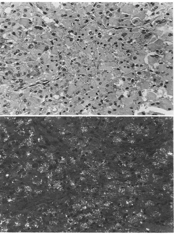
Fig 5 Sheet of macrophages, apparently without cytoplasma-tic inclusions In polarized light the same cells contain a large amount of small polyethylene particles (HE x 190)
(Figs 2 and 3); fracture of the cement was a less
con-stant finding.
The tissue curetted from the medial femur was in
continuity with the tissue around the neck and the
head of the prosthesis and presented the same
macro-scopic characters.
Two varieties were observed: a very pigmented,
gray, tissue in some cases; a white, caseous, material
in others.
Fig 4 Macrophages loaded by
opaque, stainless-steel particles. On the left side an amorphous material is present; on the right a well organized connective tissue. X-ray micro-analysis was per-formed on the same section after the coverslip had been removed
and the specimen observed with SEM (HE x 190)
Microscopically an amorphous material or a loose
connective tissue were observed, the cellular
compo-nent was formed by macrophages; two main
cytoplas-mic inclusions were found:
small (less than 1 micron), opaque particles,
which at microanalysis gave the characteristic
pat-tern of the metal alloy of the stem (stainless-steel
or Co-Cr) (Fig 4);
birefringent polyethylene particles (Fig 5).
The histological findings for each prosthesis are
re-ported in Table 2.
The level of the fracture was higher in Charnley
prostheses (average 79 1 mm from the tip of the stem)
than in Mueller prostheses ( 56 O mm) When viewed
on the back of the stem (lateral aspect) the fracture
line was oblique in all cases except two According to
the right or left side of the prosthesis the slope was
considered
when it was direct downwards
anterior-ly and + when it was downwards posterioranterior-ly; 0
marked neutral fracture slope (Table 3) Bending of
the proximal stem fragment was observed in eight
cases, all Charnley THR with stainless-steel stem.
Forward bending was always associated with a +
slope of the fracture and backward bending with
slope In two Mueller CoCrMo stems there was a
-slope of the fracture, but no bending of the stem.
Both forward and backward bending were observed
in Charnley THR; no bending was appreciable in
Mueller stems (Table 3) Fatigue striations were
evi-dent on the fracture surface in most prostheses,
al-though polishing had occurred and in some cases it
rendered recognition of striations difficult.
Fretting was evident on all proximal stem
frag-ments (Fig 8), while pitting corrosion was observed
exclusively in stainless steel prostheses (Fig 9) The
extension of the corroded area is given in Table 3 ; it
can be observed that corrosion occurred mainly in
the distal stem fragment (Fig 7), while the proximal
showed extensive involvement only in one case In
198U E Pazzaglia et al : Failure of the Stem in Total Hip Replacement 0c
at.
E v~ )0
0 O
Ca ZZ Z Z A O W v 2 O -j Q L 8 Y o O O 00 O O O o o O O .: , ; ~ t to t t~P~PP
" 8 %~ O~,
L~ 0 O O O O O O O O O Q L O O O O 4~
4 O O O O CC 0000~:
O Y Z a C' Ca P Q a M Ma 0w
Yt ; 5 CZ o o O O Z I I + I I + I + kn Ln W 'I O m O c o O 00 M CO O C 1 " O 00 00 O X 1 X) X Ca x 00 °° r-r a b a c F U U" 0 O V) 00 00 v O O O O Uh U U U U UU 199 a a. U, 0 t in Fa0 P-a E c 0 I D ;~e O '1 + O O -C" a.) I a 2 a) 00 Q z 0 Ca a.) MP
C.) 0 ,0 .0C o Ca Co Y Yr-ec. 0 Cq D Y 'r G > CZ 0 o U a 0 2 i-0
E
0 a. U a) r. t7
0 O2
0) C a Y a.) WAd
F
4 Pd
O
6
ri g P
cr
4
A; E
4
d F
6 " d
.4
tf
dr o
c;
d-;
-q (
cli ;
C-i (l
U E Pazzaglia et al : Failure of the Stem in Total Hip Replacement
Fig 9 Pitting corrosion of a stainless-steel stem (SEM x 1700)
Fig 6 M D : right Charnley prosthesis slope of the fracture
line on the back of the stem (+) and forward bending of the proximal fragment
Fig 7 Diagram illustrating the mechanism of forward and
backward bending when the loss of bone stock extends an-teriorly or posan-teriorly
Fig 8 Failed stem: fretting is evident on the proximal
frag-ment, while areas of pitting corrosion are observed on the distal stem
the latter fretting was superimposed on pitting
corro-sion.
Discussion
Fatigue fracture of the stem occurs by combination of
defective support of the proximal part with a firm
bonding of the lower part: bending cantilever fatigue
or mode IV according to the classification of Gruen
et al l 10 l.
The loss of support is due to the resorption of
bone in the medial part of the proximal femur or
cal-car area; the load on the proximal stem is the
resul-tant of flexion (in the sagittal plane) and forward
bending (in the coronal plane) when the loss of bone
stock extends anteriorly, the opposite when
resorp-tion extends posteriorly The stem is therefore
sub-jected to a combination of bending and torsion and a
crack is started on the tensile surface Bending of the
proximal stem in stainless-steel prostheses results
from plastic deformation of the metal, which
pre-cedes brittle fracture Since cracks develop
approxi-mately at right angles to the direction of the principal
tensile stress, the + slope of the fracture always
matchs forward bending and viceversa The absence
of appreciable bending in Mueller stems may be the
result of design, fracture level (which is lower than in
Charnley) and lower plasticity of the Co-Cr-Mo
alloy; however in two Mueller's stems the
slope of
the facture suggests a backward load, the O slope in
the other two the absence of a component in the
co-ronal plane.
Factors as positioning of the stem and patient's
weight may increase the load on the stem, but only
bone resorption in the medial part of the proximal
femur can start the bending cantilever fatigue
mecha-nism.
The factors which control local bone resorption
are not yet fully understood As regards the loss of
bone stock in the calcar area of prosthetic hips two
theories are in the field: mechanical overload in the
area or foreign-body reaction to particulate materials
produced by prostheses
No definitive evidences
have been so far produced to exclude one of them.
There is however a large weight of evidence that
foreign-body reaction may be one of the factors that
cause bone resorption around loosened prostheses
200U E Pazzaglia et al : Failure of the Stem in Total Hip Replacement
l 14, 17 l In this study reactive tissue to particulate
materials was found in all case in the sites where bone
resorption occurred.
Failure of the cement for mechanical or structural
causes has been reported as a possible ethiological
factor of calcar resorption, loosening and stem failure
l 3, 12 l Our findings however do not substantiate this
hypothesis since the granulation tissue found in the
calcar area was histologically characterized by
poly-ethylene or metal debris.
Radiographic evidences of cement breakage were
observed late, when osteolysis of the calcar was far
advanced.
In Mueller total hip replacements wear of the cup
was the more constant finding, in accord with the
very high quantity of polyethylene particles found in
the tissues; metal particles in these cases are the
re-sult of fretting or polishing of the fracture surface,
therefore they are produced when the process is far
advanced and the stem already brocken.
In Charnley total hip replacements corrosion of
the stem was the most remarkable finding: although
polyethylene also was produced by these prostheses,
the tissues were mainly loaded by metal particles
(only two cases showing an equal mixture of the two
type of material).
Corrosion has been cited as a possible cause of
crack propagation leading to the fracture of the metal
l 5 l, however the aspect that is of interest here is the
corrosion as a source of metal particles in the early
stages of proximal loosening Corrosion was
ob-served mainly on the distal stem fragment, which in
this type of loosening remains firmly anchored in the
bone; when corrosion was observed in the proximal
fragment, fretting was superimposed, therefore
cor-rosion precedes fretting of the stem against the
ce-ment and, in time sequence, it is the first source of
metal particles.
The question arises how the metal particles can
escape from the cement envelope that surrounds the
stem Fracture of the cement is a late phenomenon
and it is not worth in this case It is possible that an
incomplete envelope of cement surrounds the stem,
leaving exposed areas of metal; in this case however
one would expect an histiocytic reaction nearby the
source of foreign material, namely distally in the
femoral diaphysis A shrinkage of the cement after
polymerization is known to occur; this, together with
micromovement, which probably follows the
settle-ment of the implant in its bony bed with the passage
of time, may lead to formation of a narrow gap
be-tween the metal and the cement, sufficient for the
passage of small size metal particles These particles
are then pumped out around the neck, which is the
critical site of bone resorption.
Support for this hypothesis is given by the
occur-rence in a few cases of a thin fibrin film between the
cement and the metal surface in prostheses firmly
bonded to their cement envelope; the same
observa-tions have been reported by Fornasier and Cameron
l 7 l.
In the studied population fracture of the stem
oc-curred late; it is possible that earlier stem failures
have a different cause However in the two patients
of this series where fracture of the stem occurred
after 2 years and 1 month and 3 years and 6 months
the same reactive tissue was found Also Charnley l 3 l
described in his patients a necrotic, caseous material
with foreign body reactive tissue on the medial aspect
of the proximal femur.
It seems therefore that foreign body reaction
could be the common instigating factor which leads
to stem failure in all cases.
Acknowledgements This paper was supported by C N R , Gruppo per lo Studio dei Tessuti Calcificati (grant no. 82.02034 04) The authors are grateful to Dr J A S Pringle and to Dr M J Wilkinson for assistence with S E M.
References
1 Carlsson AS, Gents CF, Stenport J ( 1977) Fracture of the femoral prosthesis in hip replacement according to Charnley Acta Orthop Scand 48:650-655
2 Chao EYS, Coventry MB ( 1981) Fracture of the femoral component after total hip replacement J Bone Joint Surg lAml 63:1078-1094
3 Charnley J ( 1975) Fracture of femoral prostheses in total hip replacement Clin Orthop 111:105-120
4 Collis DK ( 1977) Femoral stem failure in total hip replace-ment J Bone Joint Surg lAml 59:1033-1041
5 Dobbs HS, Scales JT ( 1979) Fracture and corrosion in stainless steel total hip replacement stem In: Syrett BC, Acharya A (eds) Corrosion and degradation of implanted materials ASTM STP 684 American Society for Testing
and Materials, pp 254-258
6 Ducheyne P, De Meester P, Aernoud E ( 1975) Fatigue fractures of the femoral component of Charnley and Charnley-Mueller type total hip prostheses J Biomed Mat Res (Symp) 6: 199-219
7 Fornasier VL, Cameron HU ( 1976) The femoral stem-ce-ment interface in total hip replacestem-ce-ment Clin Orthop
116:248-252
8 Galante JO ( 1980) Causes of fracture of the femoral com-ponent in total hip replacement J Bone Joint Surg lAml
62: 670-673
9 Galante JO, Rostocker W, Doyle JM ( 1975) Failed femoral stems in total hip prostheses J Bone Joint Surg lAml 57:230-236
10 Gruen TA, McNeice GM, Amstutz HC ( 1979) Modes of failure of cemented stem-type femoral components Clin
Orthop 141:17-27
11 Jaeger JH, Glaesener R, Briot B, Kempf I, Nessius A, Mondoloni JL ( 1974) Ropture en service des arthroplasties totales de hanche au niveau de la piece femorale Acta Or-thop Belg 40:861-876
U E Pazzaglia et al : Failure of the Stem in Total Hip Replacement 12 Lee AJC, Ling RSM, Vangala SS ( 1978) Some clinical
var-iables affecting the mechanical behaviour of bone cement. Arch Orthop Trauma Surg 92:1-18
13 Martens M, Aernoudt E, De Meester P, Ducheyne P, Mulier JC, De Langh R, Kestelijn P ( 1974) Factors in the mechanical failure of femoral component in total hip pros-thesis Acta Orthop Scand 45:693-710
14 Pazzaglia UE, Ceciliani L, Wilkinson MJ, Dell'Orbo C ( 1975) Involvement of metal particles in loosening of metal-plastic total hip prostheses Arch Orthop Trauma Surg 104:164-174
15 Rostocker W, Chao EYS, Galante JO ( 1978) Defects in failed stems of hip prostheses J Biomed Mater Res 12:635-651
16 Ruju A, Pazzaglia UE, Mora R ( 1978) Considerazioni sul meccanismo di rottura della stele metallica di una protesi totale d'anca Minerva Ortopedica 29:297-303
17 Willert HG, Semlitsch M ( 1970) Tissue reactions to plastic and metallic wear products of joint endoprostheses total hip prostheses In: Gschwend N, Debrunner HV (eds) Total hip prosthesis Huber, Bern
18 Wroblewski BM ( 1979) The mechanism of fracture of the femoral prosthesis in total hip replacement Int Orthop 3: 137-139
19 Wroblewski BM ( 1982) Fractured stem in total hip replace-ment A clinical review of 120 cases Acta Orthop Scand 53:279-284
Received November 7, 1986 / Accepted February 3, 1988 202