I
DOTTORATO DI RICERCA IN
S
CIENZE
C
HIRURGICHE
Ciclo XXVI
Settore Concorsuale di afferenza: 06/A4 Anatomia Patologica Settore Scientifico disciplinare: MED/05 - MED/08
The vulnerable carotid plaque.
Identification of endothelial markers predictive of plaque
weakness, rupture predisposition and neoangiogenesis.
Tesi Presentata da: Dott. Francesco Vasuri
Coordinatore Dottorato Relatore
Chiar.mo Prof. Andrea Stella Chiar.mo Prof. Gianandrea Pasquinelli
II Ancora una volta, a Debbie, con tanto amore…
…e alla mia famiglia, che mi ha sempre sostenuto in questo lunghissimo percorso!
III
Background. Neoangiogenesis is crucial in plaque progression and instability. Previous
data from our group demonstrated that intra-plaque neovessels show both a Nestin+/WT+ and a Nestin+/WT1- phenotype, the latter being correlated with complications and plaque instability.
Aims. The aims of the present thesis are: (i) to confirm our previous results on
Nestin/WT1 phenotype in a larger series of carotid atheromatous plaques, (ii) to evaluate the relationship between the Nestin+/WT1- neoangiogenesis phenotype and plaque morphology, (iii) to evaluate the relationship between the immunohistochemical and histopathological characteristics and the clinical instability of the plaques.
Materials and Methods. Seventy-three patients (53 males, 20 females, mean age 71
years) were consecutively enrolled. Symptoms, brain CT scan, 14 histological variables, including intraplaque hemorrhage and diffuse calcifications, were collected. Immunohistochemistry for CD34, Nestin and WT1 was performed. RT-PCR was performed to evaluate Nestin and WT1 mRNA (including 5 healthy arteries as controls). Results. Diffusely calcified plaques (13 out of 73) were found predominantly in females
(P=0.017), with a significantly lower incidence of symptoms (TIA/stroke) and brain focal lesions (P=0.019 and P=0.013 respectively) than not-calcified plaques, but with the same incidence of intraplaque complications (P=0.156). Accordingly, both calcified and not calcified plaques showed similar mean densities of positivity for CD34, Nestin and WT1. The density of Nestin and WT1 correlated with the occurrence of intra-plaque hemorrhage in all cases, while the density of CD34 correlated only in not-calcified plaques.
Conclusions. We confirmed that the Nestin+/WT1- phenotype characterizes the
IV
complications, albeit they do not influence symptomatology and plaque vulnerability. Female patients show a much higher incidence of not-complicated or calcified plaques, receiving de facto a sort of protection compared to male patients.
V
The principal aims of the current PhD program is the evaluation of the neoangiogenesis in carotid atheromatous plaques, in term both of neovessel density and phenotype, and its correlation with plaque morphology and instability.
We performed a morphological and immunohistochemical study on 73 carotid plaques, in order to evaluate the phenotype of the intra-plaque neovessels, and its relationship with plaque morphology and clinical presentation. In previous studies conducted during the PhD program, we found that specific vasa vasorum of normal arteries co-express Nestin and WT1, two markers of progenitor endothelia and vascular proliferative potential. Those results were published in 2012 [Vasuri F, Fittipaldi S, Buzzi M,
Degiovanni A, Stella A, D'Errico-Grigioni A, Pasquinelli G. Nestin and WT1 expression in small-sized vasa vasorum from human normal arteries. Histol Histopathol 2012;27:1195-1202], and gave us a theoretical basis for the evaluation of Nestin and
WT1 in plaque neovessels. During the second year of the PhD program, we evaluated CD34, Nestin and WT1 in 49 specimens, and we found that intra-plaque neovessels express both the Nestin+/WT1+ phenotype and the Nestin+/WT1- phenotype, i.e. the WT1/Nestin ratio was inferior than 1. Moreover, the Nestin+/WT1- phenotype correlated with the occurrence of intra-plaque histological complications (and plaque vulnerability) regardless the density of CD34-positive vessels. These results are under revision [FittipaldiS, VasuriF, DegiovanniA, PiniR, Mauro R, Faggioli G, D'Errico-Grigioni A, Stella A, Pasquinelli G. Nestin and WT1 expression in atheromatous plaque neovessels: association with vulnerability. Histol Histopathol].
The results exposed in the present thesis are the progression of our study, with an enlargement of the carotid specimen series (n=73), and special focus on the different neoangiogenetic profiles in neovessel of plaques with different morphology, and on the correlations with clinical data.
VI
1. INTRODUCTION pag. 1
1.1 FUNCTIONAL ANATOMY OF THE NORMAL
ARTERIAL INTIMA pag. 2
1.1.1 Prevalent cell types in the normal intima pag. 3
1.1.2 Extracellular matrix in the normal intima pag. 4
1.2 PATHOBIOLOGY OF ATHEROSCLEROSIS pag. 6
1.2.1 Atherosclerosis as an inflammatory disease pag. 6
1.2.2 Main risk factors pag. 7
1.2.3 Plaque progression and neoangiogenesis pag. 8
1.3 PATHOLOGIC ANATOMY OF THE INTIMAL
LESIONS: THE AMERICAN HEART
ASSOCIATION (AHA) CLASSIFICATION pag. 11
1.3.1 AHA classification of intimal lesions: initial and
intermediate lesions pag. 11
1.3.2 AHA classification of intimal lesions: advanced lesions pag. 14
1.3.3 Modifications to the original classification pag. 18
1.4 PREVIOUS RESEARCH OF OUR GROUP pag. 21
1.4.1 Nestin and WT1 as markers of active neoangiogenesis pag. 21
VII
1.4.3 Nestin and WT1 expression in healthy vasa vasorum pag. 23
1.4.4 Nestin and WT1 in neoangiogenesis of atheromatous plaques pag. 25
1.5 AIMS OF THE THESIS pag. 27
2. MATERIALS AND METHODS pag. 28
2.1 PATIENTS SELECTION AND CLINICAL DATA pag. 29
2.1.1 Indications for endarterectomy pag. 29
2.1.2 Clinical data and follow-up pag. 29
2.2 HISTOPATHOLOGICAL ANALYSIS pag. 30
2.3 IMMUNOHISTOCHEMISTRY pag. 33
2.3.1 Manual procedure pag. 33
2.3.2 Automatic procedure pag. 34
2.3.3 Evaluation of the "density" of positivity
with Immunohistochemistry pag. 34
2.4 RT-PCR pag. 36
2.4.1 Trizol RNA extraction pag. 36
VIII
3. RESULTS pag. 39
3.1 PATIENTS AND CLINICAL DATA pag. 40
3.2 HISTOPATHOLOGICAL ANALYSIS pag. 41
3.3 IMMUNOHISTOCHEMISTRY pag. 44
3.4 RT-PCR pag. 46
3.5 CORRELATIONS AMONG MORPHOLOGICAL
VARIABLES pag. 47
3.6 CORRELATIONS BETWEEN
IMMUNOHISTOCHEMISTRY AND MORPHOLOGY pag. 48
3.7 IMMUNOHISTOCHEMICAL PROFILES OF THE
DIFFERENT PLAQUE TYPES pag. 50
3.7.1 Complicated plaques pag. 50
3.7.2 Calcified plaques pag. 50
3.7.3 The influence of calcifications and neoangiogenesis
IX
NEOVESSEL PHENOTYPES AND CLINICAL DATA pag. 55
3.9 THE CALCIFIED PLAQUES IN THE FEMALE PATIENTS pag. 56
4. DISCUSSION pag. 58
1
2
1.1 FUNCTIONAL ANATOMY OF THE NORMAL ARTERIAL INTIMA
(from Stary 1992)
The intima is defined as the portion of the arterial wall from the endothelium to the media margin (generally the internal elastica lamina, albeit it can be missing in branches and bifurcations). The intima/media ratio can vary from 0.1 to 1 according to the anatomical site. The arterial intima is composed by two layers, whose identification is not always easy at light microscopy (Stary 1992):
(1) An inner layer called proteoglycan layer, containing finely reticulated not-fibrous connective tissue (proteoglycan) and very few elastic fibers. The proteoglycan layer contains both RER-rich (synthetic) and myofilament-rich (contractile) smooth-muscle cells, and it was firstly described as an alcianophilic inner region of the intima in the nonhuman primates (Wight 1975).
(2) An outer layer called musculoelastic layer, with abundant contractile smooth-muscle cells and elastic fibers, and the presence of more collagen.
Figure 1 (Reprinted by Stary et al. 1992). Coronary artery from a 16-month-old boy, showing the different layers composing a healthy vessel wall. e: endothelium; pgc: proteoglycan-rich layer; me: musculoelastic layer; M: media; A: adventitia.
3 1.1.1 Prevalent cell types in the normal intima
Endothelial cells of normal arteries (both muscular and elastic) are flattened and elongated cells forming a continuous layer. Studies on animal cultured cells demonstrated that the long axis of each endothelial cell is oriented in the direction of the blood flow (Flaherty 1972; Levesque 1985). Polysaccharides, glycosaminoglycans and glycoproteins form the glycocalix that covers the luminal portion of endothelium; some of these molecules are known to bind lectins, lipoprotein lipase, and other enzymes. Moreover, endothelial cells surface shows membrane receptors for LDL (with lipoprotein-lipase activity), insulin, and histamine, among others (Dicorleto 1975; Simionescu 1981; Bar 1982; Heltianu 1982; Wang-Iverson 1982; Jaffe 1987). Endothelial cells carry out several roles, such as the transport of macromolecules (lipoproteins above all) from the blood flow to the intima, the anti-thrombotic homeostasis, the regulation of inflammation and of the response to immunity, as well as the maintenance of the vascular tone and vasoconstriction. Endothelial cytoplasms contain thrombomodulin, whose binding with thrombin inactivates the coagulation factor Va (Esmon 1988). Furthermore, the endothelial-derived prostacyclin PGI2 inhibits the platelet aggregation and cause vasodilatation (Moncada 1977). Other endothelial-derived factors are likely to promote both vasodilatation and vasoconstriction (Brenner 1989), while the same effects are obtained by soluble mediators with direct action on endothelial receptors, as for serotonin (Vanhoutte 1987), acetylcholine (Furchgott 1980), and endothelin (the latter produced by the endothelium itself) (Yanagisawa 1988).
Two types of smooth muscle cells are usually recognizable in the normal intima of humans and other species:
4
(1) The myofilament-rich smooth muscle cells, also called contractile or adult-type (Dilley 1987), are the main type in human intima.
(2) The rough endoplasmic reticulum (RER)-rich smooth muscle cells, also called synthetic, immature, modulated or ergastoplasm-rich (Dilley 1987), contain few contractile filaments, and they are more frequently visible in proteoglycan-rich intimal layer, since RER-rich smooth muscle cells can synthesize a wide set of matrix molecules (Burke 1979).
Apart from the contractile and synthetic functions, intimal smooth muscle cells are likely to play a role in the removal via phagocytosis of LDL and other lipoproteins from the intima, as it is demonstrated by the expression of LDL receptors on smooth muscle cells surface (Stary 1992).
Inactivated macrophages are physiologically present in the human intima, and their number increases from the childhood to adult life. Like in several healthy tissues, macrophages are present in intima as isolated mononuclear cells: they carry out several functions, such as the production of enzymes for matrix remodeling (elastases, collagenases), the secretion of growth factors for smooth muscle, neoangiogenesis, lymphocytes chemotaxis, phagocytosis of lipoproteins, bacteria, necrotic cells and immune complexes (Werb 1975; Nucera 2011).
1.1.2 Extracellular matrix in the normal intima
The extracellular matrix constitutes up to 60% of the intima, both in healthy condition and during intimal thickenings. Apart from molecules responsible for the cell-cell and cell-matrix adhesion, like fibronectin and laminin, the most represented molecule types in intimal extracellular matrix are proteoglycans, collagen and elastin.
5
Proteoglycans production by the endothelium cells has been demonstrated on bovine aorta cultured cells, and in particular heparan sulfate proteoglycan and, in a minor amount, dermatan sulfate proteoglycan (Kinsella 1988). In human aortic media, dermatan sulfate proteoglycan resulted to be more associated with collagen fibers, and heparan sulfate proteoglycan with elastic fibers and smooth muscle cells. The different proteoglycans in their different localizations play an important role in maintaining the viscoelastic properties of the vessel wall, and therefore in controlling the homeostasis of vascular wall cells (Wight 1989).
Collagen is involved in the vascular wall integrity by constituting the attachment site of the endothelial cells with the intimal extracellular matrix. The most represented collagen types in the artery wall are type I and type III. In children, the type III collagen (of endothelial derivation) is the prevalent type of collagen present in the subendothelial region, but with aging the synthesis of collagen type I increases, probably due to the concomitant increasing of smooth muscle cells, capable to synthesize both collagen type I and type III in vitro (Sage 1981; Sankey 1981).
Elastin in produced by endothelial cells and smooth muscle cells: in the intimal musculoelastic layer elastic fibers are prominent, similar to those present in the media layer. With aging there is a decreasing in elastin content and an increasing in collagen content (Hosoda 1984).
6
1.2 PATHOBIOLOGY OF ATHEROSCLEROSIS
1.2.1 Atherosclerosis as an inflammatory disease (from Ross 1999)
After the birth a progressive increase of the intimal thickness was described in some districts. In studies conducted on rabbits by means of tritiated thymidine, this increase is likely to be due to a mitotic numerical growth of the pre-existing smooth muscle cells (Stary 1974). Anyway it is not still clear whether the migration of smooth muscle cells from the media layer might play a role (Grotendorst 1981).
A part these adaptive changes characterizing some specific human vascular districts, the onset of atherosclerotic lesions is correlated to specific events. According to the historical definition, the first event triggering atherosclerotic lesions is a damage of the endothelial surface (Ross 1973), although more recent theories emphasized an endothelial dysfunction, rather than damage, to play a key role in the lesion initiation. The causes of endothelial dysfunction that were identified and described in the years are multiple, from LDL or homocysteine plasma levels, to diabetes and genetic diseases, from cigarette smoking to viruses such as herpervirus, Chlamydia, etc. (Ross 1999). According to Ross, after the initial damage, the endothelium acquires pro-coagulant properties (instead of anti-coagulant), and starts the production and secretion of vasoactive factors, cytokines and growth factors. The result is an actual inflammatory response to the initial stimulus, characterized mainly by lymphocytes and monocytes/macrophages (Jonasson 1986; van der Wal 1989), which is soon followed by a migration of the medial smooth muscle cells. With the maintaining of the inflammation, the artery wall thickens and dilates, resulting in a remodeling of the wall, without modifications of the lumen.
7
Different cycles of inflammatory and muscle cell accumulation, more serum fat entrapment, and the activation of fibrosis by cytokines lead to the formation of a necro-lipidic core and of a fibrous cap, which characterize the so-called advanced lesions. At this point the lesion may protrude in the lumen, leading to stenosis and symptoms.
1.2.2 Main risk factors
LDL. Circulating LDL represent the major cause of endothelium and arterial wall injury, and they are commonly found in the modified forms: oxidized, proteoglycan-associated or glycated forms, the latter most commonly in diabetes (Morel 1983; Griendling 1997). The phagocitosis by the macrophages lead to LDL oxidation, the formation of lipid peroxides and the accumulation of esters of the cholesterol; in these phase the macrophage becomes a "foam cell". The pro-inflammatory cytokines tumor necrosis factor-α and interleukin-1, among others, have shown a facilitative role in LDL binding to the endothelium surface, increasing the expression of the gene encoding LDL receptors (Stopeck 1993).
Hypertension. Hypertension is connected to the pathogenesis of atherosclerosis through different mechanisms: the activation of the renin-angiotensin system leads to systemic vasoconstriction, and a proliferative stimulus on muscle cells has been described to occur with time. The binding of angiotensin II to specific receptors on the surface of the smooth muscle cells leads also to the activation of phospholipase C, that causes muscle hypertrophy and hyperoxidation of LDL (Gibbons 1992).
Homocysteine. Homocysteine is toxic for the endothelium, and according to several studies it showes pro-thrombotic and fibrogenic activities (Harker 1993; Hajjar 1993). When serum homocysteine is elevated in patients with no deficit of homocysteine metabolism, there is an increasing risk of symptomatic
8
atherosclerosis (Verhoef 1995), while in patients with congenital enzymatic deficits which lead to an impaired homocysteine metabolism, a severe atherosclerosis can develop also in childhood (Nygard 1997).
C-Reactive Protein (CRP). Cardiovascular diseases and diabetes are inflammatory diseases, and many serum inflammatory markers have been studied in the past years (Devaraj 2009). Among them, CRP is considered the best marker to predict the severity and course of the metabolic syndrome (Ridker 2000). High-sensitivity CRP has been found to interact with a number of pro-inflammatory processes in vitro, and it is able to induce the expression of the adhesion molecules VCAM-1 and ICAM-1 in humans (Corrado 2010). C-reactive protein levels rise with increasing levels of visceral adipose tissue, and together with other adipose-derived modulators of inflammation evoke the production of acute phase reactants in the liver, implicated in thrombogenesis (Libby 2010).
Other risk factors include smoking, insulin resistance/diabetes, lack of physical activity, etc. All these factors result in direct or indirect endothelial dysfunction and inflammation of the vessel wall. The same concept is applicable for Lupus Erithematosus Systemicus and other systemic disease which may cause vasculitis.
1.2.3 Plaque progression and neoangiogenesis
The vulnerable plaques have been originally described as atheromatous plaques with a large lipid core, a thin fibrous cap, a rich infiltrate of macrophages and scarce smooth muscle cells (Kullo 1998). The erosion and rupture of the fibrous cap is the main events triggering the plaque instability. The degradation of the fibrous cap is mediated by
9
metalloproteases, such as collagenases, elastases and stromelysin (Galis 1994), mainly produced by macrophages in response to cytokines produced by activated T-cells. More recently, neoangiogenesis stood out as one of the most important pathological processes involved in the plaque progression (Hermus 2010), since neovessel formation was related to increased plaque vulnerability and to the onset of clinical symptoms (Mofidi 2001; Faggioli 2011).
Mofidi et al. have found a higher volume of intraplaque hemorrhage and a higher incidence of symptoms in those plaques with a higher neovessel density, measured with a "simple" immunohistochemistry for CD34 (Mofidi 2001).
The key mechanism triggering plaque neoangiogenesis is the hypoxia of the inner vascular layers due to the lipid accumulation and the myo-intimal thickening up to 500 µm (Moreno 2006). The chronic hypoxia activates different pathways, such as the Hypoxia Inducible Factor and the Toll-like receptor pathways (Frantz 2005), leading to a neoangiogenesis which is thought to originate from the sprouting of the endothelial cells of the post-capillary venules (Carmeliet 2003). Our recent results indicate that the
vasa vasorum located in specific adventitial "hot spots" are likely to represent the
candidate vessels of origin of the neoangiogenesis during atherosclerosis (Vasuri 2012). Anyway, the adventitial origin of the neoangiogenetic vessels is not the only origin described: for example, a previous work from Kumamoto and colleagues identified the luminal endothelium as the source of the 3.5% of the intra-plaque neovessels (Kumamoto 1995).
If the density of neovessels is likely to be directly related with plaque growth and progression (Moreno 2006), also the morphology of the neoangiogenetic structures is important for the onset of plaque instability: indeed, it was described that plaque neovessels lack extracellular junctions (Sluimer 2009), and that symptomatic plaques
10
showed larger and more irregular neoangiogenetic structures compared to the neovessels of asymptomatic plaques (McCarthy 1999).
The neoangiogenesis has been involved also in the plaque regression mechanisms, since the neovessels are responsible for the lipid transport outside the plaque core. After lipid removal, the plaque becomes fibrotic (type Vc or type VIII according to AHA, see the next chapter) or calcified (type Vb or type VII), and in these regressed plaques a minor neovessel density was described (Moreno 2006).
11
1.3 PATHOLOGIC ANATOMY OF THE INTIMAL LESIONS: THE
AMERICAN HEART ASSOCIATION (AHA) CLASSIFICATION
The original classification of the American Heart Association (AHA), after a paper by Stary et al. in 1994, sorted the intimal lesions into six bio-pathological entities, which in turn were pooled in two main groups: the so-called precursor lesions (types I, II, III), and the advanced lesions (types IV, V, VI) (Stary 1994; Stary 1995). The progression from one type to another is not demonstrated for every lesion, and studies on animal models have shown that a specific sequence of steps is required to progress from the "simple" lipid intimal accumulation (which characterize the precursor lesions) to the intimal thickening and related disorders, with the consequent deformation of the whole arterial wall (advanced lesions).
The first step of the whole process is represented by the entrapment of serum lipoproteins in the matrix intima, and their internalization by the macrophages and smooth muscle cells (Schwenke 1989; Steinberg 1990). The following oxidation of the lipoproteins represents a chemotactic stimulus for monocytes (Quinn 1987).
In the study of the atheromatous disease, a particular kind of "gelatinous lesion" has been proposed as precursor lesion: according to some authors, this lesion appears as oval-shaped intimal elevation, pink to pale grey , and soft. At histology, these thickening areas contain gelatinous scarcely-cellulated material, with little or no lipids, and constituted by abundant matrix with little collagen and elastin content (Movat 1959; Haust 1971).
1.3.1 AHA classification of intimal lesions: initial and intermediate lesions
Type I lesions ("initial lesions"). It represents the most frequent intimal lesion in childhood (Stary 1994), and it is constituted by isolated groups of macrophages
12
with cytoplasmic lipid droplets ("foam cells") (Stary 1989). These findings are observed in the same arterial regions generally prone to the hyperplasia of the eccentric type.
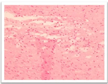
Figure 2. Isolated clusters of intimal macrophagic foam cells. Haematoxylin-Eosin stain; magnification 20x.
Type II lesions (including fatty streaks). In this step, the foam cells are not grouped in isolated clusters, but they form stratified layers in the intima. If the layers are located immediately under the subendothelial surface, the lesion in macroscopically visible as a yellow "fatty streak", but if the foam cell layers are deeper in the intima, the macroscopical appearance will be less defined (Stary 1994). In type II lesions, also intimal smooth muscle cells contain lipid droplets. At any chance, most of the lipids are still contained within cells.
A small subgroup of type II lesions are called type IIa: they raise in specific and predictable arteries with adaptive intimal thickenings, and they are called progression-prone or advanced-prone lesions. However, the majority of type II lesions belong to the type IIb group: they are also called "progression-resistant",
13
since their progression, when occurring, is slow, and occur only in subjects with very high serum lipoprotein levels (Stary 1994).
On histopathological grounds, type IIa lesions contain more smooth muscle cells than type IIb, together with more extracellular matrix and macrophages, which constitute the adaptive thickening.
Figure 3. Fatty streak. The macrophagic foam cells form stratified intimal layers. Haematoxylin-Eosin (left) and Trichrome stains (right); magnification 10x.
Type III lesions("intermediate lesions" or "preatheroma"). They are placed between type II and type IV lesions, and their morphological hallmark is the deposition of extracellular lipids, which replace the proteoglycan of the intimal matrix, but without a true lipid core. Type III lesions have different lipid content than types I and II: indeed they have more free cholesterol, triglycerids, sphingomyelin and lysolecithin (Katz 1976; Small 1988).
14 Figure 4. A type III lesion, characterized by the presence of extracellular lipids.
Haematoxylin-Eosin (left) and Trichrome stains (right); magnification 10x.
1.3.2 AHA classification of intimal lesions: advanced lesions
The so-called advanced lesions are generally associated to a higher incidence of clinical manifestations. In these phases, the accumulation of lipids, matrix, and cellular elements cause a distortion of the arterial wall. The subsequent narrowing of the lumen may reveal itself both angiographically and clinically (Stary 1995).
Type IV lesions ("atheromas"). The extracellular lipid accumulation occupies a well-defined region of the intima, of variable extension, called lipid core. The atheroma does not show complications such as hemorrhages or surface defects, but the presence of the lipid core itself causes a distortion of the arterial walls and a narrowing of the lumen, which might cause stenosis and clinical manifestations: this is why type IV lesions are included among the advanced lesions. In younger subjects, atheromas initially appear in the same anatomical regions of eccentric arterial thickenings, thus they are considered adaptive changes.
Most important (at least for the purposes of the present thesis) is that in the type IV lesions neoangiogenetic capillaries appear for the first time, initially located
15
in the peripheral margin of the lipid core ("shoulder"). A mild macrophagic infiltrate may follow the neovessels.
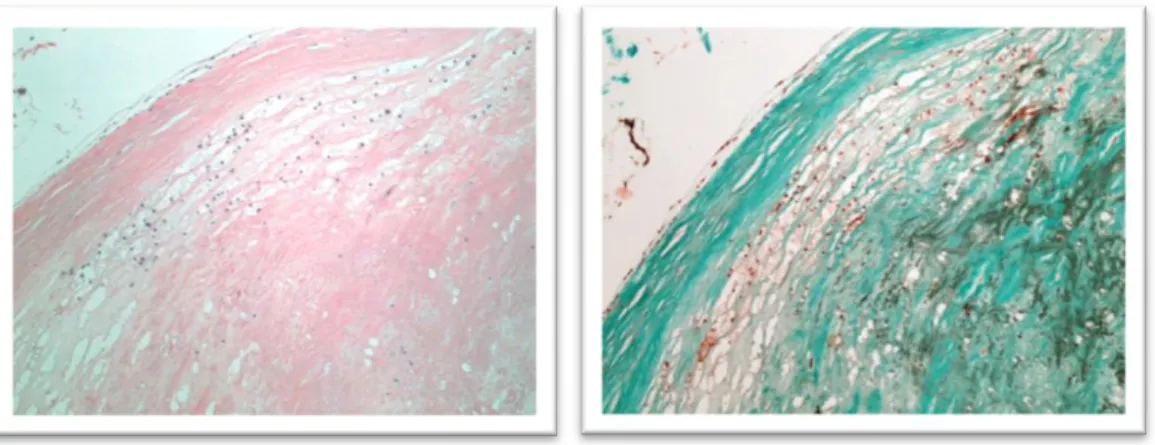
Figure 5. A type IV lesion, or atheroma, characterized by a well-visible lipid core (arrow), without a formed fibrous cap yet.
Haematoxylin-Eosin (left) and Trichrome stains (right); magnification 10x.
Type V lesions ("fibroatheromas"). In type V lesions a prominent fibrous cap appears over the lipid core, constituted by newly formed connective tissue with muscle cells rich in rough endoplasmic reticulum. The neovessels are more numerous and diffuse, and they also spread in the fibrous tissue. The type V lesions, which are also classified as type Va in 1995 classification, can be multilayered, due to the overlapping of multiple lipid cores and fibrous caps in time.
Fibroatheromas, as type IV atheromas, can undergo to histological complications, and become type VI lesions, or can regress in two different ways. Lesions showing a massive calcification of the lipid core was defined as type Vb in the 1995 classification, and type VII in the more recent; lesions showing a prominent fibrotic involution of the lipid core was defined as type Vc in the 1995 classification, and type VIII in the more recent.
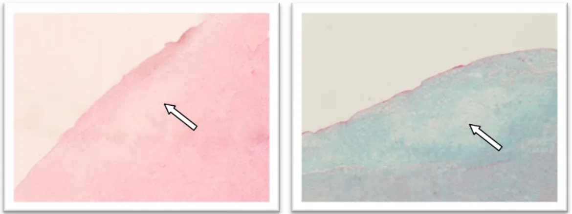
16 Figure 6. Fibroatheroma (type Va). A thick fibrous cap (arrow) is present above the
lipid core. Haematoxylin-Eosin (left) and Trichrome Stains (right); magnification 4x.
Type VI lesions ("complicated lesions"). The type VI lesions are defined as atheromatous lesions with features of type IV or V, but with (type VIa) erosion or ulcerations of the endothelial surface (type VIb), hemorrhage/hematoma or (type VIc) luminal thrombosis.
All these complications represent the morphological base of the plaque vulnerability, and will be referred in the present thesis as histological
complications. Evidences exist of a close relationship between plaque
hemorrhage and neoangiogenesis (Mofidi 2001): newly formed vessels show a weak wall, without extracellular junctions (Sluimer 2009), and their growth in the lipid content of the plaque core can easily lead to rupture. Moreover, there is evidence that the neovessels of symptomatic plaques are larger and more irregular compared to the neovessels of asymptomatic plaques (McCarthy 1999).
17 Figure 7. An example of type VI plaque, characterized by multiple hemorrhagic foci
(arrows).
Haematoxylin-Eosin stain; magnification 4x.
Figure 8. An example of type VI plaque, characterized by erosion of the endothelial surface (arrows). Haematoxylin-Eosin (left) and Trichrome Stains (right);
18 Figure 9. An example of type VI plaque, with the presence of an organizing
intraluminal thrombus.
Haematoxylin-Eosin (left) and Trichrome Stains (right); magnification 4x.
1.3.3 Modifications to the original classification
In 2000 Stary modified his own previous classification of the atheromatous lesions, in order to better define the progression (or regression) of plaques from one type to another (Stary 2000). According to Stary, type IV and type V lesions can undergo to histological complications, becoming type VI lesions or they can regress into lesions with predominant calcifications or fibrosis; on the other hand, type VI lesions can regress in the same way as well. Complying with these possible different modifications of the plaques, the previous sub-classes Vb and Vc became types VII and VIII respectively.
Type VII lesions (calcified lesions). This type of atheromatous lesions replaced the type Vb of the old nomenclature. They are characterized by a core almost fully replaced by a massive deposition of calcification.
19 Figure 10. A heavily calcified uncomplicated plaque, classifiable as type VII (or Vb according to 1995 classification). The calcium deposits completely deform the vessel's
architecture.
Haematoxylin-Eosin (left) and Trichrome Stains (right); magnification 4x.
Type VIII lesions (fibrous lesions). It replaced the type Vc of the old nomenclature. The lipid core is completely replaced by collagen.
Figure 11. A type VIII (or Vc according to 1995 classification) plaque. The lipid core is not visible, replaced by collagen.
20
The types VII and VIII plaques represent the two major ways of regressive involution to which a atheromatous plaque can undergo. This regression can involve either directly a type V uncomplicated lesion or a type VI lesion, according to the following pathway:
21
1.4 PREVIOUS RESEARCH OF OUR GROUP
1.4.1 Nestin and WT1 as markers of active neoangiogenesis
Nestin is an embryonic intermediate filament protein expressed in neural and mesenchymal stem cells, and it was recently described by Wagner and colleagues to be expressed in a variety of progenitors cells (Wagner 2006). In that paper, the role of Nestin as an angiogenic marker, expressed in newly formed microvessels, was demonstrated in endothelial cells of newly forming blood vessels after myocardial infarction (Wagner 2006), but later it was demonstrated also in both tumoral and not-tumoral neoangiogenesis (Ramasamy 2011). Afterwards numerous Nestin-positive microvessels were described also in tumoral neoangiogenesis, mainly in mouse models (Teranishi 2007; Ramasamy 2011). Moreover, a previous study showed that medial vascular smooth muscle cells in the developing arteries potently express Nestin, but its expression is abolished in adult arteries (Oikawa 2010). It is therefore believed that Nestin expression, representing an early endothelial differentiation or an endothelial progenitor phenotype, could be a reliable marker of “progenitor-committed” endothelial cells, or of a “young” endothelium, during active neoangiogenesis.
The known transcription factors regulating Nestin gene expression are: Pou, Sox, TTF-1 and WT1 (Wagner 2006; Pelizzoli 2008). The Wilms tumor suppressor (WT1) was originally described in the paediatric Wilms’ tumor of the kidney (or nephroblastoma), but its involvement has been demonstrated in a variety of other tumors and tumor cell lines (Hohenstein 2006; Wagner 2008). Some recent studies showed the involvement of WT1 as an activator of the Nestin gene (Hohenstein 2006), and the co-localization of WT1 and Nestin during the different development steps in embryogenesis (Wagner 2006).
22 1.4.2 Physiology of vasa vasorum and neoangiogenesis in diseased arteries
Vasa vasorum are small arteries present in the adventitia of the vessels of major calibre.
The principal functions of vasa vasorum are nutrient and oxygen delivery to the deeper vascular layers and waste removal (Ritman 2007; Mulligan-Kehoe 2010). In the largest vessels the so-called first-order vasa vasorum take origin directly from other arteries and run longitudinally along the adventitia (Kwon 1998). The second-order vasa
vasorum derive from a branching of the first-order vasa vasorum and reach the deeper
adventitia and media layers. In animal models the application of microscopic computed tomography in healthy vessels demonstrated that vasa vasorum form a plexus around the adventitia, with generally a lower number of second-order vasa vasorum rather than first-order. This condition changes in pathological conditions, where a dramatically increase in second order vasa vasorum was observed (Kwon 1998). In diseased arteries the vessel wall hypoxia unleashes the neoangiogenesis starting from the adventitial vasa
vasorum. In these pathological conditions vasa vasorum, and especially the
second-order vasa vasorum proliferate quickly, also regulated by local and circulating growth factors such as vascular endothelial growth factor and hypoxia inducible factor, among others (Moreno 2006). This neovasculogenic potential has been largely studied in the last decades, and the existence of endothelial progenitor cells (EPCs) within the layers of the larger vessels has been widely accepted, although their location, phenotype and roles in the vascular damage and angiogenesis are still debated (Zengin 2006; Pasquinelli 2007; Torsney 2011). Most authors agree that a population of progenitor cells is localized in the adventitia, characterized by positivity for different markers of vascular stem potential, such as CD34, c-Kit, CD105, Notch-1, SCA1, and KDR (Alessandri 2001; Hu 2004; Pfister 2005; Zengin 2006; Pasquinelli 2010; Leri 2011).
23 1.4.3 Nestin and WT1 expression in healthy vasa vasorum
In the first study that we published during the PhD program, we performed a histological and immunohistochemical analysis of vasa vasorum in 20 not-diseased arteries from 16 healthy multiorgan donors (Vasuri 2012). Our aim was to verify Nestin and WT1 expression in endothelial cells of normal adult vasa vasorum, in order to localize and quantify these microvessels that could be responsible for neovascularization in diseased arteries. In these 20 specimens immunohistochemistry for CD34, CD31, Nestin and WT1 was performed manually and the microvessel “densities” of positivity were calculated for each antibody, dividing the vascular adventitia in 1-mm2 fields with an ocular micrometer in each slide. Double immunofluorescence was used to investigate Nestin and WT1 co-localization in vasa
vasorum.
As results, the mean positivity “densities” for CD31, CD34, Nestin and WT1 were 13.63/mm2, 12.20/mm2, 8.90/mm2 and 7.98/mm2 respectively. Mean Nestin/CD31 and WT1/CD31 ratios were 0.69 and 0.63 respectively, which means that 69% and 63% of the CD31-positive neovessels also expressed Nestin and WT1 respectively. As expected, the Nestin:WT1 ratio was close to 1:1, as also confirmed by immunofluorescence, which indentified Nestin and WT1 coexpression in the same endothelial cells.
24 Figure 13 (reprinted from Vasuri 2012). On the left, schematic quantitative representation of CD31, CD34, Nestin and WT1 expression in healthy arteries, and their relationships. On the right, the immunohistochemical positivity of CD31, Nestin
and WT1 in vasa vasorum.
Finally, vasa vasorum <50 µm in diameter showed a higher percentage of Nestin/WT1-positive cells than the larger vasa vasorum, especially in “hot spots”, characterized by several small-sized arteriolar vasa vasorum, often together with nerva vasorum. Our experience indicated that WT1 and Nestin colocalize with a 1:1 ratio in the vasa
25 Figure 14 (reprinted from Vasuri 2012). Schematic picture of vasa vasorum, and their
different Nestin/WT1 phenotypes.
1.4.4 Nestin and WT1 in neoangiogenesis of atheromatous plaques
In the second step of our research, we performed a study on the expression of Nestin and WT1 in the neoangiogenetic component of the atheromatous plaques, in order to describe and quantify the protein and mRNA levels of WT1 and Nestin in the plaque neovessels and to investigate the role of neovessel phenotype in the occurrence of the histological complications correlated with plaque instability (Fittipaldi et al., under revision). For these purposes we prospectively evaluated 49 consecutive carotid endarterectomy specimens, collected the main histopathological characteristics (particularly the occurrence of histological complications) and carried out immunohistochemistry for CD34, Nestin and WT1; the density of positivity was evaluated for each marker in "regions of interest". RT-PCR was performed to assess Nestin and WT1 mRNA levels on the first 10 plaques and on 10 control arteries.
26
As results, the mean immunohistochemical densities of CD34, Nestin, and WT1-positive structures were 41.88/mm2, 28.84/mm2 and 17.68/mm2 respectively. Among the CD34+ neovessels, 68% and 42% expressed Nestin and WT1 respectively, i.e., nearly 36% of the neovessels showed a Nestin+/WT1- phenotype. Furthermore, complicated plaques (n=30) showed significantly more CD34 and Nestin-positive vessels than uncomplicated plaques (n=13; P=0.045 and P=0.009), while WT1 was less increased (P=0.139). RT-PCR confirmed that WT1 gene expression was 3-fold lower than Nestin gene in plaques (p=0.001). we concluded that plaque neoangiogenesis shows both a Nestin+/WT1- and a Nestin+/WT1+ phenotype. The Nestin+/WT1- neovessels are significantly more represented in plaques with histological complication (hemorrhage above all).
Figure 15 (reprinted from Fittipaldi, under revision). Different Nestin/WT1 phenotypes in complicated and uncomplicated atheromasic plaques.
27
1.5 AIMS OF THE THESIS
The aims of the present thesis are:
1. To confirm our previous results on Nestin/WT1 phenotype in a larger series
of carotid atheromatous plaques.
Histopathological analysis and immunohistochemistry for CD34, Nestin and WT1 will be carried out on a series of 73 consecutive carotid plaques. The plaque morphology and the immunohistochemical densities of positivity will be correlated in order to confirm our previous data on Nestin+/WT1+ and Nestin+/WT1- phenotypes in plaque neoangiogenesis.
2. To evaluate the relationship between the Nestin+/WT1- neoangiogenesis
phenotype and the main histopathological plaque characteristics (especially
complications).
The Nestin+/WT1+ and Nestin+/WT1- phenotypes will be analyzed according to the separate atheromatous types (V, VI, VII, and VIII). Special attention will be paid to histological complications (hemorrhage, surface defects, thrombosis) that identify type VI plaques, and massive calcifications, that define class VIII (ex class Vb) plaques.
3. To evaluate the relationship between the immunohistochemical and
histopathological characteristics and the clinical instability of the plaques.
The clinical data of the 73 patients who underwent endarterectomy will be collected, with focus on symptomatology (stroke and/or transient ischemic attack) and on the presence of brain lesions at computerized tomography. These data will be related to the plaque type and the neoangiogenesis phenotype.
28
29
2.1 PATIENTS SELECTION AND CLINICAL DATA
2.1.1 Indications for endarterectomy
According to the European Society for Vascular Surgery (ESVS) and the Society of Vascular Surgeons (SVS) recommendations (Hobson 2008; Liapis 2009), the patients were submitted to carotid endarterectomy (CEA) for either symptomatic carotid plaques ≥50% or asymptomatic carotid stenosis ≥70%. Symptomatic carotid stenosis was defined as the occurrence of ipsilateral cerebral ischemic events (major or minor stroke, transient ischemic attack, or amaurosis fugax) in the last 6 months.
2.1.2 Clinical data and follow-up
For each patient the following clinical data were collected:
1. Occurrence of symptoms related to the carotid disease, i.e. transient ischemic attack (TIA) and stroke.
2. Evidence of brain focal lesions by means of CT. 3. Association with chronic ischemic cardiopathy.
4. Association with chronic obstructive broncopneumonia. 5. Association with chronic renal failure.
6. Concomitant therapy with acetylsalicylic acid (asa), oral anticoagulant, or statins.
7. Percentage of carotid stenosis, and percentage of the eventual contralateral stenosis, if present.
As follow-up, TIA, stroke, myocardial infarction, hematoma or death related to these causes were evaluated as patients poor outcome after endarterectomy.
30
2.2 HISTOPATHOLOGICAL ANALYSIS
Endarterectomy specimens were sent to our Pathology Unit in formalin (Formaldehyde 4%), from gross examination, which normally includes the description and the measurement of the specimen (carotid segment or carotid bifurcation, including the common, the inner and the outer branches), the description of all plaques and visible intimal thickenings, the eventual evidence of macroscopic erosions and thrombosis. Specimens were therefore cut in cross sections, and included in toto in one or two specimen blocks.
Figure 16. An example of endarterectomy specimen. Note the deformation of the vessel wall architecture, and the endothelial irregularities.
Blocks were routinely processed and paraffin embedded. From these formalix-fixed paraffin-embedded (FFPE) samples, 2 µm-thick slices were cut for routine histopathological diagnosis with Heamatoxylin-Eosin stain. The most representative sample was also stained with Trichrome stain, which briefly evidences the elastic fibers in red (fuchsin) and the collagen in green (Light Green).
The histopathological routine analysis and the collection of the histopathological variables were performed by means of an Olympus® BX50 microscope. For given
31
variable we used an Olympus® ocular micrometer: one length unit of the micrometer is equal to 5 µm at 20x magnification, which means that an area of 100 x 100 units is equal to 0.25 mm2, and four are equal to 1-mm2.
For each case, 14 histopathological features were collected as single variables:
1. Maximum size of the fibrous cap, evaluated by means of the ocular micrometer. 2. Minimum size of the fibrous cap, evaluated by means of the ocular micrometer. 3. Extension of the lipid core, from one fourth to four fourth of the vessel section. 4. Extension of calcifications, from one fourth to four fourth, then simplified in
low-grade calcification (if absent or 1-2/4) and high-grade calcification (if 3-4/4) 5. Amount and localization of the inflammatory infiltrate. The intraplaque flogosis was semi-quantified from 0 (absent) and 1+ (mild) to 2+ (moderate) and 3+ (severe). Flogosis localization was assessed as well, whether prevalently in the shoulder, in the fibrous cap, or diffuse throughout the section.
6. Amount and localization of the neoangiogenesis. As for the flogosis, the intraplaque neoangiogenesis was semi-quantified in 0 (absent), 1+ (mild), 2+ (moderate) and 3+ (severe), with the same localizations as above.
7. Minimum diameter of the plaque neovessels, evaluated by means of the ocular micrometer.
8. Maximum diameter of the plaque neovessel, evaluated by means of the ocular micrometer.
9. Evidence of "large and winding" neovessels, by Haematoxilin-Eosin or with CD34.
32
11. Evidence of histopathological complications, which put the plaque into the AHA type VI. The complications were therefore sorted into three main groups, as follows:
12. Hemorrhage, with or without fissurations. 13. Thrombosis.
33
2.3 IMMUNOHISTOCHEMISTRY
The monoclonal antibodies used in this study are listed in Table 1.
Antibody Clone Manufacturer
Nestin 10C2 (mouse IgG) Millipore
WT1 6F-M2 (mouse IgG) Roche Ventana
CD34 QBEnd/10 (mouse IgG) Roche Ventana
Table 1. Technical characteristics of the antibodies used for immunohistochemistry.
2.3.1 Manual procedure
Immunohistochemistry for Nestin was performed manually; the first step consisted in the dewaxing of the histological slides by means of three baths in BioClear (Bio-Optica, Milan), 10 min each, followed by alcohol in decreasing dilution (100°, 96°, 70°) and rehydration with distilled water for 5 min. A citrate buffer chelating the bivalent ions, pH 6.0, was applied as antigen retrieval: slides are put in citrate buffer for four cycles 5 min each, in a 750 W microwave. Endogenous peroxidases were blocked in a solution of 3% H2O2 in methanol for 10 min. After the antigen retreval and the peroxidases inhibition, slides were equilibrated in a TBS buffer (Tris Buffered Salin pH 7.6) 1x for 10 min before the incubation with the primary antibody.
Slides were incubated with the anti-Nestin primary antibody in a wet chamber at room temperature for 1 h, and therefore washed three times or 5 min each in TBS buffer. Finally, histological sections were incubated with the NovoLink Polymer Detection System (Leica Biosystems, Newcastle Ltd Balliol Business Park, West Benton Lane, Newcastle Upon Tyne NE12 8EW, United Kingdom) as follows: (1) a first incubation lasting 30 min in the Novocastra Post Primary Block reagent, used in order to imply the
34
penetration of the second polymeric reagent; (2) a second incubation (spaced out by a 5-min bath in TBS), lasting 30 5-min, in NovoLink Polymer, which links mouse and rabbit immunoglobulins.
Afterwards, sections were incubated in DAB chromogen 3’-3’ diaminobenzidine) for 10 min. The staining reaction was blocked by means of distilled water for 5 min, and therefore the slides were counterstained with Heamatoxylin for 2 min, dehydrated with alcohol in increasing dilution (70°, 96°, 100°) and BioCLear, and then mounted with Eukitt (O.Kindler, Freiburg, Germany).
2.3.2 Automatic procedure
Immunohistochemistry for CD34 e WT1 was performed automatically, by means of Benchmark XT (Ventana Medical System), using the XT ultraView DAB v3 program. After the automatic dewaxing, the protocol included a pretreatment with the Cell Condition 1 Ventana (CC1), an EDTA buffer, pH 8.0, for 36 min at 95°C. Afterwards the slides were treated with an inhibitor at 37°C to avoid aspecific signals, and then washed. The primary antibodies were incubated at 37°C for 28 min (WT1) or 16 min (CD34). After the bath, the polymer and the chromogen (UV HRP UNIV MULT e UV DAB, Ventana Medical Systems), needed for the amplification of the antigen-antibody reaction, were added on the slides, followed by further reagents and baths in order to block the development reaction (UV DAB H2O2, UV COPPER). The last automatic step consists in the counterstaining with Haematoxilin II (Roche-Ventana Medical Systems); afterwards slides are dehydrated and mounted.
2.3.3 Evaluation of the "density" of positivity with Immunohistochemistry
The microvessel “density” per section was obtained by dividing the sum of all CD34-positive vascular structures observed by the number of counted fields in the section of
35
the total areas analyzed in all fields. Together with the microvessel density, we identified specific Regions of Interest (ROI), characterized by a major density of CD34-positive structures, and we counted the number of vascular structures expressing Nestin and WT1 in each ROI. Briefly, at 20x magnification we divided each ROI in 1-mm2 fields, using a Olympus® ocular micrometer (1 length unit = 5 µm, which means that an area of 100 x 100 units is equal to 0.25 mm2).
With these data, we could therefore evaluate the following variables:
The number of fields, positive structures and density of positivity for CD34, Nestin, WT1.
The ratio between the densities of positivity of Nestin/CD34: this value represents how many CD34-positive neovessels concomitantly express Nestin.
The ratio between the densities of positivity of WT1/CD34: this value represents how many CD34-positive neovessels concomitantly express WT1.
The ratio between the densities of positivity of WT1/Nestin: this value represents how many Nestin -positive neovessels concomitantly express WT1.
Moreover, as for the previous analysis on diseased carotid arteries, the presence of "dot-like" structures and "large winding" neovessels was assessed. The former represent small neovessels with no visible lumen, generally packed in groups; the latter are neovessel with winding and irregular lumen, of large diameter, resembling the immature neovessel of malignant tumors (Sluimer 2009). The positivity for the three antibodies by the "dot-like" and "large and winding" structures was recorded as well.
36
2.4 RT-PCR
Reverse Transcriptase-Polymerase Chain Reaction (RT-PCR) for the products of the Nestin and WT1 genes was carried out on 5 type V and on 5 type VI plaques, in order to evaluate the different expression in complicated and uncomplicated carotid plaques. A pool composed by 5 healthy arteries was used as control. Healthy arteries were kindly provided by the Cardiovascular Tissue Bank of S.Orsola-Malpighi Hospital of Bologna.
2.4.1 Trizol RNA extraction
Tissues were homogenized with an Ultraturax and incubated with 800 ul of Trizol reagent (TRIreagent, Ambion Cat #AM9738) for 5 min at RT. RNA extraction with Trizol was performed following manufacturer instructions. Mainly chloroform was added to the sample (0.2 ml: 1 ml trizol). The sample are shacked vigorously for 15 seconds and incubated for 2 min at room temperature, left for 2 min in ice, centrifuged at 12000g, 15 min at 4°C. The RNA phase was gently collected in a new Eppendorf and 500 µl of isopropyl alcohol was added with 5 µl of glicogen (2µg/µl). To allow RNA precipitation, samples were incubated at room temperature for 10 min and centrifuged at 12000g for 10 min, 4°C. Surnatant was discarded and RNA pellet was washed with 75% ice ethanol, then centrifuged at 7500g for 5 min and air dried for 20 min. Finally RNA was eluted in 15 µl RNAse free-water, heated at 65°C for 5 min in order to denature RNA and to inactivate RNases. RNA quality and concentration were measured by using an ND-1000 spectrophotometer (NanoDrop, Fisher Thermo, Wilmington, DE, USA). To compare efficiency of RNA extraction from FFPE we used as a positive control RNA extracted from fresh tissue.
37 2.4.2 RT-PCR analysis
Reverse transcription assay was performed using 2.0 µg of starting total RNA quantity per 25 µl of mix, following the manufacturer’s protocol (High capacity cDNA Archive kit, Applied Biosystem). The cDNA was stored at -20°C until RT-PCR was performed. RT-PCR was carried out following MasterMix TaqMan® Protocol (TaqMan Univ PCR MasterMix, Applied Biosystems). Four µl of neat cDNA was amplified using specific probes for WT1 (NC_000011.9), Nestin (NC_000001.10) and GUSB (NM_000181.3) in the RT-PCR mix (TaqMan® Gene Expression Assay, Applied Biosystems, respective ID assay: Hs01103751_m1, Hs04187831_g1, Hs00939627_m1). Reactions were run on ABI PRISM 7900HT Sequence Detection System (Applied Biosystems). Cycling conditions were as follows: 10 min at 95°C, 50 cycles at 95°C for 15 s and 60°C for 60 sec. Each assay was carried out in triplicate and the transcription level was normalized using βActin as a reference gene.
38
2.5 STATISTICAL ANALYSIS
All statistical analyses in the present study were carried out with SPSS® software for Windows, version 20.
All continuous variables are expressed as means, standard deviations and ranges; all categorical variables (both nominal and ordinal) are expressed as number of cases and percentages.
According to the kind of independent and dependent variable, the following statistical tests were applied:
The Spearman's test was used to compare continuous variables. It was also applied in all cases to evaluate the Spearman coefficient.
The chi-square test was used to compare categorical variables.
The Mann-Whitney test was used to compare a categorical dichotomous independent variable (e.g. type V versus type VI plaques, or calcified versus not-calcified plaques) with continuous dependent variables.
The Kruskal Wallis test was used to compare a categorical ordinal independent variable (e.g. the four different kinds of plaques) with continuous dependent variables.
As for RT-PCR analysis, a threshold at 0.2 was selected in order to be in the exponential phase. The expression values for atheromatous carotid plaques are presented as fold expression in relation to healthy arteries; the actual values were calculated using the 2−ΔΔCT equation (Yuan et al. 2006), where ΔΔCT = [CT Target − CT βActin](atheromatous sample) − [CT Target − CT βActin] (healthy sample). Due to the technical difficulty to detect WT1 gene expression, Human immortalised myelogenous leukemia line cell line (K562) were used as a positive control (Yuan et al. 2006).
39
40
3.1 PATIENTS AND CLINICAL DATA
Seventy-three patients were finally enrolled, 53 (72.6%) males and 20 (27.4%) females, mean age at the time of surgery 70.8±8.7 years (range 42-86).
Number of patients Percentage Stroke 11 15.1% TIA 18 24.6% TC positivity 25 34.2% Hypertension 65 89.0% Dyslipidemia 51 69.9% Diabetes 20 27.4% Smoke 6 8.5%
Chronic Ischemic Cardiopathy 25 34.2%
BPCO 4 5.5%
Chronic Renal Failure 4 5.5%
Aspirin use 64 87.7%
Statins use 42 57.5%
Table 2. Summary of the main clinical characteristics of the 73 patients.
Of note, 11 (15.1%) patients had a stroke as clinical presentation, and 18 (24.6%) a transient ischemic attack. So a total of 29 (39.7%) plaques were symptomatic.
At follow-up, 2 patients had a TIA, 2 had a stroke, and 3 more patients had a myocardial infarction. Two patients died at follow-up, one with stroke and one in absence of post-surgical acute comorbidities. Due to the low number of events at follow-up, no survival studies will be performed further.
41
3.2 HISTOPATHOLOGICAL ANALYSIS
Here are listed the rough results about the morphological variables that were analyzed separately in the 73 plaques:
1. Maximum cap size: 1132.8±485.6 µm (range 120-2500)
2. Minimum cap size: 284.3±199.7 µm (range 40-1125)
3. Extension of the lipid core: not evident in 8 (11.0%) cases 1/4 in 16 (21.9%)
2/4 in 18 (24.7%) 3/4 in 23 (31.4%) 4/4 in 8 (11.0%)
4. Calcifications: absent in 11 (15.1%) cases 1/4 in 21 (28.8%)
2/4 in 20 (27.4%) 3/4 in 16 (21.9%) 4/4 in 5 (6.8%)
According to this semi-quantitative assessment of the calcification extent (expressed as fourths of the lumen occupied by calcification), the plaques were therefore subdivided in two major groups: low-grade calcification (which included the plaques with up to 2/4 of extension) and high-grade calcification (including plaques with calcifications in 3/4 and 4/4 of the wall circumference). In our series, 52 (71.2%) plaques showed low-grade calcifications, and 21 (28.8%) high-grade calcifications.
42
It should be remembered that the high-grade calcification does not imply a placement in the AHA type VII, since calcified plaques with histological complications were considered as type VI.
5. Inflammation: absent in 4 (5.5%) cases mild in 10 (13.7%) moderate in 20 (27.4%) severe in 39 (53.4%)
As expected, the inflammatory infiltrate was mainly composed by monocytes, macrophages (as "foam cells" in proximity if the lipid core) and lymphocytes; the presence of granulocytes was rarely observed. The flogosis was localized in the plaque shoulder in 14 (19.2%) cases, within the cap in 19 (26.0%), intra-plaque in 11 (15.1%), and in was diffuse in all artery wall in 25 (34.2%). In 4 cases, 5.5%, flogosis localization was not evaluable.
6. Neoangiogenesis (semi-quantitative assessment): absent in 2 (2.7%) cases mild in 19 (26.0%) moderate in 21 (28.8%) severe in 31 (42.5%)
Neoangiogenesis was localized in the plaque shoulder in 19 (26.0%) cases, within the cap in 17 (23.3%), intra-plaque in 13 (17.8%), and in was diffuse in 21 (28.8%). One (1.4%) case was not evaluable.
43
8. Maximum diameter of the plaque neovessel: 224.1±210.4 µm (range 50-1500)
9. Evidence of large and winding neovessels: this kind of neovessel, very resembling the neovessel of tumoral neoangiogenesis, was recorded in 32 (43.8%) of cases.
10. Evidence of "dot-like" structures: they were seen in 31 (42.5%) plaques.
11. Histopathological complications: the presence of at least one histopathological complication, which define the type VI plaque of the AHA classification, was recorded in 52 (71.2%) plaques. In particular, plaque hemorrhage/fissuration was present in 41 (56.1%), endothelial erosion or ulceration in 22 (30.1%), and thrombosis in 4 (5.5%) cases.
44
3.3 IMMUNOHISTOCHEMISTRY
IHC for the three antibodies was evaluated in 71 cases, i.e. excluding the two cases with no visible neoangiogenesis at CD34. We initially evaluated the intraplaque neoangiogenesis by CD34, and we assessed specific areas named region of interest (ROI). ROI were defined as areas with CD34-positive neoangiogenesis, and the microvessel counting was performed for each slide in ROI, by means of an ocular micrometer. The Nestin- and WT1-positive vessels were counted in the same ROI where CD34 was evaluated.
The microvessel “density” of each antibody was obtained by dividing the sum of all positive structures observed in ROI by the number of the counted fields expressed in mm2 (number of positive vessels/mm2).
Min Max Mean St. Dev.
CD34 evaluated fields 1 33 12.4 7.3
CD34+ structures 11 391 130.7 91.4
CD34 density 3.5 22.1 10.1 3.9
Nestin evaluated fields 1 34 10.5 6.3
Nestin+ structures 7 296 73.0 59.4 Nestin density 1.4 18.5 6.8 3.7 WT1 evaluated fields 1 37 11.3 6.8 WT1+ structures 4 300 54.7 54.8 WT1 density 0.8 18.8 4.5 3.0 Nestin/CD34 ratio 0.2 1.5 0.7 0.2 WT1/CD34 ratio 0.1 1.0 0.4 0.2 WT1/Nestin ratio 0.2 1.8 0.7 0.3
45 Figure 17. Immunohistochemistry for Nestin (on the left) and WT1 (on the right). Not
46
3.4 RT-PCR
The total mean extracted mRNA from healthy tissue, type V and type VI plaques was 8764 ng, 5069.4 ng and 2172 ng respectively. The mean CT values of endogenous control GUSB were 36.28±0.21 in healthy tissue, 31.36±0.32 in type V plaques and 34.20±0.22 in type VI plaques. Mean CT for tested gene Nestin were 33.04±0.06 in healthy tissue, 32.37±0.12 in type V plaques and 34.21±0.30 in type VI plaques. Mean CT for tested gene WT1 were 41.99±1.10 in healthy tissue, 36.61±1.20 in type V plaques and 45.07±1.15 in type VI plaques. ΔΔCT Nestin and ΔΔCT WT1 were significantly different from 0 (P=0.0001 and P=0.0006) thus the null hypothesis was rejected, which indicated a change in Nestin and WT1 gene expression between healthy, type V and type VI plaques. In type V and type VI plaques, the mean ΔΔCT Nestin was respectively 4.25 and 3.25, and the mean ΔΔCT WT1 was 2.54 and 5.16. This corresponds to 2^-(ΔΔCT) of 0.05 for nestin gene expression in type V plaques and 0.11 in type VI plaques. This corresponds to 2^-(ΔΔCT) of 0.17 for WT1 gene expression in type V plaques and 0.03 in type VI plaques. The type VI plaques showed a 2-fold expression increase for Nestin gene and a 5-fold expression decrease for WT1 gene (compared to type V plaques). Thus, in complicated plaques Nestin expression increases while WT1 gene expression decreases (see Figure 18).
47
3.5 CORRELATIONS AMONG MORPHOLOGICAL VARIABLES
At cross-correlation among the morphological variables, the mean maximum size of the plaque cap was significantly related with the amount of neoangiogenesis (P=0.006, Spearman coefficient 0.325). in particular, the mean maximum cap size was 974.63±402.75 µm in plaques with low neoangiogenesis (1+/2+) and 1364.29±510.74 µm in plaques with high neoangiogenesis (3+/4+).
The extension of lipidic core was correlated with flogosis (P<0.001, Spearman coefficient 0.464), and intra-plaque hemorrhage (P=0.025, chi-square test, Spearman coefficient 0.339), since 33/40 plaques with hemorrhage had core extension ≥2+.
Moreover, the core extension was inversely correlated with calcifications (P=0.005, chi-square test, Spearman coefficient -0.324). In particular, 13/20 calcified plaques had 0/1+ of lipid core extension, versus 10/53 of the others.
Finally, the plaques with massive calcifications (3+/4+) were characterized by a lower flogosis (P=0.004, Spearman coefficient -0.332) than other plaques.
48
3.6 CORRELATIONS BETWEEN IMMUNOHISTOCHEMISTRY AND
MORPHOLOGY
As expected, the densities of positivity of the three antibodies significantly correlated each others at P<0.001 (Spearman coefficients: CD34-Nestin 0.803; CD34-WT1 0.588; Nestin-WT1 0.669).
The correlations of the immunohistochemical densities of the three antibodies with the plaque morphological variables are shown below:
CD34 (Table 4). It correlated with the lipid core extension (P=0.05, Spearman coefficient 0.234), the flogosis (P=0.002, Spearman coefficient 0.353), and it was inversely correlated with the amount of calcifications (P<0.001, Mann-Whitney test, Spearman coefficient -0.322). Notably, the density of CD34 did not correlate with the occurrence of histological complications (P=0.111, Mann-Whitney test, Spearman coefficient 0.189).
Not-calcified plaques
Calcified plaques Complicated plaques Uncomplicated plaques CD34 density 11.19±3.84 7.52±3.09 10.68±3.79 8.99±4.13 Mann-Whitney P<0.001 P=0.111 Table 4.
Nestin (Table 5). The density of Nestin-positive neovessels was correlated with flogosis (P=0.005, Spearman coefficient 0.327), and inversely correlated with the occurrence of calcifications (P<0.001, Mann-Whitney test, Spearman coefficient -0.280). Unlike CD34, Nestin density correlated with the occurrence of histological complications (P=0.015, Mann-Whitney test, Spearman coefficient 0.293).
49
Not-calcified plaques
Calcified plaques Complicated plaques Uncomplicated plaques Nestin density 7.69±3.77 4.59±2.31 7.45±3.71 5.31±3.09 Mann-Whitney P<0.001 P=0.015 Table 5.
WT1 (Table 6). It correlated with flogosis (P<0.029, Spearman coefficient 0.259), the occurrence of histological complications (P<0.033, Mann-Whitney test, Spearman coefficient -0.263), and it was inversely correlated with the amount of the calcifications (P=0.020, Mann-Whitney test, Spearman coefficient -0.280).
Not-calcified plaques
Calcified plaques Complicated plaques Uncomplicated plaques WT1 density 4.99±3.31 3.16±1.64 4.88±3.19 3.45±2.43 Mann-Whitney P=0.020 P=0.033 Table 6.