Alma Mater Studiorum Università di Bologna
DOTTORATO DI RICERCA IN FARMACOLOGIA E TOSSICOLOGIA
CICLO XXI
Settore scientifico disciplinare di afferenza: BIO/14
TITOLO DELLA TESI
LA CITOCHINA PROINFIAMMATORIA IL-6
PROMUOVE UN FENOTIPO AGGRESSIVO E
STAMINALE IN CELLULE DI CARCINOMA
MAMMARIO UMANO
Presentata da: Pasquale Sansone
Coordinatore Dottorato Relatore Prof. Giorgio Cantelli-Forti Prof. Giorgio Cantelli-Forti Correlatore Dott. Pasquale Chieco
ALMA MATER STUDIORUM UNIVERSITY OF BOLOGNA
THE ROLE OF INTERLEUCHIN 6 (IL-6) IN THE
PROMOTION OF AN AGGRESSIVE AND STEM
CELL-LIKE PHENOTYPE IN HUMAN BREAST
CANCER CELLS AND IN STEM/PROGENITOR
CELLS EXPANDED IN VITRO AS
MAMMOSPHERES
Presented in the Requirements for
the Degree Doctor of Philosophy in Pharmacology and Toxicology
By
Pasquale Sansone
*****
University of Bologna
2008
TABLES OF CONTENTS
PUBLICATIONS………...5
ACKNOWLEDGMENTS………...6
THE STEM CELL HYPOTHESIS OF BREAST CANCER………....7
MAMMARY CANCER STEM CELLS:EXPERIMENTAL EVIDENCES…………....9
IL-6: IN VITRO STUDIES IN BREAST CANCER……….………….11
IL-6 AND BREAST CANCER IN VIVO………....12
IL-6 PRODUTION AND SIGNALLING………....14
NOTCH SIGNALLING IN BREAST CANCER………...16
CHAPTER 1 ………..20
ABSTRACT………..20
LIST OF ABBREVIATIONS………..21
INTRODUCTION AND HYPOTHESIS………....22
MATERIALS AND METHODS……….25
-Chemicals and reagents………...26
-Generation of mammospheres (MS) from normal and ductal breast carcinoma tissue specimens (CHAPTER 1)………26
-Clinical samples and primary cell culture (CHAPTER 2)………27
-In vivo tumorigenicity………...27
-Immunohistochemistry………....28
-Cell cultures………...29
-Transient and stable RNA interference………30
-Expression vectors………....30
-RT-PCR analysis………...31
-Boyden Chamber Invasion assay………...31
-Gelatin zymography………...32
-Western Blot………...32
-Statistical analysis……….32
RESULTS……….34
-High levels of Interleukin 6 (IL-6) mRNA are present in mammospheres (MS) from aggressive ductal breast carcinoma and in basal-like breast carcinoma tissues……….34
-IL-6 promotes MS self-renewal and MCF-7 derived spheroids MCF-7(S) formation………..40
-The MCF-7(S) growth promoting activity of IL-6 requires Notch-3 gene……….42
-IL-6 elicits a Notch-3 dependent up-regulation of Jagged-1 mRNA expression which sustains MCF-7(S) formation and promotes MS self-renewal……….46
-IL-6 induces a Notch-3 dependent up-regulation of carbonic anhydrase IX (CA-IX) gene……….49
-IL-6/Notch-3/CA-IX axis promotes hypoxia survival in MCF-7 and MS…………51
-IL-6 triggers a Notch-3/CA-IX dependent increase in the invasiveness of MS and MCF-7 cells……….53
-Autocrine IL-6 sustains a CA-IX dependent aggressive phenotype in MCF-7-derived, hypoxia selected cells (HYPO-7)………55
-IL-6 induces an autocrine IL-6 loop which triggers a Notch-3 dependent aggressive behaviour in MCF-7 cells………58
CHAPTER 2……….62
ABSTRACT 2………..62
RESULTS 2………..63
-In vivo association between the stem cell phenotype and aggressiveness in breast cancer………..63
-IL-6 expression induces a stem-cell/basal-like phenotype in breast cancer cells.65 CHAPTER 3……….68 DISCUSSION………68 DISCUSSION 2……….73 CLINICO-PATHOLOGICAL TABLES (1-4)………...75 -Table 1………...76 -Table 2………..77 -Table 3………..78 -Table 4………..79 SUPPLEMENTARY FIGURES (1-10)………...80 REFERENCES………..92
PUBLICATIONS INHERENT TO THE THESIS
-Sansone P, Storci G, Giovannini C, Pandolfi S, Pianetti S, Taffurelli M, Santini D, Ceccarelli C, Chieco P, Bonafé M. p66Shc/Notch-3 interplay controls self-renewal and hypoxia survival in human stem/progenitor cells of the mammary gland expanded in vitro as mammospheres. Stem
Cells. 2007 Mar;25(3):807-15 Impact factor 7,5
-Sansone P, Storci G, Tavolari S, Guarnieri T, Giovannini C, Taffurelli M, Ceccarelli C, Santini D, Paterini P, Marcu KB, Chieco P, Bonafè M. IL-6 triggers malignant features in mammospheres from human ductal breast carcinoma and normal mammary gland. J Clin Invest. 2007 Dec 3;117(12):3988-4002. Impact factor 16,9
-Sansone P, Storci G, Mitrugno V, D’Uva G, Paterini P, Ceccarelli C, Tavolari S, Taffurelli M, Sanstini D, Guarnieri T, Chieco P, Bonafè M. IL-6 promotes an aggressive and stem cell like phenotype in human breast cancer cells. submitted
-Storci G, Sansone P, Trere D, Tavolari S, Taffurelli M, Ceccarelli C, Guarnieri T, Paterini P, Pariali M, Montanaro L, Santini D, Chieco P, Bonafé M. The basal-like breast carcinoma phenotype is regulated by SLUG gene expression. J Pathol. 2008 Jan;214(1):25-37. Impact factor 5,5
-AW. Studebaker, G Storci, JL. Werbeck, P. Sansone, AK. Sasser, S Tavolari, T Huang, MWY. Chan, FC. Marini, TJ. Rosol, M. Bonafè and BM. Hall. Fibroblasts isolated from common sites of breast cancer metastasis enhance cancer cell growth rates and invasiveness in an interleukin-6-dependent manner. Cancer Res 2008 Nov 1;68(21):9087-95. Impact factor 7,6
ACKNOWLEDGMENTS
This work would not be possible without the guidance of my advisor and mentor, Doctor Massimiliano Bonafè and without the help/patience of Doctor Pasquale Chieco, the Director of CRBA.
I would like also to thank all the people who were involved in the study and who work with me day after day in the lab: Gianluca Storci, Valentina Mitrugno, Gabriele D’Uva, Paola Paterini and Claudio Ceccarelli.
I would thank Prof J.F. Bromberg (Memorial Sloan-Kettering, New York, USA) for critical suggestions.
Grant supports: FIRB Project RBNE03KZRS to Pasquale Chieco and University of Bologna RFO funds-ex 60%, Cornelia Pallotti and Roberto Pallotti Fundation to Massimiliano Bonafè. We also thank Fondazione Cassa di Risparmio in Bologna for supporting the Center for Applied Biomedical Research (CRBA).
THE STEM CELL HYPOTHESIS OF BREAST CANCER
Breast cancer is one of the most common cancer in women (Edwards et al., 2005). There is increasing evidence that this cancer is originated in and maintained by a small population of undifferentiated cells with self-renewal properties (Reya et al., 2001). This small population generates a more differentiated pool of cells which represents the main mass of the tumor, resembling the hierarchical tissue organization of the normal breast. Adult normal stem cells have been isolated and studied in several tissues (Reya et al., 2001). In this regard, the existence of a reservoir of stem cells (see Figure 1), persisting throughout life and capable of multi-lineage differentiation is firmly established and well characterized for blood cells (i.e., RBCs, WBCs & platelets). Breast cancer almost always occurs in the luminal epithelial compartment, which is also where milk is produced. In 2002, Thorarinn Gudjonsson, successfully isolated cells from the human breast luminal compartment with stem cell properties (Gudjonsson et al., 2002). In the normal mammary gland, cyclical changes of estrus and pregnancy, repeated though a lifetime, require the balanced maintenance of a diversity of cell types in response to a variety of external hormonal stimuli. This may render the mammary gland susceptible to the introduction of genetic errors that may accumulate in its stem cell population and ultimately lead to malignancy and metastasis. A key property of normal mammary stem cells is their extreme migratory responsiveness to cytokinetic and hormonal signals.
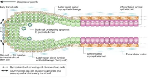
Figure 1: A schematic picture showing the architecture of a mammary gland duct. The figure depicts the different position, within the duct, of stem cells, transit cells and differentiated epithelial cells (image taken from Nat Rev Cancer 3:832, 2004).
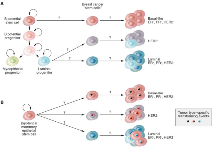
Cancer stem cells seem to share a similar phenotype with their normal counterparts but they display dysfunctional patterns of proliferation and differentiation, and they no longer respond to normal physiological controls that ensure a balanced cellular turnover. The origin of these cancer stem cells is controversial; it is not well known if they are originated from normal stem cells or from more differentiated progenitors where a de novo stem cell program is activated by the oncogenic insult (see Figure 2).
Figure 2: Breast cancer stem cells may originate from normal stem/progenitor stem cell (A) or bipotential stem cell (B) and can give raise to breast cancers over-expressing peculiar lineage markers (i.e. luminal, basal-like, epidermal growth factor 2 positive, HER2+, or epidermal growth factor 2 negative, HER2-) (image taken form Polyak, 2007 Cancer Cell).
MAMMARY CANCER STEM CELLS:EXPERIMENTAL EVIDENCES
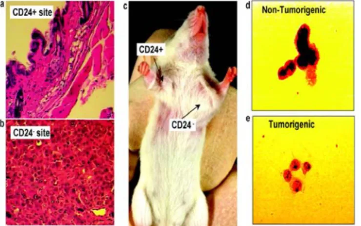
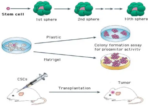
It has been recently proposed that a population of aberrant stem-like cells within a breast tumor is critically involved in the initiation of cancer disease. The tumor engraftment capacity (the ability to establish tumors in immune deficient mice mammary fat pads) of breast cancer cells is almost restricted to a sub-population of CD44 expressing cells, that have been consequently called tumor initiating cells or breast cancer stem cells (Al-Hajj et al., 2003, Ponti et al., 2005; see Figure 3). Human breast cancer stem cells can be propagated in vitro as multicellular spheroids, named mammospheres (MS) (Dontu, 2008; Farnie et al., 2007; see Figure 4). MS derived from breast tumor tissues and from breast cancer cell lines over-express the cancer stem cell marker CD44 and are extremely enriched in tumorigenic cells that show enhanced resistance to cancer therapy-induced cell death (Ponti et al., 2005; Kuperwasser, 2008). In vivo, the breast cancer stem cell phenotype is over-expressed in breast cancer bone marrow micro-metastasis and in aggressive inflammatory and basal-like breast carcinoma subtypes (Balic et al., 2006; Xiao et al., 2008). Understanding the biology of the tumor-initiating cell population in breast cancer would have a profound impact on how we design therapies, particularly those targeting cells with metastatic potential. It would also greatly enhance our ability to predict the metastatic potential of individual tumors and help patients and their physicians to select the most promising treatment options.
Figure 3: This picture shows that the tumorigenic potential of breast cancer cells in nude mice is restricted to CD24low breast cancer cells (picture taken from Al-Hajj et al., 2003 PNAS).
Figure 4: Stem/progenitor cells can be isolated from fresh breast tissue and grown in vitro in non adherent cellular condition as multicellular spheroids (mammaspheres) which can differentiate when plated in plastic or matrigel coated wells and promote breast tumorigenesis when injected and serially transplanted in nude mice.
IL-6: IN VITRO STUDIES IN BREAST CANCER
Cytokines are pleiotropic molecules who share common characteristics: a) they are biologically active at low concentrations (pg/ml-ng/ml);
b) they exert their biological effects by binding and signaling through cell surface receptors; c) they can be additive, synergistic, or antagonistic (Knupfer et al., 2006).
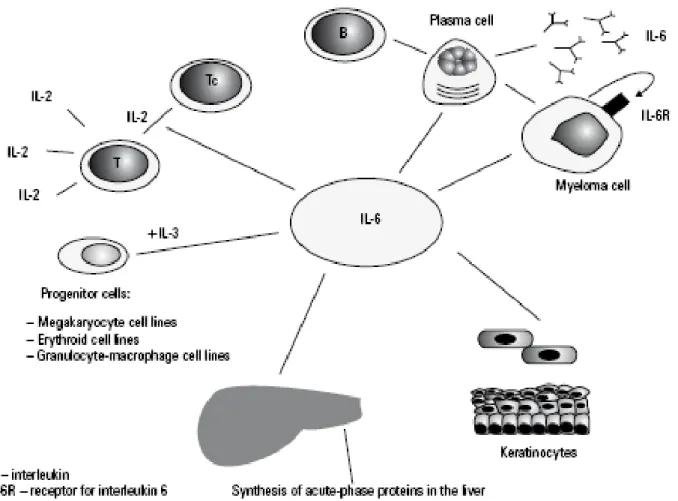
Interleukin-6 (IL-6) is a 30 kilodalton (kDa) cytokine discovered in 1986 and is known mainly for its role in mediating an immune response through expansion and activation of T cells and differentiation of B cells (Heinrich et al., 2003; see Figure 5). In addition, it is a central molecule in acute and chronic inflammatory settings (Gabay et al., 2006). While IL-6 has been studied extensively in chronic inflammatory diseases its role in breast cancer progression is less clear (Knupfer et al., 2006). IL-6 used in vitro both inhibited growth (Asgeirsson et al., 1998) and promoted growth (Honma et al., 2002; Miki et al., 1989) of breast cancer cell lines. IL-6 has been shown to augment survival of breast cancer cells through increases in pro-survival proteins such as BCL-2, BCL-xL, and MCL-1 (Brocke-Heidrich et al., 2006; Leu et al., 2003). Moreover, IL-6 production has been linked to increased drug resistance in breast tumor cells. Conze, et al showed that MCF-7 breast carcinoma cells became resistant to chemotherapeutic agents when stably transfected with IL-6 (Conze et al., 2001). Haverty et al showed that breast carcinoma cells that produced autocrine IL-6 had increased heat shock protein GP96, which promoted a drug resistant phenotype (Haverty et al., 1997). IL-6 has also been shown to increase breast cancer cell motility in vitro through decreased cell adhesions, which might contribute to cell metastasis (Asgeirsson et
al., 1998; Tamm et al., 1989,1994a,b). Additionally, IL-6 induced during hypoxia upregulates
VEGF through a specific response element upstream of the VEGF transcription start site to promote angiogenesis (Bachelot et al., 2003). This response could be extremely important in tumor progression when considering the implications of angiogenesis on tumor growth and metastasis. Overall, these observations in breast cancer are supported by an extensive body of literature documenting the ability of IL-6 to promote growth, survival, and drug resistance in multiple
myeloma, hepatocellular carcinoma, prostate carcinoma, and renal cell carcinoma cells (Cavarretta
et al., 2006; Hsia et al., 2006; Wallner et al., 2006).
Figure 5: IL-6 production and role in the immune response.
IL-6 AND BREAST CANCER IN VIVO
While the in vitro data on IL-6 in breast cancer is conflicting, the clinical literature depicts a much clearer picture of the biological impact that IL-6 has on disease progression. IL-6 has been shown to be an independent prognostic indicator of breast cancer progression (Zhang and Adachi, 1999). In addition, multiple clinical studies have demonstrated increased patient serum levels of IL-6 correlate with more aggressive disease. Two independent groups demonstrated increased IL-6 serum levels were associated with advanced breast cancer staging (Knupfer et al., 2006;
Jablonska et al., 2001). Salgado et al showed increased serum IL-6 levels correlated with the
number of metastatic sites in the patient (Salgado et al., 2003). Furthermore, Zhang, et al. showed that serum IL-6 is inversely correlated with patient response to treatment (Zhang and Adachi,
1999). Patients with complete response to therapy had average serum IL-6 of 2.4 pg/ml (+/-1.2),
partial responders were 4.1 pg/ml (+/- 1.0), non-progressive patients were 7.4 pg/ml (+/- 2.8), progressive disease patients were 36.3 pg/ml (+/- 13.2) (Zhang and Adachi, 1999). These studies demonstrate that elevation in IL-6 levels correlates with a poor prognosis in metastatic breast cancer. In fact, Bachelot, et al. looked at both VEGF and IL-6 levels in relation to survival in breast cancer patients. VEGF serum levels had no correlation to survival times. However, high IL-6 serum levels indicated a poor prognosis. Patient serum levels of IL-6 below 13 pg/ml demonstrated a median survival time of 13 months, while levels above 13 pg/ml had only 4 months. Patients with levels above 55 pg/ml had a median survival time of only 1 month, versus a median 3 years survival for patients below 55 pg/ml (Bachelot et al., 2003). In vitro data supporting the role of IL-6 in the promotion of breast cancer remains confounding and the mechanism behind these clinical observations remains unknown.
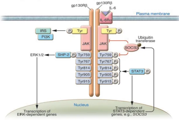
IL-6 PRODUTION AND SIGNALING
IL-6 is mainly produced by adipocytes, immune cells, and vascular endothelial cells (Heinrich et
al., 2003). When signaling, it binds first to its 80 kD extramembrane alpha receptor (IL-6Rα or
CD126) and then induces homodimerization of its 130 kD transmembrane signaling receptor (GP130). When bound by the IL-6/IL-6R complex, GP130 activates Janus kinases (Jak) and downstream signal transducer and activator of transcription (STATs), also known as the Jak/STAT pathway. STATs 1, 3, and 5 are the main mediators of IL-6 signaling (see Figure 6). These transcription factors are maintained in the cytoplasm until phosphorylated, at which time they dimerize, translocate to the nucleus, and induce gene expression (Heinrich et al., 2003). GP130 can
also activate the MAPK cascade through the adaptor protein SH2-and collagen-homology-domain-containing-protein (Shc). In addition, the PI3K cascade can also be activated by IL-6. These signaling cascades can induce many biological changes within the affected cell.
Signal transducers and activators of transcription (STATs) are a family of transcription factors that integrate cytokine and growth factor signaling to alter gene expression required for cell growth, survival, differentiation, and motility (Haura et al., 2005; Bromberg et al., 1999). Two members
of the family, STAT3 and STAT5 are increasingly associated with the progression of breast carcinoma (Clevenger, 2004). STAT3 exists in the cytoplasm of a cell in a latent form until
activated by cytokine (IL-6, LIF, oncostatin, IFN, etc) or growth factor signaling. When a cytokine or growth factor binds to its receptor dimerization occurs and induces a signaling cascade that quickly activates Janus kinases (Jaks) through autophosphorylation (Clevenger, 2004). Once
phosphorylated, Jaks recruit STAT3 to dock at the receptor/Jak complex. Jak proteins then induce tyrosine phosphorylation of STAT3 that promotes STAT3 to dissociate from the receptor complex, dimerize, and translocate into the nucleus (see Figure 6). Once in the nuclear compartment, STAT3 binds to gene promoters through a DNA specific recognition sequence, recruits activator or co-repressors, and affects gene transcription (Levy and Lee, 2002).
The first link to STAT3 and oncogenesis was the 1990s discovery that v-Src transformed cells had constitutively activated STAT3 (Yu et al., 1995). In addition, it was shown that a constitutively active mutant STAT3 was able to transform fibroblasts and allow them to form tumors in mice, establishing the role of STAT3 as an oncoprotein (Bromberg et al., 1999). Since these studies, activated STAT3 has been linked to multiple malignancies, including breast cancer. Multiple studies have shown via immunohistochemistry that activated STAT3 (nuclear STAT3) is increased in malignant tissue compared to normal controls (Dolled-Filhart et al., 2003). Evidence from in vitro findings supports these results. Many studies have demonstrated a tumor promoting role for activated STAT3 in cancer cell lines (Grandis et al., 1998). Several inhibitors of the STAT3 pathway have been tested in vitro and in vivo and show proof of concept by increased tumor cell death and decreased in vivo malignancy (Selander et al., 2004). These drugs are still in early development and it remains to be seen how they will fair in a clinical setting.
NOTCH SIGNALLING IN BREAST CANCER
Notch genes, encode highly conserved cell surface receptors (Kidd et al., 1986). The Notch signaling pathway, in which almost all elements are conserved from Drosophila to humans, consists of Notch receptors, ligands, negative and positive modifiers, and transcription factors (Kidd et al.,
1986). In mammals, these functional classes each have multiple members, and the interplay
between these molecules is not yet fully understood. Studies in Drosophila suggest, however, that Notch receptors and ligands generally influence lineage specification through two mechanisms: lateral inhibition and lateral induction (Welshons et al., 1962). In contrast, inductive signaling involves two non-equivalent cell types that express either the receptor or the ligand. The receptor expressing cell responds to ligand stimulation, triggering a cell fate decision dependent on access to the appropriate ligand(s). During the development of complex tissues, both mechanisms may be operative.
.
Figure 7: (a) Pictorial representation of a Notch protein and its signalling pathways. The extracellular domain of Notch contains between 29 and 36 tandemly repeated epidermal growth factor (EGF)-like repeats, some of which are required for the interaction of Notch with its ligands, along with three Lin-12/Notch repeats. The most prominent motifs in the intracellular domain are six cdc10/ankyrin repeats and a PEST domain close to the C-terminus of the protein. The intracellular domain also contains two functionally defined domains: the juxtamembrane RAM23 domain that mediates the interaction of the intracellular domain of Notch with CBF1, Suppressor of Hairless, Lag-1 (CSL) proteins; and a transcriptional activation domain that is C-terminal to the cdc10/ankyrin repeats. (b) The interaction of Delta, Serrate, Lag-2 (DSL) ligands (black) with EGF-like repeats 11 and 12 of Notch (dark blue and yellow) leads to two proteolytic cleavages, one extracellularly and one within the membrane, which release the intracellular domain of Notch (NICD). This fragment of Notch then migrates to the nucleus (dotted line) where it interacts with CSL proteins (orange) via its RAM23 domain to form a transcriptional activator. (c) Recent experiments have suggested that Notch can signal through a second distinct signalling pathway that requires the cytoplasmic protein Deltex (light blue). Deltex has been shown to interact directly with the cdc10/ankyrin repeats of Notch, and signalling through this pathway has been
proposed to both inhibit Jun N-terminal kinase (JNK) signalling and to sequester the transcriptional coactivator CREB binding protein (CBP)/p300 (picture taken from Brennan et al., Breast Cancer Res 2003).
An interesting aspect of Notch is its double function as a tumor suppressor and oncogene. Although the mechanism underlying this dual Notch action is being explored, the outcome of Notch signaling activity depends on signal strength, timing, cell type, and context (Maillard and Pear, 2003). The result of altered Notch signaling depends on its normal function in a given tissue. Notch thus acts as an oncogene if its normal function is as a gatekeeper of stem cells or as a regulator of precursor cell fate; its tumor suppressor activity is detected in tissues in which Notch signaling initiates terminal differentiation events (Radtke and Raj, 2003). Notch alone may not be a very efficient oncogene, however, and it must associate with another oncoprotein to cause transformation. Although such partners have not yet been identified in naturally occurring tumors, transformation can be induced in vitro in various cell types by expressing NICD with certain oncoproteins (Girard et al., 1996;
Beverly et al., 2003). Mouse Mammary Tumor Virus (MMTV) is a retrovirus that causes mammary
tumors through insertional mutagenesis of the mouse genome. Notch4 was identified as a mouse mammary tumor virus (MMTV) insertion site in mammary tumors (Nusse, 1988); provirus was inserted within the Notch4 gene (originally known as the int-3 locus) (Smith et al., 1995). This MMTV model system has proven useful for identification and characterization of genes involved in malignant transformation of normal mammary epithelium (Nusse, 1988). In the case of the Notch4 gene, provirus insertion leads to expression of a truncated Notch that lacks most of the extracellular portion of the protein but contains Notch4 transmembrane and intracellular domains (N4ICD) (Gallahan and Callahan, 1997). Transgenic mice harboring this constitutively active N4ICD under the regulation of the MMTV promoter show arrested mammary gland development and eventually develop poorly differentiated adenocarcinomas (Smith et al., 1995). Additional evidence confirms the Notch4/int-3 gene effect in mammary epithelial differentiation and mammary tumorigenesis; this is derived from studies in which N4ICD was expressed from the whey acidic protein (WAP) promoter in transgenic mice, which restricts its activity to secretory mammary epithelial cells of
pregnant mice (Gallahan et al., 1996). As predicted, secretory lobule growth and differentiation were inhibited, and mammary tumors were histologically identical to MMTV-N4ICD tumors. Enforced expression of int-3/Notch4 in cultured mammary epithelial cells induces anchorage-independent growth, matrix invasion and loss of contact inhibition (Dievart et al., 1999). Notch1 involvement in mammary tumorigenesis is being studied extensively. The first evidence that aberrant Notch1 signaling has a role in mammary tumorigenesis came from studies in the MMTV model, which attempted to identify genes that collaborate with Neu/erbB2 in mammary tumorigenesis. An MMTV insertion in the Notch1 locus in MMTV-Neu mammary tumors causes N1ICD expression (Dievart et al., 1999). HC11 mouse mammary epithelial cells expressing N1ICD-encoding cDNA are transformed, form colonies in agar, but are unable to form tumors in nude mice, indicating that acquisition of malignant characteristics requires additional genetic events (Dievart et al., 1999). Other studies showed that transgenic activation of N1ICD in mammary glands leads to development of lactation-dependent tumors that regress at weaning (Kiaris et al.,
2004; , Hu et al., 2006); with time, these regressing neoplasms apparently become non-regressing
adenocarcinomas (Kiaris et al., 2004). Stem cells thought to reside in the mammary gland are thought to renew mammary gland cells through cycles of pregnancy, lactation, and involution during a woman’s lifetime (Woodward et al., 2005). There is increasing evidence that stem cells might be targets of transformation during mammary carcinogenesis (Woodward et al., 2005). The Notch signaling pathway is implicated in the self-renewal of normal mammary stem cells (Dontu et
al., 2003, 2004), and recent works suggest a role for the Notch pathway in breast cancer (Liu et al., 2005; Yamaguchi et al., 2008).
Information on Notch in human breast cancer is scarce and indirect. Notch1-4 mRNA/proteins are expressed in selected human breast cancer cell lines and tumors (Stylianou et al., 2006). In tissue samples from breast cancer patients, Parr et al. (Parr et al., 2004) quantified NOTCH1 and NOTCH2 expression in association with clinical outcome and showed aberrant NOTCH1 and NOTCH2 levels in breast cancer tissues compared with normal breast tissue. Examination of the
clinicopathological parameters for breast cancer patients indicated that high NOTCH1 levels may be associated with poor prognosis, whereas increased NOTCH2 levels correlated with greater probability of survival (Parr et al., 2004). NOTCH1 may thus have tumor-promoting functions, whereas NOTCH2 could have a tumor-suppressive role in human breast cancer, supporting the suppression of NOTCH-1 activity as a therapeutic strategy. Tissue microarray studies showed high JAGGED1 and/or NOTCH1 expression levels in human breast cancer, associated with poor overall survival compared with patients with low levels of these genes (Reedijk et al., 2005).
Because high-level JAGGED1 and NOTCH1 co-expression showed a synergistic effect on overall survival, this type of breast tumor could also benefit from notch inhibition-based therapy.
Other studies on NUMB expression (the inhibitor of notch signaling) in human breast cancer also supports NOTCH signaling pathway involvement in breast cancer. (Pece et el., 2004; Colaluca et
al., 2008) showed that NUMB-mediated negative regulation of NOTCH signaling is lost in 50% of
human mammary carcinomas. This is due to specific NUMB ubiquitination and proteasomal degradation (Pece et el., 2004) and indicates that enhanced NOTCH signaling activity occurs in these mammary carcinomas. Overall, all these molecular and phenotypic evidences demonstrated a pivotal role for Notch in the breast cell fate determination and tumorigenesis.
CHAPTER 1 ABSTRACT
High serum levels of Interleukin-6 (IL-6) correlate with poor outcome in breast cancer patients. However no data are available on the relationship between IL-6 and stem/progenitor cells which may fuel the genesis of breast cancer in vivo. Herein, we address this issue in mammospheres (MS), multi-cellular structures enriched in stem/progenitor cells of the mammary gland, and also in MCF-7 breast cancer cells. We show that MS from node invasive breast carcinoma tissues express IL-6 mRNA at higher levels than MS from matched non-neoplastic mammary glands. We find that IL-6 mRNA is detectable only in basal-like breast carcinoma tissues, an aggressive variant showing stem cell features. Our results reveal that IL-6 triggers a Notch-3-dependent up-regulation of the Notch ligand Jagged-1, whose interaction with Notch-3 promotes the growth of MS and MCF-7 derived spheroids. Moreover, IL-6 induces a Notch-3-dependent up-regulation of the carbonic anhydrase IX gene, which promotes a hypoxia-resistant/invasive phenotype in MCF-7 cells and MS. Finally, an autocrine IL-6 loop relies upon Notch-3 activity to sustain the aggressive features of MCF-7-derived hypoxia-selected cells. In conclusion, our data support the hypothesis that IL-6 induces malignant features in Notch-3 expressing, stem/progenitor cells from human ductal breast carcinoma and normal mammary gland.
LIST OF ABBREVIATIONS IL-6 Interleukin-6 IL-6 R IL-6 receptor
MS mammospheres
MCF-7S spheroids of MCF-7 cells GPR30 G-protein coupled receptor 30
GP96 Heat shock protein GP96 MMP2 metalloproteinase 2 Anti/ -IL6 anti IL-6 antibody Anti/-Notch-3 anti Notch-3 antibody
CA-IX carbonic anhydrase IX
HYPO-7 MCF-7 hypoxia derived subpopulation IHC immunohistochemical analysis
PIAS3 Protein inhibitor of STAT3
STAT Signal transducer and activator of transcription pSTAT3 Phosphorylated STAT3
JAK Janus kinase
VEGF Vascular endothelial growth factor WHO World health organization
INTRODUCTION AND HYPOTHESIS
Interleukin 6 (IL-6), a major mediator of the inflammatory response, plays a primary role in the patho-physiology of cancer (Hodge et al., 2005; Rose-John et al., 2006). In breast cancer patients, the extent of the increase in serum IL-6 correlates with a poor disease outcome and a reduced prognosis (Knupfer and Preiss, 2007; Bachelot et al., 2003). Though it has been argued that the cytokine may be secreted by cancer cells, the source of the IL-6 in cancer patients has not yet been determined (Zhang and Adachi, 1999; Knupfer et al., 2004). Cancer cells exposed to IL-6 or which secrete the cytokine as an autocrine factor, show malignant features, such as an enhanced capacity to invade the extracellular matrix and an increased drug resistance (Conze et al.,
2001; Sehgal et al., 2001). Accordingly, the inactivation of the gp130 protein, which transduces the
signalling of IL-6 type cytokines, has been found to reduce the aggressiveness of breast cancer cells in vivo (Selander et al., 2004). On the basis of these data, the inhibition of the IL-6/IL-6 receptor interaction with specific antibodies has been proposed as a support cancer therapy (Trikha et al.,
2003).
Breast cancer has been proposed as a stem cell disease(Dontu et al., 2005). This hypothesis entails the notion that the growth of the tumour mass relies on the proliferation and self renewal capacity of a small population of cancer-initiating cells, also named cancer stem cells (Dontu et al.,
2005). Moreover, this notion helps to understand why the dys-regulation of stem cell regulatory
pathways plays a causative role in breast cancer (Reya et al., 2001). In this regard, transgenic mice over-expressing isoforms of Notch, a signalling pathway active in stem cells, are more prone to develop mammary tumours (Hu et al., 2006). Further, high levels of Notch isoforms have been found to correlate with a poorer prognostic profile and reduced survival in breast cancer patients (Reedijk et al., 2005; Stylianou et al., 2006). The molecular profile analysis of breast cancer stem tumorigenic cells revealed an up-regulation of IL-6 and of Notch-3, a stem cell regulatory gene (Shipitsin et al., 2007).
Stem/progenitor cells of the mammary gland reside in the basal cell layer (Boecker et al.,
2003) and can be expanded in vitro from normal tissues as multi-cellular spheroids, named
mammospheres (MS) (Dontu et al., 2003). Prior findings indicate that multi-cellular MS structures have a clonal origin and have the capacity to reform in vitro after trypsin dissociation. (Dontu et al.,
2003). Bi-lineage (luminal and myo-epithelial) progenitors are enriched up to eight times in MS,
compared to freshly isolated human mammary cells, and constitute virtually 100% of the cells in secondary MS (Dontu et al., 2003, 2005). MS regenerate and also form tubulo-alveolar structures in matrigel and in immunodeficient mice cleared of fat pads (Dontu et al., 2003, 2004, 2005;
Farnie et al., 2007). Similarly, MS from breast cancer tissues have been shown to proliferate in
vitro and also generate tubulo-alveolar structures composed of CD44+/CD24- cells (Farnie et al.,
2007; Ponti et al., 2005). Interestingly, the CD44+/CD24- positive cell population has been shown
to be extremely enriched in putative breast cancer stem cells (Al-Hajj et al., 2003).
Both normal and tumor MS have been shown to require active Notch signalling to sustain their survival and proliferation capacity (Dontu et al., 2004). Moreover, MS express gp130 and are potential targets of IL-6 type cytokines (Dontu et al., 2003).
In this thesis we provide evidence that IL-6 gene expression is up-regulated in MS obtained from aggressive ductal breast carcinomas and that IL-6 regulates a Notch-3-dependent signalling pathway that promotes self renewal and the invasive potentials of normal and tumour MS.
There is evidence that hypoxia affects stem cell function and survival (Cejudo-Martin et al., 2005;
Covello et al., 2006; Ramirez-Bergeron et al., 2001). In vitro, hypoxia actively maintains a stem
cell/immature phenotype, induces a loss of differentiation markers, and blocks differentiation (Cipolleschi et al., 1993; Gustafsson et al., 2005). In vivo, stem cells express higher levels of hypoxia regulated genes than the more mature progeny, as well as high levels of glycolytic enzymes (Unwin RD et al., 2006). Accordingly, stem cells reside in tissue regions (the niche) that are scarce in vasculature and are thought to provide a low oxygen environment (Nilsson et al., 2001). Furthermore, stem cells are enriched up to 1000 folds among a pool of cells (the so called side
population), which expresses high levels of the hypoxia-survival gene Bcrp-I (Krishnamurthy et
al., 2004). Recent data indicate that the stem cell regulatory Notch pathway shares in an interplay
with the hypoxia response modulator HIF-1 to promote the onset of a stem/undifferentiated phenotype (Gustafsson et al., 2005). These findings, linking stem cells with hypoxia survival, lead to hypothesise that the control of stem cell survival and the regulation of hypoxia response are intimately coupled, and that they may share common control gene/pathways. In this thesis, we also provide evidence that IL-6 induced by the exposure to hypoxic stimuli, controls the expression of the stem cells regulatory gene Notch-3. Then, we report that a IL-6/Notch-3 interplay elicits an ERK-dependent up-regulation of at least two genes: the Notch ligand Jagged-1, and the hypoxia survival gene Carbonic Anhydrase IX. Finally, we convey that IL-6/Notch-3/CA-IX axis sustains mammosphere survival in presence of hypoxia. We propose that the findings here reported may help in understanding the relationship among inflammation, hypoxia survival, cancer and stem cells at molecular level.
Chemicals and reagents.
Monoclonal Antibody (MoAb) which blocks the activation of Notch-3 protein, by inhibiting the Notch-3/Jagged-1 interaction was purchased from R&D. MoAb which blocks the IL-6 receptor/ligand interaction (anti-IL6) and recombinant human Interleukin-6 were purchased from Sigma. Desferoxamine (DFX, Sigma) was used as hypoxia mimetic, UO-126 (Sigma) was used as MEK1 inhibitor.
Generation of mammospheres (MS) from normal and ductal breast carcinoma tissue specimens (CHAPTER 1).
Seventeen fresh surgical specimens obtained from patients with ductal breast carcinoma, who underwent to quadrantectomy or mastectomy, were collected to generate mammospheres, MS (Table 1). Normal and tumor samples were hystologically characterized (as reported below) to ensure the proper classification of normal and tumor tissue. Particular care was paid to generate MS from specimens in which only normal or tumor tissues were detectable at hystological examination. The set of samples consisted in a subset of tumor specimens (n=3), in which also the mRNA of the tumor tissues from which the MS had been originated was available, and of a subset of specimens (n=14), in which even the normal tissue from the same patient was available (Table 1). MS were obtained as previously described (Dontu et al., 2003; Farnie et al., 2007), except that the methodology was downscaled to deal with low amounts of tissues 300 to 900mg. Briefly, tissues were placed in sterile Epicult (StemCell Technologies), minced with sterile scalpels and incubated for 6 to 12 hours in presence of 1000 Units of Collagenase/Hyaluronidase enzyme mix (StemCell Technologies). Samples were centrifuged at 80Xg for 2 minutes, the pellet was digested by Dispase and DNAse for 3 minutes (StemCell Technologies), and then pelletted at 450Xg for 5 minutes. Pellets were re-suspended, filtered through a 40M nylon mesh (Becton Dickinson), and plated into 1 or 3 cm2 wells low attachment plates (Corning), filled with 3 ml of Mammary Epithelial Growth Medium (MEGM), supplemented with B27 supplement, EGF 10ng/ml, bFGF 10ng/ml, 10g/ml
Insulin, Hydrocortison 10-6M, and ad hoc aliquots Gentamycin and Amphotericine (Cambrex). Primary MS started forming after 4 to 6 days, and were processed at day 10. Experimental procedures (see below) were performed on secondary MS, generated by incubating primary MS in 1X Trypsin-EDTA solution (Cambrex) for 3 minutes, followed by two washes in complete MEGM and a filtration troughout a 40m nylon mesh. Self renewal of MS was tested by assessing the capacity of primary MS to generated secondary MS after trypsin disaggregation, as previously described (Dontu et al., 2003, 2004). Secondary MS were assessed at day 7. All the procedures were approved by the local ethical committee and by the patient’s written informed consent.
Clinical samples and primary cell culture (CHAPTER 2).
43 fresh surgical specimens from patients with ductal breast carcinoma who underwent quadrantectomy were processed to obtain MS as previously described (Table 3). All procedures had been approved by the local ethical committee (protocol number 56/2006/U/Tess to M.B.) and by the patient’s written informed consent.
In vivo tumorigenicity.
Cell invasion assay was performed on breast cancer cells (3*104) and trypsin dis-aggregated MS and MCF-7(S) (5*102 cells), as previously described. Six females BALB/c nude mice were injected with 5*105 MCF-7 cells. Mice were followed up for 3 months, prior of being sacrificed. Three tumor xenografts were formalin-fixed, paraffin-embedded for immunohistochemical analysis, or immediately frozen in liquid N2 for RT-PCR analysis. All procedures had been approved by the
Immunohistochemistry.
Immunohistochemistry was performed on formalin-fixed, paraffin-embedded tumor samples (Table 1-4) and on N-/T-MS (embedded in collagen (Sigma) 2h before fixation in formalin). Tissues were histologically classified according to WHO criteria and graded (G) following Elston and Ellis’ classification (Tavassoli et al., 2003; Elston et al., 1991). The tumours were also typed by nuclear grading (NG) as follows: mild (NG1), moderate (NG2) and severe (NG3) nuclear atypia. Tumour size (pT) and axillary lymphnode involvement (pN) were also recorded using pTNM (UICC) pathological staging criteria (Elston et al., 1991). Serial sections of formalin-fixed paraffin-embedded samples were de-waxed, re-hydrated, and subjected to antigen retrieval treatment. Tumor sections were stained using monoclonal antibodies anti-estrogen receptor (ER, clone 1D5), cytokeratin-5 (CK-5 clone D5/16B4) and Epidermal Growth Factor Receptor (EGF-R, clone DAHK1-WT) obtained from DakoCytomation (Glostrup), ErbB-2 (HER-2, clone CB11) and cytokeratin-14 (CK-14, clone LL002) from BioGenex Laboratories and CA-IX (M-75), kindly provided by Dr J. Pastorek (Slovak Academy of Sciences, Bratislava, Slovak Republic). Sections of normal and tumor MS were stained with anti CK-5, CK-14, EGF-R, CK-18 (CK-18, clone KSB17, Sigma), Oct-4 (clone c-20, Santa Cruz), CD44 and CD24 (Clone 156-3C11 and Clone 24C02, Neomarkers), CD133 (Miltenyi Biotec), and E-Cadherin (clone NCH38, DakoCytomation). Antigens were unmasked with Tris-EDTA pH 9.0 at 98°C for 20 min, except for CA-IX antibody. Endogenous peroxidase activity was inhibited using a 0.5% H2O2 solution in methanol for 20 min,
and sections were processed for immunohistochemistry with a non-biotin amplified method (Novolink, Novocastra Laboratories). Stained immunoreaction was quantified by image cytometry using Cytometrica software (C&V, Bologna, Italy). Sections were independently evaluated by two pathologists, and controversial results were discussed and defined. For ER immunostaining, the percentage of the labeled nuclear area over the total neoplastic nuclear area was assessed (<10% nuclei= negative, >10% nuclei=positive). A semi-quantitative assessment was applied for CK-5, CK-14 and EGF-R evaluation: cases were considered positive when the immunopositive neoplastic
population was > 10%. HER-2 staining was scored according to the HercepTest United States Food and Drug Administration-approved grading system. The percentage of immunopositive cells in normal and tumor MS was assessed on three to five sections (accounting from 100 to 300 cells as average).
Cell cultures.
MCF-7 cells were grown in RPMI 1640 medium 10%FBS (Euroclone). Hypoxia (<0.1%O2)
was generated in 95%N2, 5%CO2 incubator (Thermo). MCF-7 derived multi-cellular spheroids,
MCF-7(S) were generated by re-suspending 1x10⁴ MCF-7 cells in complete RPMI 1640 medium and plated in 3cm2 low attachment plates (Corning). Hypoxia-resistant MCF-7 derived cells, HYPO-7 cells, have been previously described (Sansone et al., 2007).
Hypoxia induced cell death.
Cell death was induced by exposing MCF-7 cells, Hypo-7 and MS to DFX at a concentration of 100, 600 and 50M, respectively, following previously described protocols (Sansone et al.,
2007). Cell death in MS was evaluated by Trypan blue staining of single cells obtained from the
trypsin disaggregation of MS.
Transient and stable RNA interference.
Double strand RNA oligonucleotides (siRNA) directed against IL-6 (StealthTM validated RNAi DuoPaks), CA-IX and Jagged-1 (StealthTM select 3 RNAi set) mRNA, and appropriate
controls, scramble (SCR) siRNAs were purchased from Invitrogen (Carlsbad). siRNAs were transfected to adherent MCF-7 cells (105 cells in a 3 cm2 well) at a concentration of 1g/well, using Lipofectamine 2000 (Invitrogen). siRNA transfection in MS and MCF-7(S) was performed by mixing 1g of siRNA with In vitro JET-PEI reagent (Poly plus Trasfection). Notch-3 specific short hairpin RNA (shRNA) was obtained by cloning an oligonucleotide consisting of a BglII site, a
21-22-nt sense sequence (GATCCCCCT CCCCTCACCACCTAA TAAAT/ TCAAGAGATTTATTA GGTGG TGAGGG GAGTTTTTG GAAC), a short spacer (TTCAAGAGA), a 21-22-nt antisense sequence (TCGAGTTCC AAAAA CTC CCC TCA CCACCT AATAAA TCT CT TGAAT TTAT TAGGTGG TGAGGGGAGGGG), five thymidines (a stop signal for RNA polymerase III) and a XhoI site, into the pSuper-Puro expression retroviral vector (OligoEngine). The same vector encoding for a shRNA which does not match to any human known transcript (5' gatcccc AATATC CTTGGA CACAAG TTG ttcaagaga CAACTT GTGT CCAA GGATATT tttttggaac 3') was used as control (CT) for N3 shRNA. Retroviral gene transfer was performed as follows: Phoenix cells (gently provided by Dr. Gary Nolan, Stanford University, California, USA) were grown at 60% confluence and were transfected overnight with 30µg of the pSuper-Puro vector encoding a N3/CTR shRNA, using Lipofectamine 2000 (Invitrogen). Two days after transfection, the medium containing newly packaged retrovirus was collected and filtered through a 0.45µm pore size filter. After supplementation with 4µg/ml polybrene (Sigma), the augmented medium was applied to MCF-7 cells at 50% confluence for 24 hours. Successfully infected cells were selected by culturing the cells in presence of 2µg/ml Puromycin for 2 weeks.
Expression vectors.
The active form of Notch-3 (pNICD-3) was cloned by PCR, using the following primers: F-TCTTGCTGCTGGTCATTCTC; R-GGCCCCCAAGATCTAAGAAC, using Herculase Taq polymerase (Stratagene). The PCR product was inserted into pcDNA3.1/V5-His Topo TA Expression Vector (Invitrogen).
RT-PCR analysis.
Total RNA was extracted from cultured cells, MS, and from archival tissues (n=19, Table 1, samples 1-3 and Table 2) which had been frozen in liquid nitrogen at the time of surgical resection, using the RNA-extracting reagent TRIzol® (Invitrogen). Primers used in the RT-PCRs are: IL-6:
annealing temp 62°C, amplicon length 170 bp, F-5’-GAGAAAGGAGAC ATGTAACAAGAGT-3’, R-5’-GCGCAGAATGAGATGAGTTGT-3’; Notch-3: annealing temp 62°C, amplicon length 93 bp, F-5’-TCAGGCTCTCACCCTTGG-3’, R-5’-AGTCACTGGCACGGTTGTAG-3’; CA-IX: annealing temp 61°C, amplicon length 589 bp, F-5- CAGGGACAAAGAAGGGGATGAC-3’, R-5’-TTGGAAGTAGCGGCTGAAGTCA-3’; Bmi-1, annealing temp 62°C, amplicon length 220 bp, F-5’GGAGACCAGCAAGTATTGTCCTTTTG-3’, R-5’-CATTGCTGGGCATCGTAAG-3’; Jagged-1: annealing temp 62°C, amplicon length 170 bp, F-5’-TCGCTGTATCTGTCCACCTG-3’, R-5’-AGTCACTGGCACGGTTGTAG-3’; CK-5: annealing temp 55°C, amplicon length 409bp, F-5’ TAGGTGGTGGGCTCAGTGTGG-3’, R-F-5’-ACTTTGGGTTCTCGTGTCAGC-3’; CD133: annealing temp 60°C, amplicon length 286bp, F-5’- CTGGGGCTGCTGTTTATTATTCTG-3’, R-5’- ACGCCTTGTCCTTGGTAGTGTTG -3’; BCRP-I: annealing temp 62°C, amplicon length 400bp, F-5’ GTTTATCCGTGGTGTGTCTGG -3’, R-5’- CTGAGCTATAGAGGCCTGGG -3’; CD44: annealing temp 62°C, amplicon length 300bp, F-5’ CAGCAACCCTACTGATGATGACG-3’, R-5’- GCCAAGAGGGATGCCAAGATGA -3; Oct-4: annealing temp 62°C, amplicon length
169bp, F-5’ CTTGCTGCAGAAGTGGGTGGAGGAA -3’, R-5’- TGCCCGAAACCCACACTGCAG -3; Beta2 microglobulin: annealing temp 58°C, amplicon
length 180, bp F-5’-ACCCCCACTGAAAAAGATGA-3’; R-5’-ATCTTCAAACCTCCATGA-3’. PCR primers and reagents were purchased from Invitrogen.
Boyden Chamber Invasion assay.
Cell invasion into Matrigel was assessed by using Boyden chambers (New Technologies Group), containing a poly-vinyl-pyrrolidone free polycarbonate filters with 8-μm pores, coated with 15μg of Matrigel (Sigma). Cells (1 x 10⁵) and trypsin dis-aggregated MS (1-5 x 10²cells) were seeded in the upper chamber in serum-free medium, in presence/absence of IL-6 (10ng/ml) or anti-IL6 (1.5g/ml); complete medium was placed in the lower compartment as chemoattractant. In several experiments cells and MS were also transfected with appropriate siRNA for 48h and then
were collected, re-suspended in 500l in co-presence of IL-6 (10ng/ml) or anti-IL6 (1.5g/ml) and seeded in the upper chamber for 24 h at 37° C in a 5% CO2 atmosphere. At the end of incubation,
non invading cells were removed from the upper surface of the filters, and invading cells in the lower surface were fixed in ice cold methanol, stained with Toluidine Blue staining (Sigma) and scored as the mean number of invaded cells per 5 random optical fields, in three independent experiments, at a 20X magnification.
Gelatin zymography.
Metalloproteinase-2 (MMP-2) activity was determined by gelatin zymography. Briefly, proteins of collected media were precipitated with 1:4 (vol/vol) icecold methanol overnight at -20°C, solubilized with sample buffer without mercaptoethanol (1 M Tris-HCl, pH 6.8, 2% SDS, 10% glycerol) and loaded into 10% SDS-polyacrylamide gel containing 1 mg/mL gelatine (Sigma). Gel was then incubatedin a developing buffer (100 mM Tris-HCl, 10 mM CaCl2, 20 mM NaCl, pH
7.6) overnight at 37°C, stained for 2h with1% Coomassie Brilliant Blue R-250, and finally de-stained in a solution containing 10% acetic acid and 40% methanol. MMP-2 proteolytic activity was quantified using a semiautomated image analysis (GelDoc, Biorad Laboratories)
Western Blot.
Cell lysates were prepared, run and blotted using standard methodologies, and probed specific antibodies: Rabbit polyclonal anti-Notch-3 (clone M-134, Santa Cruz), Mouse monoclonal Antibodies anti-ERK and phosphorylated ERK (Cell Signalling), -Actin (Sigma) and CA-IX (clone M-75).
Statistical analysis.
Continuous variables (percentages of dead cells, number of invading cells in Boyden Chamber assays) were analysed by Anova (unequal variance assumed). Post hoc test (unequal variance
assumed) were used when comparison were > 2. Non-normally distributed variables (RT-PCR normalized values of mRNA level) were analyzed by two samples non parametric test (Mann Whitney). Categorical variables (MS and spheroid size distribution) were analyzed by Monte Carlo 2 test. All the tests were implemented in SPSS 10.1 Package (SPSS).
RESULTS
High levels of Interleukin 6 (IL-6) mRNA are present in mammospheres (MS) from aggressive ductal breast carcinoma and in basal-like breast carcinoma tissues.
MS were generated from the tumor tissues (T-MS) of 3 patients with ductal breast carcinoma (Table 1, samples 1-3 and Figure 1A).
T-MS were characterized by immunohistochemistry (IHC). We found that T-MS were composed almost entirely by CD44+ (97±3%), CD24-(<1%) cells (Supplementary Figure 1), suggesting that the majority of cells in T-MS present a CD44+/CD24- cancer stem cell phenotype (Al-Hajj et al., 2003). Further, cells in T-MS expressed Oct-4 (88±7%), which has been previously reported to be hyper-expressed in T-MS (Ponti et al., 2005), and Cytokeratine 5 (CK-5, 22±7%), which identifies the mammary gland basal cell compartment (Boecker and Buerger, 2003; Supplementary Figure 1). IHC showed also that T-MS were composed by E-Cadherin positive (97±2%), CK-14 positive (99±1%) and CK-18 positive (24±7%) cells, revealing that T-MS are composed of epithelial cells showing ductal (CK-18) and luminal (CK-14) markers (Supplementary Figure 2).
RT-PCR analysis revealed that T-MS, but not the tumour tissues which T-MS had been obtained from, expressed detectable level of IL-6 mRNA (Figure 1B). Compared to tumour tissues, RT-PCR analysis also revealed that T-MS expressed high levels of Bmi-1 mRNA, a gene associated with stem cell renewal (Liu et al., 2006), of CD44 mRNA, a gene whose expression has been associated with cancer stem cell phenotype in different organs (Al-Hajj et al., 2003; Li et al.,
2007), as well as of CK-5 and Oct-4 mRNA (Figure 1B).
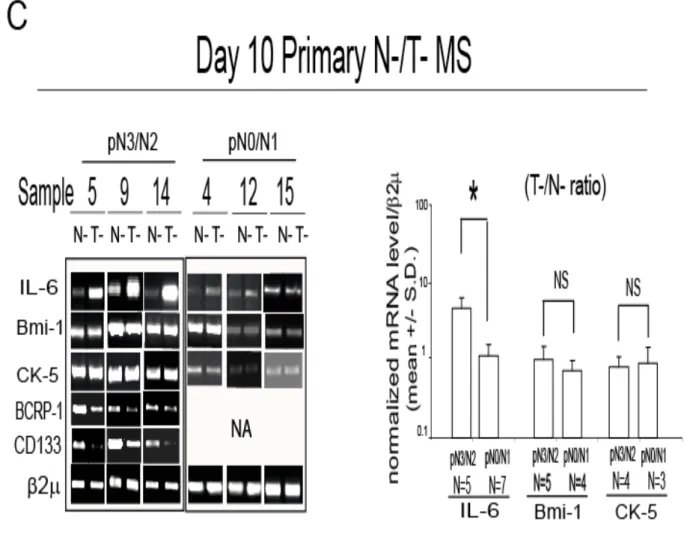
T-MS were then obtained from a set of samples (n=14), in which also the normal mammary gland tissue was available to generate normal MS (N-MS), (Table 1, samples 4-17).
Similarly to T-MS, N-MS lacked CD24 expression and contained cells expressing CD44 (95±3%), CK-5 (14±3%), CK-14 (78±7) and CK-18 (75±9, Supplementary Figure 3). The availability of N-MS and T-MS from the same patient allowed us to assess the level of IL-6 mRNA,
taking into account for variability due to the genetic make-up and age (Bonafe et al., 2001). We found that, compared to matched N-MS, T-MS from node invasive tumours (pN3/pN2) expressed increased levels of IL-6 mRNA (Figure 1C). The same comparison performed on T-MS generated from scarcely node invasive tumours (pN0/pN1) ductal carcinomas revealed a negligible difference in IL-6 mRNA level between N- and MS (Figure 1C). Notably, compared to matched N-MS, T-MS obtained from patients affected by pN3/pN2 invasive tumours expressed similar levels of Bmi-1 and CK-5 mRNA, lower levels of Breast cancer resistance protein Bmi-1 (BCRP-I) mRNA, as well as of CD133 mRNA, two antigens which have been previously associated with (cancer) stem cell phenotype (Singh et al., 2004; O'Brien et al., 2007; Ho et al., 2007; Figure 1C). The higher level of CD133 expression in N-MS compared to T-MS was also evident in IHC analysis (Supplementary Figure 4A).
We then assessed IL-6 mRNA in a set of archival breast tumour samples (Table 2), including ductal (n=10) and basal-like (n=6) breast carcinomas, a subtype of cancer showing stem cell features (Nielsen et al., 2004; Bertheau et al., 2007; Bertucci et al., 2007;
Charafe-Jauffret et al., 2006). This tumour type, similarly to MS, was characterized by the expression of
CK-5, CK-14, Epidermal Growth Factor Receptor (EGF-R) protein, as well as of Bmi-1 and CD133 mRNA (Supplementary Figure 4B and C) thereby reinforcing the notion of a tight similarity between MS and basal-like breast carcinoma cells (Charafe-Jauffret et al., 2006).
In keeping with this reasoning, we found that IL-6 mRNA was detectable in basal-like breast carcinoma tissues, but not in ductal breast carcinoma (Figure 1D). These data indicate that IL-6 expression occurs in MS obtained from aggressive ductal breast carcinoma and in basal-like breast carcinoma tissues, wherein stem cell-like phenotypes are particularly apparent.
FIGURE 1A, Phase contrast microscopy analysis of day 10 primary tumor (T-) mammospheres
FIGURE 1B, RT-PCR analysis of IL-6, Bmi-1, Cytokeratin 5 (CK-5), CD44, Oct-4, 2 mRNA
FIGURE 1C. Interleukin-6 (IL-6) mRNA is expressed in mammospheres and in basal-like breast carcinoma tissues Day 10 primary normal (N-) and T-MS obtained from the same patient
(Table 1): RT-PCR analysis of IL-6, Bmi-1, CK-5, BCRP-1 and CD133 and bar graph representation of IL-6, Bmi-1 and CK-5 mRNA level, first normalized onto 2 mRNA, and then expressed as a ratio of N-MS over T-MS, Mann-Whitney (MW) test, *p=0.031, NA: not available, NS: not significant.
FIGURE 1D, Breast carcinoma tissues from patients affected by basal-like or ductal breast
carcinoma (Table 2): Bar graph representation of RT-PCR analysis of IL-6 ratio over 2mRNA level (#p=0.001, MW test).
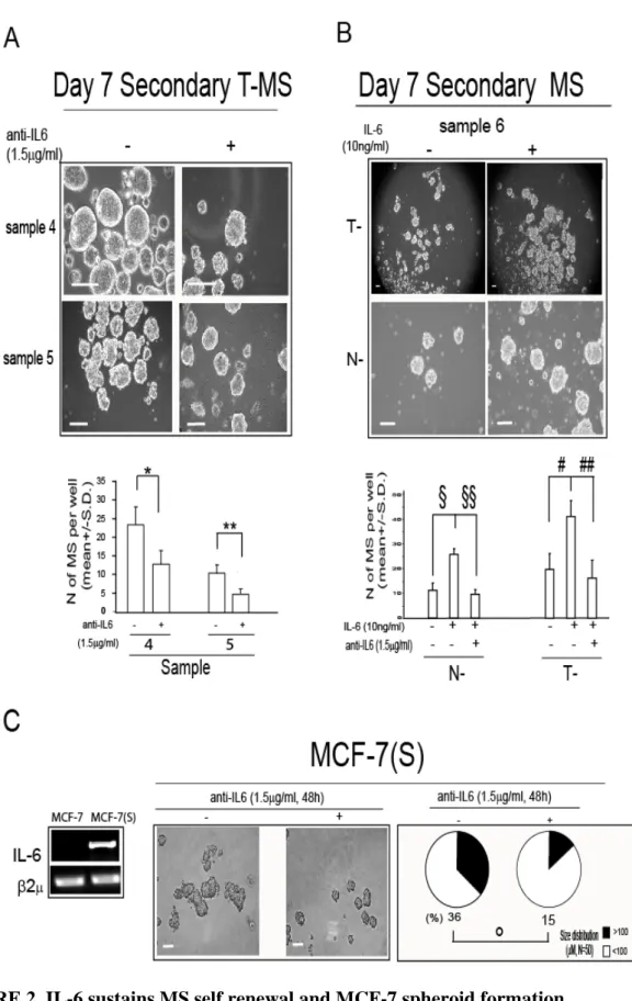
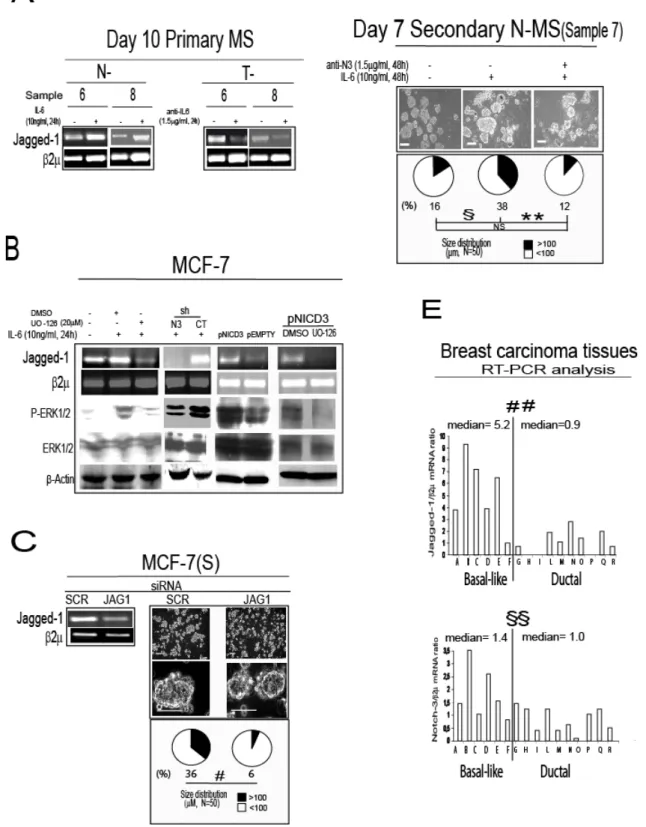
IL-6 promotes MS self-renewal and MCF-7 derived spheroids MCF-7(S) formation.
To assess the functional role of IL-6 expression in MS, we exposed secondary T-MS to a monoclonal antibody which blocks the IL-6 receptor/ligand interaction (anti-IL6, 1.5g/ml). Exposure of T-MS to anti-IL6 antibody substantially blunted their secondary regeneration capacity, a functional property which has been referred to MS self-renewal capability (Dontu et al., 2003,
2004; Figure 2A). Accordingly, we observed that, the administration of IL6 (10ng/ml) to N- and
T-MS from the same patient yielded an increase in secondary T-MS formation, compared to T-MS not exposed to the cytokine, a phenomenon which was hampered by the simultaneous addition of anti-IL-6 (1.5g/ml, Figure 2B). We further investigated this phenomenon in the context of MCF-7-derived spheroids MCF-7(S), which have been recently shown to contain a substantial proportion of CD44+/CD24- cells (Phillips et al., 2006). MCF-7(S) expressed high levels of IL-6 mRNA, whereas the mRNA of the cytokine was absent in MCF-7 cells cultured in standard conditions (Figure 2C). Moreover, the administration of anti-IL6 (1.5g/ml) caused a substantial reduction in MCF-7(S) size (Figure 2C). These data indicate that IL-6 mRNA expression promotes growth in suspension, and that both autocrine and exogenous IL-6 promotes MS self-renewal.
FIGURE 2. IL-6 sustains MS self renewal and MCF-7 spheroid formation.
A, Day 7 Secondary T-MS, generated from primary T-MS in presence/absence of a MoAb which
and number of MS per well (n=3, *p=0.029; **p=0.042, Anova test); B, phase contrast microscopy analysis and number of MS per well in day 7 secondary T- and N-MS generated from primary MS in presence/absence of IL-6 (10ng/ml) and anti-IL6 (1.5g/ml), respectively: (n=3, §p=0.027, §§p=0.020, #p=0.048; ##p=0.035, Anova test, Post Hoc tests adjusted for multiple comparisons); C,
RT-PCR analysis of IL-6 mRNA level in 7 and Day 2 7 derived spheroids and MCF-7(S) generated in presence/absence of anti-IL6 (1.5g/ml): phase contrast microscopy analysis and MCF-7(S) size distribution. N= refers to the number of spheroids counted for each sample (°p=0.02, Monte Carlo2 test). 2 was assessed as quantitative control for RT-PCR analysis. Scale
bar=100m.
The MCF-7(S) growth promoting activity of IL-6 requires Notch-3 gene.
Notch genes play a pivotal role in MS self-renewal (Dontu et al., 2004; Farnie et al., 2007). In particular, Notch-3 is highly expressed in N-MS (Dontu et al., 2003), and its blockage induces a marked reduction in MS self-renewal and survival (Supplementary Figure 5). On these basis, we tested the hypothesis that the effect of IL-6 on MS self-renewal and MCF-7(S) formation may depend upon the Notch-3 gene expression. We found that the administration of anti-IL6 (1.5g/ml) to T-MS for 24h yielded a down-regulation in the level of Notch-3 mRNA, as well as that the administration of IL-6 (10ng/ml) to N-MS for 24h elicited an up-regulation of Notch-3 mRNA (Figure 3A). A similar regulation was observed in MCF-7 cells and MCF-7/(S) exposed to IL-6 (10ng/ml, 24h), and in MCF-7(S) exposed to anti-IL6 (1.5g/ml, 24h, Figure 3B). To better characterize the role of IL-6/Notch-3 interplay in substrate-independent growth, we generated MCF-7(S) using MCF-7 cells stably transduced with a retroviral vector expressing a Notch-3 specific (shN3/MCF-7) or control (shCT/MCF-7) shRNA. We found that MCF-7(S) obtained from shCT/MCF-7 cells and generated in the presence of IL-6 (10ng/ml) showed an increase in size, whereas shN3/MCF-7 cells did not produce MCF-7(S), even in presence of exogenous IL-6
(10ng/ml, Figure 3C). These data indicate that Notch-3 signalling is of pivotal importance to sustain the IL-6 dependent growth of breast cancer cells in suspension culture.
FIGURE 3. IL-6 induces the Notch-3 gene up-regulation and the Notch-3-dependent MCF-7(S) formation. A, RT-PCR analysis of Notch-3 mRNA level in day 10 primary N-MS in
presence/absence of IL-6 (10ng/ml) and in T-MS in presence/absence of anti-IL6 (1.5g/ml) for 24h; B, RT-PCR analysis of Notch-3 mRNA level in MCF-7 cells cultured in presence/absence of IL-6 (10ng/ml), and in MCF-7(S) in presence/absence of anti-IL6 (1.5g/ml) or IL-6 (10ng/ml) for 24h; C, Day 7 MCF-7(S) generated from MCF-7 cells infected with a pSuperPuro retroviral vector
encoding a Notch-3-specific (shN3) or control (shCT) shRNA, in presence/absence of IL-6 (10ng/ml): phase contrast microscopy analysis and MCF-7(S) size distribution (N= refers to the number of spheroids counted for each sample, *p=0.034; NS=Not significant, Monte Carlo 2test);
WB analysis of Notch-3 and -Actin protein level. 2 was assessed as quantitative control for RT-PCR analysis. Scale bar=100m.
IL-6 elicits a Notch-3 dependent up-regulation of Jagged-1 mRNA expression which sustains MCF-7(S) formation and promotes MS self-renewal.
We found that Notch-3 promotes MS survival and that it regulates the expression of its ligand Jagged-1 (Supplementary Figure 5-6). Therefore we next evaluated if Jagged-1 was involved in Notch-3 dependent MS growth. Indeed either exposing N-MS to IL-6 (10ng/ml) or adding anti-IL-6 (1.5g/ml) to T-MS modulated the expression of Jagged-1 mRNA (Figure 4A). Moreover, we found that in MCF-7 cells, IL-6 elicited an up-regulation of Jagged-1 mRNA, which was blocked by the co-administration of IL-6 with the MEK/ERK inhibitor UO-126 (Figure 4B). Furthermore, we found that the up-regulation of Jagged-1, induced by IL-6 was negligible in shN3/MCF-7 cells, and that the transfection of a plasmid encoding the activated form of Notch-3 (pNICD3) into MCF-7 cells triggered an up-regulation of Jagged-1 mRNA, which was prevented by the concurrent administration of UO-126 (Figure 4B). In addition, we observed that MCF-7(S) formation was extremely reduced when MCF-7 were transfected with a Jagged-1 specific siRNA (JAG1), compared to scrambled (SCR) control siRNA (Figure 4C). Finally, we observed that an antibody blocking Jagged-1/Notch-3 interaction reduced MS regeneration capacity (see Figure 4D), indicating that the Notch-3/Jagged-1 pathway is functionally relevant for IL-6 induced MS formation. Notably, we also found that basal like breast carcinoma tissues expressed higher Jagged-1 and Notch-3 mRNA levels than ductal breast carcinoma tissues (Figure 4E). These data suggest that up-regulation of Jagged-1 via Notch-3 signalling is crucial for the growth in suspension of breast cancer cells and MS, and that this phenomenon may also occur in basal-like breast cancer tissues.
FIGURE 4. Notch-3/Jagged-1 interplay sustains MCF-7(S) formation and MS self-renewal. A, Day 10 primary N-MS and T-MS cultured in presence/absence of IL-6 (10ng/ml) or anti-IL6
(1.5g/ml) for 24h: RT-PCR analysis of Jagged-1 mRNA level; B, RT-PCR analysis of Jagged-1 mRNA level, and WB analysis of phosphorylated ERK and total ERK protein level in MCF-7 cells
exposed to IL-6 (10ng/ml) in presence/absence of the MEK1 inhibitor UO-126 (20M) or DMSO for 24h, in shN3/shCT MCF-7 cells exposed to IL-6 (10ng/ml, 24h), and in MCF-7 cells transfected with 1g pCDNA3.1 vector encoding Notch-3 intracellular active cleaved fragment (pNICD3), or empty control vector (pEMPTY) for 24h, in presence/ absence of UO-126 (20M) or DMSO; C Day 7 MCF-7(S) generated from MCF-7 cells transfected with Jagged-1 specific or SCR siRNA (1g, 72h of pre-exposure): RT-PCR analysis of Jagged-1 mRNA level, phase contrast microscopy analysis, MCF-7(S) size distribution, (#p=0.001, Monte Carlo 2test); D, Day 7 secondary N-MS
generated in presence of IL-6 (10ng/ml) and in presence/absence of a MoAb which blocks Notch-3 activity (anti-N3, 1.5g/ml): phase contrast microscopy analysis, N-MS size distribution (N= refers to the number of spheroids counted for each sample, Monte Carlo 2 test, §p=0.039, **p=0.009, Post
Hoc tests adjusted for multiple comparisons, NS=not significant); E, Bar graph representation of RT-PCR analysis of Jagged-1 and Notch-3 mRNA level (ratio over 2mRNA) in basal-likeor ductal carcinoma tissues (##p=0.005, §§p=0.042, MW test). 2 was assessed as quantitative control for RT-PCR analysis. -Actin was assessed as quantitative controls for WB analysis. Scale bar=100m.
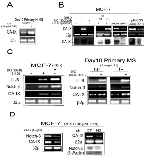
IL-6 induces a Notch-3 dependent up-regulation of carbonic anhydrase IX (CA-IX) gene. ERK up-regulation has recently been found to enhance the expression of the hypoxia survival gene carbonic anhydrase IX (CA-IX, Kopacek et al., 2005). Thus given our above observations, we next evaluated whether IL-6 signalling modulates CA-IX gene expression. Indeed adding IL-6 (10ng/ml) to N-MS induced an up-regulation of CA-IX mRNA (Figure 5A). Increased IX expression was also observed in MCF-7 cells exposed to IL-6 (10ng/ml, 24h), whereas CA-IX gene expression was markedly reduced by the administration of UO-126 (Figure 5B). Similarly to what we observed for Jagged-1 (see Figure 4B), CA-IX gene expression was inhibited in shN3/MCF-7, but not shCT/MCF-7 cells exposed to IL-6, while it was enhanced by transfection of the pNICD3 vector but not in the presence of UO-126 (Figure 5B). Because CA-IX is a hypoxia response gene (Kopacek et al., 2005), we investigated whether IL-6 plays a role in the hypoxia response. Exposure of MCF-7 cells to hypoxic stimuli (100M Desferroxamine, DFX, or low oxygen tension, <0.1%O2, 48h), as well as the exposure of N- and T- MS to 50M DFX (48h),
enhanced the expressions of IL-6, Notch-3 and CA-IX mRNAs (Figure 5C). Importantly upon blocking the up-regulation of hypoxia responsive genes with 100M DFX, the administration of anti-IL6 (1.5g/ml) to MCF-7 cells caused a down-regulation of Notch-3 and CA-IX mRNA. In addition, CA-IX mRNA was also down-regulated in shN3/MCF-7 cells exposed to 100M DFX compared to shCT/MCF-7 cells (Figure 5D). Taken together these results indicate the CA-IX gene expression is regulated by IL-6/Notch-3 pathway in MCF-7 cells and MS.
FIGURE 5. The IL-6/Notch-3 cross-talk promotes the up-regulation of carbonic-anhydrase IX (CA-IX) gene.
A, Day 10 primary N-MS cultured in presence/absence of IL-6 (10ng/ml) for 24h: RT-PCR analysis
of CA-IX mRNA level; B, RT-PCR analysis of CA-IX mRNA level and WB analysis of CA-IX (phosphorylated ERK, total ERK and -Actin protein level are reported in Figure 4B) in MCF-7 cells exposed to IL-6 (10ng/ml, 24h) in presence/absence of UO-126 (20M) or DMSO, in
shN3/shCT MCF-7 cells exposed to IL-6 (10ng/ml, 24h), and in MCF-7 cells transiently transfected with pNICD3/pEMPTY vector (1g), co-administered with UO-126 (20M) or DMSO for 24h; C, RT-PCR analysis of IL-6, Notch-3, CA-IX mRNA level in MCF-7 cells exposed to low oxygen level (<0.1%O2) or 100M Desferoxamine (DFX), and in N-/T-MS exposed to 50M DFX for 48h;
D, RT-PCR analysis of Notch-3 and CA-IX mRNA level in MCF-7 cells in presence/absence of
anti-IL6 (1.5g/ml), and in shN3/shCT-infected MCF-7 cells exposed to DFX (100M, 24h), WB analysis of Notch-3 and.-Actin protein level2 was assessed as quantitative control for RT-PCR analysis.
IL-6/Notch-3/CA-IX axis promotes hypoxia survival in MCF-7 and MS.
CA-IX gene has been found to play a crucial role in hypoxia survival of MS (Supplementary Figure 7). In keeping with these data, we observed a substantial increase in cell death of MCF-7 cells exposed to 100M DFX in the presence of anti-IL-6 (1.5g/ml), or transfected with a CA-IX-specific siRNA, compared to matched controls (Figure 6A). Further, a higher degree of hypoxia-induced cell death, accompanied by a down-regulation of CA-IX mRNA was observed in shN3/MCF-7 cells, compared to shCT/MCF-7 cells (Figure 6A). In line with these results, we found that the exposure of T-MS to anti-IL-6 or anti-N3 (1.5 and 1g/ml, respectively), or the transfection with CA-IX siRNA, in the presence of 50M DFX, increased in cell death in comparison to a matched SCR siRNA control (Figure 6B). Interestingly, detectable levels of CA-IX mRNA were found only in tissues from basal like breast carcinoma (Figure 6C). These data indicate that IL-6/Notch-3 induced CA-IX gene expression promotes hypoxia survival in MS, and support the similarity between the gene expression profiles of MS and basal-like breast carcinoma tissues.
FIGURE 6. The IL-6/Notch-3/CA-IX axis promotes hypoxia survival.
A, MCF-7 cells in presence/absence of DFX (100M, 48h): presence/absence of anti-IL6