1
UNIVERSITA‘ DEGLI STUDI DI VERONA
DIPARTIMENTO DI
SCIENZE MORFOLOGICO-BIOMEDICHE
DOTTORATO DI RICERCA IN
IMAGING MULTIMODALE IN BIOMEDICINA
CICLO XXII
NEW EVIDENCES OF INFLAMMATORY MEDIATORS IN
ABSENCE EPILEPSY
S.S.D. BIO / 16
Coordinatore:
Prof. ANDREA SBARBATI
Tutor:
Prof. PAOLO FRANCESCO FABENE
2
TABLE OF CONTENTS 1. INTRODUCTION
1.1 Definition of absence epilepsy Pag 5
1.2 Epidemiology of absence epilepsy Pag 6
1.3 Classification and clinical phenomenology of absence epilepsy Pag 7
1.3.1 Typical absence epilepsy Pag 7
1.3.2 Atypical absence seizures and absence seizures in other epilepsy syndromes Pag 11 1.3.3 Additional rare syndromes associated with typical absence seizures Pag 12
1.4 Animal model of absence epilepsy Pag 13
1.5 Theoretical considerations: classical concepts of absence Epilepsy Pag 14
1.6 Recent achievements in animal models of absence epilepsy Pag 16
1.7 The role of the cortex in generalized absence epilepsy revisited Pag 18
1.8 A focal cortical theory of absence epilepsy in WAG/Rij rats Pag 20
1.9 Experimental support of the focal cortical theory: local Deactivation Pag 23 1.10 Morpho-functional substrates of absence epilepsy in WAG/Rij rats Pag 25
1.10.1 Cytoarchitectural abnormalities of the neocortex Pag 25
1.10.2 Impairment of the Gabaergic system: immunohistochemistry and
neurophysiology Pag 27
1.10.3 Role of the Glutamatergic system in absence epilepsy Pag 30
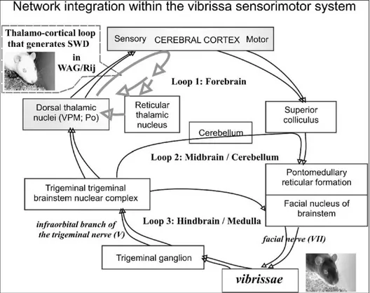
1.11 Perioral area of SmI as a generator of intrinsic oscillations Pag 32 1.12 Perioral area of the SmI as a part of an oscillatory trigeminal system Pag 36
3
2. AIM OF THE STUDY Pag 40 3. EXPERIMENT 1; STRUCTURAL AND FUNCTIONAL MRI
3.1 INTRODUCTION TO EXPERIMENT 1 Pag 42
3.1.1 T2 weighted and Diffusion-weighted imaging (DWI) analysis Pag 42
3.2.2 rCBV and rCBF maps in Wag/Rij rats Pag 43
3.2 MATERIALS AND METHODS Pag 43
3.2.1 T2 weighted and DWI maps in Wag/Rij rats Pag 44
3.2.2 rCBV and rCBF maps in Wag/Rij rats Pag 45
3.3 RESULTS Pag 46
3.3.1 T2 weighted and Diffusion-weighted imaging (DWI) analysis Pag 46 3.3.2 Regional brain volumes (rCBV) and regional brain flow (rCBF) analysis Pag 50 4. EXPERIMENT 2; MAGNETIC RESONANCE SPETTROSCOPY (MRS)
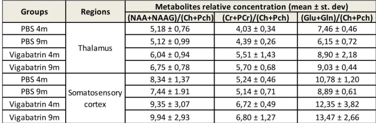
4.1 INTRODUCTION TO EXPERIMENT 2 Pag 54
4.2 MATERIALS AND METHODS Pag 55
4.3 RESULTS Pag 56
5. EXPERIMENT 3; GENE ARRAY
5.1 INTRODUCTION TO EXPERIMENT 3 Pag 58
5.2 MATERIALS AND METHODS Pag 59
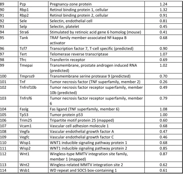
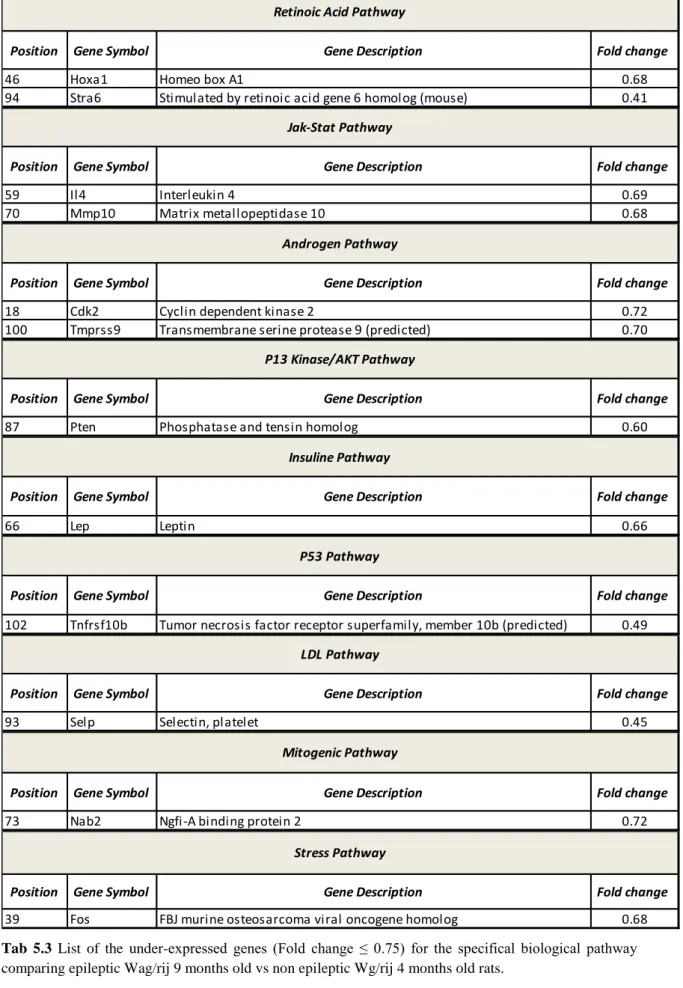
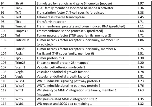
5.3 RESULTS Pag 62
6. EXPERIMENT 4; REAL TIME QUANTITATIVE PCR ANALYSIS (RT-qPCR)
4
6.2 MATERIALS AND METHODS Pag 76
6.3 RESULTS Pag 77
7. EXPERIMENT 5; EEG RECORDING AFTER CITOKINES INJECTION
7.1 INTRODUCTION TO EXPERIMENT 5 Pag 79
7.2 MATERIALS AND METHODS Pag 79
7.3 RESULTS Pag 81
8. EXPERIMENT 6; CITOKINES CONCENTRATION IN BRAIN AND PLASMA
8.1 INTRODUCTION TO EXPERIMENT 6 Pag 83
8.2 MATERIALS AND METHODS Pag 83
8.3 RESULTS Pag 84
9. GENERAL DISCUSSION Pag 85
ACKNOWLEDGMENTS Pag 92
5
-1-
INTRODUCTION1.1 Definition of absence epilepsy
Epilepsies and epileptic syndromes describe complex disease states that are accompanied by abnormal hyper excitability and/or hyper synchronicity within the central nervous system (CNS). Epilepsy, as a disease entity, has first been mentioned by the ancient Egyptians who called it nesejet, or by the ancient Babylonians who called it the bennu disease (Temkin, 1994).
About 500 BC, Hippocrates already claimed that the brain is the origin of epileptic seizure activity. Interestingly, however, this approach was consequently ignored over the millennia and there was hardly any substantial progress in understanding the etiopathogenesis of epilepsies till beginning of the modern times (Temkin, 1994). The clinical features of absence epilepsy itself were first described by Poupart in 1705 (Temkin, 1994). Later on, Tissot described a girl with absences ―[…] avec un tres leger movement dans les yeux‖ and, in addition, repetitive generalized tonic–clonic seizures (GTCS) (Tissot, 1770). The term ―epileptic absence‖ was first introduced by Calmeil (1824); shortly thereafter, Esquirol (1838) raised the term petit mal in contrast to the generalized grand mal seizure group. Interestingly, Gowers (1881) gave a very detailed description of the absence seizures ―without conspicuous convulsions‖ and Friedmann (1906) first reported a long-term favorable prognosis but originally believed that absences were not epileptic. Sauer (1916) was the first to describe an important subtype of typical absence epilepsy, i.e., pyknolepsy (pycnolepsia, pure petit mal or socalled Friedmann syndrome), which was later characterized in detail also by Adie (1924). The establishment and characterization of the electroencephalography by Hans Berger in the 1920s enabled Gibbs and colleagues to unravel absence-specific ictal discharges in clinical EEG recordings (Gibbs and Gibbs, 1935). The petit mal triad of Lennox (1945), which was misused and misunderstood, was clarified by the Commission on Classification and Terminology of the International League Against Epilepsy (ILAE) with the differentiation of typical from atypical absences (Gastaut, 1970). Later on, several groups studied absence epilepsy in greater detail using video-EEG monitoring (Penry et al., 1975; Stefan, 1982). Panayiotopoulos and colleagues (1997) described
6
syndrome-related characterization of typical absence seizures with video-EEG analysis in detail. Etiopathogenetically, absence epilepsies represent a most fascinating disease group which is probably one of the most well-characterized seizure types in humans and animal models (Danober et al., 1998; Manning et al., 2003; Steriade, 2005; Llinas and Steriade, 2006; Khosravani and Zamponi, 2006). As will be outlined below, these detailed insights fostered the development of rather effective and specific pharmacological approaches to treat absence epilepsy in humans.
1.2 Epidemiology of absence epilepsy
Nowadays, the frequency, importance and sociocultural implications of epilepsy can hardly be overestimated. Manifest epilepsy affects around 0.5–1% of the population in Europe and North America, 5% of the population perceive a single seizure in their lifetime, and about 10–15% exhibit an increased seizure susceptibility (Forsgren et al., 2005a,b; Pugliatti et al., 2007). In the US, the prevalence of epilepsy is 1.77 million (Victor and Ropper, 2001). Estimates of worldwide prevalence of epilepsy suggest 50 million people being affected with strong differences in regional distribution. Epilepsy incidence is age dependent; 70% of the patients suffering from epilepsy are below 25 years of age, with most of the seizures beginning during childhood and early adolescence. About 40% of epilepsies belong to the generalized grand mal seizure family and another 40% represent complex partial, i.e., limbic or psychomotor seizures (Matthes and Schneble, 1999). In contrast, absence epilepsy makes up approximately 10% of the epilepsies and is often accompanied by other types of generalized seizures (Panayiotopoulos et al., 1995). Estimates of the annual incidence of childhood absence epilepsy (CAE), a prototype of pure petit mal in children aged 0–15 years, range from 6.3/100000 (Olsson, 1988; Loiseau et al., 1990) to 8.0/100000 (Blom et al., 1978; Posner, 2006). The prevalence of CAE was estimated at 10% (Callenbach et al., 1998) and 12.1% (Berg et al., 1999) for children younger than 16 years. Further detailed information on current epidemiology is provided by Jallon and Latour (2005). Interestingly, CAE is two- to fivefold more frequent in females than in males (Posner, 2006). Given the severe sociocultural implications, the necessity for efficient pharmacological treatment of absence epilepsies is self-evident (Manning et al., 2003; Rogawski and Loscher,2004).
7
1.3 Classification and clinical phenomenology of absence epilepsy
1.3.1 Typical absence epilepsy
Systematically, absence epilepsies can be differentiated into typical and atypical forms with typical absences being further categorized into simple and complex ones. According to the nomenclature proposed by the Commission on Classification and Terminology of the ILAE (Sirven, 2002), typical absence seizures can be found in the following epilepsy syndromes:
a. Typical absence seizures are the archetype of childhood absence epilepsy (CAE, pycnolepsy, pycnoleptic petit mal, pure petit mal, Friedmann syndrome) (Panayiotopoulos, 1999; Panayiotopoulos, 2005b) with an age of onset of 2–8 years and a peak incidence of 5–7 years. There is an ongoing debate whether infantile forms of absence epilepsies with an onset from the 1st to the 4th year of age (early onset absence epilepsy, EOAE) should be separated from childhood absence epilepsy with an onset later on until the 12th year of onset. This debate ranks about different aetiologies, manifestation, semiology, treatment and courses compared to CAE in children over 4 years of age. Patients generally display a normal neurological development and absences exhibit spontaneous remission (>90%) before puberty even without pharmacological treatment. CAE is the prototype of an idiopathic, age-related epileptic syndrome with characteristic ictal bilaterally synchronous, high-amplitude 2.5 to 4 Hz spike-wave discharge (SWD) activity, double to triple spike-waves and slow-waves and normal interictal background activity. Epileptiform SWD episodes are brief (4–20 s) and frequent (10 to 100/day) with abrupt onset and termination. Due to its etiopathogenesis, absences in CAE and other absence entities are accompanied by severe impairment or loss of consciousness. The eyes stare or move slowly and random eyelid blinking, usually not sustained, may occur. In addition, speech and other voluntary activity stop within the first 3 s of the discharge. However, automatisms are also frequent. The diagnosis of CAE is not probable if myoclonic jerks occur shortly before the active stage of the absences, if there are clonic jerks during the absences, if
8
there is no impairment of consciousness during the SWD activity or if there are multiple spikes and ictal discharge fragmentations.
b. Juvenile absence epilepsy (JAE, spanioleptic or cycloleptic absence epilepsy) is an idiopathic generalized epilepsy (IGE), mainly characterized by typical absences that are similar to, but probably not as severe and frequent as CAE (Panayiotopoulos et al., 1997; Panayiotopoulos, 2005a). Random and infrequent myoclonic jerks as well as infrequent GTCS occur in most of the patients. The predominant age of onset is between 10 and 16 years with a peak at 10 to 12 years. JAE normally persists in adulthood, but absences tend to become less severe with age. The ictal EEG exhibits generalized, spike or multiple spike and slow-waves at 2.5 to 4 Hz. Typical exclusion criteria for JAE are mild impairment of consciousness, brief ictal discharges (less than 4 s), eyelid or perioral myoclonus and rhythmic limb jerking. Single or arrhythmic myoclonic jerks during the absence ictus are incompatible with JAE. Visual and other sensory precipitation of absences may also speak against the diagnosis of JAE as well. In rare cases however, absence episodes might be triggered by intermittent photostimulation in JAE patients who also display photosensibility.
c. Juvenile myoclonic epilepsy (JME, impulsive petit mal, bilateral massive myoclonus, Herpin-Janz syndrome, Janz syndrome) is a genetically determined, common IGE. The prevalence of JME is 5% to 11% among adults and adolescent patients with other epilepsies, and both sexes are equally affected. JME is characterized by myoclonic jerks on awakening, GTCS and typical absences in more than one third of the patients. The seizures have an age related onset (12–25 years) with absences first appearing either in childhood or early adolescence, followed by myoclonic jerks and GTCS in the middle teens. Seizure-precipitating factors like sleep deprivation and fatigue, alcohol, photosensitivity, mental and psychological arousal are prominent. JME seizures probably persist life-long, although absences may become less severe with age. Jerks and GTCS commonly improve after the fourth decade of life. Typical absences are not the predominant type of absence in JME, and they are usually mild and not associated with automatisms or localized limb jerks. Furthermore, the absence related impairment of consciousness is often subtle. Generalized discharges of 3–6 Hz spike-waves have an unstable intradischarge frequency with fragmentations and multiple spikes.
9
d. Myoclonic absence epilepsy is a rare generalized absence epilepsy of uncertain classification with a typical age of onset of 6–8 years. It is characterized by severe bilateral rhythmical clonic jerks, often associated with tonic contractions.
e. Eyelid myoclonia with absences (also called eyelid myoclonia and absences, Jeavons syndrome) is an IGE manifested with frequent, pycnoleptic-like seizures. Eyelid myoclonia consists of marked, rhythmic and fast jerks of the eyelids and is often associated with jerky upward deviation of the eyeballs and retropulsion of the head. The seizures are brief (3–6 s) and occur mainly after eye closure (eye closure sensitivity). Additionally and concomitantly, absence seizures occur. The onset is usually in early childhood at an age of 10–12 years. Interestingly, patients are highly photosensitive in childhood, but this declines with age. Infrequent GTCS are inevitable in the long term, and they are likely to occur after sleep deprivation, fatigue and alcohol indulgence. Myoclonic jerks of the limbs may emerge, but they are infrequent and random. Eyelid myoclonia with absences often turns out to be pharmacoresistant and to persist life-long. The ictal EEG manifestations mainly consist of generalized polyspikes and slowwaves at 3–6 Hz, although the latter are more likely to occur after eye closure in an illuminated room. Total darkness abolishes the abnormalities related to eye closure. Photoparoxysmal responses are recorded from all untreated young patients.
In clinical routine, the neurological symptoms of typical absence epilepsy can strongly vary among patients (Stefan, 1982; Panayiotopoulos et al., 1997; Capovilla et al., 2001; Loiseau, 2002; Panayiotopoulos, 2005a). Often, patients display a typical, sudden onset of seizure with characteristic open, glassy and staring eyes, turning pale, dropping whatever is in their hand and quivering of the eyelids. Concomitant relaxation of facial muscle often results in a typical absent impression of the patient. The impairment of consciousness may be severe and thus a predominant clinical symptom but sometimes also moderate, mild or inconspicuous so that proper detection may require additional, special cognitive testing to determine the degree of mental arrest. For the duration of ictal activity, patients typically experience a congrade amnesia. In typical simple forms, mental arrest is often associated with other manifestations, such as hypolocomotion and actions such as speaking and writing will be interrupted with no impairment of static
10
functions, i.e., posture. The ceased actions will be immediately resumed after seizure termination. Based on the impairment of consciousness, the patient will usually be unresponsive when spoken to. The attacks as outlined above typically last from a few seconds to rarely half a minute and terminate as rapidly as they commenced. In less severe absences, the patient may not entirely stop his or her activities, although reaction time and speech may considerably slow down. In their mildest form, absences may even be inconspicuous to the patient and imperceptible to the observer, as disclosed on video-EEG recordings with errors and delays during breath counting or other cognitive testing during hyperventilation. In contrast, typical ―complex‖ forms of absence seizures are often associated with more sophisticated behavioural exacerbations, such as automatisms. These can be de novo automatisms, e.g., oral automatisms or perseverative forms in which patients exhibit stereotype behaviour such as walking, swimming and writing. Automatisms are common in typical absences when consciousness is sufficiently impaired, and they are more likely to occur 4–6 s after onset. Furthermore, these automatisms are more or less coordinated, adapted, i.e., eupractic or dyspractic, involuntary movements that may be an unconscious continuation of the preservative automatisms, de novo automatisms or both. Perioral automatisms such as lip licking, smacking, swallowing, or mute speech movements are also very common. Scratching, fumbling with the clothes and other limb automatisms may also occur. Many of these symptoms of both simple and complex absences can occur in the same patient. Also, one can observe a characteristic falling back of the head or vermiform turning of the head and trunk which is typical for tonic retropulsive, versive and rotatory subtypes of typical complex absence epilepsy. Clonic and myoclonic absence epilepsy usually affects the facial muscle, sometimes also the upper limbs. During the absence itself, clonic motor manifestations, rhythmic or arrhythmic and singular or repetitive are particularly frequent at the onset and can be continuous. However, they may also occur at any other stage of the seizure; usually they present as jerking of the eyelids, eyebrows and eyeballs, together or independently, as well as random or repetitive eye closures. Fast eyelid flickering is probably the most common ictal clinical manifestation and may occur during brief generalized discharges without discernible impairment of consciousness. Myoclonia at the corner of the mouth and jerking of the jaw are less frequently observed. Myoclonic jerks of the head, body and limbs may be singular or rhythmical and repetitive, and they may be mild or violent.
11
Absences may also be tonic or atonic. In absence with atonic components, the diminution of the muscle tone is usual and may lead to drooping of the head and, occasionally, slumping of the trunk, dropping of the arms and relaxation of the grip. Rarely, tone is sufficiently diminished to cause falls. In absence with tonic components the tonic muscular contraction may affect the extensor or the flexor muscles symmetrically or asymmetrically. The head may be pulled backwards, i.e., retropulsion or to one side, and the trunk may arch. Occasionally, absence seizures can be associated with autonomic signs, so-called vegetative absence epilepsy, including dilatation or constriction of the pupils, i.e., miosis and mydriasis, a flush, sweating, hypersalivation, paleness, enuresis or encopresis. Mixed forms of these absence subtypes are the rule rather than the exception. Absences may be the only type of seizures, as in CAE, or may be mild and non-predominant, as in JME. Typical absences are fundamentally different and pharmacologically unique compared to any other type of seizures, which also results in different pharmacological treatment. Thus, AEDs effective in focal seizures may be deleterious for absence seizures. Finally, the clinical EEG manifestations of typical absences are, by definition, widespread and often not as classical as in their archetype, CAE. If untreated, these seizure events can occur several hundred times a day. This impedes the child's development, often dramatically, and hence, inadequately treated absences can have severe sociocultural and educational impact particularly in young children and bring them into dangerous situations, e.g., in traffic.
1.3.2 Atypical absence seizures and absence seizures in other epilepsy syndromes
Atypical forms of absence epilepsy are often part of or accompanied by a complex epilepsy syndrome, such as Lennox–Gastaut syndrome (about two thirds have atypical absences), atypical benign partial epilepsy/pseudo-Lennox–syndrome (about 70% with absences (Hahn et al., 2001) or Doose syndrome (about 60–80% with absences; myoclonic astatic epilepsy (Guerrini, 2005) and thus exhibit a highly differentiated behavioural and electroencephalographic phenotype, whereas simple absence seizure types display more stereotype characteristics. Atypical absences can be symptomatic, e.g., in case of tumour formation in the frontal lobe, tuberous sclerosis (Bourneville– Pringle disease), Aicardi syndrome, Angelman syndrome and neurometabolic disorders. Atypical absences differ from typical absences in the several ways:
12
1. Atypical absences occur in the context of mainly severe symptomatic or cryptogenic epilepsies of children with learning difficulties, who also suffer from frequent seizures of other types such as atonic, tonic and myoclonic seizures.
2. In atypical absences, onset and termination is not as abrupt as in typical absences and changes in muscle tone are more pronounced. In addition, seizure episodes are unusually long in duration, e.g., more than 30 s.
3. The ictal EEG of atypical absence consists of slow (<2.5 Hz), spike-and-slow waves. The discharge is heterogeneous, often asymmetrical, and may include irregular spike-wave and slow-spike-wave complexes and other paroxysmal activity. The background interictal EEG is usually abnormal. It is noteworthy that absence epilepsies in general can exhibit status-like character. Most absence seizures can further be provoked by hyperventilation or photostimulation and in contrast can often be aborted by strong auditory or other sensory stimuli, as it is known from, e.g., simple-partial seizure types. Finally, absence seizures are more common in tired than in vigilant patients.
1.3.3 Additional rare syndromes associated with typical absence seizures
Apart from those absence seizure types outlined above, there are a few rare syndromes (Panayiotopoulos et al., 1997), which should be briefly mentioned here. Perioral myoclonia with absences is an IGE with onset in childhood or early adolescence. They are characterized by rhythmic myoclonus of the perioral facial or masticatory muscles during the absence, together with a variable impairment of consciousness. The absences are frequent and brief.
This seizure type may often be resistant to medication, unremitting and possibly life-long. Phantom absences denote typical absences that are mild and often inconspicuous to the patient and imperceptible to the observer. However, video-EEG recording, breath counting or other cognitive testing during hyperventilation with brief 3–4 Hz spike or multiple spike-wave and slow-wave discharges can be used in order to disclose the occurrence of such absences. The absences are simple, occasionally with eyelid blinking. Although they may be clinically unrecognized, they usually manifest in adult life with GTCS and often with absence status epilepticus (Panayiotopoulos et al., 1997). Reflex absences represent typical absences with specific modes of precipitation, e.g.,
13
photic pattern, video games, thinking, reading, etc. (Panayiotopoulos, 2005b). Photosensitivity is estimated to occur in approximately 50% of patients with onset of absences in childhood or adolescence, and it is often associated with an unfavourable prognosis (Lu et al., 2008). The best-defined syndrome with photosensitivity is eyelid myoclonia and absences (Jeavons syndrome). Absences with single myoclonic jerks during the absence ictus are typical absences with single, often violent jerks of the head, body or limbs. They may appear in early childhood and continue in adult life—often with other types of generalized seizures. They are difficult to treat and may have a bad prognosis. Symptomatic and cryptogenic absences may be, as the name suggests, symptomatic, arising as a consequence of a known disorder of the central nervous system (Ferrie et al., 1995), or cryptogenic, i.e., resulting from a suspected, but unknown, cause. Symptomatic and cryptogenic absences may be focal or diffuse, traumatic, metabolic or inflammatory.
1.4 Animal model of absence epilepsy
Genetic rat model of absence epilepsy have been studied for many years and thought to mimic more accurately the spontaneous seizures of human epilepsy than do drug-induced animal models (Coenen et al., 2003).
GAERS (Genetic Absence Epilepsy Rats from Strasbourg) are a well-validated animal model of human Idiopathic Generalised Epilepsy (IGE), possessing a similar electrophysiological, ontogenic and pharmacological profile to the human condition (Danober et al., 1998). Historically, the GAERS colony was derived from Wistar rat and selectively bred for the seizure phenotype so that 100% of progeny spontaneously develop the epilepsy. A non-epileptic control (NEC) strain was also derived from the original colony by selectively breeding for the lack of seizure expression, providing a powerful control strain, since any differences between the two strains would have a high a priori chance of being aetiologically associated with the epilepsy. In addition to developing epilepsy, the GAERS strain also exhibits a range of behaviours indicative of affective disturbance, including increased anxiety and depressive like behaviours (Jones et al., 2008), intimating that this rat strain also models the well-documented mood disturbances observed in clinical IGE populations (Caplan et al., 1998, 2005; Davies et al., 2003; Jones et al., 2007; Ott et al., 2003; Tellez-Zenteno et al., 2007).
14
The rats strain named WAG/Rij (Wistar Albino Glaxo strain bread in Rijswijk) exhibits spontaneous SWDs that have a frequency of 7 to 11 Hz, duration of 1 to 45 s and amplitude of 200 to 1000 µV (van Luijtelaar et al.,1986; Coenen et al., 1987,1992). The spike-wave discharges become manifest in the cortical EEG at an age of 2 to 3 months, while at a younger age spike-wave discharges are not detectable. At an age of 6 months, both male and female rats show about 16–20 discharges per hour, with an average duration of about 5 s. This amounts to several hundred discharges per day (Coenen and van Luijtelaar, 1987).
Characteristics in the behaviour of WAG/Rij rats have been studied extensively and are quite similar to those of outbred Wistar rats with which they have been compared. Typical features of WAG/Rij rats are a short latency to emerge from the home cage into familiar and novel environments; low open-field defecation and high open-field ambulation; a low apomorphine-induced gnawing score; a high running-wheel activity; nondistinctive sleep percentages; good two-way, active shock-avoidance acquisition; and only slightly deficient working, albeit normal reference, memory scores in two spatial memory tasks (de Bruin et al., 2001; Festing and Bender, 1984; Harrington, 1972; van Luijtelaar and Coenen, 1988; van Luijtelaar et al., 1989).
1.5 Theoretical considerations: classical concepts of absence Epilepsy
The highly synchronous appearance of SWDs in both hemispheres led to the assumption by early researchers that SWDs could arise from a central structure with widespread cortical projections that could distribute paroxysmal activity over the entire cortex. They proposed the existence of a subcortical pacemaker for SWDs (Jasper and Kershman, 1941). A few years later the ‗‗centrencephalic‘‘ concept was introduced (Penfield and Jasper, 1954). This ‗centrencephalic integrating system‘ was thought to be located in the brain stem and diencephalon. Moreover, this system was believed to be responsible for the bilateral onset of the SWDs and the loss of consciousness. Subsequent stimulation studies in cats gave rise to the hypothesis that SWDs are likely to originate from the non-specific part of the thalamus, the intralaminar nuclei, which project diffusely to many cortical regions. The role of these nuclei in the generation of SWDs in humans remained unclear, in part because comparative anatomical studies showed that the thalamus in mammals is less developed than in humans (Ajmone-Marsan, 1965; Pollen
15
et al., 1963) and depth intracranial recordings were not an option. Despite these drawbacks, the ‗‗centrencephalic‘‘ concept was gradually transformed into a ‗‗thalamic theory‘‘. Direct evidence of SWDs beginning in the thalamus with 1–2 s cortical delay was obtained in patients during absence seizures using simultaneous thalamic and cortical local field potentials (Williams, 1953). To date, the ‗‗thalamic theory‘‘ remains valid and well accepted and, furthermore, has received recent support from modern techniques like positron emission tomography and fMRI (Prevett et al., 1995; Salek-Haddadi et al., 2003), although it can be questioned whether the temporal resolution of PET and fMRI is currently sufficient to establish temporal differences between cortical and subcortical regions. Using depth recordings in epileptic patients, Bancaud (1969, 1972) observed that the spontaneous discharges occurring during spontaneous petit mal or grand mal seizures might initially be localized to the cerebral cortex, in the vicinity of an identified lesion, particularly in the frontal lobe. This and other observations did not fit into the centrencephalic theory (Petsche, 1962). Moreover, similar attacks could be reproduced by electrical stimulation of the same cortical epileptogenic zone (Bancaud et al., 1974). Therefore, it was suggested that generalized SWDs are secondary to a diffuse focal discharge in the frontal cortex. After their cortical onset, they are rapidly propagated throughout the whole cortex through various cortico-cortical pathways (Bancaud, 1971). In addition, the bilateral synchrony during typical SWDs was not as perfect as previously believed (Lüders et al., 1980). Niedermeyer (1996) presented a new ‗‗cortical theory‘‘: primary generalized epilepsy is the expression of cortical pathology; generalized SWDs are generated in the mesiofrontal cortex, from where they rapidly spread to other cortical areas. The thalamus certainly participates, but only ‗‗plays second fiddle‘‘ in carrying out normal physiological thalamo-cortical interactions. The third theory combined thalamic and cortical processes for the initiation and maintenance of absence seizures. Gloor (1969) saw a role for the cortex in the production of these generalized discharges and proposed that a ‗‗cortico-reticular‘‘ mechanism was involved in their generation, using experimental data collected in the feline generalized penicillin absence epilepsy model (Gloor et al., 1990; Kostopoulos, 2000). In this model, systemic injections of penicillin, a weak GABAA receptor antagonist, caused a gradual dose-dependent transformation of spindles into absence seizures. SWDs could also appear after cortical application of penicillin, whereas an injection of penicillin in the thalamus does not have this effect. This indicates that the
16
epileptiformic discharges are the result of abnormal responses of the cortex and not of the thalamus (Gloor et al., 1979; Gloor and Fariello, 1988). The crucial factor responsible for SWDs here is a presumed diffuse increase in the excitability of the cortex: cortical neurons respond to the afferent thalamo-cortical volleys by producing SWDs instead of passively transferring sleep spindles. In all, this theory assumes that the mechanism responsible for the genesis of SWDs is closely linked to the thalamocortical mechanism that generates sleep spindles.
1.6 Recent achievements in animal models of absence epilepsy
It is well accepted that sleep spindles and SWDs have a common thalamic pacemaker in the reticular thalamic nucleus (RTN) (Kostopoulos, 2000). Due to specific membrane properties and reciprocal interactions with thalamo-cortical relay (TCR) cells, neurons in the RTN are capable of producing intrinsic rhythmic bursts, which manifest themselves in field potentials such as sleep spindles. Surgical isolation of the RTN abolished rhythmic bursting in TCR cells, but bursting was preserved in RTN neurons (Steriade et al., 1985). Steriade and coworkers (for review see Steriade, 2003) demonstrated that TCR nuclei could only exhibit spontaneous spindle oscillations (7–14 Hz) when they receive projections from the RTN. These and similar studies were mainly done in cats in vivo and in the ferret in vitro (Von Krosigk et al., 1993). Basic cellular properties of thalamic cells were described and modeled, such as the different firing modalities (tonic and bursting), the crucial role of Ca2+ currents, and the longlasting hyperpolarization and subsequent calcium spike, that control the basic rhythmicity of thalamo-cortical oscillations (Destexhe and Sejnowski, 2002).
In the last decade, genetic rodent models became the model of choice (Danober et al., 1998; van Luijtelaar et al., 2002; Crunelli and Leresche, 2002; Coenen and van Luijtelaar, 2003; Depaulis and van Luijtelaar, 2006). Outcomes of studies in Genetic Absence Epilepsy Rats from Strasbourg (GAERS; Liu et al., 1991, 1992; Marescaux et al., 1984, 1992; Avanzini et al., 2000; Aker et al., 2002) and WAG/Rij rats (Inoue et al., 1993), supported the idea that SWDs are triggered in the thalamus or, more specifically, in its lateral parts implying that cortical SWDs originate from the thalamus. Pharmacological studies showed that local injections of the GABAA agonist muscimol and a GABA transaminase inhibitor in the ventrobasal complex of the thalamus,
17
increases SWDs in GAERS, similar to systemic injections of GABAmimetics in GAERS and in WAG/Rij rats (Vergnes et al., 1984; Danober et al., 1998; Coenen and van Luijtelaar, 2003; Bouwman et al., 2004). Interestingly, local injections of muscimol in the RTN decreased SWDs (Liu et al., 1991), most likely by preventing local oscillatory activity (Danober et al., 1998). The results of these pharmacological studies point towards a role of thalamic GABAergic neurons in the control of SWDs and the differential role of two regions of the thalamus in their occurrence. Other evidence was based on lesion studies: SWDs in GAERS were suppressed after large electrolytic lesions of the lateral thalamus (Vergnes and Marescaux, 1992) and also after more restricted chemical lesions of the RTN (Avanzini et al., 1992, 1993). The same observations were made in WAG/Rij rats: SWDs were completely abolished after ibotenic lesions of large parts of the thalamus including the RTN (Meeren, 2002). However, rostral and caudal poles of the RTN seemed to antagonize each other in the sustaining of SWDs: Lesions in the rostral part of the RTN decreased the number of SWDs, but if lesions were restricted to the caudal and middle parts of the RTN, the incidence was unchanged or even increased (Meeren, 2002;van Luijtelaar and Welting, 2001). Most likely, there exists an antagonistic relationship between the rostral and caudal RTN; the caudal region of the RTN may inhibit the activity of a hypothetical pacemaker of SWDs located in the rostral part. This pacemaker may be disinhibited after lesions of the caudal RTN, which would explain the increase in SWDs. Nevertheless, the findings of all these studies corroborate the cortico-reticular theory that the pacemaker is located in the lateral thalamus. Although it is currently believed that SWDs are initiated in the RTN, the synaptic organization of the rostral pole of the RTN of the non-epileptic ACI rat (Agouti Copenhagen Irish, commonly used as a control strain since these rats showed no or at least very few SWDs in a comparative strain study; Inoue et al., 1990) appears to be similar to that of the epileptic WAG/Rij rat, as studied with electron microscopy (van de Bovenkamp-Janssen et al., 2004a), while GAERS also failed to show any essential structural alterations in RTN neurons compared to non-epileptic rats, no neuronal loss occurred in the RTN of epileptic animals (Sabers et al.,1996). It needs to be remarked that some physiological and anatomical features in rodents‘ brains are crucially different from the feline brain and earlier theories were exclusively based on neurophysiological studies in cats.
18
This difference might provide unique particularities to the rodent models of epilepsy, or at least account for some of its differences. First, SWDs in cats occur with frequencies from 3 to 4.5 Hz, about the same as in humans, but rats have 7–11 Hz as the intraspike frequency in a train of SWDs (Drinkenburg et al., 1993; Midzianovskaia et al., 2001; Bosnyakova et al., 2006). Second, in cats GABAergic inhibitory interneurons are present throughout the thalamus, including its relay nuclei, which receive an additional inhibitory control from RTN cells (Jones, 1985). In rats, inhibitory interneurons are absent in almost all thalamic relay nuclei (Jones, 1985), except the lateral geniculate nucleus (LGN, Ohara et al., 1983); moreover, the LGN itself gets more powerful monosynaptic inhibitory afferents from the RTN then any other part of the rats‘ thalamus (Ohara et al., 1980, 1983). Therefore, intrinsic inhibition is absent in the largest part of the thalamus in rats and the vast majority of thalamic nuclei receive only external inhibitory inputs from the RTN. This may significantly influence the oscillatory activity of thalamic neurons and the properties of thalamo-cortical networks.
Finally, the relative contributions of the cortex and thalamus and their exact mechanisms are still a matter of debate, and much of the controversies concerning the exact mechanisms can be ascribed to the usage of different experimental models (Blumenfeld, 2005). A putative disadvantage of the penicillin model is that the role of the cortex may be overemphasized because this model considers pharmacologically induced SWDs in cats, in which the seizures have slightly different mechanisms compared to spontaneous occurring SWDs in genetic rat models (Meeren, 2002). The study of mechanisms of the idiopathic types of epilepsy such as childhood absence epilepsy, however, may benefit from all the animal models that are nowadays available, including the genetic ones.
1.7 The role of the cortex in generalized absence epilepsy revisited
In their scheme for the generation of SWDs, the authors of the cortical theory did not assume a single focus or a focal area (Bancaud, 1969, 1972; Niedermeyer, 1996) nor did the authors of the cortico-reticular theory, who assumed a diffuse and global role of the cortex in the transformation of sleep spindles (Gloor, 1969; Gloor et al.,1990). Regarding the mechanisms of SWDs, both theories considered the cortex in general and did not address the specific local neurophysiological properties that could make one
19
cortical region more favourable for epileptogenesis than another, as is done in cases of focal epilepsies. Some studies in GAERS and WAG/Rij rats suggested a global role of the cortex in absence epilepsy after cortical deactivation was demonstrated by the spreading depression technique (unilateral diffusion of KCl resulted in immediate abolishment of SWDs not only in the injected cortex but also in the ipsilateral thalamus; Vergnes and Marescaux, 1992; Meeren, 2002). The same effect was found in cats with penicillin-induced SWDs during cortical spreading depression (Avoli and Gloor, 1981; Gloor et al., 1990). This observation was used as an argument for the corticoreticular theory that the cortex is a key element for generation of SWDs. The idea that an intact cortex interconnected in a thalamo-cortical network is a prerequisite for the occurrence of SWDs is also supported by the fact that surgical removal of the cortex abolishes SWDs, cortical slices do not show spontaneous, widely synchronized bursting activity, and that only in a thalamocortical slice preparation some stimulus-elicited oscillatory activity can be found if reciprocal thalamo-cortical connectivity was present (Avoli and Gloor, 1982; Tancredi et al., 2000). The cortico-reticular theory assumes that the cortex is more easily excitable in epileptic subjects than in nonepileptic ones. Consequently, the cortex produces abnormal paroxysmal responses to normal thalamic excitation. In such a way, thalamic sleep spindles, reaching the cortex through afferent volleys, could be transferred into SWDs (Kostopoulos, 2000). The penicillin model also showed that cortical blockade of GABAA receptors facilitates the presence of SWDs. Tolmacheva and colleagues (2004) investigated the excitability of the sensorimotor cortex for various types of afterdischarges and seizure types in WAG/Rij rats. No differences between WAG/Rij and ACI (non-epileptic control) rats were found in the seizure thresholds and afterdischarges irrespective of age, only the threshold for limbic seizures was reduced in WAG/Rij rats compared to ACI rats. Therefore, it does not seem likely that the cortex of WAG/Rij rats is in general more hyperexcitable than the cortex of nonepileptic control rats, at least in terms of thresholds of various types of epileptoform afterdischarges. Cortical excitability was also investigated in vitro. Some basic electrophysiological properties of cortical tissue in GAERS, WAG/Rij rats and in non-epileptic rats were not principally different. This mainly concerns passive cortical properties such as the complex waveform of the evoked local field potentials, the percentage of pyramidal neuron populations (intrinsically bursting and regular spiking
20
cells), and intrinsic membrane properties. (Luhmann et al., 1995; Avanzini et al., 1996; D‘Antuono et al., 2006).
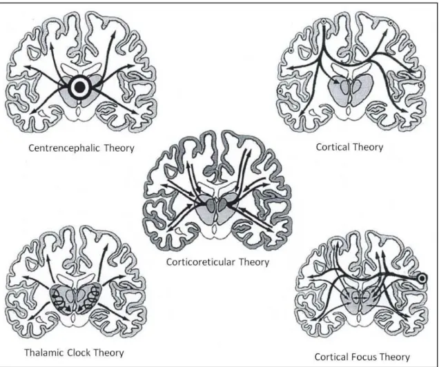
Figure 1.1. Schematic impression of the 5 theories on the origin of generalized absence epilepsy. On the
left are the thalamic theories: the centrencephalic theory of Penfield and Jasper (1954) and the thalamic clock theory of Buzsáki (1988). On the right are the cortical theories: the cortical theory of Bancaud, Lüders and collegues and Niedermeyer (1972) and the cortical focus theory of Meeren et al (2002). In the middle is the corticoreticular theory of Gloor (1968) (modified from Lüders et al.,1984).
1.8 A focal cortical theory of absence epilepsy in WAG/Rij rats
The role of the cortex in triggering SWDs became more obvious after a comprehensive study of network mechanisms responsible for the immediate onset, widespread generalization and high synchrony of SWDs (Meeren et al., 2002). Simultaneous, recorded field potentials from multiple cortical and thalamic sites were studied. The
21
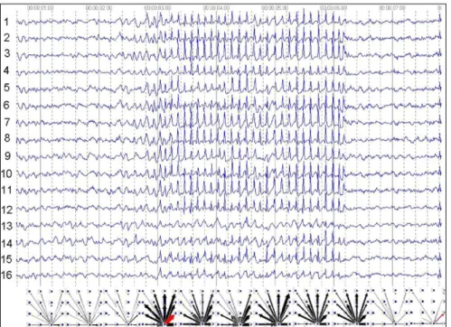
cortico-cortical, intrathalamic, and cortico-thalamic interrelationships between these field potentials were quantified using non-linear association analysis (Pijn et al., 1989). This method included direct measurements of the strength of association (the degree of correlation) and the time delay between signals recorded at different cortical and thalamic sites. These parameters together characterized functional coupling between populations of interacting neuronal populations. Fig. 1 illustrates a generalized SWD in the WAG/Rij rat and the results of the non-linear analyses, that is, the existence of a cortical focus originating from one of the 16 electrodes in the SmI (somatosensory cortex) (it is shown in graphs at the bottom, the starting point of arrows reflects the beginning of activity at the SmI in all eleven time frames). SWDs recorded at other cortical sites consistently lagged behind this focal site, with time delays that increased with electrode distance, resulting in a mean propagation velocity of about 1.5 m/s. Intrathalamic relationships were more complex and could not account for the observed cortical propagation pattern. Functionally interconnected cortical and thalamic sites appeared to influence each other, while the direction of this bidirectional coupling could vary throughout one seizure. However, during the first five hundred milliseconds the cortical focus was consistently found to lead its thalamic counterpart. Thereafter, the cortex and thalamus were found to alternately lead and lag in an unpredictable way. These results are incompatible with the common assumption that the thalamus acts as the primary driving source for the discharges. Instead, they indicate that a cortical focus plays a leading role in the origin of generalized SWDs characteristic of absence seizures in the rat. The large-scale synchronization characterizing bilateral synchronized SWDs appears to be mediated by the fast propagation of seizure activity from this focal site through cortico-cortical networks. Once the oscillation has been set into motion, however, the cortex and thalamus form a unified oscillatory network in which both structures drive each other. The role of the thalamus probably lies in providing a resonant circuitry to amplify and sustain the discharges (Meeren et al., 2002). It has been mentioned that the intrathalamic nuclei play a major role in synchronization and maintenance of neuronal oscillations (Seidenbecher and Pape, 2001). In light of these findings, it is proposed that the generation of bilaterally synchronous SWDs is only possible in an anatomically and functionally intact cortico-thalamic network that is in a suitable state. This most beneficial state is characterized by a light to moderate hyperpolarization of the intrinsically bursting cortical pyramidal cells and of the TCR
22
and the RTN cells, which makes them highly prone to produce high-frequency bursts of action potentials, as is the case during transitions from waking to sleeping, during drowsiness and light non-REM sleep, the most favorable vigilance states for the occurrence of SWDs in WAG/Rij rats (Drinkenburg et al., 1991; Coenen et al., 1991; Coenen, 1995). The initial event was the generation of an epileptic spike at the site of the cortical focus. Fig. 2 illustrates the pooled results of 8 WAG/Rij rats. In each of the 8 rats investigated, the focal zone was found to occupy a small (2-5 mm) zone in the projective area of the snout and vibrissae, as was established with functional mapping using evoked potentials. SWDs were initiated in a distinct (perioral) area of the somatosensory cortex, SmI (Meeren et al., 2002). Also, Steriade (2006) concluded that in a feline seizure model with SWDs, the latter are generated in the neocortex and thalamic involvement was instigated by cortical evoked excitation of mainly RTN neurons.
Fig. 1.2. A generalized SWD recorded from the lateral convexity of the neocortex of a WAG/Rij rat.
Results of the non-linear association analysis are presented underneath; calculations are done on EEG epochs of 500 ms. The thickness of the arrows represents the strength of the association, while the arrowheads point in the direction of the lagging site. The results of the analysis consistently suggest a cortical focus located in the projection area of the upper lip. SWDs recorded at other cortical sites lag behind the focal site with time delays that increase with electrode distance (adapted from Meeren et al., 2002).
23
Fig. 1.3. Pooled data from eight rats, in all animals leading sites of SWDs, filled symbols, located in the
somatosensory cortex. Open symbols represent lagging sites. The numbers represent the coordinates of the cortical surface in mm with zero point at the Bregma. Leading sites of SWDs occupy an area with the anterior-posterior coordinates from 2 (anterior to the bregma) to 3 (posterior to the bregma) mm, extending laterally from 6 to 8mm (adapted from Meeren et al., 2002)
1.9 Experimental support of the focal cortical theory: local Deactivation
The role of the SmI in the incidence of SWDs was examined using unilateral microinjections (1 ml) of 2% lidocaine into the vibrissal area of SmI (Sitnikova and van Luijtelaar, 2004). As is known, lidocaine temporarily blocks sodium channels and reversibly prevents neuronal activity (Malpeli and Schiller, 1979; Tehovnik and Sommer, 1997). It was hypothesized that blocking the neural activity in the SmI would unilaterally eliminate the cortical trigger of SWDs, which might result in a temporary decrease in their number. This hypothesis was tested in WAG/Rij rats. Animals were equipped with four epidural recording electrodes: bilaterally over the frontal cortex, unilaterally in the occipital area, and in the SmI adjacent to the injected site. As a control, an equal amount of saline was injected in the same area. A decrease in the number of SWDs emerged immediately after injection of 2% lidocaine as compared to saline. The difference between lidocaine and saline treated animals gradually diminished within two hours after injection. These results demonstrate that pharmacological deactivation of the driving cortical source caused a temporary reduction in the frequency of SWDs, which was observed at all recording sites
24
suggesting that deactivation of a certain cortical area is capable of decreasing generalized spike-wave activity throughout the entire cortex. Taken together, our results imply a crucial role of the vibrissae projecting area of the neocortex in the process of initiating SWDs. A bilateral blockade is much more effective than an unilateral one, at least as implied by the results of a preliminary pharmacological study: bilateral local injections in the peri-oral region of the SmI of the sodium channel blocker phenytoin in WAG/Rij rats and GAERS showed an almost complete abolishment of SWDs, while systemic administration of the drug increased SWDs. An earlier study was performed in GAERS: the anti-absence drug ethosuximide was bilaterally injected at multiple cortical and thalamic loci. The incidence of SWDs was strongly diminished if ethosuximide was injected in the SmI, and not or much less when injected in other cortical areas or in thalamic sites (e.g. the RTN or ventral basal thalamic complex). Therefore, treatment within thalamic target nuclei is insufficient for ethosuximide to have the full anti-absence effect; instead, the action of this agent may reside exclusively, or least primarily, in the cortex (Manning et al., 2003, 2004).
The outcome of these studies demonstrated that the WAG/Rij rats are not unique in the existence and location of the focal cortical origin of SWDs. In a combined EEG, in situ hybridization and immunohistochemical study in WAG/Rij rats, it was found that some types of sodium channels (Nav1.1 and Nav1.6) were up regulated and over expressed selectively at ML +6mm in the transverse plane at bregma (Klein et al., 2004). This region of the cortex approximately matches the electrophysiologically determined region of seizure onset within the perioral area of the SmI (Meeren et al., 2002). The upregulation was age dependent (only in 5–6 months old rats, not in younger ones), and this corresponds with the age-dependent increase in SWDs. The findings of this very elegant study clearly show that the age-dependent increase in SWDs is closely related to the age-dependent upregulation of certain types of sodium channels in the superficial layers of the perioral area of the somatosensory cortex only.
Neurophysiological studies showed functional differences in excitability in the somatosensory cortex between WAG/Rij rats and non-epileptic controls. An NMDAsensitive late EPSP that led to action potential discharges was found in 44% of regular bursting cells in the neocortical deep layers of WAG/Rij rats, and only in 8% of the cells in control rats in vitro (D‘Antuono et al., 2006). Inherent properties of hyperpolarisation-activated currents of pyramidal neurons (layers II–III) in the
25
somatosensory cortex of WAG/Rij rats differed in this area from those of two other rat strains (Wistar and ACI), and this modification of the physiological properties of neurons was accompanied by a reduced protein expression of subunits HCN1 (Strauss et al., 2004). It was proposed that the above-mentioned changes increase excitability especially in the perioral region of the SmI and thus facilitate the initiation of SWDs (Strauss et al., 2004). It is not clear however, whether these differences between strains are found exclusively in the perioral region of the somatosensory cortex or if other cortical regions differ between the epileptic and control rats. Therefore, it cannot be established that the perioral region of genetic epileptic rats is unique in the sense that only this region is hyperexcitable. High-level impairment of the inhibitory processes was found in the frontal cortex of WAG/Rij rats in vivo when compared to three other rat strains (ACI, APO-SUS and APO-UNSUS) in a paired pulse inhibition paradigm (sensory gating) while recording auditory evoked potentials (De Bruin et al., 2001), suggesting functional inhibitory disturbances in the neocortex of WAG/Rij rats. A functional change in the BOLD (blood-oxygen-leveldependent) signal during SWDs was found in fully conscious WAG/Rij rats. Significant increases in bold responses were found in widespread thalamic areas and the neocortex, including the SmI (Tenney et al., 2004). Other BOLD effects from functional MRI (fMRI) measurements (7 T) were measured in WAG/Rij rats under fentanyl/haloperidol anesthesia in combination with simultaneous recordings of field potentials (Nersesyan et al., 2004a, b). It was shown that spontaneous SWDs intensively involve the somatosensory area in addition to the thalamus and the electrical activity was accompanied by increased oxygen needs.
1.10 Morpho-functional substrates of absence epilepsy in WAG/Rij rats
1.10.1Cytoarchitectural abnormalities of the neocortex
It is a common belief that typical absence epilepsy is a purely ‗‗functional‘‘ disease since no structural lesion of any kind has ever been identified (Berkovic et al., 1987; Niedermeyer, 1996). An increased number of dystrophic neurons were found in neocortex and the subcortical white matter of the frontal lobe in patients with absence epilepsy (Meencke, 1989). In animal models of absence epilepsy, the cellular structure of cortical tissue appears to have entirely escaped consideration. Within the framework
26
of the focal theory of absence seizures (Meeren et al., 2002), Karpova and co-workers examined the cytoarchitecture of the anterior part of the neocortex in WAG/Rij rats, paying attention to the frontal (motor) region and to the parietal cortex, including the perioral region of the SmI (Karpova et al., 2005). The cellular composition and geometry of dendrite trees were established with the Golgi-staining technique and this was followed by qualitative and quantitative morphometric analysis. Typical for both the SmI and motor cortex of WAG/Rij rats was a disorder in the distribution of pyramidal cells in the superficial cortical layers (I–III). Apical dendrites of superficial pyramidal cells were often split in two branches, declined and went in non-perpendicular directions (Fig. 5). Quantitative morphometric measurements of dendrites such as the total length of dendrites, mean length of a dendritic segment and the size of the dendritic arbor, were increased when data from within and outside the epileptic area were compared, indicating that the pattern of dendritic arborization is abnormal in the epileptic zone in WAG/Rij rats. Disturbances in dendritic trees, a receptive part of pyramidal neurons, may cause impairment in communication between individual neurons. As mentioned, the differences were found in the superficial pyramidal cells, which, owing to long-range projections to remote cortical regions, may synchronize intrinsic cortical oscillations (Gray and McCormick, 1996). Therefore, the perioral area of SmI in WAG/Rij rats (the plausible trigger site of SWD) may express abnormal associations with other cortical areas that may facilitate synchronization and propagation of SWDs.
Age-dependent changes in the cytoarchitecture of cortical neurons may facilitate absence epilepsy. In all rodent models of absence epilepsy, SWDs appear at the age of 2–3 months and gradually increase with age (e.g. Coenen and van Luijtelaar, 1987; Willoughby and Mackenzie, 1992; Danober et al., 1998; Marescaux et al., 1992; Snead, 1992; Schridde and van Luijtelaar, 2005), which could be due to aging processes. Aging has an effect on cytomorphometric parameters of neuronal and glial populations in the SmI of the limbs and in the frontal cortex in Wistar rats (Peinado et al., 1993, 1997). Using serial sections stained with cresyl-fast-violet and quantitative morphometric techniques, the cellular composure found in younger (4–6 months old) rats was compared to that in older (30–32 months old) subjects. Cortical volume and neuronal density did not change with age, while glial density was significantly increased in older rats (mean for all layers 17%). Also, cytometric parameters of neurons in layer II–IV
27
altered with age: the shape of neurons was changed (major/minor diameter ratio was decreased) and the area of the neuronal soma was diminished. The overwhelming majority of synapses in the SmI are excitatory. The ratio between excitatory and inhibitory synapses in young adults is 4.2:1, and in older animals (32 month old) it is 4.9:1 (Poe et al., 2001). Therefore, there is a deficit in the intrinsic inhibitory circuitry of the aging neocortex that may contribute to a hyperexcitable state in the SmI and lead to absence seizures. Intracortical inhibition plays an important role in absence epilepsy, and genetic factors might influence this process of agerelated inhibitory impairment. In the following section more properties of the GABAergic system in the brain of WAG/Rij rats will be discussed.
1.10.2 Impairment of the Gabaergic system: immonocytochemistry and neurophysiology
It is already known for more than 10 years that synaptic network properties in the neocortex of WAG/Rij rats are impaired in comparison to non-epileptic Wistar controls. More precisely, the efficiency of GABAergic inhibition (as measured with intracellular recordings) is considerably reduced in the fronto-parietal cortical areas (Luhmann et al., 1995). As is known, inhibitory GABAergic interneurons form 20% to 30% of the neocortical population; their axons are relatively short and rarely spread further than 0.3mm (Peters and Jones, 1984; Markram et al., 2004). Inhibitory interneurons are subdivided into several classes: large, small and nested basket cells, chandelier cells, and double bouquet cells (for review see Markram et al., 2004). About 50% of all inhibitory interneurons are basket cells. They effectively control the firing activity of target cells (pyramidal and other interneurons): they synchronize the activity in neuronal networks. Basket cells typically express two calcium-binding proteins, parvalbumin (PV) and calbindin. PV is co-expressed with GABA in 90% of the GABAergic neurons (Miettinen et al., 1996; Celio, 1986), and PV-immunostaining is an appropriate way to mark basket cells (Kawaguchi and Kubota, 1997). This technique has been recently used in WAG/Rij rats to study the distribution of GABAergic neurons over various brain regions. It was suggested that an increase in cortical excitability in WAG/Rij rats and a low efficacy of the GABAergic inhibitory system could be the result of a smaller
28
amount of inhibitory neurons or from the impairment of metabolism of GABA in GABAergic neurons. The number of PV-immunopositive cells was quantified in various brain structures of WAG/Rij rats and compared with that in non-epileptic control ACI rats. It was found that some brain regions are much more strongly stained than others, like the molecular hippocampal layers and the thalamic nuclei, in agreement with data from, e.g. Houser et al. (1980). Both WAG/Rij and (non-epileptic control rats) ACI rats showed structures or even large cortical regions that hardly contained PVimmunoreactive cells. These cortical regions were not devoid of neurons but they simply could not be stained with the anti-PV serum. Apparently, these cells do not contain enough PV to become immunopositive. WAG/Rij rats showed a tendency to have more unstained regions than ACI rats but this difference was not statistically significant and no specific cortical area was consistently unstained in each WAG/Rij rat examined, demonstrating that there are individual differences in the affected regions. Quantification of PV-positive cells showed clear differences in the parietal (Par1) and in the forelimb area of the somatosensory cortex (FL), where ACI rats showed about two times as many PV-positive cells as WAG/Rij rats. Par1 and FL are parts of the somatosensory cortex, and Par1 contains the perioral projections. The lack of PV in these regions may destabilize intraneuronal Ca2+ homeostatic processes such as excitability, intracellular signaling and neurotransmitter release. Considering the co-localization of PV with GAD and GABA, it is assumed that the cortex contains areas with a lesser amount or even a lack of GABAergic cells in both strains (WAG/Rij and ACI). Probably, in WAG/Rij rats PV-immunoreactive (GABAergic) neurons in the somatosensory cortex and olfactory tubercle contain less PV or PV could even be absent, which would cause local cortical alterations of inhibition. A neurophysiological study aimed to establish whether differences in excitability or inhibition could underlie the presence of a focal area. Local field potentials were investigated in coronal in vitro brain slices containing Par1 and FL of the somatosensory cortex of 6-month-old WAG/Rij rats and age-matched control Wistar rats (Pitra et al., 2005). Field potentials were evoked by paired electrical stimuli (20 ms interpulse interval) applied to layer V, and they were recorded in overlying sites within layer II/III. Generally, the magnitude of the response to the second stimulus of a pair is a result of interplay between short-term dynamics of monosynaptic excitatory glutamatergic transmission and disynaptic inhibitory GABAergic transmission.
29
The amplitudes of responses to the first pulse of a pair were not different between WAG/Rij and control rats over the wide range of stimulation intensities, suggestive of a lack of difference in excitatory transmission in activated intracortical pathways of both strains. However, in WAG/Rij rats, a significantly smaller amount of paired-pulse depression of responses was observed in Par1 (Fig. 8b). This difference was not observed in the FL area. These results are consistent with a local and selective deficit in intracortical GABAergic transmission in WAG/Rij rats. In this sense it is extremely interesting that the local application of weak or strong GABAA antagonists in normal cats (penicillin and bicuculline) promotes the development of SWDs (Kostopoulos, 2000; Steriade, 2003). The role of the GABAergic system has been more extensively investigated in GAERS than in WAG/Rij rats. The hypothesis that an abnormal cortical GABAergic activity may underlie absence seizures in GAERS was confirmed by differences between GAERS and controls in thresholds for GABA receptor antagonists and inhibitors of GABA synthesis (Vergnes et al., 2000). Moreover, it was found that GAERS are more prone to seizures elicited by cortical GABA deficiency (Brailowsky et al., 1999). However, the distribution and number of neurons immunoreactive for GABA and GAD (Spreafico et al., 1993) and expressing GAD65 and GAD67 mRNAs (Danober et al., 1998) were similar in the cerebral cortex of GAERS and in age-matched controls. Also, autoradiographic studies showed that ligands binding to GABAA receptor subtypes in the cortex were not altered in adult GAERS rat, or in other rats with spontaneous SWDs (Knight and Bowery, 1992; Spreafico et al., 1993). Immunohistochemical studies of the GABAA receptor subunits showed a moderate reduction in the intensity of immunostaining of the 2,3 subunits of the GABAA receptor, selectively in the cerebral cortex of adult GAERS rats. Interestingly, this reduction was not observed before the occurrence of absence seizures, suggesting that these differences are present only after repetition of seizures in adults (Spreafico et al., 1993). A high affinity to GABAB receptors was observed in cortical membranes prepared from adult GAERS, whereas the total number of these receptors was similar in epileptic and control strains (Mathivet et al., 1996). A combined autoradiography, in situ hybridization and immunocytochemical study of the GABAB receptor in the somatosensory cortex in GAERS showed results at variance, although a clear upregulation of GABAB receptor protein in the cortico-thalamic circuit in GAERS was found (Princivalle et al., 2003). A significant increase in the number of GABAB
30
receptors occurs in the cortex of lethargic mice, although the affinity of these receptors appears unchanged (Hosford et al., 1992; Caddick and Hosford, 1996). Both alterations of GABAB receptor composition could account for an increase in cortical excitation through disinhibition by presynaptic autoreceptors. However, it is by all means not clear whether these differences in GABA-mimetic properties are present throughout the frontal cortex or mainly in the perioral region of the somatosensory cortex, or whether they are the cause of the discharges or caused by the discharges. It is also not clear whether the WAG/Rij rats and GAERS share the same deficits in the GABAergic system since comparative studies are lacking. Anyway, a deficit in the GABAergic system may have two different consequences: on the one hand, the impairment in inhibition may cause hyperexcitation of intracortical microcircuits, as was suggested for the feline penicillin model (Gloor, 1979). Then, excitatory pyramidal cells may start producing paroxysmal bursts and cause absence seizures. On the other hand, inhibitory deficits may decrease synchrony within the cortex because a sufficient amount of inhibition is needed for the synchronization of pyramidal cell bursting activity. How and in which way a reduced number of GABAergic cells available for synchronizing the burst firing of pyramidal cells contribute to more SWDs when rats are getting older remains to be established.
So, all this evidences suggest that inhibitory mechanisms in WAG/Rij rats are impaired in such a way that seizures more easily appear in local cortical regions (probably due to hyperexcitation from a lack of inhibition) and rapidly spread over the brain due to disinhibition of pyramidal neurons with long-range projections. This might imply that intracortical and cortico-subcortical network associations are altered in WAG/Rij rats.
1.10.3 Role of the Glutamatergic system in absence epilepsy
Burst firing between reciprocally interconnected glutamatergic thalamic relay neurons in the ventral basal thalamus and neocortical pyramidal neurons is synchronized by GABAergic neurons in the nucleus reticularis thalamic (NRT) (Steriade et al., 1993; Oh et al.,1995; Cox et al., 1997; Kim et al., 1997; McCormick and Bal, 1997). Glutamatergic thalamic relay neurons in fact send projections to cortex, and layer 6 of cortex sends reciprocal glutamatergic projections back to the associated thalamic nucleus (Guillery, 1969; Montero and Singer, 1984). Both of these projections pass