Index
Introduction pag. 3
The research context « 3
The research aims « 7
The research items « 9
References « 13
Results and discussion « 16
Cell and tissue localization ofβ-glucosidase during the ripening of olive fruit (Olea europaea) by in situ activity assay
S. Mazzuca, A. Spadafora, A.M. Innocenti.
Plant Science (2006), 171: 726-733. « 17
Oleuropein-specific-β-glucosidase activity marks the early response of olive fruits (Olea europaea) to mimed insect attack
S. Mazzuca, A. Spadafora, F.F. Chiappetta, A. Parise, E. Perri, A.M. Innocenti
Agricultural Science in China. (2007). Invite paper. « 27 Improvement of -glucosidase activity of Olea europaea fruit
extracts processed by membrane technology
R. Mazzei, L. Giorno, A. Spadafora, S. Mazzuca, E. Drioli.
Korean Membrane Journal (2006), 8: 58-66. « 52
Immunolocalization ofβ-glucosidase immobilized within polysulphone capillary membrane and evaluation of its activity in situ
S. Mazzuca, L. Giorno, A. Spadafora, R. Mazzei, E. Drioli.
Journal of Membrane Science (2006), 285: 152-158. « 61
Related articles « 68
β-glucosidase separation from Olea europaea fruit and its use in membrane bioreactors for hydrolysis of oleuropein
R. Mazzei, L. Giorno, S. Mazzuca, A. Spadafora, E. Drioli.
Desalination (2006), 200: 483-484. « 69
A new combined method to localize enzyme immobilized in polymeric membranes and evaluate its activity in situ
2
Desalination (2006), 199: 228-229. « 71
Protein extraction for two-dimensional electrophoresis from olive leaf, a plant tissue containing high levels of interfering compounds
W. Wang, M. Scali, R. Vignani, A. Spadafora, E. Sensi, S. Mazzuca, M. Cresti.
Electrophoresis (2003), 24: 2369-2375. « 73
Summary (in Italian) « 80
Introduzione « 80
Il sistemaβ-glucosidasi/oleuropeina nelle Oleaceae « 82
Scopo della tesi « 85
Risultati e discussione « 86
Localizzazione dellaβ-glucosidasi durante la maturazione del frutto
di ulivo (Olea europaea) attraverso saggio in situ dell’attività « 86 Attività -glucosidasica oleuropeina specifica in frutti di ulivo
(Olea europaea) in seguito ad attacco mimato di insetto « 87 Purificazione della -glucosidasica dagli estratti di frutto di Olea
europaea attraverso tecnologia a membrana « 88 Immunolocalizzazione della -glucosidasi immobilizzata su una
membrana capillare di polisulfone e valutazione della sua attività in situ « 89
Bibliografia « 90
Introduction
The research context
Olive (Olea europaea) is one of the established and largest crop in term of foodstuff production in Mediterranean countries, such as Spain, Italy, Greece. Starting from the last century, the intensive olive crop has been extended in other Mediterranean countries (e.g. Turkey, Tunisie, Maroc), and exported in several climate lands (e.g. California, Australia), thus its further expansion worldwide seems to be the tendency in the next future.
The reason of this achievement moved from the recognition of positive effects on the human health of olive food, especially virgin olive oil, a noteworthy component of the Mediterranean diet. In particular, many studies have shown that diet supplemented with olive oil daily, reduced the frequency of cardiovascular diseases, offered benefits in terms of colon cancer prevention and showed anti-inflammatory activities. However all these effects are the result of the higher levels of olive antioxidant compounds, particularly phenols[1-4].
Among olive phenols a bitter phenol glucoside, oleuropein [5], is largely accumulated in leaf and fruit and plays a key role in constitutive defence against pathogens [6] as well as in fruit ripening processes [7]. In particular, when olive tissues are damaged, an enzyme specifically hydrolyses oleuropein, producing highly reactive molecules [8].
The antioxidant and antimicrobial activities of oleuropein derivative molecules against herbivores and insect attacks has been demonstrated in planta [9] as well as against bacterial strains in vitro [10, 11] and they are recognized as pharmacologically active molecules [12].
Also during fruit ripening the same enzyme activity is involved in the progressive degradation of oleuropein, and in the release of glucose and the aglycones molecules, with the consequent physiological debittering of fruit tissues [13-15].
4
This enzyme involved in the reaction is the -glucosidase (E.C. 3.2.1.21) belongings to the Gluco Hydrolase enzyme family 1 (GH 1,
http://www.cazy.org/fam/GH1.html). The main characteristics of these enzymes are reported in Tab. 1.
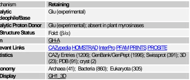
Tab. 1. Main characteristics of -glucosidases.
Mechanism Retaining
Catalytic
Nucleophile/Base Glu (experimental)
Catalytic Proton Donor Glu (experimental); absent in plant myrosinases 3D Structure Status Fold: (β/α)
Clan GH-A
Relevant Links CAZypedia HOMSTRAD InterPro PFAM PRINTS PROSITE
Statistics CAZy Entries (1206); GenBank/GenPept (1996); Swissprot (391); 3D (23); PDB (91); cryst (2)
Taxonomy Archaea (41); Bacteria (860); Eukaryota (305)
3D Display GH1_3D
-glucosidases catalyze the hydrolyis of glycosidic linkages in aryl and alkyl -glucosides and cellobiose (Fig. 1) and occur ubiquitously in plants, fungi, animals and bacteria. Since -glucosides and -glucosidases are ubiquitous in the living world, one expects to find structural and catalytic properties shared by all -glucosidases. In fact a review reported that almost all -glucosidases have subunit molecular weights of 55 to 65 kDa, acidic pH optima (pH 5-6) and an absolute requirement for a -glycoside as substrate [16].
-glucosidases from different orders and kingdoms appear to differ in their specificities for the aglycone linked to the glucosyl group by a -glycosidic bond.
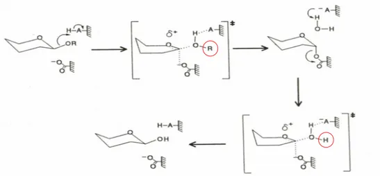
Fig. 1. Hydrolysis of the glycosidic bond. In most cases, this reaction is performed by two
catalytic residues of the enzyme: a general acid (proton donor) and a nucleophile/base.
In the past decade, considerable progress has been made on the molecular biology and biochemistry of -glucosidases. Some data have been available on very different organisms (plants, fungi, bacteria and humans) and/or related to specific problems (biomass conversion, cyanogenisis, host-parasite interactions, Gaucher’s disease). Therefore, it is of interest to underline that research on -glucosidases has significant implications both scientific and economic. In humans, one -glucosidase, commonly known as glucocerebrosidase, catalyzes the degradation of glucosylceramide in the lysosome; the deficiency of the enzyme leads to an inheredited disease, Gaucher’s disease [17].
-glucosidases of cellulolytic organisms have been the subject of much past and ongoing research. These enzymes are expected to be targets for genetic engineering to design and select -glucosidases for specific applications. In fact, fungal and bacterial -glucosidases appear as natural candidates for engineering an ideal -glucosidase to be used in the conversion of cellulose to glucose in industrial scale.
Plant -glucosidases have been known for over 170 years since the description of the action of emulsin (almound -glucosidases) on amygdalin, the cyanogenic -D-gentiobioside of almonds, by Liebig e Wöhler in 1837 [18]. In plants, -glucosidases have been implicated in several key metabolic events and
6
growth-related responses [16]. They range from defence against some pathogens and herbivores through the release of coumarins, thiocyanates, terpenes and cyanide to the hydrolysis of conjugated phytohormones (e.g., glucosides of gibberellins, auxins, abscisic acid and cytokinins) and to the fruit ripening processes.
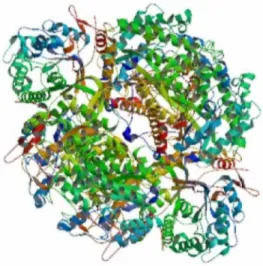
Plantβ-glucosidases have been investigated also for the specificity of their reaction sites through the crystallography and the deduced 3D structures. The omodimers of polypeptide chains arranged in a fold (β/α) structures (Fig. 2). From these data, the absolutely request of aminoacid residues of Glu 358 and Asp 374 are necessary for enzyme activity.
Fig. 2. The 3D structure of aβ/α foldβ-glucosidase from higher plant.
O. europaea tissues also contain large amounts of -glucosidase which specifically hydrolyses oleuropein [7, 9]. The detected changes in -glucosidase activity and in its products of enzymatic hydrolysis at different stages of fruit ripening [7] are strictly related to products quality [19-22]. In fact, good-tasting table olives and olive oil are greatly influenced by the phenolic compounds which are present in the fruit tissues. Thus, debittering of green olives is a major challenge in the industrial processing of fruit. In this context, investigations have been performed to test the efficiency of the enzymatic hydrolysis of oleuropein by the purified -glucosidase from almond [23] compared with the enzyme from
the crude extract of olive fruit [9-10]. More recently a bioreactor with the immobilized recombinant -glycosidase from the archaeon Sulfolobus solfataricus has been utilized to obtain high reactive molecules from oleuropein cleavage [24]. The results obtained from these investigations indicate that heterologous enzymes were not able to produce highly reactive dialdehydes from oleuropein, which strictly requests oliveβ-glucosidase.
The research aims
Aim of the present research is to investigate, by in situ β-glucosidase activity assay and by biochemical analyses
i. the mechanisms which regulate the enzyme expression and its activity on oleuropein which leads to the sweetening of the ripe olive during fruit ripening;
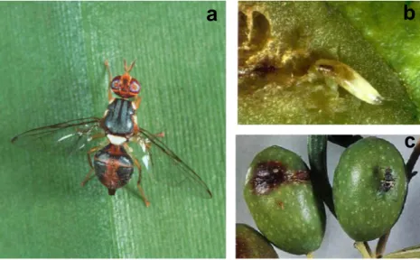
ii. the timing of enzyme activity and oleuropein content in fruit tissue following a mimed pathogen attack (Fig. 3) in two cultivars (Fig. 4) showing a different susceptibility against olive fly infestation[25].
a
b
c
a
b
c
Fig. 3. Fly female (Bactrocera oleae) during the attack on olive fruit. By the ovipositor, located
in the end part of her abdomen (a), the fly oviposes the eggs in tissues; developing larvae eat tissues and make deep tunnels trough the pulp (b) altering the integrity and the quality of fruits (c).
8
Fig. 4. Spread of the cultivars Carolea (a) and Cassanese (b).
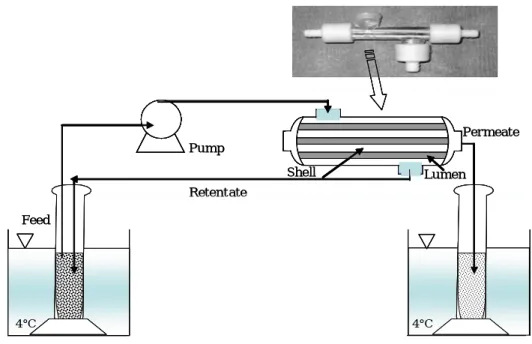
The obtained results have been also useful to determine at which ripening stage the fruit extracts show the highest enzyme activity; these extracts have been thereafter processed by membrane technology to fractionate and purify β -glucosidase (Fig. 5).
In this context the purified enzyme was used to develop a bio catalytic membrane reactor to achieve the oleuropein cleavage able to produce the bioactive molecules.
Carolea
a
Permeate Shell Lumen Retentate Pump Feed 4°C 4°C Permeate Shell Lumen Retentate Pump Feed 4°C 4°C
Fig. 5. Schematic draw of membrane diafiltration system.
Research items
Cell and tissue localization ofβ-glucosidase during the ripening of olive fruit (Olea europaea) by in situ activity assay
The cytological localization has been performed by the hydrolysis of X-Glc, a chromogenic synthetic substrate developing an insoluble blue precipitate in the cells. A strong reaction was detected within nucleus, chloroplasts and cytoplasm oil droplets. The enzymes kept in chloroplasts and oil droplets showed high specificity for the exogenous oleuropein respect to those toward X-Glc in the in situ competitive assay, thus indicating that two different oleuropein-degradative-β-glucosidases are present in these cell compartments.
Following the fruit ripening, significant variations in the number and distribution of reactive cells in mesocarp tissues have been observed. In fact, in immature fruits enzyme activity was not detectable in the outer mesocarp cells, whereas rare nuclei showed positive reaction in the inner mesocarp cells. Instead,
10
in green mature fruits a great number of reactive cells were found distributed in the whole mesocarp. The β-glucosidase activity appears preferentially localized in the outer mesocarp cells where a high activity was observable at the level of chloroplasts. Finally in black fruits numerous reactive cells were located only in the inner mesocarp close by the woody endocarp, whereas the outer mesocarp cells are devoted to polymeric anthocyanins accumulation. Thus, β-glucosidase activity level also decreased during fruit ripening.
These results clearly showed that the variations of
oleuropein-degradative-β-glucosidases activity during ripening seems to be related to a different competence of single mesocarp cells to synthesize the enzyme isoforms.
Oleuropein-specific-β-glucosidase activity marks the early response of olive fruits (Olea europaea) to mimed insect attack
Two cultivars of Olea europaea showing different susceptibilities to the fly infestation have been taken into account: a) cv Carolea, characterized by a high susceptibility to Bactrocera oleae infestation and b) cv Cassanese showing a low susceptibility. In both cultivars, the histochemical assay for β-glucosidase showed that, within 20 min after the injury, a strong enzyme activity was present in the damaged tissues. Thereafter a progressive enzyme inactivation occurred starting from tissues around the boundary of the injury and the enzyme activity disappeared after 60 min. The loss of activity decreased and stopped after 3 h, whereas the active cells limit reached the distance of 300 + 50 µm from the edge of injury. Biochemical analyses showed that in injured fruit extracts the β -glucosidase activity rapidly increased within 20 min from the injury, thereafter it decreased, reaching values comparable with those in sound fruits. Following the damage, the oleuropein contents did not change significantly in the high susceptible cultivar, while it rapidly decreased in the cultivar showing a low
susceptibility. These results strongly suggest that the olive fruit susceptibility toward the fly infestation could be related to the ability of the oleuropein-degradative-β-glucosidase to produce high reactive molecules in the damaged tissues. As a consequence of injury also a strong peroxidase activity was thereafter detected. This interesting pathway suggest that also this enzyme was involved in the following defence response.
Improvement of -glucosidase activity of Olea europaea fruit extracts processed by membrane technology
The purification of oliveβ-glucosidase is of high interest for its application in the food and pharmaceutical fields The enzyme is not yet commercially available and advanced clean and safe technologies for its purification able to maintain the functional stability are foreseen. The purification of this protein from fruit extracts has been already tempted by electrophoresis but either enzyme deactivation or high background with unclear profiles occurred. Fruit extracts obtained from the mature green phase of ripening, showing the highest enzyme activity, were processed by diafiltration and ultrafiltration. Asymmetric membranes made of polyamide or polysulphone having 50 and 30 kDa molecular weight cut-off, respectively, were tested for the diafiltration process.
Ultrafiltration membranes made of polyethersulfone with 4 kDa molecular weight cut-off were used to concentrate the diafiltered permeate solutions. The efficiency of the separation processes was evaluated by enzyme activity tests using the hydrolysis of p-D-nitrophenyl- -D-glucopyranoside (pNPGlc) as reaction model. Qualitative and quantitative electrophoresis were applied to analyze the composition of protein solution before and after the membrane separation. In addition dot blot and western blot analyses were applied to verify the presence of -glucosidase in the processed fractions.
12
The process allowed to separate and to identify a major enzyme form with molecular weight of 65 kDa identified as a putative -glucosidase by a western blot analysis and enzyme activity assay and a 20 kDa monomeric protein that in the native extract is combined to form higher molecular weight complexes. The diafiltration resulted a methodology able to guarantee the protein stability. In fact, the overall results showed that the -glucosidase functional stability was preserved during the membrane operations and the removal of 20 kDa proteins allowed to increase the specific activity of the enzyme of about 52% compared to the one present in the initial fruit extract.
Immunolocalization of β-glucosidase immobilized within polysulphone
capillary membrane and evaluation of its activity in situ
The new method we performed results from the merging of the classic in situ enzyme activity assay together with western blot technique. The results either at low than at high magnification can be easily detectable light microscopy.
-glucosidase was immobilized by physical method in asymmetric capillary membranes made of polysulphone having 30 kDa cut-off. Membranes sections processed with in situ assay showed a blue uniform staining by the insoluble reaction product. In order to verify if the colour distribution was not due to product diffusion, but corresponded to the presence of the immobilized enzyme, an immunolocalization method to localize -glucosidase specifically was also developed. As result of the antibody recognition, black spots were localized inside the membrane.
The results obtained by polyclonal antibody against -glucosidase and the synthetic substrate clearly showed a coherent correlation between the catalytic activity and the sites of enzyme immobilization.
References
1. H.K. Hamdi, R. Castellon (2005). Oleuropein, a non-toxic olive iridoid, is an anti-tumor agent and cytoskeleton disruptor. Biochemical and Biophysical Research Communications, 334 (3): 769-778.
2. A. Keys, A. Menotti, M.J. Karvonen (1986). The diet and 15-year death rate in the Seven Countries Study. American Journal of Epidemiology,
124: 903-915.
3. Willett W.C. (1990). Diet and coronary heart disease. Monographs in Epidemiology and Biostatistics, 15: 341-379.
4. World Health Organization (1990). Diet, nutrition, and the prevention of chronic diseases. Report of a WHO Study Group. WHO Technical Report Series, 797.
5. C. Soler-Rivas, J.C. Espin, H.J. Wichers (2000). Oleuropein and related compound. J. Sci. Food Agric., 80: 1013-1023.
6. M.J. Amiot, A. Fleuriet, J.J. Macheix (1989). Accumulation of oleuropein derivatives during olive maturation. Phytochemistry, 28: 67-69.
7. R. Briante, M. Patumi, S. Limongelli, F.C. Febbraio, A. Vaccaio, F. Di Salle, R. La Cara, M. Nucci (2002). Changes in phenolic and enzymatic activities content during fruit ripening in two Italian cultivars of Olea europaea L. Plant Sci., 162: 791-798.
8. A.D. Bianco, A. Piperno, G. Romeo, N. Uccella (1999). NNMR experiments of oleuropein biomimetic hydrolysis. J. Agric. Food Chem.
47: 3665-3668.
9. K. Konno, C. Hirayama, H. Yasui, M. Nakamura (1999). Enzymatic activation of oleuropein: a protein crosslinker used as a chemical defence in the privet tree. Proc. Natl. Acad. Sci. U.S.A., 96: 9159-9164.
10. G. Bisignano, M.G. Laganà, D. Trombetta, S. Arena, A. Nostro, N. Uccella, G. Mazzanti, A. Saijia (2001). In vitro antibacterial activity of some aliphatic aldehydes from Olea europaea L. FEMS Microbiol. Lett.,
198: 9-13.
11. A. Kubo, C.S. Lunde, I. Kubo (1995). A multichemical defence mechanism of bitter olive Olea europaea (Oleaceae). Is oleuropein a phytoalexin precursor? J. Chem. Ecol., 11: 251-263.
14
12. G. Bisignano, M.G. Laganà, D. Trombetta, S. Arena (2001). In vitro antibacterial activity of some aliphatic aldehydes from Olea europaea L. FEMS Microbiol. Lett., 198-199.
13. J.R. Morello, M.P. Romero, M.J. Motiva (2004). Effect of maturation process of the olive fruit on the phenolic fraction of drupes and oils from Arbequina, Farga and Morrut cultivars. J. Agric. Food Chem., 52: 6002-6009.
14. M. Brenes Balbuena, P. Garcia, A. Garrido Fernandez (1992). Phenolic compounds related to the black color formed during the processing of ripe olives. J. Agric. Food Chem., 40: 1192-1196.
15. D. Ryan, K. Robards, S. Lavee (1999). Changes in phenolic content of olive during maturation. Int. J. Food Sci. Technol., 34: 265-274.
16. A. Esen (1993). β-Glucosidases: Biochemistry and Molecular Biology. Edited by A. Esen. American Chemical Society, Washington, 1-14.
17. G. Grabowski, A. Berg-Fussman, M. Grace (1993). Molecular biology and enzymology of human acid β-glucosidase. In β-Glucosidases: Biochemistry and Molecular Biology. Edited by A. Esen, American Chemical Society, Washington, 66-77.
18. J. Liebig, F. Wöhler (1837). Annalen, 22: 11-14.
19. J.R. Morello, M.J. Moltiva, M.J. Tovar, M.P. Romero (2004). Changes in commercial olive oil (cv Arbequina) during storage with special emphasis on phenolic fraction. Food Chem., 85: 357-364.
20. F. Angerosa, N. D’Alessandro, F. Cornara, G. Mellerio (1996). Characterization of phenolic and secoiridoid aglycones present in virgin olive oil by gas chromatography-chemical ionization mass spectrometry. J. Chromatogr. A., 736: 195-203.
21. G. Cianfardini, V. Marsilio, B. Lanza, N. Pozzi (1994). Hydrolysis of oleuropein by Lactobacillus plantarum strains associated with olive fermentation. Appl. Environ. Microbiol., 60: 4142-4147.
22. V. Marsilio, B. Lanza, N. Pozzi (1996). Progress in table olive debittering: degradation in vitro of oleuropein and its derivatives by Lactobacillus plantarum. J. Am. Oil Chem. Soc., 73: 593-597.
23. R. Capasso, A. Evidente, C. Visca, L. Gianfreda, M. Maremonti, G. Greco (1996). Production of glucose and bioactive aglycone by chemical and enzymatic hydrolysis of purified oleuropein from Olea europaea. Appl. Biochem. Biotechnol., 61: 365-377.
24. R. Briante, F. La Cara, F. Febbraio, R. Barone, G. Piccialli, R. Caroll, P. Mainolfi, L. De Napoli, M. Patumi, G. Fontanazza, R. Nucci (2000). Hydrolysis of oleuropein by recombinant β-glycosidase from hyperthermophilic archeon Sulfolobus solfataricus immobilised on chitosan matrix. J. Biotechnol., 77: 275-286.
25. N. Iannotta, L. Perri, C. Tocci, F. Zaffina (1999). The behaviour of different olive cultivars following attacks by Bactrocera oleae (Gmel.). Acta Horticulturae, 474: 545-548.
16
Agricultural Sciences in China
No. 12, Zhongguancun South Street, Beijing 100081, P. R. China Tel: 86-10-62191638, 68919808, 68976244 Fax: 86-10-62118256
http://www.ChinaAgriSci.com E-mail: [email protected]
Dear Dr Mazzuca
Thank you for your contribution to our journal Agricultural Sciences in China
Your manuscript:< Oleuropein-specific- -glucosidase activity marks the early response of olive fruits (Olea europaea) to mimed insect attack) (au-2007-01330), has been evaluated by two reviewers of the journal with the following suggestions for your amendments.
Your manuscript after revised followed those suggestions will be accepted quickly. And also, you can point out your different ideas about the suggestions.
Reviewers 1:
The effects of oleuropein-specific- -glucosidase on early defense reaction to mining insect puncturing damage with a micro-needle on olive fruits of two different susceptible varieties were studied. The research topic focuses on olive fruit production practice in local area to investigate the biochemical changes on fruit after insect attack mainly using the histochemistry methods.
The paper is written in fluent English and criterion writing. The discussion in this paper is embedded based on the author’s research results. Therefore, the paper is suggested to adopt to publish
Reviewers 2:
The paper has presented an in situ -glucosidase activity detection in olive fruits after mechanically punctured by needle to mime the fly infestation in order to elucidate the early defense response of olive fruit to insect injury. It is meaningful and the paper is suggested to adopt to publish, but there still remain some points to discuss:
OMISSIS
All points have been discussed and implemented by authors in the revised version of manuscript herewith attached
As for your revising, please read the following
1) When send back your revised manuscript to the editor, a Statement for Revising in written should be attached together with and to state:
26
b) Which one has not been accepted. As the reviewers and the authors both are the experts in this field, it is possible we think that they have different views on the same issue. So if you cannot agree to the referees’ opinions, you can insist on your own viewpoint, but it is required for you to give the reasons of your own sufficiently.
2) When submitting a revised manuscript, please be sure to use the Word Highlight function to indicate the changes in the text that you have made in response to the referees.
3) If your research is supported by any foundation, please indicate it in the Acknowledgement, the foundation should be in full title, and the contact No. should be provided.
4) The information of the first author and the corresponding author including Degree, Tel, Fax and E-mail should be indicated;
5) Based on our evaluating procedure, every paper will be further reviewed by the Chief Referee upon receiving the revised version on the previous two referees for the final decision of acceptance or not.
Your any question will be welcome!
Thank you very much for your support to us!
Best regards, Zhang Juan Ph D
Oleuropein-specific-β-glucosidase activity marks the early response of olive fruits (Olea europaea) to mimed insect attack
S. Mazzuca1*, A. Spadafora1, F.F. Chiappetta1, A. Parise2, E. Perri2 and A.M. Innocenti1.
Authors address: 1
Laboratorio di Citofisiologia Vegetale, Dipartimento di Ecologia, Cubo 6b Ponte Bucci, Università della Calabria, 87030 Rende (CS), Italy
2
C.R.A. Centro di Ricerca per l'Olivicoltura e l'Industria Olearia, Contrada Li Rocchi 87030 Rende, Italy
Key words:β-glucosidase (EC 3.2.1.21), oleuropein, Olea europaea, Bactrocera oleae infestation, cultivar resistance
* Author to whom correspondence should be addressed
e-mail: [email protected] Phone number 39 0984 492967 Fax number 39 0984 492986
28
Abstract. Olive fruits might be seriously deteriorated by pre- and post-harvested
damage, due to the attack of insects, such as Bactrocera oleae which strongly alters both table olives and olive oil quality. Defence response in olive fruits injured both by pathogens and by mechanical damages, the enzyme β -glucosidase specifically hydrolyses the oleuropein, producing highly reactive molecules. In situ detection of β-glucosidase activity in olive fruit tissues following the injury, miming the Bactrocera oleae punctures is reported. The assay has been performed in two cultivars showing different susceptibilities to the fly infestation. In both cultivars, the histochemical assay for β-glucosidase showed that, within 20 min after the injury, a strong enzyme activity can be observed in the damaged tissues. Thereafter a progressive enzyme inactivation occurred starting from tissues around the boundary of the injury and the enzyme activity disappeared during 60 min after injuring. The loss of activity decreased and stopped after 3 h, whereas the active cells limit reached the distance of 300 + 50 µm from the edge of the injury. Biochemical analyses showed that in injured fruit extracts the β-glucosidase activity rapidly increased within 20 min from the injury, thereafter it decreased, reaching values comparable wit those in sound fruits. Following B. oleae damage, the oleuropein contents did not change significantly in the high susceptible cultivar, while it rapidly decreased in the cultivar showing a low susceptibility. Our results strongly suggest that the olive fruit susceptibility toward the fly infestation could be related to the ability of the oleuropein-degradative-β-glucosidase to produce the high reactive molecules in the damaged tissues. As a consequence of injury a strong peroxidase activity was thereafter detected. This interesting feature suggest that also this enzyme could play a key role in the following defence response.
INTRODUCTION
Olive (Olea europaea) is one of the established and largest crop in term of foodstuff production in Mediterranean countries, such as Spain, Italy, Greece. Starting from the last century, the intensive olive crop has been extended in other Mediterranean countries (e.g. Turkey, Tunisie, Maroc), and exported in several climate lands (e.g. California, Australia), thus its further expansion worldwide seems to be the tendency in the next future. The reason of this achievement moved from the recognition of positive effects on the human health of olive food, especially virgin olive oil. Many studies have shown that diet supplemented with olive oil daily, reduced the frequency of cardiovascular diseases, offered benefits in terms of colon cancer prevention and showed anti-inflammatory activities. However all these effects are the result of the higher levels of olive antioxidant compounds, particularly phenols, in the blood (Keys et al., 1986; Willett, 1990; WHO Report, 1990). Unfortunately many problems affect the olive grove with consequences on the product quality; namely the olive fruits are often seriously deteriorated by pre- and post-harvest damage, due to the attack of phytophagous insect, such as the larvae of the olive fly Bactrocera oleae. As it is well known, while adults of olive fly feed on nectar, honey dew, and other opportunistic sources of liquid or semi-liquid food that they found on leaves (Rice, 2003), the developing larvae, from eggs oviposed in fruit tissue by the Bactrocera oleae females, eat fruit tissues and they makes the deep tunnels trough the pulp, reaching the stone (Varela and Vossen, 2003). This parasite infestation, causing large damages in fruits, it alters the quality parameters of the olive oil inducing a decreasing of phenol fraction and of antioxidant activity (Iannotta et al., 1999a). Thus the integrate pest management for B. oleae in olive culture assumed increasing importance and economic relevance. Some biochemical methods (mass trapping, sexual confusion), together with earlier harvesting, were able to limit the parasite infestation and offered an alternative way to the use of chemicals (Iannotta et al., 1994). However, olive cultivars shown a different
30
degree of susceptibility to infestation which is ranging from high susceptibility (more than 10% of infested fruits), moderate susceptibility (3% of infested fruits) and resistance (less than 0.25 % of infested fruits), that are not influenced by culture conditions, thus suggesting that resistance or susceptibility have a source genetically determined (Iannotta et al., 1999b).
In spite of the extensive researches on pest management, very few information is available on the pattern of constitutive defence in Olea europaea. The defence molecules in olive are the phenols synthesized and accumulated in fruit tissues during growth and ripening (Amiot et al., 1989). The major component among these phenols is the phenolic secoiridoid β-glucoside oleuropein, the bitter molecule of olive fruit (Soles-Rivas 2000). This compound is responsible for many features, such as antioxidant and antimicrobial activities (Bisignano et al., 2001), and has been evoked as defence molecules mechanism against herbivores and insect attacks (Baidez et al., 2007; Kubo et al., 1993).
In particular, when olive tissues are injured by phatogens or by mechanical damage, β-glucosidase belongings to the glucohydrolase enzyme family 1 (Esen 1993), specifically hydrolyses oleuropein producing highly reactive molecules (Bianco et al., 1999). O. europaea fruits contain large amounts of β-glucosidase which specifically hydrolyses oleuropein (Briante et al., 2002; Konno et al., 1999). Previously, by in situ assay, we localized in the cell of green olive fruit mesocarp two oleuropein-degradative-β-glucosidase isoforms in the chloroplasts and in the oil droplets, respectively (Mazzuca et al., 2006). Since, oleuropein is stored in the vacuoles of mesocarp cells (Bitonti et al., 1999), the enzymes and its substrate are kept in different cell compartments. This feature provides a constitutive pest control system as in the sound fruits oleuropein may be protected from enzymatic breakdown, thus safeguarding cells from toxic derivative products. In contrast, the injury by pest destroyed the tissues and of consequence β-glucosidase, contacting oleuropein, released toxic molecules which either deterred or inhibited the entry, growth and spread of pest (Mazzuca
and Uccella, 2002; Konno et al., 1999; Kubo et al., 1993). Thus, the β -glucosidase play a key role in defence mechanism by storing and releasing the toxic chemicals from oleuropein.
However, reaction of cells and tissues in olive fruits following pest attack are at this time unknown. Aim of the present work, by using the in situ assay with the synthetic substrate X-Glc, specific for the β-glucosidase (Jefferson et al., 1987), is to define the enzyme behaviour and the oleuropein contents in olive fruit tissues submitted to injuries, miming the Bactrocera oleae punctures. We have chosen the mimed attack instead the fly punctures, because it allows to control strictly the timing of enzyme activity in tissues and to increase the number of synchronous punctures necessary to produce great effects that can be detected by biochemical analyses. Besides, to our knowledge, the ovipositor does not secret any molecule, then the response of tissue to the puncture by the microneedle is consistent with that made by the female fly. The enzyme and substrate behaviours will be studied in two cultivars showing different susceptibilities to the fly infestation in order to establish the physiological significance in the defence response and in the different susceptibility to pest, which are genetically determined in both selected cultivars. Finally peroxidase activity will also be determined in injured fruit tissues since this antioxidant enzyme is considered as important factors in fruit defence mechanisms (Valentines et al., 2005; Keck et al., 2002; Saniewski et al., 1992).
MATERIALS AND METHODS
Plant materials and fruit injuring
Among the traditional crops growing in the of South of Italy (Calabria) the cv Carolea, characterized by a high susceptibility to Bactrocera oleae infestation (10.25% infestation, 12.87 % sterile punctures) and cv Cassanese showing a low
32
susceptibility (3.62% infestation, 13.75 sterile punctures) against the same pathogen, were selected. For both cultivars sound green fruits were harvested at the end of October, when the rising of fly infestation generally occurs, from five different groves. For in situ assays each olive has been fixed on the support of a vibratome (Leica, VT1000E, Germany) and fruit mesocarp was injured up to 1-2 mm in depth by using a micro-needle (200 µm diameter). The depth of the injury has been checked by means of the red dyed sign made on the micro-needle and operating under a 10X magnification lens. Then, fruits were sectioned and processed for in situ assay of β-glucosidase and peroxidase activities immediately and at 10, 20, 60, 120 and 180 min subsequent to the injury. For biochemical analyses, each fruit was injured approximately 30 times with a group of 10 micro-needles as above described, and frozen in liquid nitrogen at the same time intervals than in situ assay. Frozen sound fruits were considered as the control.
In situ assay ofβ-glucosidase activity
Sound and injured fruits (n=5 for each time after injuring), were freshly sectioned by vibratome, thus the destruction of enzyme activities, and other deleterious effect due to fixing and embedding procedures are thus avoided. Besides, during the sectioning, fruits were fluxed with N2 and the temperature was maintained at
4 °C, in order to prevent tissue oxidation and possible enzymes degradation. Starting from epidermis the serial sections each of 80 m in thickness were obtained. Sections were immediately placed in the minivials with 1 ml of detection buffer, adapted for glucosidase (E.C. 3.2.1.21) from Jefferson (1987), containing 60 M X-Glc (5-brome-4-chloro-3-indolyl- -Dglucopyranosyde, Sigma, St Louis), 50 mM phosphate buffer pH 6.5, 1 mM potassium ferricianide, 1mM potassium ferrocianide, 10 mM EDTA pH 8.0. The synthetic substrate X-Glc, after enzymatic hydrolysis by endogenous -glucosidase, develops, after few minutes, an insoluble blue product that precipitates on the site of reaction
thus localizing the enzyme in the cells and tissues; the sections treated with the detection buffer without the X-Glc were considered as the control of the assay. Reaction reaches the stady state after 1 h of incubation in a wet chamber at 37 °C, and it was stopped by cold phosphate buffer and the sections were mounted with glycerol on slides for optical microscopy and digitalized by Leitz Dialux EB microscope equipped with a CCD camera. Image analysis on five sections for each fruit (n=25 for each time) was performed by QWTM in Image System Software (Leica). The time-dependent behaviour of enzyme activity in the damage tissues was evaluated by measuring the distances from the edge of injury to the onset of dyed cells at each time interval.
In situ assay of peroxidase activity
As a substrate for peroxidase (POX, EC.1.11.1.7), 3,3 -diaminobenzidine tetrahydrochloride (DAB) was widely used in biology (Archibald, 1992; Benayoun et al., 1981). It can form an insoluble brown polymeric non-droplet precipitate in the sites of reactions. Tissue sections from sound and injured fruits, obtained as above described, were incubated in 1 mg/ml DAB (SIGMA FAST ) at 25 °C in the dark for 1-2 h (modified from Alvarez et al., 1998). After that, sections were mounted on slides for visualization of brown precipitates by light microscopy. Simultaneous localization of β-glucosidase/POX activities was obtained by processing the DAB-treated sections with the in situ assay for β -glucosidase.
β-glucosidase activity in fruit extracts
The β-glucosidase activity in injured and sound fruit extract was performed according to Briante et al., (2002). Typically, 1 g frozen pulp of fruits (n = 5 for each time interval) was ground in liquid nitrogen using a mortar and pestle. Tissue powder was suspended in 12.5 ml 0.1 M borate buffer, pH 9.0, 6% (w/v)
34
PVP (poly-vinil pirrolidone), 1% (w/v) β-mercaptoethanol, 1.0 mM PMSF (phenylmethylsulfonyl fluoride). The suspension was shaken gently for 1 h at 4 °C and centrifuged in a minifugue at 26.000 g for 1 h. The upper oil phase was carefully removed and the aqueous phase, representing the active enzyme enriched phase, was filtered on paper and used in the enzyme assay. The β -glucosidase activity against pNPGlc (p-D-nitrophenyl-β-D-glucopyranoside) was evaluated at 37°C, by measuring the increase in absorbance at 405 nm of the reaction medium composed of 200 mM Na-phosphate buffer adjusted to pH 4.6. The linear coefficient used to calculate the concentration of the reaction product was measured by a calibration curve made with standard solutions of p-nitrophenol (Sigma-Aldrich) and corresponded to 14.0 M-1 cm-1. The enzyme specific activity was expressed as mmoles of p-nitrophenol produced per minute at 25 °C per g of fresh weight (mmol/min g). Since sound green fruits show very high enzyme activity that remains constant for many days in the lab conditions, we have considered as control the mean enzyme levels found in the sound fruit samples, prior to performe the biomimed assay. The values are reported as the dotted lines for both cultivars in the Fig 4.
Determination of oleuropein contents in injured fruits
10 g of olives pulp (pericarp and mesocarp) were homogenized in methanol (3x20 ml). The methanolic fraction was washed with n-exane (3x5ml) and evaporated under reduced pressure. The residue was dissolved in 2 ml of methanol and 10 µl of this solution were analyzed using an Agilent 1100 (Waldbronn, Germany) HPLC fitted with a C-18, reverse-phase (5 µm) column (25 cm l. x 4 mm i.d.) equipped with an Agilent UV photodiode spectrophotometer. Oleuropein was detected at 280 nm at 25°C. The flow rate was 1ml/min; the mobile phase used was 1% formic acid in water (A) vs methanol (B) for a total running time of 45 min. Quantisation of oleuropein was
performed by external calibration curve by using methanolic solutions of commercial oleuropein as external standard.
RESULTS
Longitudinal sections of olive fruit mesocarp sectioned by a vibratome after the in situ β-glucosidase assay are shown in Fig. 1. Although the cytological details are not excellently appreciable, the in situ assay is a good compromise between cell integrity and enzyme activity maintenance in the unfixed tissues. In the parenchyma cells well structured nuclei were visible together with a highly vacuolated cytoplasm, containing large oil bodies and clusters of small oil droplets (Fig. 1a). The blue precipitate, resulting from X-Glc hydrolysis, showed the endogenousβ-glucosidase localization; more than 80 % of cells were positive to the assay; a strong activity in all nuclei has been observed; the lack of activity in some cells, that appear in Fig. 1a, it derives from the position of the nuclei in the non focused plains, thus they are not visible. These effects are more appreciable in the tissue details in the Fig. 1b in which all cells, immediately below the epidermis, exhibited a strong reaction also in chloroplasts, while the nuclei have been not always detectable. (Fig. 1b, see arrows). The cells of epidermis did not react all the time. In the Fig. 2 the microphotographs of injured fruit sections at different times are reported. The punctures have destroyed the tissues and injuries appeared as the hole in the tissue transversal sections. Immediately after injury, as shown in Fig. 2a, most cells on the boundary (80 % average) showed blue spots, suggesting that an efficient cleavage of the synthetic substrate occurred (Fig. 2b). After 10 min from injury, X-Glc hydrolysis occurred only in cells located 1 ± 0.10 mm from hole edge (Fig. 2 c,d). At this time the rate of loss of X-Glc hydrolysis was 50 µm min-1, whereas 1 hours later the rate was reduced to 28 µm min-1. 3 hours after the rate of reduction reached
36
10 µm min-1and the detection of X-Glc hydrolysis was deplaced at 2.05 ± 0.65 mm from the edge of injury (Fig. 2e, f). At cellular level, just after injury, the chloroplasts appeared colourless, suggesting that the oleuropein-degradative-β -glucosidase, inside them, did not hydrolyse the synthetic substrate and the blue precipitates were observed only in the nuclei and diffused in the cytoplasm of damaged cells (Fig. 3b, see arrows). The levels of β-glucosidase activity in fruit extracts, expressed as enzyme units/fresh weight, at the different times from the injury are reported in Fig. 4. Extracts from the high susceptible cv Carolea showed an higher enzyme activity (1.2 U/g fresh weight) than those found in the cv Cassanese extracts (0.9 U/g fresh weight). However as consequence of injury in both cultivars the enzyme activity showed a similar kinetics. In fact, within 20 minutes from the injury the β-glucosidase activity rapidly increased of 0.4 and 0.5 enzyme units in the extracts of cvs Carolea and Cassanese respectively, thereafter within 3 hours it decreased, reaching the comparable values than those in sound fruits. As general rule, the oleuropein contents in the sound fruit extracts of the high susceptible cv Carolea were higher (20-26 µg/g fresh weight) with respect to the contents detected in the low susceptible cultivar (14-16 µg/g fresh weight). In the damaged tissue extracts immediately after the injury, the oleuropein content was still higher in the cv Carolea with respect to those in cv Cassanese (Fig. 5). During the first 10 min after the injury, the oleuropein levels decreased very rapidly in the low susceptible cultivar, whereas they did not change appreciably in the cv Carolea. Therefore in the cv Cassanese, the oleuropein contents decreased progressively lasting from injury and reaching a minimum values (2.8-0.1 µg/g fresh weight) after 120 minutes. On the contrary, 60 min from the injury, in the extracts of the high susceptible cultivar the oleuropein contents was higher (13.2-0.4 µg/g fresh weight) than the one found after 20 min (8.2-0.2 µg/g fresh weight), and this values remained constant in the extracts up to 3 hours after the injury. In the Fig. 6 the microphotographs of damaged tissue sections following the combinedβ-glucosidase/POX in situ assay
are shown. The insoluble brown precipitates, visible in the figures, revealed the presence of a strong POX reactions in tissue in the boundary injury just after 10 minutes (Fig. 6a). The strong POX reactions occurred in damaged tissues, when
β-glucosidase activity start to decrease (Fig. 6b, c). No differences in POX behaviours were found between the two cultivars analysed.
DISCUSSION
We have investigated the damage provide by the microneedles in olive fruit pulp, miming the injury made by the ovipositor, positioned in the terminal part of abdomen of the Bactrocera oleae females that depose the eggs in the fruit tissues. To our knowledge, the ovipositor does not secret any molecule, rather cause the spillage of the cellular juice in which the molecules, that we are investigating, are contained. Then, the response to mechanical injury has been suitable to understand what happens in tissues during early phase of fly infestation. Following the fly punctures the active molecules, contained in small droplets inside the cell tissue localized all around the oviposition hole, prevent other females from ovipositing on the same fruit by acting as the bioactive phytoalexins (Scalzo et al., 1994). Olive groves, however, showed different susceptibility toward olive fly infestation; in fact, under the same crop conditions, some olive cultivars showed lower levels of infestation than other ones, despite the equal number of fly punctures (Iannotta et al., 1999b; Iannotta et al., 2007). This indicated that fruit susceptibility or resistance to infestation are genetically determined and that the resistance source must be produced in tissues consequently to the injury. The reported results in this research indicated that the olive susceptibility toward the fly infestation is strictly related to the ability of an endogenous β-glucosidase to hydrolyzed the oleuropein in the damaged tissues, thus producing the defence reactive molecules. In fact, following the mimed fly attack, the cv Carolea, highly susceptible to fly infestation, showed a low
38
efficiency in the oleuropein cleavage. On the contrary in the low susceptible cv Cassanese, the oleuropein hydrolysis occurred very fast causing a significant decrease in the amount of this phenols in fruit extracts, just few minutes after the injury. Interestingly, at the same time theβ-glucosidase activity increased both in Carolea and in Cassanese fruit extracts, but with an opposite effect on oleuropein cleavage. Very probably, in damaged tissues of the low susceptible fruit, the oleuropein derivative chemicals might be more concentrated than in high susceptible fruit. However, it is well known that egg dispersion strategy of olive fruit fly strictly dependent on fruit chemicals. In particular, both oleuropein and its hydrolytic derivatives exhibit a strong chemotactile repulsive effect, mainly to (E)-2-hexenal. Other compounds, such as β-3,4-dihydroxyphenylethanol and other oleuropein derivatives, which exert a strong chemotactile repulsion, have been identified either in fresh olive juice or in olive mill waste water (Scalzo et al., 1994, Mazzuca and Uccella, 2002). The mimed insect attack assay allowed to analyse the timing ofβ-glucosidase activity inside the tissue and cells at the edge of the injury following the cytological localization of β-glucosidase activity by the hydrolysis of X-Glc, the chromogenic synthetic substrate developing an insoluble blue precipitate in the cells. Recent data showed that in the outer olive fruit mesocarp, immediately below the epidermis, a strong reaction is present within nucleus and chloroplasts. The enzymes kept in chloroplasts has been recognized as an oleuropein-degradative-β-glucosidase by the X-Glc in the in situ competitive assay; whereas, in nuclei, has been suggested the activity of aβ -glucosidase non-specific for oleuropein (Mazzuca et al., 2006). Consequently, the reported results obtained by enzyme in situ assay, strongly indicate that, in the early defence response against the fly injury, an oleuropein-degradative-β -glucosidase has been involved. In fact, immediately after the puncture in the cells localized around the injury, the nuclei showed a dense blue dye, which was absent in chloroplasts. This pattern could be explained assuming that the β -glucosidase highly specific for oleuropein, was not able in the damaged tissues to
hydrolyse the synthetic substrate because it was engaged in the hydrolysis of its natural substrate which was made available by tissues breakdown. However there is the need of further analyses to identify, in cv Carolea injured tissues, the factors affecting the oleuropein enzymatic hydrolysis from which the susceptibility to infestation could be strongly dependent. Another interesting aspect, related to defence mechanism in olive fruit, was the tissue browning observed in situ by the activity of the browning-related enzyme POX with its specific substrate. Namely within three hours after injury a clear increasing of the browning potential was found in the damaged tissues. It is particularly worth of note the relationship between β-glucosidase and POX activities which appear to be complementary. In fact, following the early response of olive tissues to mimed insect injury, we observed that the β-glucosidase activity decreased as soon as a progressive strong POX reactions appeared in the damaged tissues. These findings is in line with the literature data, namely it is well known the POX is considered as important factors in fruit defence mechanisms and browning (Valentines et al., 2005; Keck et al., 2002; Saniewski et al., 1992). Thus the great increase of POX activities, oxidizing endogenous phenolic substances to quinones that thereafter polymerized to polyphenols, might be responsible for the intensive staining of damaged tissues. Furthermore, POX activities are also related to the production of cellular substances (e.g. flavanoids, lignin) involved in the early defence against fly infestation. However in this case, no direct link related to the different susceptibility of cultivars to fly infestation has been found. All together our results provide evidence that in olive fruit the early defence against fly injury is mediated by the activity ofβ-glucosidase and POX which act synergically producing bioactive molecules. Their role, being played during different sequence time, underline that both of them can be considered as important factors in olive fruit reaction to damage. However only the ability of the oleuropein-degradative-β-glucosidase to cleave its substrate can be considered as a marker of olive cultivar with different degree of susceptibility to fly infestation.
40 CONCLUSIONS
An interesting implication from our research is that the different susceptibility to the fly attack depends on the release of bioactive molecules from the β -glucosidase/oleuropein reaction in the damaged tissues, with the purpose to prevent the puncture and the oviposition. This does not depend on the levels of enzyme and substrate in tissues but it depend from the ability of enzyme to cleave the oleuropein and/or from the availability of oleuropein to undergo the cleavage. On this bases, resistance competence of cultivars is produced. From this, the active molecules should be very concentrated in the cell juice of resistant cultivar, while they would be in little amount in that of the susceptible cultivars. Our results allow to suppose that the production of these molecules is maximum within 20 minutes from the attack and gradually decreases up to reach an equilibrium within the three hours from the attack. The smaller number of infected punctures in the resistant cultivar in comparison to that susceptible ones, could perhaps point out that these molecules can inhibit the development and the spread of the larvae, that take place few days after oviposition. In fact, as it is known (Konno et al., 1999), molecules from oleuropein acts as cross-linking of proteins, thus decreasing the nutritive quality of tissues and causing the death of larvae. This hypothesis seems a valid start point for further researches
ACKNOWLEDGEMENTS
Grants from Italian MIPAF Project Ricerca Innovazione Olivicoltura Meridionale (R.I.O.M.) 2005/2007 and Università della Calabria research fund are gratefully acknowledged.
REFERENCES
Amiot M.J., Fleuriet A., Macheix J.J. 1989. Accumulation of oleuropein derivatives during olive maturation. Phytochemistry, 28, 67-69.
Archibald F.S. 1992. A New Assay for Lignin-Type Peroxidases employing the Dye Azure B. Applied and Environmental Microbiology, 3110-3116.
Báidez A.G., Gómez P., Del Río J.A., Ortuño A. 2007. Dysfunctionality of the xylem in Olea europaea L. plants associated with the infection process by Verticillium dahliae Kleb. Role of phenolic compounds in plant defense mechanism. Journal of Agriculture Food Chemistry, 55, 3373-3377.
Benayoun J., Catesson A.M., Czaninski Y. 1981. A cytochemical study of differentiation and breakdown of vessel end walls. Annals of Botany, 47, 687-698.
Bianco A.D., Piperno A., Romeo G., Uccella N. 1999. NNMR experiments of oleuropein biomimetic hydrolysis. Journal of Agriculture Food Chemistry, 47, 3665-3668.
Bisignano G., Laganà M.G., Trombetta D., Arena S., Nostro A., Uccella N., Mazzanti G., Saijia A. 2001. In vitro antibacterial activity of some aliphatic aldehydes from Olea europaea L. FEMS Microbiology Letters, 198, 9-13.
Bitonti M.B., Chiappetta A., Innocenti A.M., Muzzalupo I., Uccella N. 1999. Funzionalità e distribuzione dei biofenoli nella drupa di Olea europaea L., OLIVO & OLIO 1/2, 20-29.
Briante R., Patumi M., Limongelli S., Febbraio F., Vaccaro C., Di Salle A., La Cara F., Nucci R. 2002. Changes in phenolic and enzymatic activities content during fruit ripening in two Italian cultivars of Olea europaea L., Plant Science,
162, 791-798.
Esen A. 1993. β-Glucosidases. In: A. Esen, ed, β-Glucosidases: Biochemistry and Molecular Biology, American Chemical Society, Washington. 1-14.
Iannotta N., Noce M.E., Ripa V., Scalercio S., Vizzarri V. 2007. Assessment of susceptibility of olive cultivars to the Bactrocera oleae (Gmelin, 1790) and Camarosporium dalmaticum (Thüm.) attacks in Calabria (Southern Italy). Journal of Environmental Science Health B., 42, 789-793.
42
Iannotta N., Perri, E., Sirianni R., Tocci C. 1999a. Influence of Colletotrichum gloeosporioides (Pezing) and Camarosporium dalmatica (Thum) attacks on olive oil quality. Acta Horticulturae. (ISHS), 474, 573-576
Iannotta N., Perri L., Tocci C., Zaffina F. 1999b. The behaviour of different olive cultivars following attacks by Bactrocera oleae (Gmel.) Acta Horticulturae (ISHS), 474, 545-548.
Iannotta N., Perri L., Rinaldi R. 1994. Control of the olive fly by mass trapping in Calabria. Acta Horticulturae (ISHS), 356, 411-413.
Jefferson R.A., Kavanagh T.A., Bevan M.W. 1987. GUS fusions: β -glucuronidase as a sensitive and versatile gene fusion marker in higher plants. EMBO Journal, 6, 3901-3907.
Keck M., Richter S., Suarez B., Kopper E., Jungwirth E. 2002. Activity of peroxidise in plant material infected with Erwinia amylovora. Acta Horticulturae (ISHS), 590, 343-350.
Keys A., Menotti A., Karvonen M.J. 1986. The diet and 15-year death rate in the Seven Countries Study. American Journal of Epidemiology, 124, 903-915.
Konno K., Hirayama C., Yasui H., Nakamura M. 1999. Enzymatic activation of oleuropein: a protein crosslinker used as a chemical defense in the privet tree, Proc. Natl. Acad. Sci. U.S.A., 96, 9159-9164.
Kubo A., Lunde C.S., Kubo I. 1995. A multichemical defense mechanism of bitter olive Olea europaea (Oleaceae). Is oleuropein a phytoalexin precursor? Journal of Chemical Ecololgy, 11, 251-263.
Lo Scalzo R., Scarpati M.L., Verzegnassi B., Vita G. 1994. Olea europaea chemicals repellent to Dacus oleae females. Journal of Chemical Ecology, 20, 1813-1823.
Mazzuca S., Spadafora A., Innocenti A.M. 2006. Cell and tissue localization of
β-glucosidase during the ripening of olive fruit (Olea europaea) by in situ activity assay. Plant Science, 171, 726-733.
Mazzuca S., Uccella N. 2002. β-glucosidase releasing of phytoalexin derivatives from secobiophenols as defence mechanism against pathogenic elicitors in olive drupes. Acta Horticulturae (ISHS), 586, 529-531.
Rice R. 2003. Bionomics of the olive fruit fly, Bactrocera (Dacus) olea. University of California Plant Protection Quarterly. 1-5. http://www.uckac.edu/ppq/PDF/00July.pdf
Saniewski M., Urbanek H., Puchalski J. 1992. Wound-induced phenolic metabolism in scales of Hippeastrum x hybr. Acta Horticulturae (ISHS), 325, 303-306.
Soler-Rivas C., Espin J.C., Wichers H.J. 2000. Oleuropein and related compound, Journal of Science Food Agriculture (ISHS), 80, 1013-1023.
Valentines M.C., Vilaplana R., Usall J., Larrigaudière C. 2005. Involvement of enzymatic browning and peroxidase activity as resistance mechanism in “Golden delicious” apples. Acta Horticulturae (ISHS), 682, 2041-2048.
Varela L., Vossen P. 2003. Olive fruit fly. University of California Cooperative Extension - Sonoma County.
http://cesonoma.ucdavis.edu/HORTIC/olive_fly/olive_fruit_fly.pdf
Willett W.C. 1990. Diet and coronary heart disease. Monographs in Epidemiology and Biostatistics, 15, 341-379.
World Health Organization.1990. Diet, nutrition, and the prevention of chronic diseases. Report of a WHO Study Group. WHO Technical Report Series, 797
44 Figure captions
Figure 1.
Microphotographs of olive fruit sections after the in situ assay for β-glucosidase (E.C. 3.2.1.21). The insoluble products of the X-Glc precipitated in the cells of the whole mesocarp (a). The sites of reaction were identified as blue spots inside the nuclei and chloroplasts in the mesocarp immediately below the epidermis (b); 40 x (a); 150 x (b).
Figure 2
Microphotographs of olive fruit sections following the in situ assay for β -glucosidase activity, at different times from the mimed fly attack. The mechanical injury was made by the micro needle 200 µm in diameter and processed for theβ-glucosidase in situ assay a, b) immediately after injury; c, d) 10 minutes later; e, f) 3 hours later. The bars indicate the distances from the edge of injury to the onset of dyed cells. The arrows indicate the inactive cells. 40 x (a, c, e); 100 x (b, d); 150 x (f).
Figure 3
Details of cellular localizations of β-glucosidase activity in the fruit sections immediately after the injury. (a) Cells at the edge of the injury show dense blue colour in nuclei, but not inside the chloroplasts (see arrows). (b) The colour diffuse very rapidly from the nuclei to the cytoplasm. 150 x
Figure 4
-glucosidase activity in olive fruits extracts of the two cultivars Carolea (a) and Cassanese (b) at different times after the mimed injury. Enzyme activity was evaluated toward the synthetic substrate pNPGlc at pH 4.6. Values are the mean of four independent replicates. The dotted lines indicated the mean enzyme activity values in the sound fruit populations, of both cultivars, prior to performe the biomimed attack.
Figure 5
Oleuropein amount in the olive fruit extracts at the different times after the injury in Carolea (grey bars) and in Cassanese (white bars) cultivars. Values are the mean of four independent replicates.
Figure 6
Microphotographs of olive fruit sections following the combined in situ assay for
β-glucosidase/peroxidase activity (POX, EC.1.11.1.7), at different times from the mimed fly attack. The mechanical injury was made by the micro needle 200 µm in diameter and processed for the POX in situ assay a) 10 min after injury; b) 60 minutes later; c) 3 hours later. As a substrate for peroxidase, 3,3 -diaminobenzidine tetra hydrochloride (DAB) was used. It can form an insoluble
brown polymeric non-droplet precipitate in the sites of reactions. Simultaneous localization of β-glucosidase/POX activities was obtained by processing the DAB-treated sections with the in situ assay forβ-glucosidase. 100 x.
46
Figure 1
a
48
Figure 3
ab
Figure 4
0 0,3 0,6 0,9 1,2 1,5 1,8 0 20 40 60 80 100 120 140 160 180 0 0,3 0,6 0,9 1,2 1,5 1,8 0 20 40 60 80 100 120 140 160 180 bTime after injury (min)
β -glucosid ase activi ty (U/g fresh weight) a
50
Figure 5
0 5 10 15 20 25 0 10 20 60 120 180time after injury (min)
oleur opei n (mg/g of fr e sh w eigth)
Figure 6
ab
68
80 Summary (in italian)
Introduzione
Considerata l’importanza economica che riveste nella nostra regione la coltivazione dell’ulivo, sia per quanto riguarda la produzione di olive da tavola sia per la produzione di olio, la presente tesi di dottorato é volta a caratterizzare l’attività di una β-glucosidasi endogena, che converte l’oleuropeina, un biofenolo largamente accumulato nei tessuti del frutto, in residui agliconici e glucosio. Tali residui intervengono come diretti responsabili nei meccanismi di difesa contro agenti patogeni ed erbivori ed influenzano le caratteristiche organolettiche e sensoriali del prodotto agroalimentare.
La conoscenza delle dinamiche chimiche e temporali di queste molecole offrirà, quindi, interessanti informazioni da utilizzare nel miglioramento della qualità e della produttività del prodotto agroalimentare, rappresentando per la Calabria un elemento di traino nel settore delle esportazioni di materie prime agricole e dei prodotti derivati.
Le -glucosidasi (EC 3.2.1.21), appartenenti alla famiglia 1 delle glucoidrolasi (GH 1, http://www.cazy.org/fam/GH1.html), sono enzimi ampiamente diffusi nel regno dei viventi procarioti ed eucarioti. Esse calatizzano l’idrolisi di aril e alchil-β-D-glucosidi come anche di glucosidi in solo mezzo carboidratico, come il cellobiosio. Dal momento che i β-glucosidi e le β -glucosidasi esistono dappertutto nel regno dei viventi ci si aspetta che ci sia una similitudine fra tutte le β-glucosidasi diverse per quel che riguarda la loro struttura e le loro proprietà catalitiche. Infatti, dalla letteratura si sa che quasi tutte le -glucosidasi hanno più o meno lo stesso peso molecolare compreso fra 55 e 65 kDa, un optimum di pH acido (pH 5-6) ed una assoluta richiesta di β -glucosidi come substrato [1]. Le -glucosidasi di ordini e regni diversi si
differenziano per la loro specificità all’aglicone che è legato con il gruppo glucoside tramite un legameβ-glicosidico.
Le attuali ricerche sulle β-glucosidasi hanno assunto sempre più rilevante interesse scientifico, medico ed economico.
Ad esempio, una β-glucosidasi acida umana (glucocerebrosidasi) ha attualmente acquistato un potenziale terapeutico nel trattamento della sindrome di Gaucher e nei disordini ereditari causati dalla deficienza di questa β -glucosidasi acida localizzata nei lisosomi; una β-glucosidasi citosolica umana è implicata nel metabolismo della piridossina-5’-β-D-glucoside, così come nell’idrolisi dei β-glucosidi ingeriti con alimenti di origine animale e vegetale; applicazioni di sistemi β-glucosidasi/glucosidi cianogenici trovano positivo riscontro nella terapia antitumorale [2].
Tali enzimi hanno un grande interesse in campo economico sia perché le glucosidasi di funghi e batteri appaiono come candidati naturali per creare una -glucosidasi ideale da utilizzare nella conversione della cellulosa in glucosio su scala industriale sia perchè risultano coinvolte nella modulazione delle qualità organolettiche e sensoriali di prodotti agroalimentari.
Le β-glucosidasi delle piante sono conosciute da oltre 170 anni dalla descrizione dell’azione dell’emulsina (β-glucosidasi di mandorlo) sull’amigdalina, una β-D-gentiobiside cianogenica, da parte di Liebig e Wöhler nel 1837 [3]. Solo negli ultimi vent’anni, però, è stato fatto un progresso considerevole nella biologia molecolare e nella biochimica di tali enzimi.
Nelle piante, le β-glucosidasi sono coinvolte in una varietà di eventi chiave del metabolismo ed in risposte relative alla crescita. Si passa dall’idrolisi di fitormoni coniugati e, quindi, all’attivazione degli ormoni stessi (ad esempio, glucosidi di gibberelline, auxine, acido abscissico e citochinine) al coinvolgimento nei processi di lignificazione fino alla difesa contro alcuni patogeni ed erbivori attraverso il rilascio di sostanze tossiche (quali cumarine, tiocianati, terpeni e cianidi) ed ai processi di maturazione del frutto.