ORIGINAL PAPER
©2008 European Journal of Histochemistry The presence and distribution of cells positive to orexin A (OXA) and to orexin type 2 receptor (OX2R) were investigat-ed in the gastrointestinal tract of neonatal dogs by means of immunohistochemical techniques. The orexin A-positive cells were identified with some of the endocrine cells in the stom-ach and in the duodenum; they were both of the open and closed type and were lacking in the large intestine. In the stomach, a large subset of orexin A-positive cells also showed gastrin-like immunoreactivity while, in the duode-num, many of them seemed to store serotonin. The orexin type 2 receptor-positive cells were evidenced all along the gastrointestinal tract examined, also in the large intestine, and they showed the same morphological characteristics as those positive to orexin A. Moreover, the immunohistochem-ical techniques revealed intense positivity for both orexin A and orexin type 2 receptor in the neurons and fibers of the enteric nervous system. A large subset of orexin A-positive neurons seemed to store substance P.
Key words: orexin, orexin receptors, immunohistochemistry, gastrointestinal tract, dog.
Correspondence: Cecilia Dall’Aglio, Sezione di Anatomia Veterinaria,
Dipartimento di Scienze Biopatologiche Veterinarie ed Igiene delle Produzioni Animali ed Alimentari,
Via San Costanzo 4, 06126, Perugia, Italy Tel.: +39.075.5857635.
Fax: +39.075.5857631. E-mail: [email protected]
Paper accepted on October 6, 2008 European Journal of Histochemistry
2008; vol. 52 issue 4 (October-December):229-236
Identification of orexin A- and orexin type 2 receptor-positive cells in
the gastrointestinal tract of neonatal dogs
C. Dall’Aglio,
1L. Pascucci,
1F. Mercati,
1A. Giontella,
2V. Pedini,
1P. Scocco,
3P. Ceccarelli
11
Dipartimento di Scienze Biopatologiche Veterinarie ed Igiene delle Produzioni Animali ed Alimentari,
Sezione di Anatomia Veterinaria;
2Dip. di Patologia, Diagnostica e Clinica Veterinaria, Sez. di Scienze
Sperimentali e Biotecnologie Applicate, Facoltà di Medicina Veterinaria, Perugia;
3Dipartimento di
Scienze Ambientali, Facoltà di Medicina Veterinaria, Matelica, Italy
I
t has been known for some time that theendocrine cells distributed in the gastrointesti-nal mucosa, together with those present in the pancreas, form the gastroenteropancreatic endocrine system (Calingasan et al., 1984). From the first studies describing, by histochemical tech-niques, the presence of numerous granules in the basal cytoplasmic portion of the endocrine cells scattered among epithelial gut cells (Singh, 1964) , many steps forward have been made thanks to more and more sophisticated histochemical and immuno-histochemical procedures and to the commercial availability of specific antisera against peptides and amines. This has allowed a great many peptide hor-mones, that are synthesized and stored in the gas-trointestinal endocrine cells, to be identified and the complex mechanism controlling the gastrointestinal functions and definitive body weight homeostasis to be studied (Mendieta-Zéron et al., 2008). In fact, studies in both man and laboratory and domestic animals have shown that the peptide hormones syn-thesized and excreted by the endocrine cells are involved in the control of muscular movement, in the secretion and also in the stimulation of nerve fibres located in the subepithelial connective tissue (Al Saffar et al., 1985).Recently, there has been growing interest in two neuropeptides, orexin A and orexin B, and their receptors, orexin type 1 and orexin type 2 receptors. Their distribution was initially studied in the central nervous system, where their presence was evidenced in the neurons of the rat hypothalamus (Sakurai et al., 1998).Their presence in this site has been justi-fied with their possible intervention in the control of appetite(Bernardis and Bellinger, 1996; Sakurai, 1999). In fact, it has been shown that when orexins were injected into the lateral ventricle, they caused an increase in food intake in non-fasted rats (Kukkonen et al., 2002). Moreover, it has been
shown that their production is directly correlated with diet and that a fasting status stimulates their production while an obesity condition depresses it (Horvath et al., 1999).
Some studies that have focused on embryonic age and on the early postnatal period have evi-denced the lack of orexin mRNA during embryonic development in the hypothalamus of rats, using Northern Blotting Analysis and, on the contrary, its substantial presence starting from the third week of postnatal life (De Lecea et al., 1998; Sakurai et al., 1998). More recent studies have shown that the presence of orexin mRNA in rat hypothalamus gradually increases throughout the postnatal period (Yamamoto et al., 2000). Another study showed, by immunohistochemistry technique, the presence of orexin A in the endocrine cells of the mouse gas-trointestinal apparatus, starting from the four-teenth day of gestation, and that it considerably increased during the postnatal period (Sánchez de Miguel and Burrel, 2002). The presence of orexins in the mouse gastrointestinal tract seems to be linked to such developmental changes as weaning and feeding that take place in this period of life (Yamamoto et al., 2000).
Subsequently, many studies in humans (Ehrstróm et al., 2005; Nakabayashi et al., 2003), laboratory animals (Näslund et al., 2002; Sánchez de Miguel and Burrel, 2002) and, more recently, in such domestic animals as horses (Dall’Aglio et al., in press) have shown the presence of orexins and their receptors in the peripheral tissues of adult animals and, in particular, in the endocrine cells of the gas-troenteropancreatic system and in neurones and nervous fibres localized in the gastrointestinal sub-mucosa and muscular layer. Thus, an intervention of these substances in the peripheral control of the gastrointestinal apparatus was pointed out. In par-ticular, their presence in the gastroenteropancreat-ic endocrine system was correlated with the control of gut motility and secretion. These functions could be performed by orexins alone or in association with other peptides since it has also been demonstrated that endocrine cells could co-store more than one peptide (Dall’Aglio et al., in press; Kirchgessner and Liu, 1999; Nakabayashi et al., 2003; Näslund et al., 2002). In particular orexins in endocrine cells may modulate gastrointestinal motility in col-laboration with those of the myenteric plexus (Nakabayashi et al., 2003).
Data regarding the distribution of orexins and
their receptors in many domestic animal species are lacking at the moment. Therefore with the aim of gaining information to clarify the complex mecha-nism of appetite control, we decided to study their presence and distribution in an animal species, the dog, that has a which is very similar to that of humans and, therefore, frequently subject to pathologies linked to a bad nutrition status. Moreover, since a good alimentary status in the neonatal period is assumed to be required for good condition in the later life, we decided to study the presence and distribution of orexin A and orexin type 2 receptor in neonatal dogs.
Materials and Methods
Immunohistochemistry
Considering that the OXA sequence is fully pre-served among a large variety of mammals and that OXA’s binding affinity is the same for both receptor types (Smart and Jerman, 2002), we carried out our investigation by studying the presence of orexin A and of the orexin type 2 receptor in the gastroin-testinal tract of dogs.
For this study, samples were taken from a total of 10 neonatal-dogs of the same litter (of about 3 days old), 5 males and 5 females, submitted to our Department for post-mortem examination and devoid of primitive or secondary digestive lesions. In particular, specimens from the stomach and from the different portions of the small and large intes-tine were fixed by immersion in Bouin’s fluid at room temperature for 24h. Then the tissue samples were dehydrated through a graded series of ethanols, cleared in xylene, and embedded in paraf-fin. The immunohistochemical reaction was visual-ized on 5 µm serial sections, mounted on poly-L-lysine coated glass slides, utilising the avidin-biotin-complex (ABC) and the 3,3’diaminobenzidine-4-HCl (DAB) as the chromogen. Sections were then counter-stained with Gill’s ematoxilin.
To reduce variations in staining, tissue sections from each of the above-mentioned portions were incubated together during each immunohistochem-ical procedure. In brief, dewaxed sections were microwaved for 15 minutes in 10 mM citric acid (pH 6.0) for antigen retrieval. To prevent non-spe-cific binding of primary antibodies, after a proper cooling the sections were pre-incubated for 30 min-utes with the normal serum. All subsequent steps
were carried out in a moist chamber at room tem-perature.
Subsequently, serial sections were incubated overnight with the primary antibodies: anti-OXA and anti-OX2R rabbit polyclonal antibodies.
The next day, after washing in phosphate-buffered saline (PBS), the sections were incubated for 30 minutes at room temperature with the secondary biotin-conjugated antibody (Table 1) and then processed, using the Vectastain ABC kit, for 30 minutes. Subsequently, the tissue samples were repeatedly rinsed with PBS and developed with a chromogen solution. After several rinses in PBS, the sections were dehydrated and mounted in Canada Balsam Natural (BDH, Poole, Dorset, England).
In a subsequent step, serial sections were stained with a set of primary antibodies: a monoclonal mouse serotonin, a monoclonal mouse substance P antibodies and a polyclonal goat anti-gastrin antibody. Obviously, the secondary biotin conjugated antibody used was different in reference to the primary one: a goat anti-mouse IgG for the monoclonal antibodies and a chicken anti-goat IgG for the polyclonal antibody.
Sections in which the primary antibodies were omitted or substituted with pre-immune gamma globulin were used as control of unspecific staining. The preparations were examined with a light microscope (Nikon Eclipse E800, Nikon Corporation, Tokyo, Japan) connected to a digital camera (Nikon Digital Camera DXm 1200). Images were processed using the Adobe Photoshop 6.0 soft-ware (Adobe Systems, Mountain View, CA, USA).
The working dilutions and the sources of the anti-bodies are listed in Table 1.
Statistical analysis
To count the orexin A- and orexin type 2 receptor-positive cells in the investigated gastrointestinal portions, we randomly selected ten fields of 0.5 mm2
in some sections of the different portions and, in each field, the number of positive cells was assessed. The setting for image capture was stan-dardized by subtracting the background signals obtained from the matched tissue sections which had not reacted with the primary antibodies and which were used as immunohistochemical controls. The cells were considered positive only if cytoplas-mic staining was present. Although we observed some changes in the intensity of immunolabeling for
OXA and OX2R among different portions, which may reflect the expression of the corresponding antigens, they were not estimated given the preva-lent qualitative nature of the immunohistochemical technique in the tissue sections.
Statistical analysis was carried out by “R” soft-ware (R Development Core Team, 2007). Due to the reduced sample size, non-parametric tests were used. Kruskal-Wallis’s test, followed by Wilkoxon’s test with Bonferroni’s correction, were used to com-pare OXA and OX2R in the four different portions, and Wilkoxon’s signed rank sum was used to com-pare OXA and OX2R in the same portion
Results
The immunohistochemical study for OXA and OX2R revealed the presence of endocrine cells showing cytoplasmic positive reactions, of both the open and closed type, all along the gastrointestinal tract examined; their number tended to decrease proceeding from the stomach to the rectum, with their largest concentration localized in the duode-num.
In all the tract examined, orexin A-positive cells were more numerous than those positive for the orexin type 2 receptor. In the stomach, orexin A positive cells were gathered in groups or isolated in the basal third of the tubular glands and were mainly of the closed type (Figure 1), with an oval or round shape, and contained many perinuclear gran-ules.
Immunohistochemical studies carried out on seri-al sections, evidenced that a large subset of cells, positive to OXA, in the stomach also contained gas-trin, as displayed in laboratory animals (Figure 2). In the duodenum, they were localized in the crypts and scattered among the epithelial cells along the Table 1. Sources and working dilutions of the reagents used.
Antisera Working dilutions Sources
Orexin A 1:100 Chemicon
Orexin type 2-receptor 1:100 Chemicon
Serotonine 1:100 Dako
Substance P 1:100 Santa Cruz Biotechnology
Gastrin Ready to use Dako
Goat-anti rabbit IgG 1:200 Zymed
Goat-anti mouse IgG 1:200 Santa Cruz Biotechnology ABC, Vector Elite Kit 1:200 Vector
villi and, moreover, they were prevalently of the open type, making contact with the lumen of the gut via an apical cytoplasmic process (Figure 3). On villi, in particular, they tended to be elongated and spindle shaped. Moreover, some of these cells showed cytoplasmic processes that ran along the basement membrane and made contact with neigh-bouring cells.
In the large intestine, they were less numerous and were localized in the tubular glands and scat-tered among the epithelial cells.
Immunohistochemical staining for serotonin seemed to evidence that a large subset of the orex-in-containing cells also hold this hormone, as dis-played in humans and laboratory animals (Figure 4).
The orexin type 2 receptor-positive cells showed the same morphological characteristics as those
positive to orexin A; their number was considerably lower than the orexin A-cells but, in any case, they were present not only in the stomach but also in the small and large intestine (Figure 5).
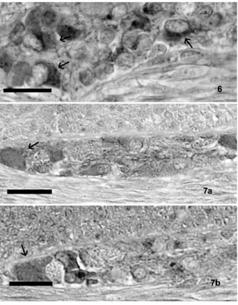
The immunohistochemical techniques revealed intense positivity for both the orexin A and the orex-in type 2 receptor orex-in the neurons and fibers of the enteric nervous system. They were localized in the submucosal and in the muscular layers all along the gastrointestinal tract examined, but with their most evident concentration in the intestine (Figure 6).
Positive neurons appeared isolated or gathered in small or more voluminous groups among the con-nective tissue of the submucosal layer and, in par-ticular, in the duodenum, and in little groups also among the characteristic glands of the submucosa. Some interconnecting orexin A positive nerve fibers were also evident.
Figure 1. A “closed-type” OXA-immunoreactive cell (arrow), with a typical triangular shape, in the epithelium of the stomach. Figure 2. (a) A cell, in the stomach (arrow) was stained with anti-OXA antibody; (b) in the subsequent section the same cell (arrow) stained with the anti-gastrin antibody.
Figure 3. A characteristic “open-type” OXA-containing cell with a typical apical cytoplasmic process (arrow) that reaches the gut lumen. Figure 4. (a) Some cells (arrows), in the duodenum, stained with anti-OXA antibody; (b) In the subsequent section, the same cells (arrows) stained with the anti-serotonin antibody.
Figure 5. A typical “open-type” OX2R-immunoreactive cell, in the duodenum, with a characteristic triangular shape (arrow). Scale bars are for 20 µm.
Further immunohistochemical studies carried out to identify the orexin A positive neurons neuro-chemically seemed to evidence a positive reaction to substance P in some of them, both in the submu-cosal and the muscular layers (Figure 7).
Staining was completely absent in the control sec-tions (data not shown).
Both the number of OXA cells (p<0.001) and OX2R (p<0.001) were influenced by anatomical portions. In particular, the number of the OXA cells was less in rectum than in the stomach (p<0.001) and in duodenum (p<0.001); the difference between stomach and duodenum was not significa-tive. Also the OX2R cells were less in rectum than in stomach (p<0.001), and in duodenum than in stomach (p<0.05). This difference was significant also for the duodenum and rectum (p<0.05) where these cells were less in number.
Both in stomach (p<0.001) and in duodenum (p<0.001) there was significative difference in the number of OXA and OX2R. This also occurs in rec-tum (p<0.05).The variations in the number of OXA and OX2R positive cells in the different tract of gastrointestinal tract are clearly visualized in Figure 8.
Discussion
In the present study, cells positive to OXA and OX2R were identified in the mucosa, submucosa and muscular layers of the alimentary tract in neonatal dogs.
This important result permitted us to identify a peripheral production of orexin A and its receptor, principally localized in several tracts of the gas-trointestinal apparatus also in neonatal dogs, as previously evidenced in horses (Dall’Aglio et al., in press). This allowed us to hypothesize that orexin A has a peripheral action in the digestive apparatus linked to a local production of orexin A and orexin type 2 receptor.
Orexin A containing cells were numerous in the stomach; their number then decreased in the duo-denum. In the gut, immunostained OXA cells were more numerous in the small intestine than in the large one, as occurs in adults in different animal species (Sánchez de Miguel and Burrel, 2002).
They showed morphological features that are typ-ical of endocrine cells: moreover, the presence of serotonin in a large subset of OXA-positive cells in
the duodenum, evidenced by immunohistochemical staining on serial sections, allowed us to consider these cells as entero-chromaffin cells. These results are in agreement with those in humans, laboratory animals (Kirchgessner et al., 1992; Yamamoto et Figure 6. A group of OXA positive neurons (arrows) in the submu-cosa layer of the duodenum.
Figure 7. (a) A group of neurons (arrow), in the duodenal submu-cosa, positive to OXA; (b) In the subsequent section, some of these cells (arrow) stained with anti-substance P antibody. Scale bars are for 20 µm.
Figure 8. Mean number of endocrine cells positive to orexin A and orexin type 2 receptor antibodies, in the dog gastrointestinal tract. 12 10 8 6 4 2 0
Stomach Duodenum Rectus
Orexin A Recpt. Ox A
al., 2000) and in horses (Dall’Aglio et al., in press). Moreover, a large subset of OXA-positive cells in the stomach also contains gastrin and this confirms our finding, that also in neonatal dogs these cells may contain more than one peptide and that these substances may act synergistically, answering to luminal stimuli, to check the digestive functions both acting in synergism in the control of muscular or secretory functions and favouring the secretion of the other peptides (Dall’Aglio et al., in press; Kirchgessner, 2002).
Furthermore, in neonatal animals the co-expres-sion of orexin A with gastrin and serotonin in the enteroendocrine cells could find an explanation in the possible involvement of orexin in gastrointesti-nal development (Zabielski, 2007).
A restricted number of orexin type 2 receptor positive cells was evidenced in the stomach and then in the small and large intestine. In any case, they always followed a decreasing expression from the stomach to the terminal gut tract.
Considerable immunoreactivity for both sub-stances was evidenced in some neurons and in nerv-ous fibers localized in the submucosal and muscu-lar layers, in the different gastrointestinal tracts. The latter observation, even if in disagreement with a recent report that questions the presence of orex-ins in murine and human enteric neurons (Baumann et al., 2007), finds confirmation in numerous reports present in the literature. Nevertheless, the discrepant results between Baumann’s analysis and our immunohistochemistry findings in the canine gastrointestinal tract may be partly due to the dif-ference of the antibody for Orexin A used and/or the species reactivity (Nakabayashi et al., 2003).
Neuron positivity for OXA and OX2R and their pattern of distribution in the gut tract of neonatal dogs are the same as in adult animals of different species, as evidenced in the literature. In humans, in particular, it has been shown that OXA-immunore-activity does not change in the gut neurons from birth to adult (Nakabayashi et al., 2003).
Some of the neurons positive to orexin A seemed to co-store substance P; the latter is a marker of neurons that have been shown to project to the mucosa and to respond to sensory stimuli from the gut (Kirchgessner et al., 1992), carrying informa-tion from the gut lumen to the submucosal myen-teric ganglia. Even if these neurons are a small part of total neurons present in the submucosa of gas-trointestinal tract, they seem to be important to
guide motility and secretion and are essential to start peristaltic activity (Cooke,1998; Gershon et al., 1994). In addition to these data, it is reported in the literature that neurons positive to orexin A were also positive to leptin receptor antibody and this could demonstrate a possible intervention of orexin A on receiving signals coming from the adi-pose tissue, to integrate them with those from the mucosa (Kirchgessner et al., 1992). In any case, at the moment, this datum is not available for dogs but it could be of interest to study it also in this ani-mal species.
In conclusion, the results of the present work show that orexin A and orexin type 2 receptor are present in the gastrointestinal tract of young dogs, in the early stages of their postgestational life. Their distribution is superimposable on that of the mouse at the same stage of life and this suggests that orexin A could be associated with some devel-opmental changes, like weaning, feeding and sleep/wakefulness states (Yamamoto et al., 2000).
Acknowledgments
The authors wish to thank Mrs. G. Mancini for her excellent technical assistance.
References
Al Saffar A, Hellstrom PM, Nylander G. Correlation between peptide YY-induced myoelectric activity and transit of small-intestinal con-tents in rats. Scand J Gastroenterol 1985;20:577-82.
Baumann CR, Clark EL, Pedersen NP, Hecht JL, Scammel TE. Do enteric neurons make hypocretin? Regul Pept 2008;147:1-3. Bernardis LL, Bellinger LL. The lateral hypothalamic area revisited:
ingestive behavior. Neurosci Biobehav Rev. 1996;20:189-287. Calingasan NY, Kitamura N, Yamada J, Oomori Y, Yamashita T.
Immunocytochemical study of the gastroenteropancreatic endocrine cells of the sheep. Acta Anat 1984; 118: 171-80.
Cooke HJ. “Enteric tears”: chloride secretion and its neural regula-tion. New Physiol Sci 1998;13:269-74.
Dall’Aglio C, Pascucci L, Mercati F, Giontella A, Pedini V, Ceccarelli P. Immunohistochemical identification and localization of Orexin A and Orexin type 2 receptor in the horse gastrointestinal tract. Res Vet Sci 2008 (in press)(To authors: please complete, if possible) De Lecea L, Kilduff TS, Peyron C, Gao X-B, Foye PE, Danielson PE.
The hypocretins: Hypothalamus-specific peptides with neuroexcita-tory activity. Proc Natl Acad Sci USA 1998;95:322-7.
Ehrstróm M, Gustafsson T, Finn A, Kirchgessner A, Grybäc P, Jacobsson H. Inhibitory effect of exogenous orexin A on gastric emptying, plasma leptin, and the distribution of orexin and orexin receptors in the gut and pancreas in man. J Clin Endocrinol Metab 2005;90:2370-7.
Gershon MD, Kirchgessner AL, Wade PR. Functional anatomy of the enteric nervous system. In Physiology of the Gastrointestinal Tract, Third Edition, LR Johnson et al., eds Raven Press, New York, 1994; pp. 381-422.
hypocretin (orexin) and neuropeptide Y cells in the rodent and pri-mate hypothalamus: a novel circuit implicated in metabolic and endocrine regulations. J Neurosci 1999;19:1072-87.
Kirchgessner AL. Orexins in the brain-gut axis. Endocr Rev 2002;23: 1-15.
Kirchgessner AL, Liu M. Orexin synthesis and response in the gut. Neuron 1999;24:941-51.
Kirchgessner AL,Tamir H, Gershon MD. Identification and stimulation by serotonin of intrinsic sensory neurons of the submucosal plexus of the guinea-pig gut: activity-induced expression of Fos immunoreac-tivity. J Neurosci 1992;12:235-49.
Kukkonen JP, Holmqvist T, Ammoun S, Åkerman KEO. Functions of the orexinergic/hypocretinergic system. Am J Physiol Cell Physiol 2002; 283:1567-91.
Mendieta-Zéron H, López M, Diéguez C. Gastrointestinal peptides con-trolling body weight homeostasis. Gen Comp Endocr 2008; 155: 481-95.
Nakabayashi M, Suzuki T, Takahashi K, Totsune K, Muramatsu Y, Kaneko C, et al. Orexin-A expression in human peripheral tissues. Mol Cell Endocrinol 2003;205:43-50.
Näslund E, Ehrström M, Ma J, Hellström PM, Kirchgessner AL. Localization and effects of orexin on fasting motility in the rat duo-denum. Am J Physiol Gastrointest Liver Physiol 2002;282:470-9. R Development Core Team. A language and environment for statistical
computing. R Foundation for Statistical Computing, Vienna, Austria, 2007. ISBN 3-900051-07-0, URL http://www.R-project.org
Sakurai T, Amemiya A, Ishii M, Matsuzaki I, Chemelli RM, Tanaka H, Williams SC et al. Orexins and orexin receptors: a family of hypo-thalamic neuropeptides and G protein-coupled receptors that regu-late feeding behavior. Cell 1998;92:573-85.
Sakurai T. Orexins and orexin receptor: implication in feeding behavior. Regul Pept 1999; 85: 25-30.
Sánchez de Miguel MJ, Burrel MA. Immunocytochemical detection of orexin A in endocrine cells of the developing mouse gut. J Histochem Cytochem 2002; 50: 63-9.
Singh I. A modification of the Masson-Hampler method for staining of argentaffin cells. Anat Anz 1964; 115: 517-8.
Smart D, Jerman J. The physiology and pharmacology of the orexins. Pharmacol Ther 2002;94:51-61.
Walsh JH. Gastrointestinal hormones. In Physiology of the Gastrointestinal Tract, Third Edition, LR. Johnson et al., eds Raven Press, New York, 1994, pp. 1-128.
Yamamoto Y, Ueta Y, Hara Y, Serino R, Nomura M, Shibuya I, et al. Postnatal development of orexin/hypocretin in rats. Mol Brain Res 2000;78:108-19.
Zabielski R. Hormonal and neural of intestinal function in pigs. Livest Sci 2007;108:32-40.