Management of uncommon secondary trigeminal neuralgia related to a rare
Arnold Chiari type I malformation
Zafar Ali Khan
1*, Ammar Ahmed Siddiqui
2*, Jamaluddin Syed
3*, Andrea Cicconetti
4, Alessandro
Orefici
4, Giovanni Falisi
5, Francesca Fabiano
6, Luigi Santacroce
7, Patrizio Bollero
8*, and Francesco
Inchingolo
9*1. Department of Oral & Maxillofacial Surgery, College of Dentistry, University of Hail, KSA
2. Department of Dental Public Health, College of dentistry, University of Hail, KSA
3. Advanced Technology Dental Research Laboratory, Department of Oral Basic Sciences, Faculty of
Dentistry, King Abdul Aziz University, Jeddah, KSA
4. Department of Oral and Maxillofacial Sciences, School of Dentistry, Sapienza University, Rome, Italy
5. Department of Life, Health and Environmental Sciences, School of Dentistry, University of L'Aquila,
L'Aquila, Italy
6. Department of Engineering, University of Messina, Messina, Italy
7. Department DISIGEM-Jonian, University of Bari, Italy
8. Unit of Oral Pathology, Department of Systems Medicine, University of Rome “Tor Vergata”, Italy
9. Department of Interdisciplinary Medicine, University of Bari, Italy
*: These authors equally contributed to this article
1072
BRIEF REPORT
Please cite this paper as: Ali Khan Z, Siddiqui AA, Syed J, Cicconetti A, Orefici A, Falisi G, Fabiano F, Santacroce L, Bollero P, Inchingolo F. Management of uncommon secondary trigeminal neuralgia related to a rare Arnold Chiari type I malformation. AMJ 2017;10(12):1072–1076. https://doi.org/10.21767/AMJ.2017.3276
Corresponding Author:
Francesco Inchingolo
Department of Interdisciplinary Medicine University of Bari, Italy
Email: [email protected]
ABSTRACT
Background
Trigeminal neuralgia (TN) may sometimes present secondary to an intra-cranial cause. Arnold Chiari Malformation (ACM) is downward herniation of the cerebellar tonsils through the foramen magnum that may be a cause of TN like pain in very rare cases.
Aims
The aim of this brief report is to suggest the proper
management of uncommon secondary trigeminal neuralgia related to a rare Arnold Chiari type I malformation.
Methods
A male patient presented electric shock like stabbing pain on the right side of the face for more than ten years. The symptoms were typical of trigeminal neuralgia except that there was loss of corneal reflex on the right side and the patient also complained of gait & sleep disturbances. Complex and multilevel diagnosis was made.
Results
A multiplanar imaging through brain acquiring T1/T2W1 revealed ACM Type I Malformation with caudal displacement of cerebellar tonsils through foramen magnum.
Conclusion
Dental surgeons and oral and Maxillofacial Surgeons should exclude intra-cranial causes by Magnetic Resonance Imaging(MRI) in patients of TN presenting with loss of corneal reflex, gait and sleep disturbances due to night time pain episodes.
Key Words
Arnold chiari malformation, trigeminal neuralgia, corneal reflex
1073
Implications for Practice:
1. What is known about this subject?
Trigeminal neuralgia (TN) is most commonly related to vascular compression of the trigeminal nerve. Trigeminal neuralgia associated with Chiari's malformation is rare. 2. What new information is offered in this report?
The severe trigeminal neuralgia could be related to ACM also thanks to the unusual finding of the loss of corneal reflex.
3. What are the implications for research, policy, or practice?
This case highlighted how a correct diagnosis must first exclude the severe intra-cranial pathology in case of TN.
Background
Trigeminal neuralgia (TN) is a chronic condition characterized by pain presenting in brief episodes, in one or more distributions of the trigeminal nerve. Episodes of pain secondary to TN are triggered by specific or not specific stimuli, such as chewing, shaving, or touching the face. A common cause of TN is compression of the trigeminal nerve root entry zone by an artery or vein, many cases of TN are idiopathic. Trigeminal neuralgia (TN) a sudden, usually unilateral, severe, brief, stabbing, recurrent pain in the distribution of one or more branches of the fifth cranial nerve is one of the most painful conditions of the face and has profound impact on quality of life as this prevents the patient from speaking, eating, drinking, touching or washing of the face and brushing teeth.1,2 TN patients often initially presents to dental surgeons who subsequently refer them to Oral & Maxillofacial Surgeons.2 Patients with TN are at times misdiagnosed and undergo misguided, unnecessary procedures and ineffective treatments. Some clinicians consider the diagnosis of TN to be purely history and clinical examination based without the need for any further investigation.1,3
In majority of cases TN may be primary (idiopathic) but rarely may arise secondaryto an intracranial cause.
Typically, TN is diagnosed by the patient’s clinical history, supported by a negative neurologic exam, and after a positive response to a trial of carbamazepine. NMR studies are often useful, especially when the diagnosis is uncertain or neurologic disorders are present. Many conditions are triggered by the compression of the Trigeminal nerve, even if the compression is within a few millimetres before to entry into the pons. Neuroimaging may help in
differentiating between primary and secondary TN by identify an intracranial cause in up to 15 per cent of patients and thus help correct diagnosis and management.4-6
TN may rarely be associated with Arnold Chiari 1 malformation (ACM).7 ACM is a group of complex brain abnormalities traditionally defined as downward herniation of the cerebellar tonsils through the foramen magnum. Diagnosis of ACM with MRI evaluation of posterior cranial fossa is confirmed if the cerebral tips have exceeded 5mm below the foramen magnum. It occurs in about 0.4:1000 live births and has formed 3 per cent of all abortions.8-10
Case details
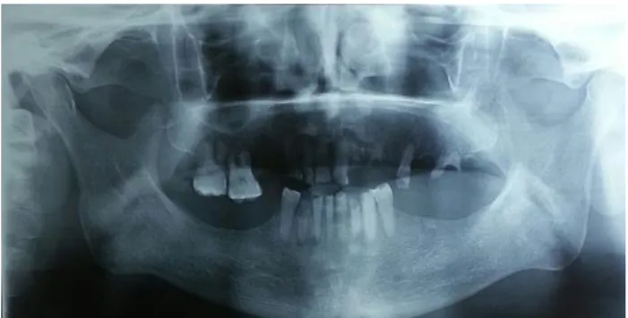
A 65-years-old male presented electric shock like stabbing pain on the right side of the face for more than ten years. He was unable to eat, drink, speak or sleep properly because of the severity and multiple episodes of pain during day and even at night during sleep. He was taking carbamezapine 200mg and gabapentine 300mg twice daily without much relief in pain. On clinical examination there was loss of corneal reflex on the right side and the patient also complained of gait disturbances. On intra oral examination he had lost most of his teeth because of his inability to maintain proper oral hygiene due to the triggering of pain while brushing. He lost some of his teeth due to the TN pain mimicking as a tooth ache during the early stages of his disease as shown in his panoramic radiograph (Figure 1).
Based on history and clinical examination, secondary TN was suspected, and an MRI brain scan was an advised to identify probable intra cranial cause for the pain. A multiplanar imaging through brain acquiring T1/T2W1 revealed caudal displacement of cerebellar tonsils through foramen magnum (Figure 2). There was compression of cervico-medullary junction with syrinx in the cervical cord suggesting Arnold Chiari malformation type I (Figure 3). The patient was subsequently referred to the neurosurgery department for the decompression (occipital craniectomy and laminectomy).
Discussion
Chiari malformations (CM) named after Hans Chiari, an Austrian pathologist, who first identified this pathology 1891. After that Ulius Arnold further elaborated on malformation it became now to be known as Arnold-Chiari malformation (ACM). ACM is classified into four types.9-11 Specifically, type 1 is characterized by herniation of cerebellar tonsils alone, radiologically as simple tonsillar
1074
herniation 5mm or greater, below the foramen magnum.9-12Presentation of TN secondary to ACM is extremely rare as a very small number of such cases have been reported in scientific literature. In this specific clinical report, we first describe a TN secondary to ACM in whole country of Pakistan. The pathophysiology of TN secondary to ACM is hypothesized to be due to a variety of problems that may include vascular compression at the nerve root entry zone (NREZ) of fifth cranial nerve, demyelination of the trigeminal NREZ, micro-ischemic changes and direct brainstem compression.13 In ACM spinal tract of the trigeminal nucleus is more susceptible as it is located dorsally and is poorly myelinated.14,15 A variety of treatment options are available for TN secondary to ACM that may include microvascular decompression (MVD), retro-sigmoid craniotomy, endoscopic third ventriculostomy and ventricular shunt procedures.14-18 Craniocervical decompression has been reported with 73 per cent resolution of pain symptoms.15 It could be interesting to approach future therapies by using of mesenchymal stem cells, given their paracrine immunoregulatory effects.19 Several risk factors should be also avoided in TN, as some factors could be involved in the processes of cytopathic hypoxia and of cellular oxidative stress.20-23 Finally, the diagnosis must even consider, and exclude, oncological issues,24,25 syndromic conditions especially those related to oral and maxillofacial regions,26 or micro- and macro-trauma, eventually treated with traditional or innovative autologous scaffolds.27-30
Conclusion
Chiari I malformation usually presents with headache, numbness, weakness and gait abnormalities. This case report is an addition to the very few reported cases of TN secondary to ACM. Dental surgeons and oral & Maxillofacial Surgeons should consider this diagnosis in patients of TN presenting with loss of corneal reflex, gait and sleep disturbances due to night time pain episodes; the pathophysiology of TN in these cases may be due to neurovascular conflict. MRI in all patients presenting with TN is mandatory, which may provide valuable etiological information, prevent the progression of the disease, and avoid erroneous diagnosis and unsuitable treatment interventions. This report supports early surgical intervention for symptomatic patients to achieve long-term surgical benefit.
References
1. Ko AL, Lee A, Raslan AM, et al. Trigeminal neuralgia without neurovascular compression presents earlier
than trigeminal neuralgia with neurovascular compression. J Neurosurg. 2015;123(6):1519–27. doi: 10.3171/2014.11.JNS141741.
2. Zakrzewska JM. Differential diagnosis of facial pain and guidelines for management. Br J Anaesth. 2013;111(1):95–104. doi: 10.1093/bja/aet125.
3. Wang Y, Zhang S, Li P, et al. Gamma Knife Surgery for Recurrent Trigeminal Neuralgia in Cases with Previous Microvascular Decompression. World Neurosurg. 2017. doi: 10.1016/j.wneu.2017.11.062.
4. Khan M, Nishi SE, Hassan SN, et al. Trigeminal Neuralgia, Glossopharyngeal Neuralgia, and Myofascial Pain Dysfunction Syndrome: An Update. Pain Res Manag. 2017;2017:7438326. doi: 10.1155/2017/7438326. 5. Toller MO, Uzun E, Incesu L. Clinical and magnetic
resonance imaging evaluation of facial pain. Oral Surg Oral Med Oral Pathol Oral Radiol Endod. 2004;97:652–8. DOI: 10.1016/S1079210403005997
6. Gronseth G, Cruccu G, Alksne J, et al. Practice Parameter: The diagnostic evaluation and treatment of trigeminal neuralgia (an evidence-based review). Neurology. 2008;71(15):1183–1190. doi: 10.1212/01.wnl.0000326598.83183.04.
7. Loch-Wilkinson T, Tsimiklis C, Santoreneos S. Trigeminal neuralgia associated with Chiari 1 malformation: symptom resolution following craniocervical decompression and duroplasty: Case report and review of the literature. Surg Neurol Int. 2015;6:S327–9. doi: 10.4103/2152-7806.161407.
8. Langridge B, Phillips E, Choi D. Chiari Malformation Type 1: A Systematic Review of Natural History and Conservative Management. World Neurosurg. 2017;104:213–219. doi: 10.1016/j.wneu.2017.04.082. 9. Nagy L, Mobley J, Ray C. Familial Aggregation of Chiari
Malformation: Presentation, Pedigree, and Review of the Literature. Turk Neurosurg. 2016;26(2):315–20. doi: 10.5137/1019-5149.JTN.10471-14.1.
10. Milhorat TM, Nishikawa M, Kula RW, et al. Mechanisms of cerebellar tonsil herniation in patients with Chiari malformations as a guide to clinical management. Acta Neurochir. 2010;152:1117–1127. doi: 10.1007/s00701-010-0636-3.
11. Galarza M, Lopez-Guerrero AL, Martinez-Lage JF. Posterior fossa arachnoid cysts and cerebellar tonsillar descent: short review. Neurosurg Rev. 2010;33:305–314. doi: 10.1007/s10143-010-0262-9.
12. Elster AD, Chen MY. Chiari I malformations: clinical and radiologic reappraisal. Radiology. 1992;183(2):347–53. PMID: 1561334
13. Papanastassiou AM, Schwartz RB, Friedlander RM. Chiari I malformation as a cause of trigeminal neuralgia: Case
1075
report. Neurosurgery. 2008;63:E614–5. doi:10.1227/01.NEU.0000324726.93370.5C.
14. González-Bonet LG, Piquer J. Trigeminal Neuralgia: A Symptom of Chiari I. Neurosurgery. 2012;71:911–2. DOI: 10.1227/NEU.0b013e318266214b
15. Than KD, Sharifpour M, Wang AC, et al. Chiari I malformation manifesting as bilateral trigeminal neuralgia: Case report and review of the literature. J Neurol Neurosurg Psychiatry. 2011;82:1058–9. doi: 10.1136/jnnp.2009.196121
16. Vince GH, Bendszus M, Westermaier T, et al. Bilateral trigeminal neuralgia associated with Chiari’s type I malformation. Br J Neurosurg. 2010;24:474–6. doi: 10.3109/02688691003587421.
17. Teo C, Nakaji P, Serisier D, et al. Resolution of trigeminal neuralgia following third ventriculostomy for hydrocephalus associated with chiari I malformation: Case report. Minim Invasive Neurosurg. 2005;48:302–5. doi: 10.1055/s-2005-915597.
18. Gnanalingham K, Joshi SM, Lopez B, et al. Trigeminal neuralgia secondary to Chiari’s malformation treatment with ventriculoperitoneal shunt. Surg Neurol. 2005;63:586–8. PMID: 15936398.
19. Tatullo M, Falisi G, Amantea M, et al. Dental Pulp Stem Cells and Human Periapical Cyst Mesenchymal Stem Cells In Bone Tissue Regeneration: Comparison Of Basal And Osteogenic Differentiated Gene Expression Of A Newly Discovered Mesenchymal Stem Cell Lineage. J Biol Regul Homeost Agents. 2015;29(3):713–8. PMID: 26403412.
20. Tatullo M, Gentile S, Paduano F, et al. Crosstalk between oral and general health status in e-smokers. Medicine (Baltimore). 2016;95(49):e5589. doi: 10.1097/MD.0000000000005589
21. Tatullo M, Simone GM, Tarullo F, et al. Antioxidant and Antitumor Activity of a Bioactive Polyphenolic Fraction Isolated from the Brewing Process. Sci Rep. 2016;6:36042. doi: 10.1038/srep36042
22. Marrelli M, Gentile S, Palmieri F, et al. Correlation between Surgeon's experience, surgery complexity and the alteration of stress related physiological parameters. PLoS One. 2014;9(11):e112444. doi: 10.1371/journal.pone.0112444.
23. Tatullo M, Marrelli M, Scacco S, et al. Relationship between oxidative stress and "burning mouth syndrome" in female patients: a scientific hypothesis. Eur Rev Med Pharmacol Sci. 2012;16(9):1218–21. PMID: 23047505
24. Tatullo M, Marrelli M, Amantea M, et al. Bioimpedance Detection of Oral Lichen Planus Used as Preneoplastic Model. J Cancer. 2015;6(10):976–83. doi:
10.7150/jca.11936. eCollection 2015.
25. Inchingolo F, Tatullo M, Abenavoli FM, et al. Non-Hodgkin lymphoma affecting the tongue: unusual intra-oral location. Head Neck Oncol. 2011;3:1. doi: 10.1186/1758-3284-3-1.
26. Inchingolo F, Tatullo M, Abenavoli FM, et al. Non-syndromic multiple supernumerary teeth in a family unit with a normal karyotype: case report. Int J Med Sci. 2010;7(6):378–84 doi:10.7150/ijms.7.378
27. Aulino P, Costa A, Chiaravalloti E, et al. Muscle extracellular matrix scaffold is a multipotent environment. Int J Med Sci. 2015;12(4):336–40. doi: 10.7150/ijms.10761
28. Perniconi B, Coletti D, Aulino P, et al. Muscle acellular scaffold as a biomaterial: effects on C2C12 cell differentiation and interaction with the murine host environment. Front Physiol. 2014;5:354. doi: 10.3389/fphys.2014.00354.
29. Marrelli M, Pujia A, Palmieri F, et al. Innovative approach for the in vitro research on biomedical scaffolds designed and customized with CAD-CAM technology. Int J Immunopathol Pharmacol. 2016;29(4):778–783. doi: 10.1177/0394632016646121
30. Paduano F, Marrelli M, Amantea M, et al. Adipose Tissue as a Strategic Source of Mesenchymal Stem Cells in Bone Regeneration: A Topical Review on the Most Promising Craniomaxillofacial Applications. Int J Mol Sci. 2017;18(10). doi: 10.3390/ijms18102140.
ACKNOWLEDGEMENTS
None
PEER REVIEW
Not commissioned. Peer reviewed.
CONFLICTS OF INTEREST
The authors declare that they have no competing interests.
FUNDING
None
ETHICS COMMITTEE APPROVAL
This study was approved by internal committee of Department of Oral & Maxillofacial Surgery, College of dentistry, University of Hail, KSA.
1076
Figure 1: The Patient lost some of his teeth due to the TNpain mimicking as a tooth ache during the early stages of his disease as shown in his panoramic radiograph
Figure 2: MRI Brain showing caudal displacement of cerebellar tonsils through foramen magnum
Figure 3: Compression of cervico-medullary junction with syrinx in the cervical cord suggesting Arnold Chiari malformation type I