1
UNIVERSITY OF NAPLES
“FEDERICO II
”
PhD in Chemical Sciences
XXXI Cycle
2015-2018
Eumelanin exploitation in bioelectronics:
ionic-electronic charge transport in eumelanin
organic thin films
Dr Ludovico Migliaccio
Supervisors
Dr Alessandro Pezzella (University Tutor)
Dr Paolo Tassini (ENEA Tutor)
Assessor PhD Coordinator Prof. Antonio Roviello Prof. Luigi Paduano
2
Index
Aknowledgements ... 4 Abstract ... 6 1 Introduction ... 14 1.1 Introduction on melanins ... 14 1.2 Interest in melanins ... 16 1.3 Synthesis of eumelanins ... 181.4 Eumelanin physicochemical properties ... 21
1.5 Eumelanin-based organic (bio) electronics ... 23
1.6 Thin film fabrication ... 25
1.7 References ... 28
2 Integration of Eumelanin within Conductive Polymers ... 40
2.1 Introduction ... 40
2.2 Pigment functionalization: tailoring Eumelanin conductivity properties by hybrid buildup ... 44
2.3 Eumelanin integration within PEDOT:PSS conductive polymer and structural organization... 48
2.4 Exploitation of Eumelanin-PEDOT:PSS as polymeric anode for the design and fabrication of ITO-free OLED ... 63
3
3 Enhancement of electrical conductivity of Eumelanin through vacuum
processes ... 91
3.1 Introduction on high vacuum annealed DHI Eumelanin ... 91
3.2 High vacuum annealing of DHI Eumelanin thin films: synthesis and characterization ... 93
3.3 References ... 120
4 Eumelanin as a photocatalytically-active bio-material... 124
4.1 Introduction ... 124
4.2 Stable Photocatalyst for oxygen reduction reaction (ORR) to hydrogen peroxide ... 127
4.3 Eumelanin as Photoelectrocatalyst for efficient hydrogen peroxide photosynthesis ... 135
4.4 Application of DHI Eumelanin for hydrogen evolution reaction ... 139
4.5 Experimental section ... 143
4.6 References ... 146
Conclusions and future directions 151 List of Publications ... 153
List of communications at conferences ... 156
List of schools, seminaries and courses ... 158
4
Aknowledgements
First of all I would like to thank my two tireless supervisors, Alessandro Pezzella and Paolo Tassini without whom every single experiment would have been difficult and with complicated scientific answers. I still know I have to learn a lot from them, but during these years I have tried to steal a lot of ideas that I hope will serve me for my future as a researcher. Furthermore, it is a duty to thank Mariagrazia Maglione and Paola Manini who also helped me in situations that were outside the experiments related to my PhD topic and that were always on the front line during the entire journey.
A small parenthesis of my Ph.D, which turned out to be a modification of my life, was the experience as a visiting student at the LOE group in Norrköping (Sweden), for which opportunity I thank Alessandro Pezzella who believed in me, and it is also a duty to thank Eric Głowacki, a big scientist and also a good friend, who was my supervisor during this period, and his group that welcomed me and considered me an integral part of the team. For all the things that they have done, it is more than fair to thank them individually, so I would like to thank Vedran Đerek, one of the most meticulous scientists I have ever met in my path, practically a “factotum” with an uncommon brain. I would also like to thank Marie Jakešová, a true friend and a wonderful person, not only scientifically but also humanly. And I also thank Maciej Gryszel, my experiment collegue, with whom, despite the countless moments of desolation, we were also able to laugh during the experiments. It was a really nice period that made me stronger. It was not easy for me to fight against the cold, the snow, the dark and so on, for me that if I look out of the office I see the sea, but it helped me to improve myself because I am of the opinion that before being a good scientist, you have to be a good person and appreciate the things you have in front of you. For this I would like to thank the biggest piece of my heart, my "Bella famiglia", that has been beside me from the happiest days to the saddest
5 ones of this journey. It was not easy for them to fight against my ugly behaviour resulting from days of inconclusive experiments, but they never made me weigh and I take this opportunity to apologize to them of my attitude. But one of the best moments of the day was to come back home, my fortress, with my trusted and loved people, so I would like to thank them by giving a few lines from the opening speech of the film "Patch Adams", which I saw when I was in Sweden and immediately I shared that thought and meaning attributed to home and in my case family:
“All of life is a coming home. Salesmen, secretaries, coal miners, beekeepers, sword swallowers, all of us. All the restless hearts of the world, all trying to find a way home. It's hard to describe what I felt like then. Picture yourself walking for days in the driving snow; you don't even know you're walking in circles. The heaviness of your legs in the drifts, your shouts disappearing into the wind. How small you can feel, and how far away home can be. Home. The dictionary defines it as both a place of origin and a goal or destination. And the storm? The storm was all in my mind. Or as the poet Dante put it: In the middle of the journey of my life, I found myself in a dark wood, for I had lost the right path. Eventually I would find the right path, but in the most unlikely place.”
Of course I would like to thank my old date friends, Antonio, Carlo and Nicola who have always supported me in everything I have done, believing in me and strengthening my constancy and audacity in my activities.
Finally I thank "you", THE MUSIC, the perfect accompanying lady of every single moment of my life. I consider myself to be a rather solitary person, but the music and the Guitar have softened me and they are a company that never tires, from racing to take trains, to long walks back home, to air flights, to countless experiments.
6
Abstract
Eumelanin, the mammal pigment originating via the oxidative polymerization of 5,6-dihydroxyindole (DHI) and/or 5,6-dihydroxyindole-2 carboxylic acid (DHICA), for more than forty years have been the focus of uncommon interest in biomedical research due to their central relevance to skin and eye photoprotection.
During the last two decades, several peculiar properties of eumelanin-inspired polymers have attracted the attention of the materials science community too, following the realization of device quality thin films through controlled polymerization of DHI and derivatives. The properties of these materials include: a) broadband optical absorption in the visible range; b) efficient UV-dissipative mechanisms; c) photoconductivity in the solid state; d) electronic-ionic hybrid conduction properties.
Anyway, despite a burst of interest in the use of synthetic eumelanin for organic electronics and bioelectronics, the implementation of competitive eumelanin based technology has so far been hindered by several drawbacks. These ones include: complete insolubility in all solvents, preventing the development of standardized and reproducible synthetic procedures, low conductivity, and the lack of a solid
7 conceptual frame of structure–property–function relationships. In particular, the high molecular heterogeneity of the eumelanin, whose impact on their electrical performances has not been still systematically assessed, generates several critical consequences like, for example, the lack of well-defined HOMO–LUMO gaps.
This PhD project aims to identify Structure Property Function relationships in order to propose a mechanistic model describing the ionic-electronic charge transport in eumelanin thin films and finally fabricate eumelanin based devices.
In Chapter 1, is described an overview related to the physico-chemical properties of Eumelanin and the different methods used to obtain it , and this chapter will present also a description related to the recent explosion of interest for Eumelanin applications in bio-electronics and organic-electronics fields that will be one of the main focus with which this thesis will deal.
In Chapter 2, to increase the eumelanin conductivity performances, hybrid materials were designed, adding eumelanin to conductive materials (in this case, PEDOT:PSS (poly(3,4-ethylenedioxythiophene):polystyrene sulfonate)). Resulting material properties have been studied through spectroscopic characterizations, and by means of X-rays, SEM, UV-VIS, contact angle, profilometry, and electrical measurements on different devices architectures.
Figure 1: Part of the instruments used to characterize material properties
Contact angle Scanning electron
microscopy Four point probe
UV/VIS Spectrophotometer
Optical profilometer
Contact profilometer Probe station X-Rays Process chamber for thin film
8 Thin films are ideal candidates to be studied, because they are easily accessible to chemical and morphological characterizations and potentially susceptible to device applications. The first step in the PhD study has involved the design and characterization of hybrids made from blends of Clevios™ PEDOT:PSS PH1000, a conductive polymer, and melanin pigments. The aim of this blend is to have a material which is conductive or satisfactorily conductive as the PEDOT:PSS and, at the same time, resistant to the possible conditions under which the PEDOT: PSS is unstable, for example, exposure to water and other degrading agents such as oxygen. The melanin is able to ensure factors such as stability to degrading agents, but also adhesion to the substrate, which does not make the PEDOT:PSS.
The designed and prepared blends differ in the variable DHI / PEDOT:PSS ratio; the solvent used for the solubilization of the DHI was found to be the propan-2-ol. The blends were prepared in the presence and in the absence of a secondary dopant. A primary dopant differs from a secondary dopant (better defined as “additive”) since the former has a reversible effect, while the effect of the secondary doping is permanent and remains even when the additive is removed. Here, the agent of the secondary doping was dimethylsulfoxide (DMSO).
Capitalizing on a recently developed protocol to prepare high quality eumelanin coatings, herein it is reported the design and the integration of standard commercial PEDOT:PSS with eumelanin pigment (see chapter 2). During this study, were prepared different blends with different DHI/PEDOT ratios in order to understand and define an electrical conductivity trend, with eumelanin content increase in the chemical system. The study of electrical properties of these blends was mandatory in order to define and satisfy a good blend able to work as polymeric anode in an Organic light emitting diode device (OLED) that is one of the ideas of application of this material for organic-electronics and bio-electronics purposes. The blend used is the one with DHI/PEDOT ratio = 0.4 w/w. This blend has allowed to get water stable transparent thin films, capable to operate as electrodes for organic devices, complementing the PEDOT:PSS electronic conductivity with the peculiar
9 eumelanin properties, including adhesion, water stability and ionic-electronic conductivity.
As a proof of concept, an unprecedented ITO (Indium tin oxide)-free organic light emitting diode (OLED) implementing an eumelanin-PEDOT layer as the anode was fabricated and characterized. To the best of our knowledge, this is the first evidences of an OLED device based on an anode layer integrating eumelanin. To prepare the mixture of eumelanin and PEDOT:PSS (Eu-PH), a protocol involving in situ eumelanin generation was designed. This approach allowed not only to circumvent the actual insolubility of eumelanin in any solvent, but also to gain over the PEDOT molecular organization. The target was to obtain a material with good conductivity (not less than 300 S*cm-1) and a work function at least comparable with the ITO (whose commonly reported around 4.4÷4.7 eV) or larger for an efficient hole injection, to be effectively used as the anodic material in organic electronic devices. The Eu-PH thin films were thus obtained by exposing the DHI-PEDOT:PSS films, obtained via spin-coating onto glass substrates, for 1 h to air-equilibrated gaseous ammonia (AISSP (Ammonia induced solid state polymerization) protocol)), from an ammonia solution (28% in water) inside a sealed chamber at 1 atm pressure and at controlled temperature (25°C÷ 40°C). For the OLEDs, a hole injection layer (HIL), CLEVIOS PEDOT:PSS PVP Al 4083, used as-is, was deposited via dip-coating (thickness 90 nm). Final steps of the OLEDs fabrication were carried out under inert atmosphere. All the substrates were loaded into a glove box system (N2 atmosphere, O2 < 1 ppm, H2O < 1 ppm),
and transferred into an in-line evaporation chamber. The active area of the devices was 7 x 10⁻⁶ m2. The final OLED architecture includes: the glass substrate; the anode (ITO; or CLEVIOS PH 1000; or Eu-PH); the HIL; the HTL; the ETL-EML and the cathode.
10 Figure 2: Schematic section of the OLED devices prepared
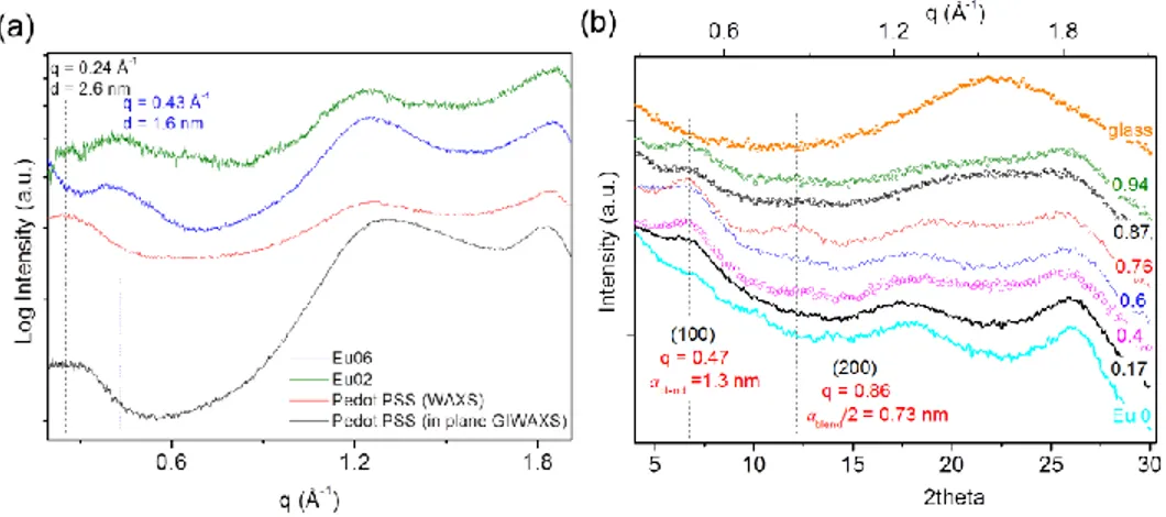
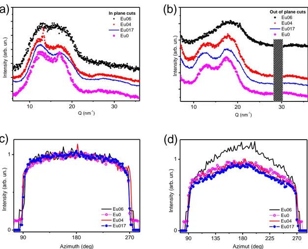
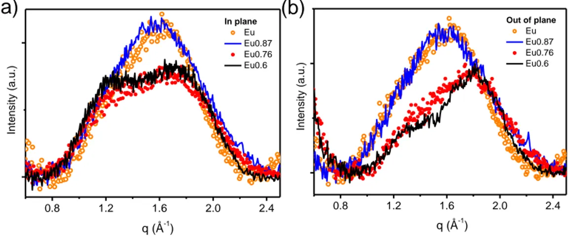
Because a more in-depth investigation is required to fully address the eumelanin-PEDOT integration, the effect of the eumelanin on conductivity of the blends was thus related to structural features observed in Wide Angle X-ray Scattering (WAXS) and X-ray diffraction (XRD) patterns of films with different eumelanin content and fixed PEDOT:PSS ratio. Analysis of free-standing films in transmission geometry (WAXS) allowed to get rid of substrate scattering contribution, and mainly probe crystallographic directions parallel to the film plane. On the other hand, reflection geometry (XRD, GIWAXS) allowed probing structural order in both directions, in and out of the film plane, and can be used for the as-prepared films, laying on glass substrates, to better relate structural and electrical properties. The effect of the eumelanin integration within PEDOT:PSS layers was investigated in terms of the changes in the hierarchical structure of the PEDOT:PSS films. The results of the X-ray scattering characterization clearly demonstrate that the presence of the eumelanin affects the PEDOT component of the blend, inducing an overall increase of the crystalline order at low eumelanin contents. This effect is associated with a smaller distance between the PEDOT chains. Moreover, when eumelanin percent content increases, a less steep decay of the blend conductivity was observed, than the one expected on the basis of the data reported on the conductivity of PEDOT ternary mixtures. At the same time, the introduction of the eumelanin gives noteworthy properties to the Eu-PH blend, including a strong adhesion on inorganic substrates, and water stability, which open
11 to an efficient exploitation of the Eu-PH for conductive coatings within biointerfaces for application in bioelectronics.
As said, higher values of the eumelanin electrical conductivity are needed for applications in organic electronics, thus several studies explored the integration of the pigment with conductive materials. But, these approaches actually modify the chemistry of the layers. Other approaches also exploited severe modifications of the eumelanin-like materials to gain a graphene-like material, as for example by pyrolitic treatment of polydopamine under hydrogen atmosphere.
The mechanism of charge transport in eumelanin is still not fully clear, but several evidences are concurring to sustain a hybrid ionic-electronic behaviour, where the electronic contribution depends on the presence, extent and the redox properties of the delocalized aromatic systems, while the ionic part is largely dictated by the hydration level of the pigment (i.e. humidity in the measuring environment). Basing on the concurring evidences disclosing the correlation between the chemical physical properties of the eumelanins and the polyindole -system staking, as well as the packing of the molecular constituents within the pigment, we speculated about the modulation of the electronic conductivity, by acting on the polyindole packing in eumelanin thin films. Here (Chapter 3), preparation and characterization of eumelanin thin films showing conductivity up to 318 S/cm are discussed. Highly conductive films were prepared via the oxidative polymerization of films of 5,6-dihydroxyindole (DHI), the ultimate monomer precursor in the formation pathways of natural and synthetic eumelanins, then treated under high vacuum thermal annealing. We name the obtained material as High Vacuum Annealed Eumelanin, HVAE.
During the 3rd year of the PhD course, I have had a research visiting period in the Laboratory of Organic Electronics of the University of Linköping in Norrköping under the supervision of Dr. Eric Daniel Głowacki. During this
12 time, I worked on a project concerning the photo(electrochemical) properties of the eumelanin. We report (see Chapter 4) that eumelanin is a photo-catalytic material. Though photoconductivity of eumelanin and its photochemical reactions with oxygen have been known for some time, eumelanins have not been regarded as photofaradaic materials. We found that eumelanin shows photocathodic behaviour for both the oxygen reduction reaction and the hydrogen evolution reaction. Eumelanin films irradiated in aqueous solutions at pH 2 or 7 with simulated solar light, photochemically reduce oxygen to hydrogen peroxide with accompanying oxidation of sacrificial oxalate, formate, or phenol. Auto-oxidation of the eumelanin competes with oxidation of donors. Deposition of thin films on electrodes yields photoelectrodes with higher photocatalytic stability, compared with the case of pure photocatalysis, implicating the successful transport and extraction of holes from the eumelanin layer. These results open up new potential applications for eumelanin as a photocatalytically-active biomaterial, and inform the growing fundamental body of knowledge about the physical chemistry of the eumelanin.
From all of this work, as result of this finding, we demonstrate new potentialities of Eumelanin in organic-electronics and bio-electronics field, developing new methods and new recipes either to increase electrical conductivity in order to exploit this material for several applications and purposes either to use it as photocatalytically-active material. Although a conclusive picture about the conductor vs semiconductor behavior of the eumelanins and insights about the mobility of charge carriers will require further investigations, results related to annealed thin films of Eumelanin, here reported, radically modify the actual picture of the eumelanin charge transport properties, reversing the paradigm according to which eumelanin conductivity increases with the water content of the pigment. Indeed, when
13 eumelanin molecular constituents are rearranged in conductive layers, the contribution of electronic current is demonstrated to be largely preeminent with respect to the ionic one, allowing to get unprecedented conductivity and to consider the mammalian pigment as an actual conductor.
14
1. Introduction
1.1 Introduction on melanins
The term Melanins (μέλας = black)1 has been used since 1840 to denote the broad class of pigments found throughout nature, from human beings to invertebrates, plants and fungi.
Melanin is a predominantly indolic macromolecule2. There are many different types of melanin, including eumelanin, pheomelanin, neuromelanin and allomelanin. Eumelanin and pheomelanin are both found in the skin, hair and eyes of many animal species, including humans, where they act as photo-protectants (absorbing harmful ultraviolet and visible radiation).
Eumelanin is known to be a macromolecule built up from 5,6-dihydroxyindole (DHI) and 5,6-5,6-dihydroxyindole-2-carboxylic acid (DHICA), and is black to brown in colour3. Eumelanin is the most extensively studied of all melanins since it is the primary pigment found in human skin, although it also forms the major component of squid ink and is responsible for the dark colouration in feathers4. Pheomelanin is a sulfur containing macromolecule composed of 1,4-benzothiazine units, and is red to yellow in colour (pheomelanin is responsible for the colouration of human red hair and chicken feathers5, 6). This thesis deals essentially with eumelanins.
The other varieties of melanins (including allomelanins and neuromelanins) will not be described in this thesis, but briefly allomelanins are pigments
15 nitrogen free found largely in plants such as certain fungi and seeds, and also in soil. Neuromelanins are found in the brainstem and inner ear of humans and higher primates where its role is unknown, although it is thought to have some biological significance; neuromelanin is decreased or absent in individuals with Parkinson’s disease, for example7, 8. It is not clear whether the relationship between neuromelanin and Parkinson’s disease is causal, although it is suggested that the pigment might modulate neurotoxic processes through interaction through iron, binding of drugs or reaction with free radicals and free radical producing species9. Additionally, albinism often leads to deafness in animals, suggesting biological functionality10.
16
1.2 Interest in melanins
Paradoxically, although melanin is a photoprotectant, it has also been implicated in the chain of events that lead to malignant melanoma skin cancer11-13, although this link is very poorly understood. Highly pigmented skin is more protected from carcinogenesis than un-pigmented skin14, but it has been suggested that pheomelanin may actually function as a photosensitizer15, and has been shown to actually enhance DNA damage in cells in response to ultraviolet radiation16, 17. The skin is the most common site of cancer in humans14, and although melanoma is one of the rarer types of skin cancer, it causes the majority of skin cancer related deaths18. According to the World Heath Organisation, approximately 48,000 melanoma related deaths occur each year19. This makes understanding the biological functionality of melanin and its role in melanoma a health priority, particularly for countries with high levels of solar radiation such as Australia. Melanin also plays a central role in a variety of highly visible and inconvenient pigmentary disorders such as albinism and vitiligo20.
Melanin is also of interest as a model system for understanding disorder in biological systems. Disorder is thought to be an essential part of the biological functionality of melanin, which is unlike other much more thoroughly studied biomolecular systems such as DNA and proteins. Disordered systems in biology have yet to receive a great deal of attention, likely due to the difficulties inherent in studying them. This makes melanin a fascinating novel system with the potential for development of techniques applicable to a wide range of important biosystems21-23.
As a third point of interest, melanins exhibit interesting physical and chemical properties such as anti-oxidant and free-radical scavenging
17 behaviour, metal and drug binding properties24-27, broad band ultraviolet and visible absorption and strong non-radiative relaxation of photo-excited electronic states28. These physical chemical properties, together with the natural pigment bio-compatibility, are the basis for the great interest in the realization of eumelanin based electronic devices. Possible examples include highly efficient broadband single photon counters (superconductor based bolometers with a thin eumelanin film as the sensitising pigment), extremely sensitive humidity sensors (based on the extreme sensitivity of eumelanin solid state conductivity to hydration), organic semiconductor electronics and solar cells29, 30. Despite a burst of interest in the use of synthetic eumelanins and related biopolymers for organic electronics and bioelectronics, the implementation of competitive eumelanin based technology has so far been hindered by several drawbacks, expecially related to the low conductivity of the material. One of the main focuses with which this thesis will deal is thus related to the possibility to modulate the electrical conductivity of this bio-polymer in order to make it suitable for its application as material for devices fabrication.
18
1.3 Synthesis of eumelanins
Biochemically, eumelanin is derived from tyrosine in a reaction catalysed by the enzyme tyrosinase, as outlined in the scheme depicted in scheme 1.1. Analysis of this synthetic process and final eumelanin structure is extremely difficult due to the highly unstable nature of the reaction intermediates, and extremely stable and insoluble nature of the final product31. Nevertheless, intermediates have been identified, largely due to the pioneering work of Raper and Mason32. The enzyme tyrosinase acts on dopa and tyrosine to produce dopaquinone, which reacts to form dopachrome (a red compound). Dopachrome is rather stable, but will spontaneously decompose to form DHI (5,6-dihydroxyindole), giving off CO2. If the enzyme dopachrome
tautomerase is present (Dct), dopachrome will instead tautomerise to give mostly DHICA (5,6-dihydroxyindole-2-carboxylic acid), retaining the carboxylic acid group33. Hence the availability of Dct will determine the relative amounts of DHI and DHICA produced, and therefore the ratio of these components in the final eumelanin macromolecule. This ratio will also be affected by other enviromental factors, and hence varies widely depending upon the source of the eumelanin under study34. It is widely accepted that eumelanin is a heterogeneous macromolecule of DHI and DHICA. Melanin must be extracted from biological systems for study, and the extensive amount of bound protein must be removed. This is often achieved via harsh processes such as acid/base treatment, which is known to severly disrupt the properties of the pigment35.
Much milder enzymatic methods are also available to extract melanin from hair, skin and eye tissue yielding melanin that is closer to its natural state36. It has become common practice to use eumelanin extracted from the ink sac of
19 the cuttlefish Sepia officinalis as a model eumelanin pigment since it is readily available and easily extracted via mild techniques which yield a uniform black pigment that is appropriate for use as a standard37. Material that is termed ‘synthetic melanin’ is also often used as a standard for natural melanins. Synthetic melanin is formed under a variety of conditions in vitro including:
• Biomimetic conditions - oxidation by DHI with enzyme tyrosinase in aqueous buffer at neutral pH. This would seem to be the best synthetic method since it is closest to the natural system, but unexpectedly eumelanin formation occurs very slowly under these conditions, even in the presence of large amounts of enzyme38. Additionally, it is challenging to control and measure the activity of tyrosinase during this reaction, giving poor reproducibility3. Enzymatic preparations may also be performed with the alternate substrates of dopa and tyrosine.
• Autoxidative dopa eumelanin may be prepared by simply allowing alkaline solutions of dopa to come into contact with air. The solution may then be acidified to precipitate the eumelanin31.
• A peroxidase/H2O2 couple induces a complete and rapid conversion of DHI to
eumelanin in aqueous buffer at biomimetic pHs39. Similarly, a synthetic eumelanin is commercially available that has been prepared by persulfate or peroxide oxidation of tyrosine31.
The resulting black pigments show great similarity to natural eumelanin, although proof that they are identical to natural eumelanin remains lacking3.
Synthetic eumelanin is convenient as a model system for pioneering new methods or theories to treat this difficult material, and since they are produced under controlled conditions and contain known monomeric units they offer advantages for analysis over the more complex naturally extracted melanins. Throughout this thesis we use DHI eumelanin as a synthetic analogue for natural eumelanin in the way to be able to circumvent the
20 multiple problems related with the low processability of the Eumelanin following the monomer route40 as will be described in 1.5.
Scheme 1.1. Schematic view of eumelanin synthesis from tyrosine or dopa.
21
1.4 Eumelanin physicochemical properties
Working on eumelanins has usually been regarded as an intriguing, though experience40. This is due to several challenging features of the system, including almost complete insolubility in all solvents, an amorphous particulate character, and extreme molecular heterogeneity. Eumelanin does however possess a number of physicochemical properties28 that can be used to identify and quantify the system, such as a persistent electron paramagnetic resonance (EPR) signal, broadband monotonic optical absorption, peculiar excitation and emission properties41, 42, and time dependent photodynamics43-45.Standard vibrational methods such as infrared absorption and Raman spectroscopy46, 47 and more recently inelastic neutron scattering spectroscopy48 have also been applied with varying degrees of success to study the vibrational finger-print of eumelanin precursors. Controlled chemical degradation giving traces of pyrrolic acids has been exploited mainly for pigment analysis in tissues49, 50 yielding only limited information as to the basic aspects of eumelanin primary-level structure3. Yet to-date eumelanins fundamental structure (if indeed the term “structure” can rightly be applied to such a highly heterogeneous material), is still under intense scrutiny40, 51.
These properties – which include features such as anti-oxidant and free-radical scavenging behaviour, broad band UV and visible absorption and strong non-radiative relaxation of photo-excited electronic states – are defined by the molecular, supramolecular and aggregate-level structure. Establishing the relationships which link structure and properties is a common goal in many branches of materials physics and chemistry. Understanding how these structure–property relationships define biological
22 function is the realm of molecular biophysics and molecular biology. The ultimate goal of melanin research is to link all these facets together to gain a full mapping of how molecular and cellular scale structure relate to macroscopically observable properties and functions. Such a process will allow us to understand melanin related disease states, and create meaningful medical interventions. Exactly this process has led to the genomics and proteomics revolutions of the last decade.
23
1.5 Eumelanin-based organic (bio) electronics
The pigment melanin, more precisely eumelanin, was reported to have semiconducting properties already in the 1960’s and 70’s56-59. Properties of eumelanin that caught the attention of material scientists and physicists include a persistent electron spin resonance signal, indicating stable free radicals60, strong optical absorption with a featureless spectrum61, and the hydration-dependent (photo- )conductivity of eumelanin pellets. These properties were mostly explained within the framework of the amorphous semiconductor model, building on the discovery of reversible threshold switching of eumelanin pellets by McGinness in 197459. Most recent works referring to the electrical properties of eumelanin still consider eumelanin as an amorphous semiconductor54, 61-65. However, there have also been reports about mobile ions, in particular protons, in eumelanin67-69. In 2012, Mostert et al. resumed the debate on the origin of the electrical conductivity of eumelanin68a. Their work on eumelanin pellets seems to disprove the amorphous semiconductor model and suggests that both electrons and protons are mobile in eumelanin pellets.
Santato et al. have elaborated a charge transport model in a series of papers showing the relationship between protons as the major charge carrier and the paramagnetic species observed in eumelanin68b. Definitive proof of electron and proton transport over device-relevant distances, insights into the relative contribution of electrons and protons to the electrical current at different sample hydration states, and a new model for the charge carrier transport mechanism in eumelanin are still missing to date. In particular, reports on the electrical properties of hydrated eumelanin films are very sparse. The characterization of eumelanin is complicated by its high degree of chemical
24 and structural disorder and its insolubility in common solvents70. Furthermore, many properties of eumelanin are strongly affected by hydration71. The structure of eumelanin depends on (bio-)synthetic conditions and precursors70, 72. Chemical and structural disorder also implies energetic disorder and makes any structure-property correlation challenging. The insoluble nature of eumelanin defies many conventional techniques for the characterization of organic molecules and it is furthermore an obstacle for the preparation of eumelanin thin films. Indeed, strategies for the solution processing of eumelanin films have been suggested only recently73, 74. Thin films enable the use of a wider range of characterization techniques and facilitate the integration in device architectures. The next step in this line of research is to optimize film processing, characterize film structure and functional properties, and to investigate the interaction of eumelanin thin films with other device components. The development of organic bioelectronics revived the interest in eumelanin as a functional material53, 54,
75, 76
. The electrical and chemical properties of eumelanin in combination with its intrinsic biocompatibility have encouraged researchers to explore the use of eumelanin in tissue engineering65, biocompatible batteries77, memory devices78, and sensors66, 79. The possibility of mixed ionic-electronic conduction makes eumelanin an interesting candidate for ion-to-electron transduction68.
25
1.6 Thin film fabrication
The employment of eumelanin in organic electronics requires effective technologies for device fabrication, in particular thin film preparation. A milestone in film fabrication is given by the report on device-quality synthetic melanin thin films by Bothma and de Boor in 200874, using eumelanin solution obtained by alkaline treatment. It has to be noted here that such treatments also produce serious chemical modification in the structural backbone of the pigment80, 81. A number of other papers also report melanin film preparation by spin-cast procedures.
Generally, these procedures require alkaline treatment of eumelanin samples69, 82 or very harsh synthetic procedure such as benzoyl peroxide-promoted oxidation of L-DOPA in dimethyl sulfoxide (DMSO) over days83,
84
. Bettinger and Bruggeman reported good biocompatibility of eumelanin films spin-cast from either alkaline solution or DMSO66.
More recently, a series of studies addressed novel technologies for film preparation. Abbas and Ali reported the use of electrospray deposition86. A similar procedure has been adopted previously62. The first deposition of biomimetically prepared eumelanins was achieved by the use of laser deposition matrix-assisted pulsed laser evaporation (MAPLE), as reported by Bloisi and Pezzella, yielding melanin films featuring a high structural integrity at the molecular level87, 88.
Electrochemical methods were also used for the self-assembly of eumelanin films on Au and graphite surfaces, using alkaline suspensions of eumelanin aggregates79, 84, 89. A totally different approach is the polymerization of melanin precursors on substrates. Subianto and Will obtained eumelanin free-standing films by electrochemical oxidation of DOPA solution on
26 indium tin oxide (ITO) glass electrode90. Electrochemical polymerization of DHI on ITO substrates was obtained by using cyclic voltammetry and constant potential methods84. Also, dopamine was used as precursor to obtain films of melanin-like polymers, namely, polydopamine. By simple immersion in dopamine solution, a large variety of substrates could be coated91. The thickest films were produced in alkaline medium85, 92.
These works demonstrate the significant progress made in eumelanin film fabrication during the last years. In this thesis the approach used for the fabrication of eumelanin films was the use of DHI as the eumelanin precursor in place of commonly used dopa or dopamine, for the following reasons: (1) DHI is soluble in organic solvents and is the ultimate monomer precursor in the pathways of natural and synthetic eumelanins, ensuring the generation of homopolymers rather than copolymers of various intermediates, as in the case of dopa and dopamine melanins72, 93, 94;
(2) the mode of polymerization of DHI and its dimers and oligomers95-99, the mechanisms of aggregation underlying particle growth, and the optical and free radical properties of DHI melanin93-100 have all been clarified in detail;
(3) the electrical properties of DHI melanin suspensions have been characterized using an organic-electrochemical transistor101;
(4) DHI melanin can be used to prepare thin films by a variety of methodologies, although their morphological properties are not always of high quality87, 88.
The manifold problems associated with the limited processability of insoluble eumelanins were then overcome by rational development of a procedure, referred to as ammonia induced solid state polymerization (AISSP), which is based on the uniform deposition by spin coating of the soluble DHI monomer as highly homogeneous thin films, followed by solid state polymerization induced by exposure to gaseous ammonia in an air-equilibrated atmosphere. Film fabrication on suitable quartz substrates
27 allowed to easily follow the polymerization process by UV-vis spectroscopy, observing the spectral evolution with time.
28
1.7 References
1. P. A. Riley, Melanin. The International Journal of Biochemistry & Cell Biology, 29(11), 1235, 1997. DOI= 10.1016/S1357-2725(97)00013-7
2. G. A. Swan, Structure, chemistry and biosynthesis of the melanins. In Progress in the Chemistry of organic natural products (Springer-Verlag, New York, 1974). 3. G. Prota. Melanins and Melanogenesis (Academic Press, San Diego, 1992). 4. N. Kollias, R. M. Sayer, L. Zeise, and M. R. Chedekel, New trends in
photobiology: Photoprotection by melanin. Journal of Photochemistry and Photobiology B Biol 9, 135, 1991. DOI= 10.1016/1011-1344(91)80147-A
5. V. J. Hearing, Biochemical control of melanogenesis and melanosomal organization. Journal of Investigative Dermatology 4, 25, 1999. DOI= 10.1038/sj.jidsp.5640176
6. V. J. Hearing, The regulation of melanin production. In J. J. Norlund, R. E. Boissy, and V. J. Hearing, eds., The Pigmentary System: Physiology and Pathophysiology, pp.423–438 (Oxford University Press, New York, 1998).
7. T. B. Fitzpatrick, A. Z. Eisen, K. Wolf, I. M. Freeberg, and K. F. Austen, Biology of Melanocytes (McGraw-Hill Book Co., Sydney, 1987), 3rd ed.
8. Y. M. Chen, W. Chavin, Radiometric assay of tyrosinase and theoretical considerations of melanin formation. Analytical Biochemistry 13, 234 1965.DOI= 10.1016/0003-2697(65)90194-6
9. M. D’Ischia, G. Prota, Biosynthesis, structure, and function of neuromelanin and its relation to parkinson’s disease: A critical update. Pigment Cell Research 10, 370, 1997. DOI= 10.1111/j.1600-0749.1997.tb00694.x
10. B. J. R. Nicolaus, A critical review of the function of neuromelanin and an attempt to provide a unified theory. Medical Hypotheses 65, 791 (2005). DOI= 10.1016/j.mehy.2005.04.011
11. L. Zeise, M. Chedekel, and T. Fitzpatrick, Is melanin photoprotective or photosensitising? (Vladenmar Press, Overland Park, KS, 1995).
29 12. J. Nofsinger, S. Forest, and J. Simon, Explanation for the disparity among
absorption and action spectra of eumelanin. Journal of Physical Chemistry B. 103, 11428, 1999. DOI= 10.1021/jp992640y
13. I. A. Menon and H. F. Haberman, Mechanisms of action of melanins. British Journal of Dermatology 97, 109, 1997. DOI= 10.1111/j.1365-2133.1977.tb15439.x 14. J. Y. Lin and D. E. Fisher, Melanocyte biology and skin pigmentation. Nature 445,
843, 2007. DOI= 10.1038/nature05660
15. C. Kennedy, J. ter Huurne, M. Berkhout, N. Gruis, M. Bastiaens, W. Bergman, R. Willemze, and J. N. B. Bavinck, Melanocortin 1 receptor (mc1r) gene variants are associated with an increased risk for cutaneous melanoma which is largely independent of skin type and hair color. Journal of Investigative Dermatology 117, 294, 2001. DOI= 10.1046/j.0022-202x.2001.01421.x
16. M. C. Scott, K. Wakamatsu, S. Ito, A. L. Kadekaro, N. Kobayashi, J. Groden, R. Kavanagh, T. Takakuwa, V. Virador, V. J. Hearing, and Z. A. Abdel-Malek, Human melanocortin 1 receptor variants, receptor function and melanocyte response to UV radiation. Journal of Cell Science 115, 2349, 2002. PubMed ID: 12006619
17. E. Wenzel, G. P. V. der Schans, L. Roza, R. M. Kolb, A. J. Timmerman, N. P. Smit, S. Pavel, and A. A. Schothorst, (Pheo)melanin photosensitizes UVA-induced DNA damage in cultured human melanocytes. Journal of Investigative Dermatology 111, 678, 1998. DOI= 10.1046/j.1523-1747.1998.00357.x
18. L. A. G. Ries, D. Melbert, M. Krapcho, A. Mariotto, B. A. Miller, E. J. Feuer, L. Clegg, M. J. Horner, N. Howlader, M. P. Eisner, M. Reichman, and B. K. Edwards, SEER cancer statistics review, 1975-2004. National Cancer Institute, 2006.
19. R. Lucas, Global burden of disease of solar ultraviolet radiation, environmental burden of disease series. News release, World Health Organization 13, 2006. 20. G. Prota, M. D’Ischia, A. Napolitano, The chemistry of melanins and related
metabolites. In J. J. Norlund, R. E. Boissy, V. J. Hearing, R. A. King, and J. P. Ortonne, eds., The Pigmentary System: Its physiology and Pathophysiology, chap. 24, pp. 307–332 (Oxford University Press, New York, 1998).
30 21. H. Xie, S. Vucetic, L. I. C. J. Oldfield, A. K. D. Z. Obradovic, and V. N. Uversky,
Functional anthology of intrinsic disorder. 3. Ligands, post-translational modifications, and diseases associated with intrinsically disordered proteins. Journal of Proteome Research 6(5), 1917, 2007. DOI= 10.1021/pr060394e
22. K. Shimizu, Y. Muraoka, S. Hirose, K. Tomii, and T. Noguchi, Predicting mostly disordered proteins by using structure-unknown protein data. BMC Bioinformatics 8, 78, 2007. DOI= 10.1186/1471-2105-8-78
23. A. Loettgers, Getting abstract mathematical models in touch with nature. Science in Context 20(1), 97, 2007. DOI= 10.1017/S0269889706001153
24. M. G. Bridelli, A. Ciati, and P. R. Crippa, Binding of chemicals to melanins re-examined: Adsorption of some drugs to the surface of melanin particles. Biophysical Chemistry 119, 137. 2006. DOI= 10.1016/j.bpc.2005.06.004
25. L. Hong, Y. Liu, and J. D. Simon, Binding of metal ions to melanin and their effects on the aerobic reactivity. Photochemistry and Photobiology 80, 477, 2004. DOI= 10.1111/j.1751-1097.2004.tb00117.x
26. A. Samokhvalov, Y. Liu, and J. D. Simon, Characterization of the Fe(III)-binding site in Sepia eumelanin by resonance Raman confocal microspectroscopy. Photochemistry and Photobiology 80, 84, 2004. DOI= 10.1111/j.1751-1097.2004.tb00053.x
27. B. S. Larsson, Interaction between chemicals and melanin. Pigment Cell Research 6, 127, 1993. DOI= 10.1111/j.1600-0749.1993.tb00591.x
28. P. Meredith, T. Sarna, The physical and chemical properties of eumelanin. Pigment Cell Research 19(6), 572, 2006. DOI= 10.1111/j.1600-0749.2006.00345.x 29. P. Meredith, B. J. Powell, J. Riesz, R. Vogel, D. Blake, S. Subianto, G. Will, I. Kartini, Broad band photon-harvesting biomolecules for photovoltaics. In A. Collings and C. Critchley, eds., Artificial Photosynthesis: From Basic Biology to Industrial Application, chap. 3, p. 37, 2005.
30. L. Panzella, A. Pezzella, A. Napolitano, and M. d’Ischia, The first 5,6-dihydroxyindole tetramer by oxidation of 5,5’,6,6’-tetrahydroxy-2,4’-biindolyl and an unexpected issue of positional reactivity en route to eumelanin-related polymers. Organic Letters 9(7), 1411-1414, 2007. DOI= 10.1021/ol070268w
31 31. N. Kollias, R. M. Sayer, L. Zeise, M. R. Chedekel, New trends in photobiology:
Photoprotection by melanin. Journal of Photochemistry and Photobiology B Biol 9, 135, 1991. DOI= 10.1016/1011-1344(91)80147-A
32. H. S. Raper, The aerobic oxidases. Physiology Reviews 8, 245, 1928. DOI= 10.1152/physrev.1928.8.2.245
33. S. Ito, A chemist’s view of melanogenesis. Pigment Cell Research 16(3), 230, 2003. DOI= 10.1034/j.1600-0749.2003.00037.x
34. A. Pezzella, M. D’Ischia, A. Napolitano, A. Palumbo, G. Prota, An integrated approach to the structure of sepia melanin. Evidence for a high proportion of degraded 5,6-dihydroxyindole-2-carboxylic acid units in the pigment backbone. Tetrahedron 153, 8281, 1997. DOI= 10.1016/S0040-4020(97)00494-8
35. S. Ito, Re-examination of the structure of eumelanin. Biochimica Biophysica Acta 883, 155, 1986. DOI= 10.1016/0304-4165(86)90146-7
36. Y. Liu, V. R. Kempf, J. B. Nofsinger, E. E. Weinert, M. Rudnicki, K. Wakamatsu, S. Ito, J. D. Simon, Comparison of the structural and physical properties of human hair eumelanin following enzymatic or acid/base extraction. Pigment Cell Research 16, 355, 2003. DOI= 10.1034/j.1600-0749.2003.00059.x
37. G. Prota, Progress in the chemistry of melanins and related metabolites. Medical Research Reviews 8, 525, 1988. DOI= 10.1002/med.2610080405
38. M. d’Ischia, A. Napolitano, K. Tsiakas, G. Prota, New intermediates in the oxidative polymerisation of 5,6-dihydroxyindole to melanin promoted by the peroxidase/H2O2 system. Tetrahedron 46, 5789, 1990. DOI= 10.1016/S0040-4020(01)87775-9
39. A. Pezzella, M. Barra, A. Musto, A. Navarra, M. Alfè, P. Manini, S. Parisi, A. Cassinese, V. Criscuolo, M. D’Ischia, Stem cell-compatible eumelanin biointerface fabricated by chemically controlled solid state polymerization. Mater. Horiz., 2, 212, 2015. DOI= 10.1039/c4mh00097h
40. W. L. Cheun, J. D. Simon, S. Ito, The chemical structure of melanin. Pigm. Cell Res., 17, 422 – 424, 2004. DOI= 10.1111/j.1600-0749.2004.00165_1.x
32 41. S. P. Nighswander-Rempel, J. Riesz, J. Gilmore, P. Meredith, A quantum yield
map for synthetic eumelanin. J. Chem. Phys., 123, 194901, 2005. DOI= 10.1063/1.2075147
42. S. P. Nighswander-Rempel, J. Riesz, J. Gilmore, P. Meredith, Quantitative fluorescence excitation spectra of synthetic eumelanin. J. Phys. Chem. B, 109, 20629 – 20635, 2005. DOI= 10.1021/jp053704+
43. J. B. Nofsinger, T. Ye, J. D. Simon, Ultrafast Nonradiative Relaxation Dynamics of Eumelanin. J. Phys. Chem. B, 105, 2864 – 2866, 2001. DOI= 10.1021/jp004045y
44. J. B. Nofsinger, J. D. Simon, Different Molecular Constituents in Pheomelanin are Responsible for
45. Emission, Transient Absorption and Oxygen Photoconsumption. Photochem. Photobiol., 74, 31– 37, 2001. DOI= 10.1111 ⁄ j.1751-1097.2007.00281.x
46. S. P. Nighswander-Rempel, I. Mahadevan, H. R. Rubinsztein-Dunlop, P. Meredith, Time-resolved and steady-state fluorescence spectroscopy of eumelanin and indolic polymers. Photochem. Photobiol., 83, 1449 – 1454, 2007. DOI= 10.1111/j.1751-1097.2007.00186.x
47. S. A. Centeno, J. Shamir, Surface enhanced Raman scattering (SERS) and FTIR characterization of the sepia melanin pigment used in works of art. J. Mol. Struct., 873, 149 – 159, 2008. DOI= 10.1016/j.molstruc.2007.03.026
48. S. P. Nighswander-Rempel, S. Olsen, I. B. Mahadevan, G. Netchev, B. C. Wilson, S. C. Smith, H. Rubinsztein-Dunlop, P. Meredith, Solvochromic effects in model eumelanin compounds. Photochem. Photobiol., 84, 613 – 619, 2008. DOI= 10.1111/j.1751-1097.2007.00290.x
49. J. Tomkinson, J. Riesz, P. Meredith, S. F. Parker, The vibrational spectrum of indole: An inelastic neutron scattering study. Chem. Phys., 345, 230 – 238, 2008. DOI= 10.1016/j.chemphys.2007.06.007
50. S. Ito, K. Wakamatsu, Quantitative Analysis of Eumelanin and Pheomelanin in Humans, Mice, and Other Animals: a Comparative Review. Pigm. Cell Res., 16, 523 – 531, 2003. DOI= 10.1034/j.1600-0749.2003.00072.x
33 51. A. Napolitano, M. R. Vincensi, P. Di Donato, G. Monfrecola, G. Prota,
Microanalysis of Melanins in Mammalian Hair by Alkaline Hydrogen Peroxide Degradation: Identification of a New Structural Marker of Pheomelanins1. J. Invest. Dermatol., 114, 1141 – 1147, 2000. DOI= 10.1046/j.1523-1747.2000.00977.x
52. G. Prota, Melanins, Melanogenesis and Melanocytes: Looking at Their Functional Significance from the Chemist’s Viewpoint. Pigm. Cell Res., 13, 283 – 293, 2000. DOI= 10.1034/j.1600-0749.2000.130412.x
53. M. Irimia-Vladu, “”Green” electronics: biodegradable and biocompatible materials and devices for sustainable future,” Chemical Society Reviews, vol. 43, no. 2, pp. 588–610, 2014. DOI= 10.1039/c3cs60235d
54. P. Meredith, C. J. Bettinger, M. Irimia-Vladu, a. B. Mostert, P. E. Schwenn, “Electronic and optoelectronic materials and devices inspired by nature,” Reports on Progress in Physics, vol. 76, no. 3, p. 034501, 2013. DOI= 10.1088/0034-4885/76/3/034501
55. C. J. Bettinger, Z. A. Bao, “Biomaterials-based organic electronic devices,” Polymer International, vol. 59, no. 5, pp. 563–567, 2010. DOI= 10.1002/pi.2827 56. H. Longuet-Higgins, “On the origin of the free radical property of melanins,”
Archives of Biochemistry and Biophysics, vol. 86, no. 2, pp. 231–232, 1960. DOI= 10.1016/0003-9861(60)90410-0
57. A. Pullman, B. Pullman, “The band structure of melanin,” Biochimica et Biophysica Acta, vol. 54, no. 2, pp. 384–385, 1961. DOI= 10.1016/0006-3002(61)90389-4
58. J. E. McGinness, “Mobility gaps: a mechanism for band gaps in melanins,” Science, vol. 177, no. 52, pp. 896–897, 1972. DOI= 10.1126/science.177.4052.896 59. J. McGinness, P. Corry, P. Proctor, “Amorphous semiconductor switching in melanins.” Science, vol. 183, no. 127, pp. 853–855, 1974. DOI= 10.1126/science.183.4127.853
60. B. Commoner, J. Townsend, G. E. Pake, “Free radicals in biological materials,” Nature, vol. 174, no. 4432, pp. 689–691, 1954. DOI= 10.1038/174689a0\
34 61. P. Crippa, V. Cristofoletti, N. Romeo, “A band model for melanin deduced from
optical absorption and photoconductivity experiments,” Biochimica et Biophysica Acta (BBA) - General Subjects, vol. 538, no. 1, pp. 164–170, 1978. DOI= 10.1016/0304-4165(78)90260-X
62. M. Abbas, F. D’Amico, L. Morresi, N. Pinto, M. Ficcadenti, R. Natali, L. Ottaviano, M. Passacantando, M. Cuccioloni, M. Angeletti, R. Gunnella, “Structural, electrical, electronic and optical properties of melanin films,” European Physical Journal E, vol. 28, no. 3, pp. 285–291, 2009. DOI= 10.1140/epje/i2008-10437-9
63. M. Piacenti da Silva, J. C. Fernandes, N. B. de Figueiredo, M. Congiu, M. Mulato, C. F. de Oliveira Graeff, “Melanin as an active layer in biosensors,” AIP Advances, vol. 4, no. 3, p. 037120, 2014. DOI= 10.1063/1.4869638
64. T. Ligonzo, M. Ambrico, V. Augelli, G. Perna, L. Schiavulli, M. A. Tamma, P. F. Biagi, A. Minafra, V. Capozzi, “Electrical and optical properties of natural and synthetic melanin biopolymer,” Journal of Non-Crystalline Solids, vol. 355, no. 22-23, pp. 1221–1226, 2009. DOI= 10.1016/j.jnoncrysol.2009.05.014
65. C. J. Bettinger, J. P. Bruggeman, A. Misra, J. T. Borenstein, and R. Langer, “Biocompatibility of biodegradable semiconducting melanin films for nerve tissue engineering,” Biomaterials, vol. 30, no. 17, pp. 3050–3057, 2009. DOI= 10.1016/j.biomaterials.2009.02.018
66. M. E. Lynge, R. van der Westen, A. Postma, B. Stadler, “Polydopamine–a nature-inspired polymer coating for biomedical science,” Nanoscale, vol. 3, no. 12, pp. 4916–4928, 2011. DOI= 10.1039/c1nr10969c
67. P. J. Gon¸calves, O. B. Filho, C. F. O. Graeff, “Effects of hydrogen on the electronic properties of synthetic melanin,” Journal of Applied Physics, vol. 99, no. 10, p. 104701, 2006. DOI= 10.1063/1.2201691
68. a) A. B. Mostert, B. J. Powell, F. L. Pratt, G. R. Hanson, T. Sarna, I. R. Gentle, P. Meredith, “Role of semiconductivity and ion transport in the electrical conduction of melanin,” Proceedings of the National Academy of Sciences, vol. 109, no. 23, pp. 8943–8947, 2012. DOI= 10.1073/pnas.1119948109; b) J. Wünsche, Y. Deng, P. Kumar, E. Di Mauro, E. Josberger, J. Sayago, A. Pezzella, F. Soavi, F. Cicoira,
35 M. Rolandi, C. Santato, Protonic and Electronic Transport in Hydrated Thin Films of the Pigment Eumelanin. Chem. Mater., 27, 436–442, 2015. DOI= 10.1021/cm502939r
69. M. Ambrico, P. F. Ambrico, A. Cardone, T. Ligonzo, S. R. Cicco, R. D. Mundo, V. Augelli, and G. M. Farinola, “Melanin layer on silicon: an attractive structure for a possible exploitation in bio-polymer based metal-insulator-silicon devices,” Advanced Materials, vol. 23, no. 29, pp. 3332–3336, 2011. DOI= 10.1002/adma.201101358
70. M. D’Ischia, A. Napolitano, A. Pezzella, P. Meredith, T. Sarna, “Chemical and structural diversity in eumelanins: unexplored bio-optoelectronic materials,” Angewandte Chemie (International Edition), vol. 48, no. 22, pp. 3914–3921, 2009. DOI= 10.1002/anie.200803786
71. P. Meredith, T. Sarna, “The physical and chemical properties of eumelanin,” Pigment Cell Research, vol. 19, no. 6, pp. 572–594, 2006. DOI= 10.1111/j.1600-0749.2006.00345.x
72. M. D’Ischia, K. Wakamatsu, A. Napolitano, S. Briganti, J.-C. Garcia-Borron, D. Kovacs, P. Meredith, A. Pezzella, M. Picardo, T. Sarna, J. D. Simon, S. Ito, “Melanins and melanogenesis: methods, standards, protocols,” Pigment Cell & Melanoma Research, vol. 26, no. 5, pp. 616–633, 2013. DOI= 10.1111/pcmr.12121
73. S. N. Dezidério, C. A. Brunello, M. I. N. da Silva, M. A. Cotta, C. F. O. Graeff, “Thin films of synthetic melanin,” Journal of Non-Crystalline Solids, vol. 338-340, pp. 634–638, 2004. DOI= 10.1016/j.jnoncrysol.2004.03.058
74. J. P. Bothma, J. de Boor, U. Divakar, P. E. Schwenn, and P. Meredith, “Device-quality electrically conducting melanin thin films,” Advanced Materials, vol. 20, no. 18, pp. 3539–3542, 2008. DOI= 10.1002/adma.200703141
75. A. Pezzella, J. Wunsche, “Eumelanin: an old natural pigment – a new material for ¨ organic electronics. Chemical, physical, and structural properties in relation to potential applications,” in Organic Electronics: Emerging Concepts and Technologies, F. Cicoira and C. Santato, Eds. Weinheim, Germany: Wiley-VCH Verlag GmbH & Co. KGaA, 2013. DOI= 10.1002/9783527650965.ch05
36 76. P. Meredith, K. Tandy, A. B. Mostert, “A hybrid ionic–electronic conductor:
melanin, the first organic amorphous semiconductor?” in Organic Electronics: Emerging Concepts and Technologies, F. Cicoira and C. Santato, Eds. Weinheim, Germany: Wiley-VCH Verlag GmbH & Co. KGaA, 2013. DOI=
10.1002/9783527650965.ch04
77. Y. J. Kim, W. Wu, S.-E. Chun, J. F. Whitacre, C. J. Bettinger, “Biologically derived melanin electrodes in aqueous sodium-ion energy storage devices,” Proceedings of the National Academy of Sciences, vol. 110, no. 52, pp. 20 912–20 917, 2013. DOI= 10.1073/pnas.1314345110
78. M. Ambrico, A. Cardone, T. Ligonzo, “Hysteresis-type current–voltage characteristics in Au/eumelanin/ITO/glass structure: Towards melanin based memory devices,” Organic Electronics, vol. 11, no. 11, pp. 1809–1814, 2010. DOI= 10.1016/j.orgel.2010.08.001
79. A. G. Orive, Y. Gimeno, A. Creus, D. Grumelli, C. Vericat, G. Benitez, R. Salvarezza, “Electrochemical preparation of metal–melanin functionalized graphite surfaces,” Electrochimica Acta, vol. 54, no. 5, pp. 1589–1596, 2009. DOI= 10.1016/j.electacta.2008.09.046
80. W. Korytowski, T. Sarna, Bleaching of melanin pigments. Role of copper ions and hydrogen peroxide in autooxidation and photooxidation of synthetic dopa-melanin. J. Biol. Chem., 265, 12410, 1990. PubMed ID: 2165063
81. C. C. Felix, J. S. Hyde, T. Sarna, R. C. Sealy, Melanin photoreactions in aerated media: Electron spin resonance evidence for production of superoxide and hydrogen peroxide. Biochem. Biophys. Res. Commun., 84, 335, 1978. DOI= 10.1016/0006-291X(78)90175-4
82. L. Sangaletti, P. Borghetti, P. Ghosh, S. Pagliara, P. Vilmercati, C. Castellarin-Cudia, L. Floreano, A. Cossaro, A. Verdini, R. Gebauer, and A. Goldoni, Polymerization effects and localized electronic states in condensed-phase eumelanin. Phys. Rev. B, 80, 174203–174209, 2009. DOI= 10.1103/PhysRevB.80.174203
37 83. M.I.N. da Silva, S.N. Deziderio, J.C. Gonzalez, C.F.O. Graeff, M.A. Cotta,
Synthetic melanin thin films: Structural and electrical properties. J. Appl. Phys., 96, 5803, 2004. DOI= 10.1063/1.1803629
84. I. G. Kim, H. J. Nam, H. J. Ahn, D. Y. Jung, Electrochemical growth of synthetic melanin thin films by constant potential methods. Electrochim. Acta, 56, 2954, 2011. DOI= 10.1016/j.electacta.2010.12.095
85. F. Bernsmann, B. Frisch, C. Ringwald, V. Ball, Protein adsorption on dopamine-melanin films: Role of electrostatic interactions inferred from ζ-potential measurements versus chemisorption J. Colloid Interf. Sci., 344, 54, 2010. DOI= 10.1016/j.jcis.2009.12.052
86. M. Abbas , M. Ali , S. K. Shah , F. D'Amico , P. Postorino , S. Mangialardo , M. Cestelli Guidi , A. Cricenti , R. Gunnella, Control of structural, electronic, and optical properties of eumelanin films by electrospray deposition. J. Phys. Chem. B, 115, 11199, 2011. DOI= 10.1021/jp2033577
87. F. Bloisi, and A. Pezzella, M. Barra, F. Chiarella, A. Cassinese, L. Vicari, Matrix assisted pulsed laser deposition of melanin thin films. J. Appl. Phys., 110, 026105– 026108, 2011. DOI= 10.1063/1.3602084
88. F. Bloisi, A. Pezzella, M. Barra, M. Alfe, F. Chiarella, A. Cassinese, L. Vicari, Effect of substrate temperature on MAPLE deposition of synthetic eumelanin films. Appl Phys a-Mater, 105, 619-627, 2011. DOI= 10.1007/s00339-011-6603-x 89. P. Diaz, Y. Gimeno, P. Carro, S. Gonzalez, P. L. Schilardi, G. Benitez, R. C.
Salvarezza, A. H. Creus, Electrochemical self-assembly of melanin films on gold. Langmuir, 21, 5924, 2005. DOI= 10.1021/la0469755
90. S. Subianto, G. Will, P. Meredith, Electrochemical synthesis of melanin free-standing films. Polymer, 46, 11505, 2005. DOI= 10.1016/j.polymer.2005.10.068 91. H. Lee, S.M. Dellatore, W.M. Miller, P.B. Messersmith, Mussel-inspired surface
chemistry for multifunctional coatings. Science, 318, 426, 2007. DOI= 10.1126/science.1147241
92. M. Muller, B. Kessler, Deposition from dopamine solutions at Ge substrates: an in situ ATR-FTIR study. Langmuir, 27, 12499, 2011. DOI= 10.1021/la202908b
38 93. L. Panzella, G. Gentile, G. D'Errico, N. F. Della Vecchia, M. E. Errico, A.
Napolitano, C. Carfagna, M. d'Ischia, Atypical structural and π-electron features of a melanin polymer that lead to superior free-radical-scavenging properties. Angew. Chem., Int. Ed., 52, 12684–12687, 2013. DOI= 10.1002/anie.201305747
94. J. Liebscher, R. Mrowczynski, H. A. Scheidt, C. Filip, N. D. Hadade, R. Turcu, A. Bende, S. Beck, Structure of polydopamine: A never-ending story? Langmuir, 29, 10539–10548, 2013. DOI= 10.1021/la4020288
95. A. Pezzella, O. Crescenzi, L. Panzella, A. Napolitano, E. J. Land, V. Barone, M. d'Ischia, Free radical coupling of o -semiquinones uncovered. J. Am. Chem. Soc., 135, 12142–12149, 2013. DOI= 10.1021/ja4067332
96. A. Pezzella, L. Panzella, A. Natangelo, M. Arzillo, A. Napolitano, M. d'Ischia, 5,6-Dihydroxyindole tetramers with "anomalous" interunit bonding patterns by oxidative coupling of 5,5′,6,6′-tetrahydroxy-2,7′- biindolyl: Emerging complexities on the way toward an improved model of eumelanin buildup. J. Org. Chem., 72, 9225–9230, 2007. DOI= 10.1021/jo701652y
97. A. Pezzella, L. Panzella, O. Crescenzi, A. Napolitano, S. Navaratman, R. Edge, E. J. Land, V. Barone, M. d'Ischia, Short-lived quinonoid species from 5,6-dihydroxyindole dimers en route to eumelanin polymers: Integrated chemical, pulse radiolytic, and quantum mechanical investigation. J. Am. Chem. Soc., 128, 15490–15498, 2006. DOI= 10.1021/ja0650246
98. M. D'Ischia, A. Napolitano, A. Pezzella, E. J. Land, C. A. Ramsden, P. A. Riley, 5,6-Dihydroxyindoles and indole-5,6-diones. Adv. Heterocycl. Chem., 89, 1–63, 2005. DOI= 10.1016/S0065-2725(05)89001-4
99. S. Reale, M. Crucianelli, A. Pezzella, M. d'Ischia, F. De Angelis, Exploring the frontiers of synthetic eumelanin polymers by high-resolution matrix-assisted laser/desorption ionization mass spectrometry. J. Mass Spectrom., 47, 49–53, 2012. DOI= 10.1002/jms.2025
100 L. Ascione, A. Pezzella, V. Ambrogi, C. Carfagna, M. d'Ischia, Intermolecular π-electron perturbations generate extrinsic visible contributions to eumelanin black chromophore in model polymers with interrupted interring conjugation. Photochem. Photobiol., 89, 314–318, 2013. DOI= 10.1111/php.12003
39 101. G.Tarabella, A. Pezzella, A. Romeo, P. D'Angelo, N. Coppede, M. Calicchio, M.
d'Ischia, R. Mosca, S. Iannotta, Irreversible evolution of eumelanin redox states detected by an organic electrochemical transistor: En route to bioelectronics and biosensing. J. Mater. Chem. B, 1, 3843–3849, 2013. DOI= 10.1039/c3tb20639d
40
2. Integration
of
Eumelanin
within
Conductive Polymers
2.1 Introduction
Since the initial discovery of conducting polymers (CPs) in the 1970s1, the field of organic electronics has seen significant development. The application of CPs at the interface with biology is an exciting new topic in the field of organic electronics2. The term organic bioelectronics3 refers to the coupling of CP (and conducting small molecule)- based devices with biological systems, in an effort to bridge the biotic/abiotic interface. Applications to date include (but are not limited to) biosensing, diagnostics, tissue engineering, and neural interfacing. The field of organic electronics has progressed a great deal in the past4 years Here will be shown the advantage of the unique functionalities associated with CPs, as opposed to traditional biomaterials and electronic materials. Due to the versatility of polymer synthesis, there is a large catalog of CPs and small molecules, with champion materials optimized for the various applications. Some common CPs
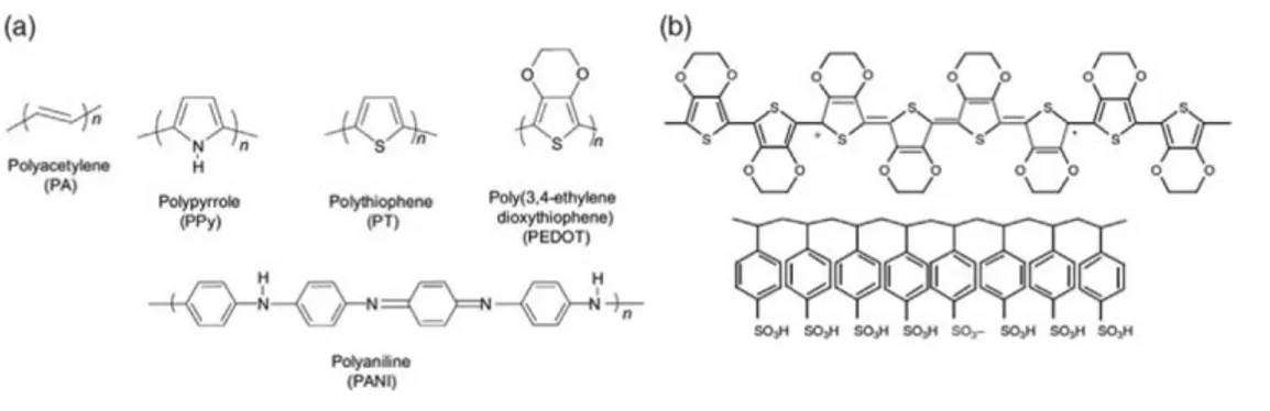
41 used for biological applications are shown in Figure 2.1. The conjugated bonding structure of the polymers shown in Figure 2.1 gives rise to their metal-like, semiconducting properties. However, dopants are necessary to raise the room-temperature electrical conductivity to practical levels (100 S/cm and above). For p-type doping, in which the material is oxidized into a more conductive state, the dopant can be any form of anion. When in close proximity with the conjugated polymer, the negative charge will be compensated with a mobile hole along the conjugated polymer backbone. Such is the case for polypyrrole (PPy) an poly(3,4-ethylenedioxythiophene) (PEDOT), which are both considered hole conductors. Figure 2.1b shows how a polaron on the backbone of a CP chain neutralizes the SO3- on the PSS molecule, a common macromolecular dopant. A common
small-molecule dopant is the anion of p-toluenesulfonic acid (pTS), sometimes referred to as tosylate (TOS).
Figure 2.1 (a) Chemical structures of common conducting polymers used in biological applications:
polypyrrole, polythiophene, poly (3,4-ethylenedioxythiophene), and polyaniline. (b) Chemical structure of PEDOT doped with PSS, showing a delocalized hole in the form of a positive polaron. The anion on the PSS chain acts as the dopant (acceptor).
42 Conducting polymers serve as the active layer in a variety of different applications. The attributes of CPs that make these materials uniquely suited for interfacing with biological systems include soft mechanical properties: the soft mechanical properties allow for compatibility with flexible substrates and good mechanical matching with delicate biological tissue. In applications for neuroprobes or functional substrates for cell growth, these materials better mimic in vivo environments compared to their inorganic counterparts.
Mixed conduction and ideal interfaces: The unique ability of organic electronic materials to conduct ions, in addition to electrons and holes, facilitates their communication with biological systems, which rely heavily on ion fluxes.
Freedom in chemical modification: The nature of polymer synthesis allows for a level of chemical variation not achievable with inorganic materials. Various moieties can be covalently added to a polymer chain for the purpose of increased biological functionality. In situ polymerization enables physical entrapment of desired molecules, including large polyanions and bulky proteins. Overall, the extensive catalog of available chemistries is extremely useful in optimizing materials for various applications.
Ease of processing: Along with the advantages unique to bioelectronics, the benefits of organic electronics as seen in other fields are maintained. Namely, commercially available CP inks and monomers are extremely adaptable to a wide
43 range of processing techniques based on solution- and vapor-phase deposition methods. The ease of processing facilitates deposition on a variety of substrates with unique mechanical properties and form factors, including extreme aspect ratios4.
Moreover, easily scaled-up processing techniques, such as spray coating and other roll-to-roll compatible techniques, lower the cost of the final product. In developing single-use devices for point-of-care diagnostics, low cost remains extremely important.
In this chapter will be discussed the integration of PEDOT:PSS as conductive material within eumelanin, in order to investigate the electrical properties and the structural organization of this blend in the way to create a merge between the unique properties of both of these two polymers.
44
2.2 Pigment functionalization: tailoring Eumelanin
conductivity properties by hybrid buildup
The growing interest toward biocompatible and bioinspired materials is boosting the investigation and the engineering of natural products as active components in electronic devices. Capitalizing on a recently developed protocol to prepare high quality eumelanin coatings, herein will be discribed the design and the integration of standard commercial poly(3,4-ethylenedioxythiophene) with the poly(styrenesulfonate) (PEDOT:PSS) with eumelanin pigment.
The growing advancements of organic (bio)electronics3, 5 together with a number of concurrent needs6, such as processability, and issues7, as the scarcity of the indium8, a critical raw material9, are spurring the search for an alternative to replace the indium tin oxide (ITO) in organic electronic devices10. To date, other transparent conductors cannot compete with the ITO, particularly on its transparency, conductivity, and electronic properties, and on the performances of the devices basing on it. A number of different solutions are under investigation, including metal nanowires7, 11, graphene and/or graphene oxide12, conductive polymers13, carbon nanotubes14, and fullerenes15, each one of these carrying pros and cons. The applications in bioelectronics have particularly fuelled the