1
Index
CHAPTER 1 Pag.
CONGENITAL MUSCULAR DYSTROPHY (CMD)………... 3
CLASSIFICATION OF CMD ... 5
CMD with Collagen VI Deficiency (Ullrich Disease, UCMD) ... 7
CMD due to Integrin α7 Deficiency ... 8
Merosin-Deficient CMD Type 1A (MDC1A, Laminin α2 Related CMD)….. 9
LMNA-Related CMD... 10
Rigid Spine CMD (RSMD, SEPN1-CMD)……….... 11
Dystroglycanopathies... 12
EPIDEMIOLOGY AND MOLECULAR DIAGNOSIS OF CMD……….... 16
CHAPTER 2 CONGENITAL MUSCULAR DYSTROPHIES IN THE UK POPULATION: CLINICAL AND MOLECULAR SPECTRUM OF A LARGE COHORT DIAGNOSED OVER A 12-YEARS PERIOD Introduction... 17
Patients and methods... 18
Results……... 20
Discussion……... 25
CHAPTER 3 COMPLETE MOLECULAR CHARACTERIZATION OF PATIENTS AFFECTED BY CONGENITAL MUSCULAR DYSTROPHIES USING NEXT GENERATION SEQUENCING STRATEGIES IN SICILY Introduction... 31
Clinical protocol and Methods... 33
Results……... 38
2
CHAPTER 4
CARDIAC INVOLVEMENT IN A PATIENT WITH CMD RELATED TO POMT2 GENE MUTATION
Introduction ... 42
Clinical History ... 43
Conclusions ... 45
CHAPTER 5 MUTATIONS IN THE SYNE-1 GENE CAUSE AUTOSOMAL RECESSIVE CMD. THE FIRST CASE REPORT. Introduction... 47
Materials and methods... 49
Results……... 50
Discussion……...51
3 CONGENITAL MUSCULAR DYSTROPHY
Congenital muscular dystrophies (CMD) are a highly heterogeneous group of conditions clinically characterized by muscle weakness with onset at birth or shortly after, and variable involvement of eyes, heart and central nervous system [1]. Some forms of CMD can be fatal in the first years of life, whereas others have a milder course and survival into adulthood is possible [2].
While the phenotypes of CMD have first been described over 100 years ago by Frederick
Eustace Batten [3], a significant progress in the understanding of pathophysiological
mechanisms has been made in the past 20 years, first and foremost attributable to the substantial advances in genetic research [4-6].
Following the initial classification of CMD based on clinical features and country of origin, it was soon recognized that different CMD show significant clinical overlap and marked genetic heterogeneity. In the past few years, the improved understanding of the molecular mechanisms underlying these diseases has allowed a genetic classification based on the function of the involved protein [7, 8]. Figure 1 summarises the location of a number of proteins described in this thesis. It is also now recognised that allelic mutations in genes responsible for CMD can cause milder phenotypes and in particular limb girdle muscular dystrophies (LGMD) too [9-11].
Although CMD have been associated with mutations in more than 20 genes, the number of clinically and histologically proven CMD cases without a genetic diagnosis is not entirely defined, but it is considered to be significant [12].
An accurate genetic diagnosis in CMD is of paramount importance not only for improved phenotype-genotype correlation, but also to facilitate genetic and prenatal counselling and for prognosis and management, as well as to facilitate clinical trials and possible future treatments specific for an individual genetic subtype or mutation.
4
Fig.A. Location of some muscle proteins involved in the pathogenesis of CMD. In particular α-dystroglycan, laminin α2, the integrin complex and collagen VI can be visualised. (Mercuri et al. Ann. Neurol. 2012)
5 CLASSIFICATION OF CMD
Recent classifications are based on combined clinical, genetic, and pathological data [8, 13]:
• Forms of CMD due to mutations in genes encoding for structural proteins of the basal lamina or extracellular matrix or receptors for extracellular matrix proteins. They include CMD variants due to mutations in the collagen 6A1, 2, and 3 genes, laminin α2 (LAMA2, one of the components of the merosin trimer), integrin α7 (ITGA7), and the more recent variant due to integrin α9 gene (ITGA9) deficiency. Mutations in DAG1 have also been described recently, only in cases of limb girdle muscular dystrophies.
• Abnormalities of nuclear envelope proteins (LMNA and nesprin).
• Abnormalities of proteins with as yet unknown function localized in the endoplasmic reticulum, which include the form with rigid spine syndrome (RSMD1) secondary to mutations in SEPN1 • Forms secondary to genes encoding for putative or demonstrated glycosyltransferases that affect the glycosylation of α-dystroglycan (α-DG). They include a wide spectrum of phenotypes that range from the severe Walker-Warburg syndrome to Muscle-Eye-Brain syndrome and Fukuyama CMD with severe structural muscle, brain, and often eye involvement, to mild forms of limb girdle muscle dystrophy. These are commonly referred to as secondary dystroglycanopathies. The first six genes identified represent as a group the vast majority of the cases and are genes involved in the O-mannosyl-linked glycosylation of α-DG. These genes are: protein-O-mannosyl transferase 1 (POMT1), protein-O-mannosyl transferase 2 (POMT2), protein-Omannose 1,2-N-acetylglucosaminyltransferase 1 (POMGnT1), fukutin (FKTN), fukutin-related protein (FKRP), and LARGE.
• CMD with mitochondrial structural abnormalities (CMDmt).
Table A gives an overview of the most important genetically recognized congenital muscular dystrophies.
6 Tab. A
Phenotype Gene Locus Protein Clinical features CNS involvement
Inh
Collagen VI and integrin related dystrophies Ullrich CMD Intermediate phenotypes Bethlem myopathy COL 6A1 COL 6A2 COL 6A3 21q22, 21q22 2q37
Collagen VI Characteristic phenotype with proximal contractures and distal joint hyperextensibility Variable degree of weakness, with no ambulation possible in severe cases
No AD, AR
Integrin α7 deficiency
ITGA7 12q13 Integrin α7 Rare
Delayed motor milestones Cognitive impairment possible
Probably mostly cardiac phenotype
No AR
Laminin α-2 related dystrophy Merosin-deficient
CMD
LAMA2 6q22 Laminin α-2 Early onset of weakness with proximal and facial pattern Delayed motor development Ambulation rarely achieved Epilepsy in 30%
Normally no CI
White-matter changes possible
AR
LMNA-related dystrophy LMNA-CMD
“Dropped-head syndrome”
LMNA 1q22 Laminin A/C Early onset, characteristic, and severe weakness of axial and neck muscles, precludes often free sitting
Contractures
Cardiac involvement with conduction defects common
No AD
SELENON-related myopathy Rigid spine syndrome Multiminicore myopathy SELENON (SEPN1)
1p36 Selenoprotein N Rare, early axial muscular weak- ness, scoliosis, rigidity of spine Ambulation achieved in most patients
Early respiratory insufficiency
No AR
α-Dystroglycan-related dystrophies
Walker–Warburg syndrome
POMT1 9q23 Protein O-mannosyltransferase Very severe congenital weakness, often without motor development
Severe CI
Varying degree of ocular involvement
Lissencephaly type II, pachygyria Brainstem hypoplasia Occipital encephalocele Cerebellar atrophy
AR POMT2 14q24 Protein O-mannosyltransferase 2
POMGnT1 1p34 O-linked mannose β-1,2-N- acetylglucosaminyltransferase LARGE 22q12 Acetylglucosaminyltransferase-
like-protein FKTN 9q31 Fukutin
FKRP 19q13 Fukutin-related protein ISPD 7p21 Isoprenoid synthase domain
containing protein
B4GAT1 11q13 Beta-1,3-acetylgalactosaminyl- transferase 2 1
AR
MEB disease POMGnT1 FKR, FKTN ISPD
Severe CMD
Ambulation often precluded Wide spectrum of ophthalmo- logical malformations Epilepsy is common
Lissencephaly type II, pachygria Cerebellar/ pontine hypoplasia
AR
TMEM5 12q14 Transmembrane protein 5 B3GALNT2 1q42 Beta-1,3-N-acetylgalactosami-
nyltransferase 2
AR
Fukuyama CMD
FKTN High incidence in Japan
Often precludes ambulation Epilepsy, severe CI Overlap with MEB
Lissencephaly type II, pachygyria Cerebellar/pontine hypoplasia
7 Phenotype Gene Locus Protein Clinical features CNS
involvement
Inh
CMD/LGMD FKRP Early onset of CMD or LGMD Mild cortical abnormal- AR with MRI POMT1 phenotype ities possible, often
POMT2 CI frequent normal
ISPD Microcephaly possible
GMPPB
CMD/LGMD FKRP Milder phenotype of early onset No AR without MRI FKTN weakness
ISPD Ambulation often possible
Also early onset LGMD phenotype
No CI GMPPB 3p21 GDP-mannose pyrophosphory-
lase B
Cardiac involvement possible Congenital myasthenia possible in GMPPB
LARGE-related LARGE Severe CMD White-matter changes AR
CMD CL possible
Overlap with MEB/WWS Pachygyria Cerebellar/pontine hypoplasia
Table A. Overview of the most important genetically recognized congenital muscular dystrophies. (Schorling D.C. et. al. Neuropediatrics 2017)
CMD with Collagen VI Deficiency (Ullrich CMD, UCMD)
Collagen VI forms a micro-fibrillar extracellular network that, among other possible functions, may link extracellular proteins with basement membrane around muscle cells [14].
Collagen VI is composed of 3 chains encoded by 3 separate genes: COL6A1 and COL6A2 on chromosome 21q22, and COL6A3 on chromosome 2q37.
Although original Ullrich CMD description [15] was thought to be due to recessive mutations, whereas the milder allelic disorder, Bethlem myopathy, was secondary to dominant mutations, it is now recognised that both disorders can be associated with autosomal recessive mutations or de novo dominant variants that may occur in each of the 3 genes. [16-18]
Muscle biopsy can show a variable pathology ranging from myopathic changes to clearly dystrophic patterns, with abundant adipose tissue. A marked deficiency or absence of collagen VI is a clear marker of Ullrich CMD, but in several patients the deficiency is subtle and affects specifically the basal lamina expression of collagen VI, but not its interstitial deposition. In these
8 cases, careful double labeling with another marker of basal lamina integrity such as perlecan is necessary [19]. Immunohistochemical analysis of collagen VI can also be performed in skin biopsy derived fibroblast culture and may be more sensitive [13].
Serum creatine kinase (CK) is often normal or only mildly elevated.
Diagnosis is typically based on the clinical presentation. The distinct phenotype of Ullrich CMD is characterized by congenital generalized muscular weakness and typical hypermobility with marked distal hyperlaxity of fingers and ankle in association with proximal joint contractures, torticollis and hip dysplasia. Some children may only show delayed milestones as the presenting sign, and the mean age at onset is around 12 months. Cognitive development is normal. Additional features may include a prominent calcaneus, kyphoscoliosis, congenital dislocated hips, and follicular hyperkeratosis [8]. Affected children show significant delay in motor development, although ambulation may be achieved in milder forms. The natural course often shows progression of contractures, muscle weakness and rigidity of the spine, often associated with scoliosis, with concomitant decline in respiratory function (about 2.6 % per year). The average age at onset of non-invasive ventilation is 11 years [20]. In contrast to the Emery–Dreifuss muscular dystrophy, which also presents with proximal contractures, cardiac function is not affected in UCMD. A characteristic pattern of selective muscle involvement on muscle MRI has also been described [21].
CMD due to Integrin α7 Deficiency
Integrin α7 is a transmembrane laminin α2 receptor (Fig. A). Primary deficiency of integrin α7 appears to be an exceptionally rare form of CMD. So far only three patients with normal laminin α2 but absent integrin α7 were found to carry causative mutation in the integrin α7 (ITGA7)gene [22].
9 Hayashi et al. reported 3 male children who had delayed motor milestones, 2 of them achieved independent ambulation, whereas the third had a more severe phenotype showing progressive weakness, scoliosis, and respiratory impairment, requiring NIV at the age of 12 years. [23].
No additional patients with this condition have been reported to date, suggesting that this condition is very rare.
Merosin-Deficient CMD Type 1A (MDC1A, Laminin α2 related CMD)
The laminin α2 is a subunit of laminin, an extracellular protein consisting of three subunits that links extracellular matrix with α-dystroglycan (Fig. A).
Phenotypes of mutations in the LAMA2 gene, which encodes the heavy α2 chain of merosin, can be classified by the amount of merosin in muscle biopsy: complete absence of this protein leads to a severe CMD, whereas patients with partial merosin deficiency may have milder phenotypes [24,25]. The muscle biopsy shows a classical dystrophic pattern and immunohistochemical techniques can readily demonstrate the reduction or absence of laminin α2 chain. In most CMD cases, the protein is totally absent or only present in traces. The laminin α2 chain is processed into 2 fragments on immunoblots of 80 and 300 kDa, and a reduction is often easier to observe on immunohistochemistry with antibodies that recognise the 300 kDa fragment, although it is recommended to use both antibodies in diagnosis. [26-27]
The diagnosis of MDC1A should be genetically confirmed by the identification of mutations in the LAMA2 gene. [24]
Clinical findings in patients with congenital or early onset, during the first months of life, are characterized by marked muscular hypotonia, involvement of facial muscles and sometimes presence of distal contractures resembling arthrogryposis. Serum CK is typically elevated. Although cognitive development typically is not affected, virtually all patients show white-matter changes in T2-weighted brain magnetic resonance imaging (MRI) in later course (>6 months) and rarely
10 occipital structural anomalies [28]. Epilepsy is found in up to 30% of patients but is not necessarily associated with MRI anomalies. A demyelinating neuropathy may occur since laminin α2 is also expressed in Schwann cells [29].
Respiratory function is invariably reduced by the end of the first decade, and night-time hypoventilation is frequently observed even in early childhood [25]. Feeding problems are also frequent, and failure to thrive occurs in >80% of affected children [30] .
In children with absent merosin the maximal motor ability achieved is sitting unsupported. In patients with merosin reduction, in contrast, the severity of the phenotype can vary significantly, but is often milder and they can reach the ability to walk [8].
LMNA-Related CMD
Mutations in LMNA gene, encoding for lamins A and C (Fig. A), intermediate filaments of the inner nuclear membrane in almost all cells, are most commonly associated with dominant form of Emery–Dreifuss muscular dystrophy (EDMD) [31], which is characterized by scapulohumeral– peroneal muscular weakness, cardiomyopathy, and multiple contractures. However, mutations in LMNA gene can cause a wide spectrum of diseases, sometimes called “laminopathies, including an early onset muscular dystrophy (termed LMNA-CMD), first described in 2004 [32]. Inheritance is autosomal dominant, but very rare autosomal recessive cases have been described [33, 34].
Children with LMNA-CMD present a congenital or early infantile onset of severe axial and neck muscles weakness, denoted by the term dropped head syndrome [35]. Further characteristic features include: involvement of the extremities, that is more proximal in the upper limbs and distal in the lower limbs, early lumbar hyperlordosis, multiple contractures and cardiac function is also affected. Free sitting is commonly achieved, except in the most severe presentations, but ambulation can be variable and respiratory involvement can occur. Cardiac damage with typical atrial and ventricular
11 arrhythmias, conduction system disease, and eventually dilated cardiomyopathy can lead to sudden death and best addressed by implantable cardioverters. The intellectual development is normal [13]. Muscle biopsy does not show any peculiar key diagnostic aspect, and may variable range from a myopathic pattern with atrophic fibers, mostly of type 1, to more obvious dystrophic findings. Inflammation with conspicuous cellular infiltration can be found in some rare cases. Immunohistochemical examination and Western blot analysis of LMNA in the biopsy are normal [36]
Rigid Spine CMD (RSMD, SEPN1-CMD)
The selenoprotein N1 (SEPN1) gene encodes a selenocysteine-containing protein, which is thought to have a redox function and to regulate calcium levels in the endoplasmic reticulum [37].
Recessive mutations in SEPN1 gene have been reported in patients with a consistent clinical phenotype characterized by weakness and rigidity of the spine which typically leads to major scoliosis and life-threatening respiratory insufficiency in the second or third decade of life and contrasts with relatively preserved limb strength and ambulation [38].
By contrast, the morphologic spectrum of SEPN1-CMD is large, encompassing at least 4 different myopathologic patterns: 1. minimal dystrophic changes, 2. multiminicores disease, 3. congenital fiber-type disproportion (CFTD) and desmin-related myopathy with Mallory-body–like inclusions. This heterogeneity of morphologic features, together with the lack of immunohistochemical markers, can delay early diagnosis of SEPN1-CMD. [2]
Delayed and poor head control is the most common presenting sign; dropped head is virtually constant and noticeable from the age of 3 months, whereas other motor milestones are relatively normal. Most patients achieve the ability to walk at the normal age but are never able to lift their head from the supine position. The patients frequently have a nasal, high-pitched voice and a variable degree of facial weakness. Typically, scoliosis begins in the cervical region and it is associated with dorsal lordosis and a lateral trunk deviation, frequently
12 requiring spinal fusion. Restrictive respiratory failure is mainly determined by diaphragmatic dysfunction and not correlated to the degree of general weakness; most patients require non-invasive ventilation (NIV) while still ambulant [2,39]. The diagnosis of RSMD should be genetically confirmed by the identification of mutations in the SEPN1 gene.
Dystroglycanopathies
Dystroglycanopathies are clinically and genetically heterogeneous muscular dystrophies caused by defective glycosylation of α-dystroglycan (α-DG) [40].
The dystroglycan complex is encoded by the DAG1 gene and translated as a pro-peptide proteolytically cleaved into α and β subunits. α-DG is an extracellular peripheral protein (Fig. A) that undergoes extensive N- and O-linked glycosylation. The post-translational modifications are essential for the interactions of α-DG with extracellular proteins, such as laminin-2 (LAMA2), agrin, perlecan and neurexin [40]. Diagnosis of dystroglycanopathy is based on the detection of hypoglycosylated α-DG by immunolabeling and/or Western blot on muscle biopsy, whereas the core α-DG protein is normally expressed.
Primary dystroglycanopathies with mutations in DAG1 gene remain extremely rare [41, 42]. As the defect in this group of conditions relates to genes involved in the glycosylation of α-DG, a more appropriate terminology is that of secondary dystroglycanopathies; however we will continue to use only the term dystroglycanopathy for simplicity. The class of dystroglycanopathies is ever-growing, with mutations in 20 genes reported to date [40]. Phenotypes range from severe CMD, often associated with brain and eye defects to milder adult onset limb girdle muscular dystrophies (LGMD).
Suspicion of a dystroglycanopathy is based on clinical, brain imaging and pathological findings [12].
13 Since mutations in all genes involved affect glycosylation modifications on α-DG, diagnosis of dystroglycanopathies is established upon the detection of hypoglycosylated α-DG at the sarcolemma of skeletal muscle fibres by immunolabelling and/or Western blot.
Muscle biopsies show alterations in labelling with antibodies against glycosylated α-DG with preserved staining for the α-DG core protein by immunohistochemistry (monoclonal IIH6 and V1A4-1) [43].
Diagnosis is confirmed by identification of pathogenic mutations in one of the known dystroglycanopathy genes. Some of these genes (such as FKTN or POMGnT1) more commonly cause CMD, while others (in particular FKRP and GMPPB) show a wider phenotypic spectrum, with LGMD being the most prevalent phenotype. However, since no firm genotype-phenotype correlations have been demonstrated and there are no specific clinical or pathological markers to guide molecular investigations, the diagnosis must be achieved through the analysis of a large panel of genes.
These genetically heterogeneous disorders frequently include central nervous system (CNS) pathology and encompass a striking range of clinical severity. At the most severe end of the clinical spectrum there are the conditions: Walker–Warburg syndrome (WWS), muscle–eye–brain disease (MEB) and Fukuyama congenital muscular dystrophy (FCMD) [6].
They are characterised by CMD with severe structural brain and eye abnormalities [13]:
- WWS delineates the most severe phenotype within the continuum of α-dystroglycanopathies. It is characterized by muscular dystrophy in combination with ocular and cerebral malformations. The latter may involve occipital encephaloceles, Dandy– Walker cysts, pontocerebellar hypoplasia, hydrocephalus, cortical heterotopias, fusion of hemispheres, and type II lissencephaly (also referred to as “cobblestone lissencephaly) [44]. Prognosis is poor with only little psychomotor development and exitus occurs mostly during the
14 - MEB disease is characterized by severe CMD with elevated CK levels, brain malformations similar to FCMD that additionally include hypoplasia of brainstem and cerebellum, and presence of a wide spectrum of ophthalmological malformations, ranging from marked congenital myopia to congenital glaucoma, optic nerve atrophy, progressive retinal dysplasia, and dysgenesis of the anterior chamber [45]. Epilepsy is common, and cognitive impairment is often severe.
- FCMD is characterized by general muscular weakness, severe cognitive impairment and elevated levels of serum CK [46]. Motor development is substantially delayed, and ambulation is predominantly not achieved. Deep tendon reflexes are decreased or absent. Characteristic MRI findings include cortical malformations, especially micropolygyria resulting in a pachygyric appearance (lissencephaly type II or cobblestone lissencephaly), and cerebellar and pontine hypoplasia. Affected individuals may be microcephalic, and epilepsy occurs in 50%. There is considerable clinical overlap with MEB disease, but eye involvement is considerably more prominent in MEB.
Conversely, individuals at the mildest end of the clinical spectrum may present in adult life with limb girdle muscular dystrophy (LGMD) and without associated brain or eye involvement. A number of intermediate phenotypes between these above mentioned extremes have also been described with the best characterised being MDC1C, in which affected children do not typically have brain or eye involvement despite the relative severe skeletal muscle involvement [6].
The anatomical abnormalities of the central nervous system that are apparent on MRI can be helpful for a preliminary diagnosis. Patients with dystroglycanopathies, mainly according to the degree of structural and functional brain involvement, have been more recently subdivided in the following groups [7]:
15 WWS: this group includes patients with severe structural brain abnormalities including complete agyria or severe lissencephaly with only rudimentary cortical folding, marked hydrocephalus, severe cerebellar involvement and complete or partial absence of the corpus callosum.
MEB/FCMD-like: this group encompasses CMDs with brain abnormality less severe than that seen with WWS. MRI findings include pachygyria with preferential fronto-parietal involvement, polymicrogyria, cerebellar hypoplasia, cerebellar dysplasia and frequent flattening of the pons and brainstem. Eye abnormalities are often seen.
CMD with cerebellar involvement (CMD-CRB): this category included CMD with mental retardation and cerebellar involvement on MRI scan as the only structural abnormality. Cerebellar abnormalities may include cysts, cerebellar hypoplasia or dysplasia.
CMD with mental retardation (CMD-MR): patients with isolated microcephaly or minor white matter changes on MRI are included in this group, however, they usually show mental retardation and structurally normal brain.
CMD with no mental retardation (CMD-no MR): several patients within this group have had no neuroimaging but had entirely normal intellectual function.
EPIDEMIOLOGY AND MOLECULAR DIAGNOSIS OF CMD
In terms of incidence and prevalence of CMD, there were only few studies available until few years ago, mostly of which focusing on small regions [47-49]. In the last two years many progresses have been done, with emerging new studies referring to larger geographical areas [34, 50]. In addiction a significant proportion of patients are still genetically undiagnosed.
Before the advent of next generation sequencing (NGS), an integrated clinical, pathological and imaging approach was required to better address the genetic analysis. In the last years the NGS technologies have revolutionized the approach to the diagnostic work-out, leading to the discovery of several new genes and therefore reducing the number of cases without a genetic confirmation.
16 Of the above mentioned genes of dystroglycanopathies, ten (DPM2, DPM3, DOLK, B3GNT1, B3GALNT2, GTDC2, ISPD, TMEM5, SGK196 and GMPPB) have been discovered in the last few years through NGS. Moreover, further new genes have been discovered by NGS in other CMD forms such as CHKB related to CMDs with mitochondrial structural abnormalities [51]. The rapidly increasing identification of several genes responsible for different forms of CMD has dramatically expanded the spectrum of the known forms, allowing a better understanding of the individual forms and different underlying pathomechanisms.
The aims of the following studies were to better understand the frequency of the various forms of CMD in large populations, like the UK and our geographic area, providing a complete molecular characterization of patients.
Furthermore we report the results of genetic screening performed using NGS strategies and describe new peculiar clinical features identified in patients in whom a genetic diagnosis was reached.
CHAPTER 2
Congenital Muscular Dystrophies in the UK population: clinical and molecular spectrum of a large cohort diagnosed over a 12-years period
INTRODUCTION
Information on the incidence and prevalence of CMD in various populations is limited. Point prevalence of CMD in Northern England was calculated as 0.9/100.000 [49] while a recent study on the Italian population showed slightly lower figures at 0.563/100.000 [50]. Relative frequency of individual types seems to differ among populations often because of founder mutations [48, 52]. For example, in the Japanese population the most common type is Fukuyama CMD, due to a founder
17 insertion of a 3-kb retrotransposition element in the 3’ untranslated region of fukutin (FKTN) gene [52], while this mutation is virtually absent outside Japan.
In the UK, there is the Highly Specialised Services (HSS) commission for the diagnosis and management of rare neuromuscular diseases, with the Dubowitz Neuromuscular Centre (DNC) in London providing a comprehensive clinical, histopathological and molecular service for diagnosis of CMD. In the UK, genetic analysis of CMD genes is offered at the DNC only, with the exception of the FKRP and LMNA gene, also tested at the HSS for LGMD in Newcastle in view of the most common and milder phenotypes (LGMD2I and AD-EDMD, respectively).
We have previously reported the relative frequency of CMD among children specifically referred for a clinical opinion to the DNC Centre who had been assessed by the multidisciplinary team [53]. A genetic diagnosis was reached in 53 patients (46%); the commonest diagnosis in this selected population was collagen VI disorders (19%), followed by dystroglycanopathies (12%) and
laminin-α2 related CMD (MDC1A) (10%). However our previous work did not take into account patients
with a clinical or pathological diagnosis of CMD who were referred to the DNC for genetic testing only.
In the present study we have extended our previous observation and report the result of genetic screening of CMD genes in all the UK patients referred to the two HSS diagnostic laboratories between 2001 and 2013, with the main aim of investigating frequency and relative prevalence of CMD subtypes in the UK population. We also summarise observed genotypes and novel mutations identified in the CMD genes.
PATIENTS AND METHODS
Patients
Between 2001 and 2013, 3734 DNA samples were sent to the DNC and the HSS in Newcastle for genetic testing of CMD genes. Among these, 1042 samples were referred to the DNC for sequencing of the 14 CMD genes offered for analysis until 2014 (LAMA2, POMT1, POMT2,
18 POMGnT1, FKRP, FKTN, LARGE, ISPD, GMPPB, B3GALNT2, COL6A1, COL6A2, COL6A3 and SEPN1), while further 2692 samples were sent to the HSS in Newcastle for analysis of the LMNA and FKRP genes only. These figures also include samples of patients with milder allelic phenotypes, such as LGMD and BM. All patients were resident in UK at the time of this study. Written informed consent for diagnostic genetic testing was obtained from patients and/or parents prior to DNA collection by referring clinicians. Genetic testing was gate kept by clinical members of the two services based on relevant medical history, clinical features and clinical investigations (including serum CK values, EMG, muscle biopsy analysis, brain and/or muscle MRI) provided in clinical referral forms. Clinical information was collected retrospectively from the clinical referral forms only for patients in whom pathogenic mutation/s were identified. Patients were divided into two clinic subgroups: a) CMD and b) milder phenotype. A diagnosis of CMD was confirmed, in presence of 1) presentation before 2 years of age with hypotonia, weakness, contractures, delayed motor milestones or characteristic structural eye or brain abnormalities; 2) dystrophic or myopathic changes on the muscle biopsy, with exclusion of other specifically identifiable neuromuscular disorders, in accordance to the proposed diagnostic criteria for CMD [12]. Increased creatine kinase (CK) levels were not considered an inclusion criteria as not all CMD have raised CK levels. Patients who did not fulfil the diagnostic criteria for CMD but who had a clear dystrophic muscle pathology phenotype were included in the milder phenotype subgroup.
Because FKRP gene was analysed in both HSS diagnostic laboratories, we did account for possible overlap regarding all patients referred to the two Centres and we have cross checked all patients in whom a genetic diagnosis was reached, to avoid possible duplications.
The spectrum of clinical severity for dystroglycanopathy patients was described using the classification proposed by Godfrey et al.: MEB/FCMD = Muscle-Eye-Brain/Fukuyama Congenital Muscular Dystrophy Like; CMD-MR = Congenital Muscular Dystrophy with Mental Retardation; CMD-NOMR = Congenital Muscular Dystrophy with No Mental Retardation; CMD-CRB = Congenital Muscular Dystrophy with cerebellar Involvement; LGMD-MR = Limb Girdle Muscular
19 Dystrophy with Mental Retardation; LGMD-NOMR = Limb Girdle Muscular Dystrophy with No Mental Retardation [7].
Molecular analysis
Molecular genetic analysis of the LAMA2, POMT1, POMT2, POMGnT1, FKRP, FKTN, LARGE, ISPD, GMPPB, B3GALNT2, COL6A1, COL6A2, COL6A3 and SEPN1 genes was performed at the Viapath Molecular Laboratory at Guys and St Thomas’ Trust, London, as part of Highly Specialised Service (HSS) for CMD, while the HSS in Newcastle performed analysis of the FKRP and LMNA genes. Sequencing of gene/s by a candidate gene approach was guided by phenotypic features, brain and muscle MRI and muscle biopsy review. In case of suspicion of dystroglycanopathy, DNAs underwent analysis of the following 9 α-DG genes (POMT1, POMT2, POMGnT1, FKRP, FKTN, LARGE, ISPD, GMPPB, B3GALNT2).
The entire coding regions and flanking intronic regions of each gene were sequenced from peripheral genomic DNA. Mutation nomenclature was based on the reference sequence, with nucleotide number 1 corresponding to the first base of the translation initiation codon. To investigate whether novel changes could represent pathogenic sequence variants, in silico analysis was performed using the Alamut mutation analysis software (Interactive Biosoftware, v1.5 http://www.interactivebiosoftware.com ). In particular, novel variants were considered to probably be pathogenic if they affected a moderate to highly conserved nucleotide or a highly conserved residue and if the resulting physiochemical difference between the wild-type and mutant amino acid was at least moderate. Where available, affected and unaffected family members were assessed for segregation of variants identified in the proband.
RESULTS
Molecular analysis
A confirmed genetic diagnosis was reached in 440 unrelated patients (Table 1). Detection rate for the totality of the nationally referred patients was 12% (440/3734). We identified 362 mutations,
20 160 of which were novel (Table 1). Detailed clinical data were available for 418 of the 440 unrelated patients with mutations. A total of 249/418 (59.6%) patients fulfilled the diagnostic criteria for CMD (CMD subgroup), whereas 169/418 (40.4%) patients showed a milder phenotype. Details of phenotype prevalence for each group of genes are indicated in Figure 1.
CMD population subgroup
The most common clinical subtype of CMD was MDC1A, accounting for 93 unrelated patients (37.35%), followed by dystroglycanopathies (n= 66, 26.5%), UCMD (n=39, 15.7%), RSMD1 (n=29, 11.65%) and LMNA-CMD (n=22, 8.8%; Fig. 2).
Detailed data regarding onset of symptoms in MDC1A patients were available for 72/93 subjects. Most common presenting symptoms were hypotonia at birth or in the first week of life (n=51; 72%) or delayed motor milestones during the first year of age (n=20, 28%). Among the 47 patients with complete absence of laminin-α2 protein on muscle biopsy, only one achieved independent ambulation. Conversely, among the 23 patients with partial laminin-α2 deficiency, 14 achieved ambulation, while 3 were not yet walking at age 6 years. Clinical information of the remaining 6 patients was limited. Median CK value was 2100 UI/L, with two patients aged less than 4 years having normal CK.
The majority of the 66 CMD patients with genetically confirmed dystroglycanopathy carried mutations in the POMGnT1 gene (n=18, 27.3%), FKRP gene (n=15, 22.8%) and POMT1 or POMT2 genes (n=10; 15.1%), with a single patient only carrying mutations in LARGE (Fig. 3). Analysis of detailed clinical data (available for 58/66 patients) showed a wide spectrum of clinical severities, ranging from WWS (n= 12), MEB/FCMD–like (n= 16) and CMD-CRB (n= 9) to the less severe CMD with (CMD-MR) (n= 11) or without mental retardation (CMD-without MR; n= 10) (Table 2 and supp. tables 2-4, Sframeli et al. 2017) [34]. All patients with B3GALNT2 mutations showed a severe CMD phenotype (1 WWS or 3 MEB/FCMD-like disease), while only 2 patients with ISPD (1 WWS and 1 CMD-MR) and 2 with GMPPB gene mutations (1 MEB/FCMD-like and 1 CMD-MR) fulfilled CMD inclusion criteria. Serum CK values ranged from 200 to 13000 UI/L,
21 with a total of 9 patients with values <1000 UI/L (with mutations in POMGnT1, POMT1 and B3GALNT2). Moreover we identified three patients with normal or very mildly elevated serum CK levels (173, 228 and 267 UI/L, normal range being 38-234 UI/L). These 3 patients had a clinical diagnosis of MEB/FCMD-like with mutations in POMGnT1 gene (in order, patient 1 being compound heterozygous c.511C>T/p.(Arg171*) andc.1895+1G>T mutations, patient 2 compound heterozygous for the c.385C>T/p.(Arg129Trp) andc.1460delG/p.(Gly487fs) changes and patient 3 homozygous for c.33_34delGCinsA/p.(Phe13fs) mutation). The clinical phenotype of these three patients was characterised by early onset of weakness, axial hypotonia and predominant upper motor neuron features with brisk reflexes and ankle clonus. In addition, one patient had hydrocephalus at birth, severe myopia, infantile spasms, evolving to cerebral palsy with limb spasticity. Brain MRI revealed cobblestone lissencephaly and cerebellar polymicrogyria. The second child presented with reduced visual attention, poor feeding and hypotonia since birth. She had brisk tendon reflexes, myopia and esotropia. Brain MRI at 3 months of age showed lissencephaly and widespread polymicrogyria with cerebellar cysts. The third patient also presented with horizontal nystagmus, convergent squint and learning difficulties. Her brain MRI at age 2 years showed cobblestone polymicrogyria involving frontal and temporal lobes and dysplastic cerebellar cysts with cerebellar and pontine hypoplasia.
Clinical features of UCMD and RSMD1 patients were typical (data not shown). Among UCMD patients, 21 had AR inheritance and 18 AD. Twenty patients (51%) carried mutations in the COL6A1 gene (12 dominant and 8 recessive), 14 (36%) had mutations in the COL6A2 gene (1 dominant and 13 recessive) and the remaining 5 patients (13%) had dominant mutations in the COL6A3 gene (Fig. 4). Twenty-two of the 36 patients (61,1%) with LMNA gene mutations showed a severe phenotype with early onset CMD, with characteristic axial and neck muscle weakness. All carried single dominant heterozygous changes, occurring de novo in all patients but two children, where the mutation was inherited from a healthy somatic mosaic parent.
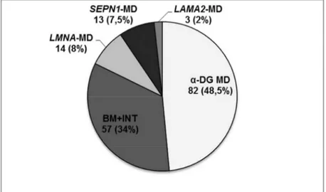
22 Dystroglycanopathy was the most common subtype (n=82 patients, 48.5%) followed by collagenopathies (n=57, 33.7%), LMNA related MD (n=14, 8.3%), milder forms of RSMD1 (n=13, 7.7%) and LAMA2 gene related MD (n=3, 1.8%) (Fig. 5).
Seventy-five patients with a clinical diagnosis of LGMD carried mutations in FKRP gene; all but two showed the common c.826C>A/p.(Leu276Ile) mutation in homozygosity (n=45) or compound heterozygosity (n=28) (Supp. Table 2) [34]. Two patients, one with a novel homozygous missense mutation c.1327G>A/p.(Glu443Lys) and another patient compound heterozygous for the two novel missense changes c.607C>T/p.(Arg203Cys) and c.1223G>A/p.(Ser408Asn), showed onset of symptoms at the age 6 and 11 years respectively. Mutations in GMPPB (4 patients) or ISPD (3 patients) genes were associated with a variable range of clinical severities (Fig. 3).
Forty-two of the 57 patients with collagenopathies had BM and a further 15 showed an intermediate UCMD/BM phenotype (Fig. 4) [20]. The majority of patients (45/57, 79%) had AD inheritance. The majority of patients with intermediate phenotype showed AD inheritance (12/15), with 8 patients carrying mutations in COL6A1. About half of BM patients (20/42) had mutations in the COL6A3 gene, (17 dominant and 3 recessive), 12 in COL6A1 (10 dominant and 2 recessive) and 10 in COL6A2 genes (6 dominant and 4 recessive).
Patients with a milder phenotype with a LMNA gene mutation had a diagnosis of Emery–Dreifuss muscular dystrophy (EDMD). Inheritance was dominant in all patients but one patient was carrying the c.674G>A/p.(Arg225Gln) pathogenic variant in homozygosity. This child presented in early childhood with mobility difficulties, upper and lower limb weakness, progressive elbow and Achilles tendon contractures, normal cardiac function but reduced respiratory function, and MRI features suggestive of EDMD. One patient carried the c.1930C>T/p.(Arg644Cys) change in the LMNA gene. This patient presented at the age of 12 years with typical AD-EDMD and no other pathogenic changes were identified in LMNA or any other genes investigated. This change was also identified in two additional patients who however carried pathogenic changes in alternative genes, and were not included in this study.
23 Patients with SEPN1 gene mutations showed onset of symptoms ranging from 2.5 to 20 years (average 7.85 years). Among the 3 patients with LAMA2 mutations and partial protein deficiency, the first had an onset at 35 years and showed clear reduction of laminin-α2 on muscle biopsy, the second had epilepsy and leukoencephalopathy and the third, an 11 year old girl, showed mild proximal weakness and electrophysiological findings compatible with a diagnosis of sensorimotor demyelinating polyneuropathy, and she had been previously reported [54].
Molecular results of the entire cohort (including both CMD and milder phenotypes)
The 9 genes responsible for α-DG were the most commonly mutated (n=162 unrelated patients; 36.8%) followed by the COL6A1-A2-A3 genes (n=102; 23.2%), LAMA2 (n=96; 21.8%), SEPN1 (n=44; 10%) and finally the LMNA gene (n=36; 8.2%) (Fig. 6).
- α-DG genes: The FKRP gene (n=94/162, 58%) was the most frequently mutated gene, followed by POMGnT1 (n=23/162, 14%). 15 patients carried mutations in ISPD, GMPPB or B3GALNT2 (15/162, 9%). A total of 125 different pathogenic variants were identified (50 novel), 39 in the FKRP (31%) and 25 in the POMGnT1 (20%) genes only (Table 1 and Supp. Table 2, Sframeli et al. 2017) [34]. Missense mutations were the most common, with one initiation codon -loss mutation c.2T>G/p.(Met1Arg) identified in POMGnT1. Eighty-four patients carried the recurrent c.826C>A/p.(Leu276Ile) mutation in the FKRP gene (50 in homozygosity and 34 in compound heterozygosity) (Supp. Table 2, Sframeli et al. 2017) [34].
- COL6A genes: Twenty-nine mutations (18 novel) were identified in the COL6A1 gene. Inheritance was autosomal recessive (AR) in 10 patients and dominant (AD) in 32 (Table 1). Similarly, 29 mutations were identified in the COL6A2 gene (17 novel), with AR inheritance in 19 patients and AD in 10. Thirty pathogenic variants were found in the COL6A3 gene (21 novel), inheritance was AR in 5 patients and AD in further 26. The following mutations were recurrent: c.1272+1G>A (2 homozygous and 1 compound heterozygous patients) and c.877G>A/p.(Gly293Arg) (in 3 heterozygous patients) in COL6A1; c.2839_2850del/ p.(Leu947_Gly950del) in COL6A2 (in 4 homozygous patients) and c.7447A>G/p.(Lys2483Glu) in COL6A3 (in 3 compound heterozygous
24 patients). Missense and splice site mutations were the most common in all three COL6 genes (Table 3, Sframeli et al. 2017) [34].
-LAMA2 gene: A total of 89 mutations were found (36 novel), including 71 altering the reading frame. One mutation was a synonymous variant predicted to affect splicing: c.4860G>A/p.(Lys1620Lys). There were four recurrent mutations: c.2049_2050delAG/ p.(Arg683Serfs) in 3 homozygous and 5 compound heterozygous patients; c.5562+5G>C in 6 compound heterozygous patients; c.2556delT/p.(Phe852fs) in 7 compound heterozygous patients; c.7881T>G/p.(His2627Gln) in 5 homozygous patients and the c.3283C>T/ p.(Arg1095*) and c.1306+2T>G mutations in homozygosity in two patients.
-SEPN1 gene: 32 mutations (5 novel) were identified. The c.943G>A/p.(Gly315Ser) mutation recurred in 15 patients (6 in homozygosity and 9 in compound heterozygosity) and the c.713_714insA/p.(Asn238fs*) mutation recurred in two patients in compound heterozygosity.
-LMNA gene: We identified 28 mutations (13 novel), mostly missense. Inheritance was dominant in the majority of patients, with only one patient being homozygous for a novel missense change in exon 4, c.674G>A/p.(Arg225Gln), segregating from carrier unaffected parents. One mutation, c.745C>T/p.(Arg249Trp) recurred in 4 patients.
DISCUSSION
In this study we present clinical and molecular data for 440 UK unrelated patients with confirmed mutations in genes responsible for CMD, from over 3700 patients referred for genetic testing. We then concentrated on patients presenting with typical clinical features of CMD and provided relative frequency data of different CMD subtypes.
Our data indicate that MDC1A is the most common CMD in the UK (37.35% of patients). This finding refines our previously reported study on patients clinically referred to the DNC, where only 10% of patients had this diagnosis [53]. This difference is explained by the fact that MDC1A is now more readily diagnosed pathologically also in secondary centres, as immunohistochemistry for
25 laminin-α2 can be easily performed and brain MRI readily shows characteristic features. Because of this, patients are less likely to be referred directly to the DNC for a tertiary clinical opinion, while genetic testing is still channelled to our service following DNA referral. Our result agrees with early studies reporting MDC1A as the most common CMD in other European populations, with an average frequency of 30–40% among CMDs [20] and with that observed in the North of England, where MDC1A represents the most prevalent CMD (0,6/100.000) [49].
Dystroglycanopathy is the second most common CMD in our study group, being diagnosed in 26.5% of patients. Among these, POMGnT1 is the most frequently mutated gene (27.3 %), confirming the result found in our previous study [53] but different from that observed in other European populations where POMT1 is the most frequently mutated [50, 55-56]. In our cohort of CMD with dystroglycan glycosylation defect, FKRP gene is the second most commonly mutated gene (22.8%), with phenotypes ranging from severe CMD with cerebellar involvement (n=6) to milder CMD-without MR (n=7) [Suppl. Table, Sframeli et al. 2017) [34]. No patient with FKRP gene mutations showed WWS or MEB/FCMD-like phenotype. Patients with the common c.826C>A/ p.(Leu276Ile) mutation in compound heterozygosity showed CMD-without MR phenotype, confirming that FKRP mutations other than the common c.826C>A/p.(Leu276Ile) variant associate with a more severe phenotype [12, 57-58]. Interestingly, we also identified one homozygous c.826C>A/p.(Leu276Ile) patient who presented at the age of 22 months apparently with a clinical picture of CMD, but no further detailed information was available and this subject was therefore included in the CMD-unclassified group. CMD with mutations in the FKTN, B3GALNT2, ISPD, GMPPB and LARGE genes appear rare, in part similar to what reported in other populations [50, 56]. B3GALNT2 mutations are confirmed to cause more severe CMD phenotypes (WWS or MEB/FCMD-like disease), as also indicated by another recent report of a CMD patient with structural pontine and cerebellar abnormalities and B3GALNT2 mutations [59]. Among dystroglycanopathies, the most frequent phenotype is MEB/FCMD-like (27.5% families), similar to the 30% rate reported by Mercuri et al. [56] and most frequently caused by mutations in POMGnT1
26 gene. The wide clinical spectrum, the variable degree of reduction of α-DG labelling (from absent to minimal changes only) on muscle tissue as well as the secondary reductions of α-DG labelling sometimes observed in other forms of CMD, such as in MDC1A, can make dystroglycanopathy a challenging diagnosis for non-specialist centres. A further complicating factor is the observation of rare MEB/FCMD-like CMD patients with minimally raised or normal serum CK, suggesting that the diagnosis of dystroglycanopathy should not be excluded based on CK levels alone. Of note, since 2014, we have identified further 3 patients with similar clinical pictures, normal or mildly elevated CK and dystroglycan glycosylation defect, further supporting that this is not an exceptional finding among patients with CMD. In this group of patients other conditions characterised by isolated brain malformations were the leading diagnosis, until the possibility of a dystroglycanopathy was also considered.
UCMD is the third most common form of CMD in the UK. Despite an overall similar mutation rate for the 3 collagen genes (COL6A1 41%, COL6A2 29% and COL6A3 30%) in the totality of patients diagnosed (Table 1), among UCMD patients we observed a higher prevalence of mutations in COL6A1 (51%) and COL6A2 (36%) compared to COL6A3 gene (13%, Fig. 4A). Interestingly, we also noticed an overall similar number of dominant and recessive mutations in UCMD patients (18 vs 21), with mutations in COL6A2 being mostly recessive (13 AR vs 1 AD) (Fig. 4B). It is now known that it is rather the effect of the mutation on protein function (and not mode of inheritance) that affects severity of the phenotype [14, 16-18]. Our findings corroborate that de novo dominant mutations in collagen genes are a common cause of UCMD but also suggest that, at least in our population, dominant mutations in COL6A2 are not a common cause of UCMD, while mutations in COL6A3 (dominant or recessive) are more frequent in milder phenotypes (Fig. 4B, C).
We were not able to accurately assess point prevalence for CMD in the UK, because of study methodology and absence of longitudinal clinical data. We are also aware of a limited number of CMD patients from Scotland who might have had LMNA and FKRP testing elsewhere and were therefore not included in this study. However, since the DNC and the HSS in Newcastle were the
27 only UK centres offering molecular testing for the CMD genes free of charge for NHS patients, there was no financial disincentive to obtaining a genetic confirmation of the diagnosis. Hence we believe that we have captured the great majority of patients with a genetic diagnosis of CMD until 2013 and in view of this we felt the available data was sufficiently reliable to make a tentative evaluation of prevalence figures for the UK. Considering a current estimated UK population of 64.1 million, our study suggests a combined prevalence of genetically confirmed CMD of 0.39 per
100,000 total UK population. As we did not include in this study patients with clinical/pathological
diagnosis of CMD who were not referred for genetic testing, this prevalence figure is likely to be underestimated. A recent publication on Italian CMD patients showed that despite major advances in diagnostic technologies and genetic testing, a considerable proportion of CMD patients remain genetically undiagnosed (35%) [50]. We could hypothesize that a similar figure is also valid for the UK population. Interestingly, the total number of genetically confirmed CMD patients is similar in the UK and Italy (249 vs 220 patients) [50], countries that show similar population sizes (64 vs 60 million; 2013 estimate).
The 40% of patients with mutations in one of the CMD genes did not fulfil strict diagnostic criteria for CMD. In this group, LGMD2I due to FKRP gene mutations was the most common phenotype (44%). BM and intermediate BM/UCMD phenotypes were the second most frequent diagnosis (33.53%; Fig. 5). Diagnostic application of muscle MRI and quantitative collagen analysis on fibroblasts might have improved diagnostic abilities in milder forms of collagenopathies, where COL VI protein immunohistochemistry is not helpful, increasing frequency of milder COL6 phenotypes versus UCMD [60].
In this study, we identified 15 families with mutations in the more recently identified dystroglycanopathy genes (ISPD, GMPPB and B3GALNT2), and among these, GMPPB was the most frequently mutated (6/15). Interestingly, patients with ISPD and GMPPB mutations showed a wide phenotypic spectrum ranging from severe WWS or MEB/FCMD like disease to milder
28 phenotypes with or without MR. This finding is in concordance with recent studies describing wide inter and intra-familial clinical variability with GMPPB and ISPD gene related conditions [61-64]. The 12% mutation detection rate in the whole cohort of patients is lower compared to what is reported elsewhere. As detailed clinical analysis of negative patients was not performed, we are unable to comment on detection rate only for patients with a clinical diagnosis of CMD. In the previous study by Clement et al., a genetic diagnosis was reached in 46% of CMD patients referred to the DNC for a clinical opinion (2012) [50]. This higher detection rate may be related to the integrated diagnostic approach applied to patients referred to the DNC which includes clinical, pathologic and often brain and muscle MRI assessment in addition to muscle histopathology. Despite strict gatekeeping criteria on acceptance of samples, a specialised clinical review at a tertiary centre, such as the DNC, is a major factor in improving diagnostic ability. Further factors might have affected our detection rate. Firstly, we did not sequence all 15 genes in each patient as gene analysis was phenotype driven, and we did not assess intronic mutations or larger deletions/duplications. Also, we did not analyse CMD genes which have been described more recently, such as for example CHKB, MICU1, GTDC2, SGK196, TMEM5, B3GNT1 or TRAPPC11[65-69]. Finally, the undiagnosed cohort also included patients with milder phenotypes with wide differential spectrums including LGMD, congenital or later onset myopathies.
We have identified a large number of mutations, further expanding the allelic spectrum associated with CMD genes. Mutations leading to loss of protein function (frameshift, splice and stop mutations) were the most common in the LAMA2 gene (71/89), while missense changes were prevalent in dystroglycanopathy genes (73/125), in keeping with known pathomechanisms of these conditions. Three patients carried two null alleles in the ISPD or B3GALNT2 genes, but we did not identify null FKRP and POMT2 mutations, further supporting the findings that null mutations in these genes are rare [70-73].
In conclusion, this study represents a comprehensive report on relative frequency of CMD in the UK population, indicating MDC1A as the most common CMD subtype (37.35%). We have
29 identified 160 novel mutations in 15 genes and we further expanded the clinical and genetic spectrum associated with mutations in these CMD genes. Our data confirm that MEB/FCMD-like is the prevalent phenotype among CMD patients with dystroglycanopathy and mutations in POMGnT1 gene are the most common. We also describe a number of patients with dystroglycanopathy and normal CK which is an important factor to appreciate as this might complicate the diagnostic path for this subgroup of patients, in whom isolated cobblestone lissencephaly (such as GPR56-related bilateral frontoparietal polymicrogyria) is in differential diagnosis. We also show that B3GALNT2 gene mostly associates with a severe CMD phenotype, while mutations in the ISPD and GMPPB more often cause milder phenotypes. Finally we show that dominant COL6 gene mutations are a common cause of UCMD. As more than 2/3 of patients remain genetically undiagnosed, further CMD genes needs to be identified. While an integrated clinical, pathological and imaging approach has improved diagnostic abilities, in view of the major clinical and genetic variability associated with CMDs, a multiple gene approach, by parallel panel gene sequencing of known genes, or targeted/whole exome gene sequencing, will possibly improve diagnostic pathways for this complex group of patients.
30
CHAPTER 3
INTRODUCTION
Focusing on the Italian patients, two population studies have been published so far [50, 56]. The first one was a systematic genetic screening of dystroglycanopathies that showed a detection rate of roughly 53% of patients, suggesting that other genes are implicated in the pathogenesis of this form of CMD. In that Italian cohort, mutations in POMT1 gene were the most prevalent (21%), followed by POMT2 (11%), POMGnT1 (10%), and FKRP (9%). One patient carried two heterozygous mutations in fukutin and only one case harbored a new homozygous variant in LARGE.
Moreover, a significant bulk of evidence showed that no clear-cut genotype–phenotype correlation could be observed with each gene, resulting in a wide spectrum of clinical phenotypes. The more
31 severe phenotypes, however, appeared to be consistently associated with mutations predicted to result in a severe disruption of the respective genes. (56, 72, 74-75).
The most recent nationwide population study [50] of patients with CMD in Italy, included 336 patients and showed a point prevalence of 0.563 per 100000. Mutations were identified in 220 of the 336 (65.5%). The cohort was subdivided according into diagnostic categories based on the above mentioned CMD classification. The most common forms were those with alpha-dystroglycan deficiency (40.18 %) followed by those with merosin deficiency (24.11 %) and those with collagen VI deficiency (20.24%). The forms of congenital muscular dystrophy related to mutations in the SEPN1 and LMNA were less frequent (6.25% and 5.95% respectively).
Before the advent of NGS, an integrated clinical, pathological and imaging approach was required to better address the genetic analysis.
In the last years the technologies of next generation sequencing (NGS) have revolutionized the approach to the diagnostic work-out and led to the discovery of several new genes.
A significant proportion of patients (around 50%) are still genetically undiagnosed, however only few of them have been screened for the new emerging genes by few tertiary referral centres. This issue could be addressed as technology progresses are making feasible and cost effective the analysis of large set of disease-related genes, full set of coding exons (whole exome sequencing, WES) or even the entire genome.
A Telethon grant (GUP 13004) has been presented by the Italian CMD network and funded aiming at harmonizing the genetic screening of undiagnosed patients by NGS towards Italy.
The main aims of this project was:
1) To collect and review clinical, MRI and hystopathological findings of our cohort of CMD patients without a genetic confirmation and, in strict collaboration with other neurological centres in the South of Italy, to acquire data of other CMD patients .
32 In collaboration with the CMD Italian network:
2) to provide a complete molecular characterization of these genetically undefined forms using the modern technologies offered by NGS technical platforms
3) to contribute to better define genotype-phenotype correlations of known forms of CMD including those associated with the recently discovered genes and those with milder phenotypes ;
4) to identify new disease genes by applying WES strategies, reducing the number of cases without a genetic confirmation.
On our centre population:
5) to establish the spectrum of clinical, MRI and hystopathological findings, particularly focusing patients with mutations in the recently discovered genes;
6) to provide the prevalence of different forms for our geographic area;
7) to identify and report cases with distinctive features not previously described.
Our centre is a tertiary referral centre for neuromuscular disorders, offering specialised clinical and physiotherapist assessments combined with pathological, biochemical testing and imaging techniques. Furthemore, as a partner of the Italian CMD network, this centre has been trained to use, as part of the previous studies, structured proforma with detailed description of the phenotypic characteristics of CMD patients that they are still recruiting as part of the Italian national registry. The proforma required details of clinical, morphological, and brain imaging features.
This punctual collection of data was helpful in both targeting gene testing and identifying new phenotypes. To this end, the network used technologies of NGS that have revolutionized the approach to the exploration of the human genome. Moreover NGS technology, combined with various methods of target enrichment, has been rapidly applied to the molecular diagnosis in a clinical setting especially in rare diseases where a high level of heterogeneity implies that multiple causative genes need to be sequenced.
33 The working hypothesis of this project was that the use of modern methodologies of NGS, such as targeted-exome enrichment and sequencing, for all currently known etiologies in CMD might increase our chances to define the full spectrum of gene variations. In cases with no identifiable mutation, even more high-throughput strategies such as WES analyses could serve to unveil new genes possibly involved in the pathogenesis. We might adopt WES analysis in small families grouped for phenotypes or even in single patients whose phenotype is particularly intriguing. Resulting genetic variants were accurately filtered and prioritized to match criteria of pathogenicity.
CLINICAL PROTOCOL AND METHODS Clinical protocol
We included all individuals with CMD subdivided in two groups: with normal and with low α-DG, as evidenced by unequivocal morphological, biochemical or molecular findings.
New patients underwent a complete clinical and radiological characterization. Cardiac and respiratory functions as well as cognitive profile were also assessed.
If possible patients were reviewed to our centre, if it had not been possible, all relevant clinical information of patients assessed elsewhere were collected from a mandatory clinical referral form. Detailed clinical, morphological, and neuroradiological data were collected in a structured proforma For genetic studies a targeted resequencing method in NGS that included 95 genes associated with CMD, LGMD or related diseases has been used and performet at the Molecular Medicine and Neurogenetics- IRCCS Fondazione Stella Maris.
Full genetic analyses of known genes were performed using a “targeted” re-sequencing tool, customized to spot more rapidly mutations with NGS methodologies.
Search for copy number variations (CNV) was also performed in the whole set of known genes associated with low α-DG adopting commercially available MLPA strategies or customized aCGH arrays, depending of what was more practicable, in cases presenting a single mutation or a suspected deletion/duplication. After this genetic screening, we matched genetic findings with
34 clinical features to define genotype-phenotype correlations and to delineate the prevalence of individual gene mutation in the Italian population and in our region.
Methods
Clinical data collection.
The muscle biopsies of patients who were previously assessed to the UOC of Neurology and Neuromuscular Diseases - Clinical Center NEMO SUD of AOU Policlinico “G. Martino”, Messina, have been re-evaluated, while the new patients underwent a muscle biopsy if not previously performed.
Skeletal muscle samples were obtained by open biopsy procedure and assessed using a variety of histological, histochemical and immunohistochemical techniques.
Frozen samples of skeletal muscle were studied using haematoxylin and eosin (H&E) staining and a routine battery of histochemical stains. The immunohistochemical examination, using antibodies for merosin, COL6, and glycosylated α-DG was particularly helpful to provide a definitive diagnosis in most of cases. Abnormal labelling for α-DG was common to all the dystroglycanopathies and it was an indicator that the mutation may reside in one of the known or unknown genes involved the same pathway [36]. Glycosylation defects were revealed by loss or reduction of immunoreactivity to two antibodies VIA41 and IIH6, which recognise carbohydrate moieties of α-DG. The extent of α-DG reduction could vary from severe to very subtle and most of cases were also verified by immunoblot.
Laminin α2 protein on normal immunoblot appears as 80 and 300 kDa fragments; in our practice it was assessed with antibodies to both fragments.
Abnormal expression of Collagen VI was detected in muscle tissue using antibodies that recognize human COL6 and double labelled with another antibody (perlecan) in order to detect subtle abnormalities of collagen VI expression in the basal lamina.
35 Brain magnetic resonance was performed in all patients with a diagnosis of CMD due to α-DG reduction, allowing us to classify them according to the recently established classification [7]: WWS (Walker-Walburg syndrome); MEB/FCMD (Muscle-Eye-Brain/Fukuyama Congenital Muscular Dystrophy Like); CMD-MR (Congenital Muscular Dystrophy with Mental Retardation and normal MRI); CMD-NO MR (Congenital Muscular Dystrophy with No Mental Retardation and normal MRI); CMD-CRB(Congenital Muscular Dystrophy with cerebellar Involvement).
Muscle magnetic resonance, used to determine patterns of muscle involvement, was performed only in few patients, in whom the clinical diagnosis and the muscle biopsy results were not conclusive.
A structured proforma including all the relevant informations on clinical and radiological features and muscle biopsies findings was already available from our previous studies. The proforma was reassessed at the beginning of the project in order to identify possible updates due to recent findings in these disorders. This has been filled during clinical assessment of patients and constituted the backbone of the database.
Genetic analysis
All patients who met the diagnostic criteria of CMD were included on the research project (GUP 13004) in order to reach a complete molecular characterization.
A blood peripheral sample from patients and available family members was collected at the tertiary centres.
The Genomic DNA was prepared, according to standard protocol, and sent to Molecular Medicine and Neurogenetics- IRCCS Fondazione Stella Maris, where they were stored, checked for duplicates, and analyzed. A customized solution-based target enrichment kit designed using Sure Design Software (Agilent) was used to capture coding exons regions of the following genes:
a) first and second generation etiologies: LAMA2, COL6A1, COL6A2, COL6A3,SEPN1, LMNA, POMGNT1, POMT1, FKTN, POMT2, FKRP, LARGE, DPM3, DAG1, DPM2, DOLK;