Annuls of Oncology 12: 1765-1766,2001.
Letters to the editor
Gastrointestinal metastases in
lobular breast cancer
In a previous issue of Annals of Oncology, Bamias et al. re-ported the clinical case of a patient with rectal metastasis from lobular carcinoma of the breast (ILC) [1], In their summary of the literature, our series of 17 patients with colorectal metas-tases reported in 1992 is mentioned [2], However, five of the_ seven patients with rectal localisation selected for their table, suffered from additional infiltration of parts of the colon. In only two cases the rectum was the sole localisation of meta-static gastrointestinal involvement. We would like to add some more information on this subject to put it into perspective.
One patient (50 years) developed diarrhoea caused by rectal stenosis 15 months after the diagnosis of locally advanced, lobular breast cancer. Endocrine treatment resulted in local response until death due to cerebral metastases 22 months later.
The second patient (51 years) had diarrhoea and weight loss due to severe rectal obstruction, seven years after the treat-ment of ILC . Anthracyclin based chemotherapy resulted in a fair response of 9 months without the need for a colostomy. She eventually died 11 months after the diagnosis of rectal involvement due to diffuse peritoneal metastases.
Solitary rectal metastasis of breast cancer is a rare condi-tion. In contrast, gastric involvement is a more common event, also prefentially occurring in ILC [3]. In our series 36 of 51 patients with gastric metastases had ILC [4]. Endoscopy showed mainly a diffuse linitis plastica-hke infiltration (57%), but also localised lesions (18%) such as ulceration and a polypoid mass, or stenosis due to extrinsic compression at the cardiac junction or the pylorus (25%) were present. Symptoms were non-specific: anorexia (71%), epigastric pain (53%) and vomiting (41%). The interval between primary breast cancer and intestinal complaints was prolonged (median 62 months, range 2-104). Metastases at other sites were present in 48 of 51 patients. The presenting site of metastatic disease was: skeleton (43%), stomach (27%), lung (8%) and liver (4%). The overall response to systemic therapy was fairly good with 46% despite the pre-treatment in half of the patients. Calculated from the detection of gastric metastases the median survival was 11 months with some long-term survivors leading to a two-year survival of 23%.
Based on tumour invasion primarily in the subserosa, it not surprising that the endoscopic biopsies might be negative. In our series of gastric metastases endoscopic biopsies were positive in 35 patients; in six cases a second endoscopy re-vealed tumour cells, while in 10 patients with a negative histology the diagnosis was based on circumstantial evidence from the characteristic endoscopic features along with metas-tases at other sites. Also, in the case of Bamias et al. repeated biopsies were helpful [1].
In conclusion: especially in lobular type of breast cancer metastases in the gastrointestinal tract occur, often with metastases at other sites as the skeleton. Gastric metastases occur more frequently than colorectal metastases.
B.G.Taal,* H. Boot & H. Peterse
Netherlands Cancer Institute/Antoni van Leeuwenhoek
Hospital, Department of Gastroenterology and Pathology,
Amsterdam, The Netherlands; * Author for correspondence
(e-mail: [email protected])
References
- Bamias A, Baltayiannis G. Kamina S et al Rectal metastases from lobular carcinoma of the breast: Report of a case and literature review. Ann Oncol 2001; 12: 715-8.
2. Taal BG, den Hartog Jager FCA, Steinmetz R, Peterse H. The spectrum of gastrointestinal metastases of breast carcinoma: 11. The colon and the rectum. Gastrointest Endosc 1992; 38: 136-41. 3. Taal BG, den Hartog Jager FCA, Steinmetz R, Peterse H. The
spectrum of gastrointestinal metastases of breast carcinoma: 1. Stomach. Gastrointest Endosc 1992, 38: 130-5.
4. Taal BG, Peterse H. Boot H. Clinical presentation, endoscopic features, and treatment of gastric metastases from breast carci-noma. Cancer 2000; 98. 2214-21.
Detection of tyrosinase mRNA in
tumor tissue microdissections from
classic Kaposi's sarcoma
Classic Kaposi's sarcoma (CKS) is a systemic, multifocal, angiomatous, rare tumor [1], CKS is more common in the Mediterranean area (mostly, in people of Italian and Greek origin), with the highest incidence found among Jews in Israel [2]. Although Kaposi's sarcoma-associated herpesvirus (KSHV or human herpesvirus 8) has been causally linked to CKS, the factors involved in the tumorigenesis and prognosis of the disease are still unidentified [1]. In addition, very little is known about the molecular mechanisms underlying the prevalence of clinical and pathological correlation between CKS and malig-nant melanoma (MM) [2]. In our previous study, circulating melanoma-associated (MA) markers were detected in peripheral blood of CKS patients by reverse transcriptase-polymerase chain reaction (RT-PCR) [3], Briefly, 13 (62%) out of 21 CKS patients were found positive to at least one of the two most specific MA markers used in that study [in particular, 3 of 21 (14%) and 11 of 21 (52%) were found positive to tyrosinase and MelanA/Martl markers, respectively] [3]. To evaluate whether presence of MA markers in peripheral blood of CKS patients could be due to abnormal expression of such mRNAs in CKS tumor cells, we performed a RT-PCR analysis on a subset of microdissected tumor tissues from CKS patients.
Histological samples were selected from 16 patients with ascertained diagnosis of CKS (archival stained slides were further evaluated in order to confirm previous diagnosis and better define the specific histological type). Serial 4-um sections were cut from formalin-fixed, paraffin-embedded-tissues and slides were stained with methyl green before laser capture microdissection using a PixCell laser capture microscope (Arcturus Engineering, Mountain View, California) as pre-viously described [4], Total cellular RNA was isolated and subsequently amplified by RT-PCR with primers specific for
1766
tyrosinase and MelanA/Marti markers following our reported
experimental protocols [5].
Amplification of GAPDH mRNA (a housekeeping gene
product as positive control) gave no PCR product in six tumor
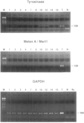
samples of our series. Figure 1 shows RT-PCR results in the
remaining 10 CKS tissues with expression of GAPDH mRNA.
Specific amplification for individual MA markers was only
observed for tyrosinase in cases 2, 10, and 12 of our series (as
also confirmed by Southern blot hybridization) (Figure 1).
For eight of such patients (cases 2, 3, 7, 9, 10. 12, 14, and 15 of
Tyrosinase M 1 2 3 10 12 14 15 T N - 1 2 5 Melan A / Marti M 1 2 3 4 7 9 10 12 14 15 T N - 1 2 8
Figure 1), peripheral blood sample was obtained (after a written
informed consent) and RT-PCR was performed. Tyrosinase
was detected in one CKS patient (case 2; 12.5%), whereas
MelanA/Martl marker was found in three blood samples
(cases 3, 7, and 9; 37.5%) (data not shown).
Considering the RT-PCR results on microdissected tumor
tissues, detection of tyrosinase in 3 of 10 (30%) samples and
absence of amplification for MelanA/Martl is a strong
indica-tion that these two markers present a different specificity as
melanoma-associated antigens. However, we found expression
of both MA mRNAs in peripheral blood of CKS patients (at
rates comparable to those we have previously reported [3]).
While positivity to tyrosinase in peripheral blood from CKS
patients seems to be due to the presence of metastatic tumor
cells expressing this MA marker, there is no explanation for the
detection of circulating MelanA/Martl. One could speculate
that in CKS patients cells of melanocytic lineage expressing
MelanA/Martl might be somehow induced to dissemination
and, thus, found in peripheral blood. Further improvements of
bio-technologies are awaited to systematically perform
addi-tional studies on large collections of CKS tumor tissues in
order to confirm our findings.
G. Palmieri,
1A. Cossu,
2A. Lissia,
2L. Leoncini,
3S. Lazzi,
3P. A. Ascierto,
4G. Castello
4& F.Tanda
2*
^Institute of Molecular Genetics, C.N.R., Algliero;
2Insti-tute of Pathology, University of Sassari; ^InstiInsti-tute of
Pathology, University of Siena; * Division of Clinical
Immunology, National Tumor Institute, Naples, Italy;
*Author for correspondence (e-mail: [email protected])
M l 2 3 4 7 9 10 12 14 15 T N Nc
- 1 6 0
Figure I RT-PCR results on microdissected tumor samples from CKS patients. PCR products were separated by electrophoresis on a 2% agarose gel and directly visualized by ethidium bromide staining. T -human melanoma-derived cell line SK-MEL-29 as positive control. N - PCR reagents and primers without RNA as reaction negative control. Nc - RT reagents and random examers without RNA as cDNA negative control: M - marker.
References
1. Redondo P, Sanchez-Carpintero I.Vazquez-Doval J, Quintanilla E. Classic Kaposi's sarcoma and vascular endothelial growth factor. Acta Derm Venereol 2000; 80: 218-9.
2. Davidovici B, Karakis I, Bourboulia D et al. Seroepidemiology and molecular epidemiology of Kaposi's sarcoma-associated herpes-virus among Jewish population groups in Israel. J Natl Cancer Inst 2001; 93: 194-202.
3. Palmieri G, Ascierto PA, Satriano SMR et al. Circulating mela-noma-associated markers detected by RT-PCR in patients with classic Kaposi's sarcoma. Ann Oncol 2000; 11: 635-6.
4. Emmert-Buck MR, Bonnert RF. Smith PD et al. Laser Capture Microdissection. Science 1996; 274: 998-1001.
5. Palmieri G. Ascierto PA, Cossu A et al. Detection of occult melanoma cells in paraffin-embedded histologically-negative sen-tinel lymph nodes using a reverse transcriptase-polymerase chain reaction assay. J Clin Oncol 2001: 19: 1437-43