Università degli Studi di
Ferrara
DOTTORATO DI RICERCA IN
"Biochimica, Biologia Molecolare e Biotecnologie"
CICLO XXVI
COORDINATORE Prof. Bernardi Francesco
Improved Adoptive T-cell Therapy Protocols for
EBV-driven Malignancies
Settore Scientifico Disciplinare BIO/11
Dottorando Tutore
Dott. Faè Damiana Antonia Prof. Di Luca Dario
_______________________________ _____________________________
- 1 -
- 2 - Summary
INTRODUCTION ... 2
EBV-biology ... 2
EBV: the dark side ... 3
EBV-driven lymphomagenesis ... 3
EBV and carcinomas ... 7
Nasopharyngeal carcinoma (NPC) ... 7
Immunotherapy strategies against EBV-associated malignancies ... 10
EBV-specific immunotherapy in PTLD ... 12
EBV-specific immunotherapy in NPC and HL ... 13
BARF1 as novel target for NPC immunotherapy ... 15
AIM OF THE STUDY... 21
MATERIALS AND METHODS ... 23
RESULTS ... 29
DISCUSSION ... 44
- 3 - INTRODUCTION
EBV biology
Epstein-Barr Virus (EBV) is a large enveloped virus, belonging to the γ-Herpesviridae subfamily. It is commonly widespread and it infects about 95% of world population. EBV preferential targets are B cells, although other lymphoid and epithelial cells may be also infected by this virus. EBV entry in B lymphocytes occurs through interaction with complement receptor type 2/CD21, whereas the mechanism of epithelial cell infection is still not completely known. It is still unclear whether EBV infects B cells or epithelial cells first, but it seems that, for an efficient infection of polarized basal epithelial cells, a cell-to-cell contact with B cells is necessary (1, 2). Moreover, it has been shown that virions released from B cells infect efficiently epithelial cells but have less affinity for these lymphocytes, whereas EBV particles released from epithelial cells infect efficiently B cells (3, 4). These findings suggest that EBV shuttles from B to epithelial cells during persistence, and that epithelial cells undergoing lytic cycle provide a source of virions for virus spreading to other individuals. Upon its primary infection in the oropharyngeal cavity, EBV can cause a lytic infection to produce progeny or it can establish a latent infection in B lymphocytes with different program-gene usage depending on cell type and on the differentiation stage of the infected cells. These are referred to as latency programs, which are necessary for the long-life persistence of the virus in the infected hosts, but similar programs are also detected in the various EBV-driven tumors.
In physiological conditions, EBV life cycle may be described by the Germinal Center Model (GCM) (5) (Figure 1). EBV enters the lymphoid tissue of the Waldeyer’s ring and at some stage it infects naïve B cells, where the virus establishes the growth program, also known as Latency III. During this phase, EBV expresses the EBERs (small non coding mRNA) and all the 9 latency genes, including the EBNA gene family (Epstein-Barr Nuclear Antigens 16) and the LMP gene family (Latent Membrane Protein1, 2A and -2B). Naïve B cells activated into proliferating infected blasts move to germinal center (GC) where EBV switches its Latency III program into more restricted forms (Latency II, or the “default” program). This phase is characterized by a restricted gene expression pattern (only LMPs and EBNA1 are expressed), which promotes cell survival of EBV-infected memory B cells. The memory compartment is indeed considered the site of viral long-term persistence, characterized by the Latency I program in which only EBNA1 or
- 4 -
occasionally LMP-2 may be expressed. In these memory B cells, EBV can even completely silence the transcription of genes encoding for latency proteins (putative Latency 0). At any time, EBV can enter the lytic cycle in a small subset of terminally differentiated plasma cells producing a progeny that allows the virus to spread within and outside the infected host. Although this model is supported by several experimental data, the actual role of EBV in GC formation and the involvement of LMPs on the maturation of memory B cells are still debated (5).
Normally, in healthy individuals EBV is carried by a stable number of B cells in the blood, and virus replication is constantly monitored by immunological surveillance through a specific subset of EBV-specific cytotoxic T lymphocytes (CTLs) and antibodies (6). However, when the complex and delicate balance between immune system and EBV is altered, several types of EBV-related disorders, including malignancies, may occur.
EBV, the dark side.
EBV seroconversion is usually asymptomatic, especially when it occurs in childhood, but individuals infected in late teens or early twenties may develop Infectious Mononucleosis (IM), an acute infection caused by a vigorous immunopathologic response to EBV-infected cells. After IM, all EBV-EBV-infected B cells display a type III latency pattern.
More importantly, since its discovery in a lymphoblasts culture of a Burkitt’s lymphoma, EBV was found to be related to a wide variety of malignancies (Table 1), and it is considered a “group I carcinogenic agent” since 1997 by the world Health Organization (WHO) (7). In particular, EBV is causally associated mainly with tumors of lymphoid origin, although particular types of carcinomas can be also EBV positive.
EBV-driven lymphomagenesis.
EBV has elegantly evolved different strategies to promote cell proliferation and transformation leading to specific type of lymphomas (8). The different EBV latency patterns include the expression of proteins that may variably contribute to lymphomagenesis (Table 1). In the broad Latency III, the full set of EBV latency proteins is expressed and contribute to lymphomagenesis by acting cooperatively. On the other hand, these proteins, mainly EBNA-3, -4, and -6, are strongly immunogenic and mediate the prompt recognition and elimination of EBV-infected cells by EBV-specific CTLs.
- 5 -
Consistently, most EBV-associated lymphomas show prevalently Latency I or II, whereas EBV+ lymphoproliferations of immunosuppressed individuals usually show a Latency III pattern. The main transforming EBV protein is LMP-1 (9), whose expression may depend on EBNA-2. LMP-1 exerts multiple oncogenic mechanisms, including a functional mimicry with a constitutively active CD40, being able to activate the NF-κB pathway (B-cell growth signal) and induce the transcriptional activation of telomerase reverse transcriptase (TERT) (10-12). LMP-1 can also interact with other crucial cell signaling pathways, such as JAK/STAT, MAPK, Wnt and IRF4, which are key molecules for B-cell growth and survival. Notably, it was demonstrated that LMP-1 up-regulates Bcl-2 and A20, thus blocking p53-mediated apoptosis. Moreover, LMP-1 may also impair the B-cell differentiation to plasma cells through down-regulation of BLIMP1α, thus avoiding the lytic cycle entry and maintaining EBV invisible to the immune system. On the other hand, even though LMP-2 is not required for B-cell transformation, its functions partially overlap with those of LMP-1 in promoting cell survival (13, 14).
EBNA-1 is a DNA binding factor, responsible for the maintenance of the EBV episome in infected cells. It can cause genomic instability and alteration of DNA repair mechanisms, thereby promoting cell cycle progression despite the occurrence of severe DNA damages. Other EBNA proteins such as EBNA-2 and EBNA-LP are able to modulate the expression and/or function of c-myc, CD21, CD23 and cyclin-D2 (15, 16). Besides the direct interaction with cell cycle and survival regulatory signals, EBV can also modulate the activity of innate and adaptive immunity. In particular, the viral non-coding RNAs, EBERs, highly expressed in all latency programs, can modulate innate immune responses affecting different pathways, including Toll-like receptors, type I-Interferon signaling and may deregulate pro-inflammatory cytokines (17). Moreover EBV expresses a viral homologue of 10 (18) and can up-regulate human 6, 8 and IL-10 through LMP-1 (19). EBV is also able to escape immune recognition by limiting the number of antigens that can be presented by HLA class-I molecules, or through the expression of viral antigens that have an impaired ability to be processed by proteasome as the case of EBNA-1 (20).
- 6 - Table 1. EBV associated lymphomas.
B-cell Lymphoma in immunocompeten t hosts EBV frequen cy (%) Latenc y progra m Mainly interested geographic area Current Therapy Prognosis Endemic Burkitt’s Lymphoma 100 Type I Equatorial Africa,
Papua New Guinea High-intensity Short-duration chemotherapy EPOCH-R (experimental) Remission in >85% (High toxicity) 100% of remission Sporadic Burkitt’s lymphoma 20-30 Type I EBV-positive Diffuse large B-cell lymphoma of the elderly (DLBCL) 100 Type II World Wide Chemotherapy Poor prognonsis (Adoptive T cell Therapy should be considered) Poor prognosis 5-Year overall survival in 26% of patients DLBCL associated with chronic inflammation 70 Type II Pyotorax-associated lymphoma (classically associated with DLBCL) 70 Type II Japan Surgical resection Chemotherapy Radiotherapy Classical Hodgkin’s Lymphoma (HL) 40 Type II Long term remission in the majority of cases Poor prognonsis in EBV+ relapsing cases (new therapeutic strategies are needed) Lymphomatoid granulomatosis (LYG) 100 Type II Chemotherapy EPOCH-R (grade III) IFN (grade I-II)
(experimental approaches) Frequent relapse, poor prognosis for Grade III disease. Better prognosis for grade I-II. Promising results with IFN
- 7 - Peripheral T cell
lymphoma, NK tumors, and
EBV-associated haematophagocyti c syndrome (HS) 100 Type II (EBER s and LMP1 only) Japan- South-East Asia Aggressive NK
leukemia >90 Type II Far East
Poor prognosis
Extranodal NK-T cell Lymphoma
(Nasal Type)
100 Type II Asia, Central-South America Radiotherapy (no standard treatment) No effective chemotherapy New clinical trials with SMILE (L-aspariginase, methotrexate, dexamethasone, etoposide, ifosfamide) Extremely poor when treated with anthracyclin es ……… … ORR 74%, Complete remission rate 38% Inflammatory pseudo-tumor-like follicular dendritic cell tumor IPLFD
Near 100 Type II Angioimmunoblast ic T-cell lymphoma (AITL) and associated peripheral T cell lymphoma (PTCL)
>90 Type II North America Europe Tipical Lymphoma chemotherapy (no standard treatment) Frequent relapses (new therapeutic strategies are needed) Lymphomas in immonocomprom ised hosts EBV frequen cy (%) Latenc y progra m
Current Therapy Prognosis
Post transplantation lymphoproliferativ e disorders (PTLD) B-cells Near 100 Type III Rituximab Immunotherapy Responses in 35-70% Great success PTLD NK/T cells >70 Type III Burkitt’s
Lymphoma (HIV) 25-35 Type I
BL classical treatment Hodgkin’s
lymphoma (HIV) >80 Type II
Classical HL for general population treatment More aggressive of HL Primary effusion
Lypmhoma (PEL) >80 Type I
No effective
therapy exists Very poor Plasmablastic Lymphoma Near 70 Type I-II No standard therapy Poor Plasmablastic Lymphoma, oral type (HIV) 100 Type I
- 8 - Primary CNS lymphoma (HIV) 100 Type III Combined chemotherapy (no standard treatment) 5 years survival 20-30% NHLs with primary immune disorders >90 Type III Treatment for NHL Iatrogenic immunodeficiency lymphoma 40-50 Type III Therapies for autoimmune disease and reduced immunosuppressiv e regimen Sometimes, spontaneous remission for EBV+ cases
EBV and carcinomas.
The complex interactions between EBV and genetic or environmental factors triggering EBV-mediate carcinogenesis, are still controversial and not completely understood. In this regard, it was recently demonstrated that at least a 10% of total gastric cancers (GC) is associated to EBV (21), while, to date, no evidence support the possible correlation between EBV and breast cancer.
Conversely, available evidence strongly supports the association between EBV and nasopharyngeal carcinoma.
Nasopharyngeal carcinoma (NPC).
Nasopharyngeal carcinoma is an epithelial malignancy that arises from the lateral nasopharyngeal recess (Rosenmϋller’s Fossa). WHO classification distinguishes two NPC histopathologic variants, a Squamous Cell Carcinoma, the well-differentiated keratinizing NPC and a keratinizing NPC, divided into differentiated non-keratinizing carcinoma and undifferentiated carcinoma of Nasal Type NPC (UNPC)(22), which is typically paired with a considerable infiltration of normal chronic inflammatory lymphocytes.
NPC is endemic in South-East Asia, Southern China and in Alaska. High incidence is also found in Northern Africa, Taiwan, Vietnam and the Philippines. Low incidence (below 1/100,000) is found in most western countries, especially in Europe and North America, even though the presence of immigrant from the endemic zones significantly increases the incidence of NPCs in south Italy and in France (23).There are multiple risk factors related to NPC onset, including diet, smoke, complex genetic predisposition (24) and different environmental factors (Table 2). Notably, despite oncopathogenic mechanisms are still
- 9 -
not completely understood, EBV infection seems to be invariably associated to NCP development, since virus genome is virtually present in all NPC biopsies of all histological subtypes (25).
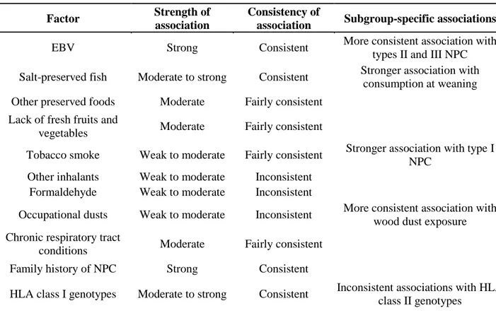
Table 2: Summary of possible risk factors associated to NPC development
Factor Strength of
association
Consistency of
association Subgroup-specific associations EBV Strong Consistent More consistent association with
types II and III NPC Salt-preserved fish Moderate to strong Consistent Stronger association with
consumption at weaning Other preserved foods Moderate Fairly consistent
Lack of fresh fruits and
vegetables Moderate Fairly consistent
Tobacco smoke Weak to moderate Fairly consistent Stronger association with type I NPC
Other inhalants Weak to moderate Inconsistent Formaldehyde Weak to moderate Inconsistent
Occupational dusts Weak to moderate Inconsistent More consistent association with wood dust exposure Chronic respiratory tract
conditions Moderate Fairly consistent Family history of NPC Strong Consistent
HLA class I genotypes Moderate to strong Consistent Inconsistent associations with HLA class II genotypes
NPC cells characterized by an EBV Latency II program, in which LMP-1 protein is often detected also in pre-invasive lesions and in overt tumors, underlying the possible role of this viral protein in the initial phases of malignant transformation(25). LMP-1 is also responsible for the deregulation of cellular genes, such as Bcl-2, NFκB and STAT3, which are involved in cell proliferation/survival and tumor progression.
Since epithelial cell infection by EBV can be demonstrated in vitro (Borza CM, 2002), but it has not been convincingly documented in vivo, the etiology of this tumor is still obscure. Genetic predisposing factors and and dietary carcinogens, are currently thought to be of relevance in the development of NPC. With regard to immunologic factors, it is well known that, generally, vigorous humoral and cellular immune responses control the proliferation of EBV-infected cells in healthy virus carriers. Indeed, both non-specific (NK-cell mediated) and EBV-specific (T-cell mediated) responses were shown to play important roles during primary infection, while EBV-specific T-cells appear to be
- 10 -
critically involved in restraining the proliferation of EBV infected cells during life-long persistent infection. On these grounds, different studies have confirmed that T-cells specific for EBV antigens expressed during latent and productive infection are maintained in the blood of healthy carriers at relatively high frequencies throughout life (26). Moreover, direct evidence of the importance of these EBV-specific T-cells and controlling the oncogenic capacity of the virus is provided by the occurrence of EBV-associated immunoblastic lymphomas in patients where this activity is impaired by congenital immunodeficiency, immunosuppressive therapy or HIV infection (27). Thus, these EBV-associated lymphomas can be prevented or even cured by adoptive transfer of in vitro activated and expanded EBV-specific T cells (28), suggesting that the reconstitution of EBV-specific immunity could also be a useful strategy in the management of NPCs. Early after the discovery of EBV association with NPC, a deregulation of the EBV-specific immune response with elevated IgA titers against the virus was documented (29). This indicated that the immune response at the site of tumor development was changed, and that the tumor might influence local microenvironment to facilitate its growth. Indeed, conclusive studies supported the notion that local immune suppression rather than a systemic deficiency in EBV-specific immune control may contribute to NPC development. In these studies, EBV specific CD4+ and CD8+ T-cell responses could be reactivated from peripheral blood of NPC patients (30). Even though LMP-1- and LMP-2-specific CD8+ T-cells were enriched in tumor infiltrating lymphocytes, their cytotoxicity and cytokine secretion was impaired. This impairment could be due to the presence of CD4+CD25+FoxP3+ natural Treg cells in the tumor tissue, which could suppress EBV-specific immune responses against NPC even after correct homing of effector T cells(31). In addition to active T-cell suppression at the tumor site, the efficiency with which NPC can present antigens to T-cells might also be compromised. While earlier studies based on a limited number of NPC cell lines suggested that antigen processing for HLA-I presentation was intact in NPC cells (32), a more recent study on primary tumor tissues suggested that the MHC-I antigen processing machinery is down-regulated in the majority of tumors (33). Even though no functional deficiency of MHC-I antigen presentation could be tested in this latter study, this makes possible to speculate that in addition to active immune suppression at NPC tumor site, the recognition of tumor cells by CD8+ T-cells could be also impaired. Together, these data suggest that NPC impairs EBV-specific immune control locally, while allowing efficient systemic immune responses against this virus.
- 11 -
The prognosis of NPC is strongly related to carcinoma histotypes and to the stage of the disease, with a survival rate of 70% at 2-years and 30% at 5-years (34, 35). Accordingly, conventional treatments for NPC are still unsatisfactory and are often accompanied by severe long-term side effects (30). Therefore, the strict association of NPC with EBV infection and the expression of immunogenic viral antigens in tumor cells, has stimulated intense efforts to develop strategies of immune intervention that could complement or even substitute current therapeutic regimens for a better control of this malignancy.
Immunotherapy strategies against EBV-associated malignancies.
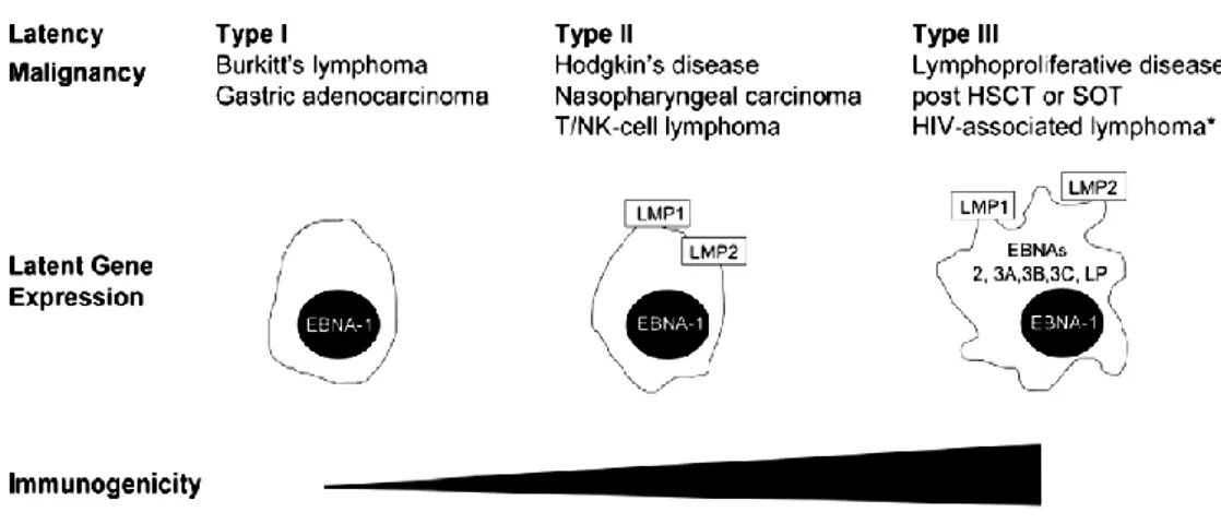
The development of EBV-associated malignancies may be favored by an underlying defect in virus-specific CTL immunity and function. Much work has been focused in the last years on the reconstitution of CTL immunity to EBV in transplant patients, who are rendered susceptible to PTLD by iatrogenic immune suppression modalities. Moreover, recent data indicates that other EBV-associated diseases such as NPC, HL, and chronic active EBV infections (CAEBV) can potentially be treated by immunotherapeutic approaches. Indeed, virus infection in these tumor cells is characterized by the expression of a limited set of EBV latent proteins, thus limiting tumor immunogenicity (Figure 1), since they may serve as targets for specific immunotherapy.
Figure 1. EBV latent protein expression and immunogenicity of common EBV-associated malignancies. Only EBV latent protein expression is shown. In EBV latency types I-III EBV
- 12 -
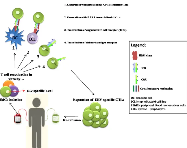
To date, there are only limited experiences with human EBV vaccines (36, 37). Although potentially ideal for preventing EBV-associated malignancies, vaccines providing life-long immunity against primary EBV infection may not be feasible, because the type of immunity required to prevent repeated infection through mucosal surfaces is not clearly defined. Moreover, repeated infections with different EBV strains have been described, suggesting that the natural immune response to EBV is not sufficient to protect healthy EBV-positive individuals from recurrent infections. Vaccine strategies for the immunotherapy of EBV-related tumors should seek to elicit or boost specific cellular immune response against EBV antigens expressed in these malignancies. Individuals likely to benefit from this approach are EBV-seronegative patients prior to solid organs transplant (SOT) or patients affected by EBV-associated malignancy with a low tumor burden or in remission. However, vaccine strategies are unlikely to be the optimal method to enhance EBV-specific T-cell responses for patients who are immunocompromised due to immunosuppressive therapies after transplantation or as a result of HL. In such cases, the adoptive cell therapy (ACT) with ex vivo activated EBV-specific CTL seems to be more promising, especially because it could benefit from T-cells engineering, able to enhance effector cells’ specificities and functions. Briefly, ACT consists in the infusion of autologous or donor-derived tumor/virus-specific T-cells in patients, upon an ex-vivo enrichment and expansion of antigen-specific effectors, in order to reconstitute or boost CTLs functions, with the final aim to kill tumor cells and avoid relapses (Figure 2). Furthermore, in the last years, ACTs took advantage of molecular biology techniques to improve effector cells’ specificities by CTLs engineering either with T-cell receptors (TCRs) or Chimeric Antigen Receptors (CARs) specific for a particular tumor associated antigen (TAA) (38). These new strategies are intriguing and confer high specificity to CTLs prior to infusion in patients, but they require higher production costs and strictly regulated manufacturing controls.
- 13 -
Figure 2. Adoptive T-cell therapy. Peripheral blood mononuclear cells are isolated from buffy-coat
derived from patients or donors; anti-tumor specific T-cells could be selectively reactivated with 1) dendritic cells loaded with particular peptides or proteins, or transfected with vectors expressing protein of interest 2) with autologous LCLs (most frequent in case of EBV targeted immunotherapy) naturally expressing antigens of interests or transfected with vectors; alternatively, 3) CTLs could be engineered with TCR or 4) with CAR, to confer to the effectors a high-TAA specificity. Specific anti-tumor effectors are expanded in vitro and the re-infused in patients.
EBV-specific immunotherapy in PTLD.
EBV infection poses a significant problem in transplant patients who are greatly immunosuppressed in order to prevent chronic organ rejection. Risk factors for the development of PTLD include EBV-seronegativity in the transplant recipient, the type of organ transplanted (highest in lung and heart and lowest in liver and kidney), and the level and type of immune suppression. PTLD emerges as either of recipient or donor origin, depending on the type of transplant. For example, bone marrow transplant (BMT) patients develop PTLD of donor origin, as EBV-infected B cells derived from the donor
- 14 -
marrow proliferate uncontrollably into lymphoma. Conversely, SOT patients develop PTLD of recipient origin, as EBV released from the transplanted organ infects the recipient’s B cells. On these grounds, initial studies investigated the potential of EBV-specific CTLs to treat PTLD in BMT patients, as CTLs could be easily generated from EBV-seropositive, immunocompetent donors. Pioneering studies (39, 40) demonstrated that PTLD was resolved after adoptive transfer of EBV-specific CTLs grown from donor peripheral blood mononuclear cells. The method developed to stimulate and expand large numbers of EBV-specific CTLs utilized the donor’s autologous EBV-immortalized lymphoblastoid B-cell lines (LCLs), which were co-cultured with donor PBMCs in the presence of interleukin-2 (IL-2). Similar to PTLD tumor cells, LCLs also have a latency III phenotype and can activate polyclonal EBV-specific CTLs with a broad reactivity to a range of EBNA-derived p epitopes. The resulting EBV-specific CTLs used in these studies killed donor LCLs in vitro, did not compromise allograft function, and most importantly, eradicated tumors. More recently, further studies obtained similar results from a group of SOT patients (37, 41), however graft versus host disease (GvHD) or severe local inflammation and tissue damages frequently occurred after non-autologous T cell infusion (37, 42).
EBV-specific immunotherapy in NPC and HL.
Clinical evidence accumulated so far indicates that adoptive therapy with EBV-specific CTLs (EBV-CTLs) is safe, well tolerated and particularly effective in the case of most immunogenic tumors like PTLD (43). In latency II EBV-associated malignancies, however, the more restricted pattern of viral latent antigen expression strongly limits the therapeutic potential of EBV-CTLs obtained by conventional protocols based on the use of autologous LCLs as a source of viral antigens. In fact, the infusion of EBV-targeted autologous CTLs was shown to enhance specific immune responses and to induce objective clinical responses only in a proportion of NPC and HL cases (44, 45). This is probably due to the weak immunogenicity of LMP-1 and LMP-2. To improve protocols for in vitro expansion of T-cells specific for the EBNA-1, LMP-1 and LMP-2 antigens, which are present in these malignancies, recombinant viruses encoding for these EBV products have been utilized to expand specific CD8+ T-cells, which could protect against LMP-positive tumor growth in mice (46). However, these T-cell lines, targeting a select subset of EBV antigens, are just now starting to be tested in patients. As an alternative to
- 15 -
passive immunization, adoptive T-cell transfer of EBV antigen loaded DCs has been evaluated for inducing protective CD8+ T-cell responses against NPC. Although LMP-2-specific CD8+ T cells could be expanded after peptide-pulsed DC injection in NPC patients, these responses were too weak or transient. Thus, vaccine approaches that primarily target CD8+ T cells have not yielded sufficient therapeutic success against EBV-associated lymphomas. Learning from these trials and as a result of a better understanding of the crucial role for CD4+ T cells in assisting CD8+ T cell immunity, more recent vaccine formulations aim to incorporate both CD4+ and CD8+ T-cell antigens. In addition to CD4+ T-cell help for CD8+ T-cell responses, CD4+ T-cells can also target EBV-transformed B cells directly, adding to their value as vaccine targets. As previously observed, many of these immunization strategies target DCs, which have been shown to be more efficient than LCLs in expanding EBV specific T cells and are capable of priming protective CD4+ and CD8+ T cell responses against EBV transformed B cells in vitro (47). CD4+ and CD8+ T cells, expanded with DCs, which had been infected with a recombinant adenovirus encoding LMP2, were able to kill NPC cells (48). Finally, considering that NPC’s and HL’s malignant cells have functional antigen processing machinery and express HLA and co-stimulatory molecules (49, 50), the demonstration that other viral latent proteins expressed by these neoplastic cells may serve as tumor-associated antigens could provide the rational background to improve the clinical efficacy of adoptive immunotherapy protocols in this setting.
Adoptive immunotherapy with EBV-specific CTLs has proven to be an effective strategy in many PTLDs (51) to reconstitute EBV-specific immunity, prevent the development of PTLD (52) and treat patients with established PTLD. For other EBV-associated malignancies, the use of EBV-specific CTL has proven less efficacious; however the results obtained so far are sufficiently encouraging to justify continued active exploration of this approach. Novel approaches are being developed to enhance the potency of EBV-specific immunotherapy by targeting CTL to subdominant EBV proteins and by genetically modifying these effector cells to render them resistant against inhibitory cytokines or immunosuppressive therapies. Notably, such strategies could have broad implications for the adoptive immunotherapy of a broader spectrum of human cancers with defined tumor antigens. All these approaches open promising avenues to enhance or prime protective EBV-specific immune responses (53), which have been suppressed by the tumor cells itself or by their microenvironment, and whose absence might predispose for the development of EBV-associated malignancies.
- 16 - BARF-1 as novel target for NPC-immunotherapy.
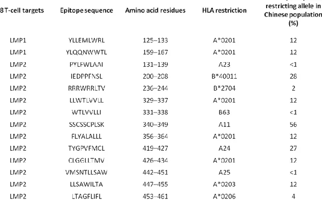
As previously discussed, EBV-specific CTLs have been successfully used for the prophylaxis and treatment of the highly immunogenic PTLDs, as demonstrated by a large number of phase I and phase II trials (54). Conversely, the clinical experience for other EBV-associated malignancies, such as HL and NPC, is limited and the results obtained so far indicate that EBV-specific CTLs are less effective in these settings (Table 3 (54)). EBV-driven tumorigenesis encompasses not only the coordinate activity of latent viral proteins but also to the ability of the virus to inhibit host immune responses directed towards EBV-carrying lymphocytes. The EBNA-1 protein was initially considered to be invisible to the immune system due to the long internal glycine-alanine repeat domain that hampers the proteasome-mediated processing of the protein, thus preventing the efficient generation of peptides that can bind to HLA class I (20). More recent evidence however indicates that EBNA-1-specific CD8+ and CD4+ T cells can be successfully generated from patients with PTLD or HL for therapeutic purposes (55, 56). Another interesting question is why LMP-1 and LMP-2 expression is tolerated in latency II or III malignancies, despite the fact that these viral proteins carry CTL target epitopes restricted through common HLA alleles (table 4). Analysis of virus-specific CTL responses at the tumor site of EBV-positive HL patients showed that infiltrating CTLs are functionally impaired and unable to eliminate the neoplastic cells (57). Therefore, decreased CTL efficacy is not only due to the ability of EBV to generate an immunosuppressive microenvironment, by local secretion of inhibitory cytokines, but it also involves defects in antigen processing or presentation by tumor cells and a selective down-regulation of immunodominant EBV proteins. On these grounds, one of the possible approaches to overcome these limitations is the identification of additional viral proteins expressed by tumor cells and that may serve as tumor-associated antigens to be targeted by improved CTL induction and expansion protocols.
- 20 -
The BamHI-A fragment of the EBV genome encodes for the BARF1 gene, located at nucleotide positions 165449-166189, of the B.95.8 strain. The BARF1 gene is translated into a 221 amino acids long protein, with a calculated mass of 31-33 kDa (58). This protein may play different functions in immunomodulation and oncogenicity. In particular, it has been demonstrated that BARF1 functions as a soluble receptor for human colony-stimulating factor 1 (hCSF-1) (59), and recombinant BARF1 inhibits the ability of hCSF-1 to induce proliferation of bone marrow macrophage progenitor cells. Notably, hCSF-1 is known to have a number of other activities, including induction of mononuclear cells to release cytokines, such as interferon alpha (IFN-α), tumor necrosis factor alpha (TNF-α), granulocyte colony-stimulating factor (G-CSF), and IL-1 (60). Thus, the ability of BARF1 to block hCSF-1 activity might impair cytokine release from mononuclear cells, thereby reducing cellular immune response to EBV. BARF1 could also act as an oncogene when stably expressed in mouse fibroblasts and monkey kidney cells (61) being able to induce the expression of the c-myc proto-oncogene and the CD21 and CD23 B-cell activation antigens (62). Interestingly, BARF1 was found in EBV-immortalized epithelial cells, without the expression of LMP1, which is essential for B-cell immortalization (63) and was also capable of inducing malignant transformation in Balb/c3T3 cells and in human Louckes and Akata B-cell lines (62, 64, 65). Moreover, Cohen and colleagues showed that both recombinant and EBV-derived BARF1 protein were able to inhibit IFN-α production by human monocytes (66). Therefore, BARF1 might also play an important role in modulating the innate host response to promote survival of virus-infected cells in vivo. Although BARF1 is thought to be a lytic gene in B-lymphocytes, since it is not expressed in BL cell lines (67), its expression was detected in NPC and EBV-positive gastric carcinoma (GC) tissues in the absence of the expression of other lytic genes (68). This suggests that BARF1 may be expressed as a latent gene in EBV-associated epithelial malignancies. Notably, computer analysis of BARF1 sequence predicted a cleavage site after the 20th N-terminal amino acid. The secretion of a 29 kDa BARF1-coded polypeptide (69) from human B cells was already reported by Strockbine et al. (70, 71) suggesting that almost all BARF1 protein is secreted in culture medium rendering its detection difficult in intracellular compartments. Thus, one possible mechanism of oncogenic transformation induced by BARF1 might be autocrine/paracrine cell cycle activation by the secreted form of its translation product. Finally, BARF1 is also able to induce humoral responses in EBV-seropositive individuals and may serve as a target for antibody-dependent cellular cytotoxicity in NPC patients (72).
- 21 -
- 22 - AIM OF THE STUDY.
Although adoptive infusion of EBV-specific T-cell lines constitutes a promising strategy for the treatment of patients with NPC or HL, the clinical benefit of current protocols is still unsatisfactory. One major limitation is constituted by the restricted number of EBV antigens that can be targeted in malignancies carrying a latency II (or I) and their poor immunogenicity. The oncogenic EBV protein BARF1 is expressed in the majority of NPC cases and may constitute an attractive therapeutic target. In fact, we have previously demonstrated that NPC patients have strong spontaneous CD4 and CD8 T-cell responses against BARF1 protein and derived epitopes. Moreover, BARF1-specific cCTLs can be easily generated from EBV+ donors, an important prerequisite to exploit BARF1 immunogenicity for immunotherapeutic purposes.
The present study aims at developing a new optimized protocol for adoptive immunotherapy of NPC, based on the generation of T-cell lines enriched in BARF1-specific effectors. To this end, we had to devise strategies to up-regulate BARF1 in LCLs without inducing cell apoptosis or a complete EBV lytic replication in order to allow these cells to effectively present BARF1 peptides together with other EBV target epitopes. On these grounds, we investigate different EBV lytic cycle inducers used at suboptimal concentrations for their ability to up-regulate BARF1 expression in LCLs without compromising cell survival. This approach was chosen in the light of the relative simplicity of use of drugs already adopted in the clinic or easily up-gradable to GMP standards.
BARF1-expressing LCLs were then used as antigen presenting cells to generate specific donor- and patient- derived CTLs potentially able to kill more efficiently NPC cells in a HLA-A*0201 restricted fashion . We have demonstrated that, as compared with the other drugs investigated, doxorubicin (DX) (an anthracycline family member) is able to induce a more specific expression of BARF1 mRNA in LCLs at concentrations that do not affect cell survival.
CTLs generated with DX-LCLs (DX-CTLs), showed higher specificity for targets loaded with BARF1 peptides or endogenously expressing this target protein. Intriguingly, responses against LMP1 were also enhanced in several instances. Consistently with this findings, DX-CTLs displayed a higher content in granzyme-β granules. Considering that DX is able to induce immunogenic cell death (73-76), we also investigated whether DX treatment induced the expression of molecules potentially able to enhance the
- 23 -
immunogenicity of LCLs, even at doses inducing complete apoptosis only in a minority of cells.
Feasibility and effectiveness of the protocol developed were also verified on LCLs and T lymphocytes derived from NPC patients. In particular, cytotoxicity assay demonstrated that DX-CTLs generated from NPC patients achieved a similar efficiency in terms of antigen-specific lysis as donor-derived DX-CTLs. These findings further confirm that BARF1 CTL could be successfully exploited to potentially enhance the clinical efficacy of NPC adoptive immunotherapy, and provide the rationale for a rapid up-grading at the GMP level of this innovative protocol.
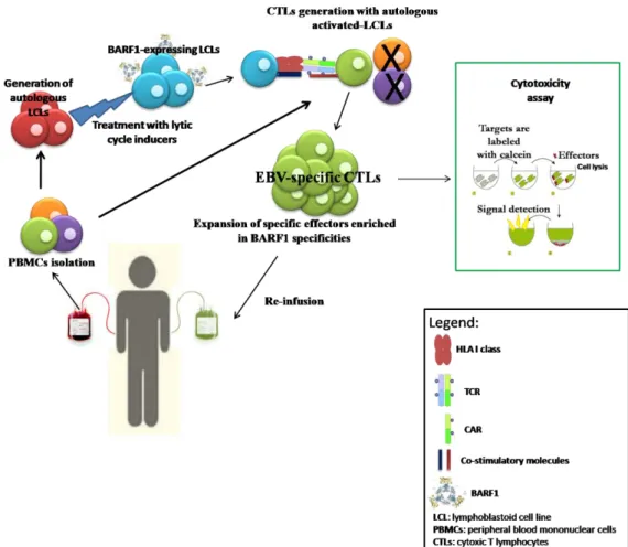
Figure 3. Representation of our new approach of immunotherapy protocol. Peripheral blood
mononuclear cells are isolated from buffy-coat derived from patients and properly criopreserved; Autologous LCLs were generated with B.95.8 EBV strain and subsequently treated with lytic cycle inducers, in order to induce BARF1 expression. PBMCs were then co-cultured with treated-LCL and autologous anti-BARF1 T-cells could be selectively reactivated and expanded. By this way, not only BARF1-specific effectors could be selected, but other cytotoxic T-cells enriched in EBV specificities will be included in the culture, and this could be further improve spectrum of T-cell killing and increase the therapeutic potential. Standard calcein AM release were performed to verify efficiency of the new immune effectors, then specific CTLs could be further expanded prior re-infusion in patient.
- 24 - 1. MATERIAL AND METHODS NPC patients and healthy donors
Four blood samples were obtained from NPC patients. All NPC cases investigated were EBV-associated as shown by in situ hybridization for EBERs. Buffy coats from 5 EBV-seropositive healthy donors were also collected and included in present study. Peripheral blood mononuclear cells (PBMCs) were freshly isolated on Ficoll-Hypaque density gradient (Lymphoprep, Freseniu Kabi Norge Halden, Norway) and cryopreserved immediately using standard procedures and viably frozen at -180°C until use. HLA-A and -B typing was performed in all cases by sequence-based typing, according to standard high-resolution typing techniques.
Reagents and antibodies
Hsp70, Hsp90, Myd88 (D80F5), and cleaved Caspase 3 (D175) antibodies were from Cell Signaling Technology (Cell Signaling Technology, Inc., Boston, MA 02241-3843); GAPDH antibody was from Abcam (Abcam, Cambridge, UK); Parp (F2), Zebra (BZ1) and β-tubulin (H-235) from Santa Cruz Biotechnology (Dallas, Texas, U.S.A.).
Cell lines and culture conditions
The following HLA-A*0201-restricted cell lines were used in the study: the DG75 human Burkitt’s lymphoma; the Granta-519 human mantle cell lymphoma; donor-derived EBV-transformed LCLs, generated in vitro by transformation of B cells using the standard EBV isolate B.95.8; the transporter associated with antigen-processing-deficient T2-A2 cells and the c666.1-A2 NPC cell line transfected in vitro with the HLA-A*0201 gene. The Ramos human BL and the c666.1-Wt NPC cell lines were used as non HLA-A*0201 controls. Phoenix cell line were used as packaging cell line for infection protocol. All cell lines were cultured in RPMI-1640 (Gibco, Grand Island, NY), containing 10% fetal bovine serum (Gibco, Grand Island, NY), 2 mM L-glutamine, 100 g/ml streptomycin and 100 IU/ml penicillin (Sigma Aldrich, St Louis, Missouri, US), with the exception of Granta-519 cell line and Phoenix, which was cultured in complete Dulbecco’s Modified Eagle’s Medium (DMEM, Cambrex Bio Science Walkersville, MD). Donor- and patient-derived EBV-specific CTL lines were cultured in CellGro® GMP DC (CellGenix GmbH, Am Freiburg, Germany), supplemented with 100 lg/ml streptomycin and 100 IU/ml penicillin (Sigma Aldrich, St Louis, Missouri, US).
- 25 - Transfection-infection protocol.
For the expression of the HLA-A*0201 allele in c666.1Wt NPC cell line, HLA-A*0201 gene was inserted into pBABE-Puro retroviral vector (Add gene), cloned into One Shot Top-10 chemically competent E.coli (InvitrogenTM life technologies) and purified using the PureYeld Plasmid Maxiprep System (Promega) according to manufacturer recommendations. Phoenix were transfected by calcium phosphate methods using a calcium phosphate Profection kit (Promega) or with DOTAP (InvitrogenTM life technologies) with 20 μg of pBABE-Puro-A2 vector accordingly to manufacturer recommendations. Briefly, Phoenix were first incubated with calcium phosphate/vector precipitate or DOTAP/vector for 24 hr at 37°C 5%CO2 in DMEM + 10%FCS. Medium
was then replaced with fresh RPMI1640 and cells were newly incubated 24 hr 37°C 5%CO2. After the secondary incubation, viral medium was collected and used to infect
c666.1 NPC cells. C666.1-Wt cells were plated in 6-well plates (106cells/ml) and incubated twice with viral medium, first, 2 hr at 32°C 5% CO2and then over night with
fresh viral medium at 32°C 5% CO2. After incubations, viral medium was replaced with
fresh RPMI1640. Selection in puromicin started into 3 days.
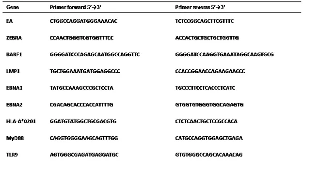
RNA extraction, cDNA synthesis and quantitative Real-time PCR (qRT-PCR). One-to-3x10⁶ cells were collected and washed twice in PBS. Total RNA was extracted from cells by QIAGEN RNeasy Mini Kit. Quantification and integrity of mRNA were determined through the Experion Automated Electrophoresis system (BIO-RAD, Hercules, CA, US). One µg of RNA was retro-transcribed into cDNA using the Iscript RT OneTube Supermix (BIO-RAD, Hercules, CA, US) according to manufacturer’s recommendations. Quantitative real-time PCR were performed in a Thermal Cycler CFX96, using SsoFast EvaGreen Supermix (BIO-RAD, Hercules, CA, US) accordingly to manufacturer’s recommendations. Primers (table 5) were designed with Primer3 (version 0.4.0) and specificity controls were performed by BLAST alignment tool. Primer were from Sigma Aldrich, St Louis, Missouri, US.
Primer sequences for the housekeeping genes 18S, beta-actin, beta-2-microglobulin and HPRT were kindly provided by BIO-RAD. Normalized fold expression was calculated with “delta-delta Ct” method.
- 26 -
Table 5: primers used for qRT-PCR. Primers were designed with primer3 web tool
(http://primer3.ut.ee/). Then were tested at different concentration of both, target and primers, before analysis.
Immunoblotting analysis.
Whole cell lysates were prepared in lysis buffer [50 mmol/L Tris-HCl (pH 7.5), 150 mmol/L NaCl, 2 mmol/L EDTA, 2 mmol/L EGTA, 2 mmol/L sodium orthovanadate, 25 mmol/L h-glycerophosphate, 25 mmol/L sodium fluoride, 1 mmol/L phenylmethylsulfonyl fluoride, 1 Amol/L okadaic acid, 5 Ag/mL leupeptin, 5 Ag/mL aprotinin, 0.2% Triton X-100, and 0.3% NP40] and lysed for 30 minutes on ice. Total protein extracts were obtained by centrifugation at 13,000 rpm for 15 minutes and protein concentration was determined by the Biorad Bradfor Protein Assay (Milan, Italy). Proteins were fractionated using SDS-PAGE and transferred onto nitrocellulose membranes. Immunoblotting was performed using the enhanced chemiluminescence plus detection system (PerkinElmer, Massachussets, U.S.A.) through Chemidoc XRS+ instrument (Biorad, Hercules, CA, US).
Induction of EBV lytic cycle in lymphoblastoid cell lines.
Lymphoblastoid B-cell lines were seeded at the concentration of 5x10⁵ cells/ml and the induction of EBV lytic cycle was achieved by incubation of cells with either with 1) TPA+NaB: 20 ng/ml of 12-O-tetradecanoyl-phorbol-1-acetate, TPA, and sodium butyrate, NaB, both from Sigma Aldrich, St Louis, Missouri, US, in complete
RPMI-- 27 RPMI--
1640 medium for 48 hr at 37 °C 5% CO2; 2) Doxorubicin (DX): 25 nM DX for 6 hr at 37
°C 5% CO2 and 3); CSP: 5 µM CSP for 6 hr at 37 °C 5% CO2. After DX and CSP CRO
treatment the cells were washed once and cultured in fresh complete RPMI-1640 medium for further 24 hr. DX and CSP were both provided from pharmacy of our istitution.
Generation of EBV-specific CTL lines.
Autologous donor- and patient-derived EBV-specific CTLs were generated and weekly re-stimulated using as antigen presenting cells (APCs) LCLs treated or not with suboptimal concentration of TPA+NaB, DX and CSP in order to induce mainly abortive EBV lytic cycle. After the treatment, LCLs were γ-irradiated 80 Gy, before the first stimulation, and 40 Gy, before each CTL culture re-stimulation. IL-2 (3 ng/ml) was added to the culture medium starting from day 14th and fresh medium was added every 3 days. Effector cells were co-cultured with APCs at a 40:1 T-cells:LCLs ratio and CTL lines differentiation/memory phenotype was monitored at day 10th and at 35th.
Flow cytometry.
The following fluorescent-conjugated monoclonal antibodies were used: Fluorescein Isothiocyanate (FITC) or Phycoerythrin-TexasRed (ECD) α-CD3 (mouse IgG1, clone UCHT1), Cyanine5 α-CD4 (PC5; mouse IgG1, 13B8.2), Phycoerythrin-Cyanine7 α-CD8 (PC7; mouse IgG1, SFCI2IThy2D3), ECD α-CD45RA (mouse IgG1, 2H4LDH11LDB9) all from Beckman Coulter, Fullerton, CA, USA; Phycoerythrin α-CD197 (CCR7) (PE; rat IgG2a, 3D12) from BD Pharmingen, Becton Dickinson, Franklin Lakes, NJ, USA; PE α-CD284 (TLR-4) (mouse IgG2a, HTA125) from Affimetrix eBioscience, San Diego, CA, USA; PE α-HLA-A2 (mouse IgG2b, BB7.2) from Acris, Herford, Germany); PE α-CRT (calreticulin) (mouse IgG1, FMC.75) from Abcam (Cambridge, UK).
Properly labelled isotypic antibodies were used as negative controls. All antibodies were used in an appropriate volume of 10% Rabbit Serum (Dako, Glostrup Denmark) and Phosphate Buffer Saline (PBS, Biomerieux, Marcy l'Etoile, France) to reduce nonspecific signal. Cytofluorimetric analysis was performed with a Cytomics FC500 (Beckman Coulter) and data were analyzed with CXP software (Beckman Coulter).
- 28 -
Cytotoxic activity of peptide-specific CTLs was evaluated using peptide-loaded T2-A2, c666.1-Wt, c666.1-A2, Granta-519 and K562 cell lines as targets in calcein-AM release assay. T2-A2 cells were pulsed for 2 hours at 37 °C at 5% CO2 either with
HLA-A*0201-restricted BARF1 peptides (p23 and p49) or with the LMP-1 epitope YLQ (ref). All target cells were resuspended in Hanks Balanced Salt Solution without phenol red (HBSS), supplemented with 5% FCS, labelled with 5 µM (T2-A2 and K562) or 7.5 µM (c666.1-Wt, c666.1-A2 and Granta-519) of calcein-AM (Calbiochem, Darmstadt, Germany) and incubated 30 minutes at 37°C, 5% CO2. Labelled cells were washed 3 times and seeded in
96-wells plate at a concentration of 5x10³ cells/well. EBV-specific CTLs were added at 20:1, 10:1, 5:1 effector:target ratio. All tests were performed in triplicate. The HLA-A*0201-specific mAb cr11.351 was added to the target cells and incubated at room temperature for 30 min to assess the HLA-A*0201 restriction of CTL responses. To obtain total calcein-releasing cells, targets were incubated with 100 µL/well of lysis buffer (25 mM sodium perborate, 0.1% Triton-X100 in HBSS, pH 9.0). Spontaneous release was determined by seeding target cells and adding 100 µL/well of HBSS. Plates were incubated for 4 hours at 37°C and 5% CO2 in a volume of 200 µL/well. Following incubation, the content of each well was mixed, plates were centrifuged and 100 µL of the supernatant was transferred to a 96-well black culture plate. Fluorescence intensity was measured by reading the plates from the top using Tecan Infinite 200 Pro (Tecan Group Ltd, Männedorf). Excitation and emission filters were 485 and 535 nm, respectively and gain was set at 70. The percentage of lysis was calculated as follows:
- 29 - Multispectral imaging flow cytometry.
The following fluorescent-conjugated monoclonal antibodies were used: PC7 α-CD8 (mouse IgG1; clone SFCI21Thy2D3) from Beckman Coulter, PE α-CD19 (mouse IgG1; clone HIB19) from eBioscience and FITC α-granzyme β (mouse IgG1; clone GB11) purchased from BD PharmingenTM, BD Biosciences. To determine the T:APC conjugate formation (CD8+ T cells and autologous B-LCL) and to quantify the specific recognition and killing of our cytotoxic cell cultures by granzyme β granules formation we performed a cytotoxicity assay, as previously described. Briefly, after 2 hours co-culture, autologous LCL+T cells (1.5x10⁶ cells/condition) were stained with α-CD8-PC7 and α-CD19-PE monoclonal antibodies in an appropriate volume of 10% rabbit serum and PBS to reduce nonspecific. Following surface molecules staining, cells were fixed and permeabilized with fixation/permeabilization buffer for 30 minutes at 4°C, washed twice, and labeled with α-granzyme β antibody in the presence of 2% rabbit serum in PBS at 4°C for at 45 minutes and, after two washes, cells were re-suspended in PBS with 1% paraformaldehyde. The cells were run on ImageStreamX cytometer using the INSPIRE software (Amnis Corporation, Seattle, WA) and images were analyzed using the IDEAS software (Amnis Corporation, Seattle, WA). Cells were excited using a 488 nm laser with intensity of 50 mW. Brightfield, side scatter, fluorescent cell images were acquired at 40× magnification. Only events with brightfield areas greater than 30 μm2 (excluding debris) and non-saturating pixels were collected. In T:APC binding experiments, 3x10⁴ events were collected for each sample. In particular, cells were gated for focused populations and doublets containing at least one T cell were gated from among all cells. Intracellular granzyme β granules formation upon specific T cell activation was determined by sub-cellular localization and spot count experiments. Thirty thousand events were collected for each sample. The cytoplasmic localization of the granules was measured using the “internalization algorithm” of the IDEAS software, defined as the ratio between intensity inside the cell and the intensity of the entire cell. The inside of the cell is defined by the “erosion mask” that fits the cell membrane. Cells containing small concentrated fluorescent spots have positive scores, whereas cells showing little and diffuse fluorescence have negative scores. Only viable cells were selected on the basis of morphologic features. Single-stained compensation controls were used to compensate fluorescence between channel images on a pixel-by-pixel basis.
- 30 - RESULTS
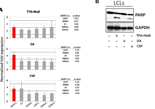
Doxorubicin up-regulates BARF-1 mRNA expression in LCLs.
Considering that BARF1 is mainly expressed in B lymphocytes undergoing EBV reactivation, we investigated the ability of different EBV lytic cycle inducers to up-regulate BARF1 expression in LCLs. As a first step, we used TPA and NaB that synergically activate EBV lytic cycle in EBV-infected cells (77, 78). In particular, healthy donor-derived LCLs were treated for 48 hours with suboptimal concentrations of TPA and NaB to induce a mainly abortive lytic cycle and the expression of several EBV latent and lytic genes was assessed by quantitative reverse transcription PCR (qRT-PCR). As shown in figure 4A (upper graph),BARF1 mRNA expression was significantly enhanced after TPA and NaB treatment (TPA+NaB) if compared to untreated cells, with a mean of 2.5 fold increase (p≤0.05). The treatment also significantly enhanced the mRNA expression of both latent (LMP1, EBNA1, EBNA2) and lytic (BZLF1/ZEBRA, EA) EBV genes, with a 2 to 3 fold-increases (p≤0.05). Therefore, at the various concentrations used, TPA and NaB treatment induced a generalized enhancement of EBV gene expression including a strong induction of genes responsible for EBV lytic reactivation, being thus unsatisfactory for our purposes (Figure 4A).
We then investigated DX and CSP as less potent lytic cycle inducers for their ability to up-regulate BARF1 expression at concentrations mainly leading to abortive EBV replication (79). These experiments disclosed that DX and CSP treatment (Figure 4A, central and lower graph) up-regulate EBV lytic and latent gene expression, but more interestingly DX-treated LCLs showed a significantly higher expression of BARF1 mRNA and a lower, although significant (p≤0,05), up regulation of the other EBV genes investigated (Figure 4A, central graph).
Furthermore, at concentrations used, DX was found to more efficiently preserve cell viability as compared to the other drugs used. In fact, LCLs treated with TPA+NaB or CSP, but not with DX, displayed late phase of apoptosis as assessed by PARP cleavage in western blot (Figure 4B).
- 31 -
Figure 4. A. DX treatment specifically enhancesBARF1 expression. qRT-PCR on EBV lytic and latent
gene performed on LCLs treated with TPA+NaB- (20ng/ml and 3mM respectively) DX- (25 nM) or CSP- (5 µM). mRNA fold expression is referred to Ctrl-LCLs (=1). 2-ΔΔCq method, was used to normalize gene expression, using 18-S as reference gene. The data represent a mean of 3 independent experiments. p value was calculated through Student t-test. B. DX-LCLs did not show a late phase of apoptosis . Apoptosis stage was assessed by PARP cleavage. Whole cell lysates corresponding to 50 μg of proteins were analyzed by immunoblotting analysis for the indicated proteins. GAPDH shows equal loading of protein for each lane.
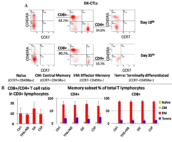
LCLs treated with doxorubicin does not affect the differentiation of healthy donor-derived EBV-specific CTLs.
EBV-specific CTL lines were generated by priming healthy donor-derived PBMCs either with untreated (Ctrl) or treated (TPA+NaB, DX, CSP) autologous LCLs. CD8+/CD4+ T cell ratio and phenotype were monitored by multiparametric flow cytometry analysis (Figure 5A) at day 10 and at day 35 of culture. The number of CD8+ T cells ranged from 49-to-82% after the first and the last stimulation, respectively. In particular, we observed a high prevalence of CD8+ in our CTLs cultures at day 35th, ranging between 6-11% (±5%) for CD4+ and 67-77% (±7%) for CD8+ (Figure 5B, left histogram). No difference in CD8+ T-cells percentage was observed among the four CTLs cultures. Combined
- 32 -
analysis of CCR7 and CD45RA receptors demonstrated that the 80-90% of CD8+ and 69-77% of CD4+ T cells displayed an Effector Memory phenotype (EM, CCR7-CD45RA-, Figure 5B, mid and right histograms respectively). Again, no significant differences were observed with regard to the differentiation features of the four CTL cultures.
Figure 5. DX-treated LCLs do not affect CTLs differentiation phenotype. A. Differentiation (memory)
status of CD3+CD4+ and CD3+CD8+ through CCR7 and CD45RA expression, in early stage of culture generation (day 10th) and after the last re-stimulation (day 35th). Percentages of Temra (CCR7-CD45RA+) EM (CCR7-CD45RA-) and CM (CCR7+CD45RA-) are shown. B. Left histogram: CD8+/CD4+ ratio within
CD3+ lymphocytes among the at day 35th. Mid and the right histograms represent CD4+ and CD8+ T-cells phenotype, respectively (data represent a mean of 3 experiments).
A
- 33 -
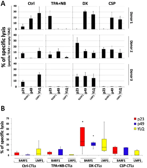
DX-CTLs specifically kill T2-A2 cells loaded with BARF1-derived peptides and tumor cell lines endogenously expressing BARF1.
EBV-specific CTL cultures were tested for their ability to recognize and kill T2-A2 target cells loaded with EBV-derived peptides (Figure 6A) in a HLA-A*0201 restricted fashion. In particular, T2-A2 cells were loaded either with two BARF1-derived peptides, p23 and p49 (80), and the LMP1-derived YLQ peptide (49). DX-CTLs were able to specifically kill both BARF1- (median=25.8% and median=21.0%, p23 and p49 respectively) and LMP1-loaded T2A2 (median=14.9%) inducing higher percentage of specific lysis if compared to Ctrl- and TPA+NaB- or CSP-CTLs (p23 peptide, median= 0, 5.7, 6.0% respectively; p49 peptide, median= 0, 11.2, 0.3% respectively; YLQ peptide, median= 2.6, 0.3, 0.3%, respectively) (Figure 6B).
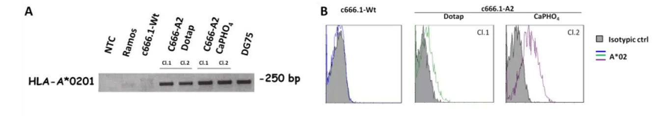
Considering that the c666.1 NPC cells do not express HLA-A molecules, we have generated a derived cell line stably expressing HLA-A*0201(c666-A2) using the pBABE_HLA-A*0201 retroviral expression vector. To confirm HLA-A*0201 gene expression in c666-A2 cell line mRNA was isolated from these cells and specific PCR was performed on cDNA. As shown in Figure 7A, both, c666-A2 CaPHO4 and DOTAP clones expressed the HLA-A*0201 transcript as the HLA-A*0201 naturally expressing DG75 cell line. HLA-A*0201 expression on cells surface of infected cells was confirmed by flow cytometry (Figure 7B). Since the c666.1-A2 cl.2 CaPHO4, displayed a significant
HLA-A*0201 cell surface expression if compared to the parental cell line, cytotoxicity assays were performed using this clone as specific target model. As shown in Figures 8A and B. DX-CTL cultures were also able to efficiently recognize and specifically kill BARF1 endogenously expressing NPC cells. In particular, these effectors showed a median 37% (mean= 58%) of specific lysis against the NPC c666.1-A2 cell line, whereas no killing was achieved against the HLA-A* lacking parental c666.1 cell line. Conversely, Ctrl-, TPA+NaB- and CSP-CTLs induced specific lysis only at low levels against c666.1-A2 (median= 5.8, 30.3, 19.3%, respectively) (Figure 8B). Finally, only low killing was observed against the myelogenous leukemia K652 cell line, thus excluding unspecific NK-like cytotoxicity.
- 34 -
Figure 6. A. DX-CTLs specifically kill T2-A2 cells loaded with BARF1-derived peptides. Induction of
BARF-1 (p23 and p49) and LMP1 (YLQ) peptide-specific T-cell responses from 3 HLA-A*0201+ healthy donors. Cytotoxicity assays were performed at 20:1 E:T ratio. Specific lysis was calculated subtracting the lysis of empty T2-A2 condition. B. Box plot represent cytotoxicity assay performed on T2-A2 cells loaded with BARF1 or LMP1 peptides. Statistical analysis were performed with Student t-test (*=p≤0.05 versus the other CTL lines).
- 35 -
Figure 7. A. c666.1-A2 cells stably express HLA-A*0201. HLA-A*0201 gene expression was assessed by
PCR on cDNA of the following cell lines: NPC cell lines, c666.1 –Wt and c666.1–A2 transfected with DOTAP or CaPHO4 technique (clones 1 and 2, respectively); Burkitt’s lymphoma cell lines, Ramos (HLA-A*0201 negative cell lines) and DG75 (HLA-(HLA-A*0201 positive cell lines). B Flow cytometry analysis of c666.1 Wt NPC cell line, or c666.1 infected (DOTAP or CaPHO4 clones) with the retroviral vector carrying the HLA-A*0201 gene. The isotypic control is shown (gray area).
The enhanced lytic activity showed by DX-CTLs prompted us to investigate intrinsic characteristics of these effector cells. In particular, intracellular content of granzyme-β granules was analyzed by spot counting through multispectral imaging flow cytometry (Figure 9A). These experiments were carried out using as stimulators autologous LCLs pulsed with two different BARF1 peptide epitopes or the YLQ LMP-1-derived peptide. Upon stimulation with empty LCLs, CTLs generated with DX-LCLs displayed the highest number of intracellular granzyme-β granules. Moreover, when co-cultured with BARF1-peptide loaded LCLs, DX-CTLs showed a marked increase in the number of granules, with most of effectors with 2-3 positive spots/cell. This effect was also observed in CTLs generated with TPA+NB or CSP, although with a lower number of total granzyme-β spots (Figure 9A). Notably, DX-CTLs showed the highest intracellular content of granules also when stimulated with LCLs pulsed with the LMP-1 peptide epitope (Figure 9A). We also exploited the ability of multispectral imaging flow cytometry to enumerate LCL-T cell doublets. As shown in Figure 9B, DX-CTLs were also able to achieve the highest frequency of doublets with BARF1-peptide pulsed LCLs. Stimulation with LMP1-loaded LCLs elicited comparable numbers of doublets in all cultures except for Ctrl-CTLs (Figure 9B).
- 36 -
Figure 8. A. DX-CTLs show high specific killing against BARF1 endogenously expressing tumor cell lines. Cytotoxic activity of CTLs derived from 2 HLA-A*0201+, against c666.1-Wt (used as negative control) and c666.1-A2 NPC cell lines, Granta-519 MCL cells and K562 (used to exclude unspecific NK-like cytotoxicity). Tests were performed at 20:1 E:T ratio. HLA-A*0201 restriction was confirmed by the use of the anti-HLA-A*0201 cr11.351 monoclonal antibody. B. Box plot represent cytotoxicity assay performed on BARF1 endogenously expressing tumor cell lines.(*=p≤0.05 in respect to other targets; **= p≤0.05 in respect to responses of the other CTLs against c666.1-A2).
- 38 -
Figure 9. A. Enumeration of granzyme β granules and LCL-T cell doublets in DX-CTLs. A.
Peptide-loaded autologous B-LCL were co-cultured with DX-CTLs and labeled with CD19, CD8 and α-granzyme β monoclonal antibodies to identify LCL-CTL doublets and α-granzyme β content. Results are indicated as the number of granzyme β spots within CD8+ T cell population. Some representative images of granzyme β-FITC, CD19-PE and CD8-PC7 positive cells are displayed. Lower histograms represent granzyme β granule content in Crtl-, TPA+NaB- and CSP-CTLs. B. The quantification of LCL-T cell doublets was performed on CTRL-, TPA+NaB-, DX- and CSP-CTL lines. The y axis displays the normalized frequency of LCL-CTL doublets containing at least one T-cell. The histograms are representative of a single experiment. The right panel displays some representative cell images (CD19-PE and CD8-PC7). LCL-CTL conjugates were acquired at 40× magnification.
DX treatment enhances LCLs immunogenicity.
In an attempt to elucidate the mechanisms underlying the enhanced functional properties of DX-treated LCLs as antigen presenting cells, we assessed whether DX was able to up-regulate HLA Class I expression. Considering that γ-irradiation is currently used to inactivate LCLs before stimulation and that is also able to modulate the expression of HLA molecules (81, 82), we investigated the possible synergism between DX treatment and γ-irradiation. We therefore monitored HLA-A*0201 expression in LCLs by qRT-PCR before γ-irradiation (NI=Not Irradiated) and 24 hours after γ-irradiation (t24) (Figure 10). DX-treated LCLs revealed a marked increase in HLA-A*0201 expression (mean=2.3 fold increase before irradiation), an effect that persisted also 24 hours after irradiation. No significant change in HLA-A*0201 mRNA was detected in TPA+NaB- and CSP-LCLs compared to untreated LCLs (mean=1.3 and 1.0 fold expression, respectively) in NI samples, whereas a decreased expression in TPA+NaB-LCLs was observed at t24 (mean=0.8 fold change). The higher expression of HLA-A*0201 in DX-LCLs was also confirmed at the protein level by flow cytometry analysis at t24 (Figure 11A).
- 39 -
Figure 10. DX treatment enhances HLA-A*0201 expression in LCLs. Relative quantification by
qRT-PCR on HLA-A*0201 allele in LCL before (NI=Not Irradiated), and 24 hr after γ-irradiation (t24). Data are relative to Ctrl-LCLs (=1), 2-ΔΔ-Ct method was used to calculate and normalize fold expression. 18-S was used as reference gene.
Figure 11. HLA-A*0201 and CRT surface expression were increase inDX-LCLs. Flow cytometry on
treated LCLs, isotypic control (dashed line) Ctrl-LCLs (gray area) and treated-LCLs (black line) are shown for all graphs. x-mean for every condition is reported A. Flow cytometry analysis on HLA-A*02 were performed at t24. B. Calreticulin (CRT) was investigated in LCLs after 3 hr of treatment.
Recent data demonstrated that DX is able to induce an immunogenic cell death both in vitro (76) and in vivo (74, 83). Although our experimental conditions were set to preserve cell viability, we hypothesized that DX could enhance immunogenicity of treated cells (84) also when used at concentrations inducing only minimal apoptotic effects. As a first
- 40 -
step, we assessed by flow cytometry the membrane translocation of calreticulin (CRT), after 3 hours of treatment, since this is a very early event occurring after exposure of tumor cells to anthracyclines (85). After DX treatment, a higher percentage of cells displayed membrane localization of CRT (49.8%; x-mean is reported in Figure 11), whereas Ctrl-LCLs and CSP-LCLs maintained a similar percentage of positive cells (35% and 33% respectively), and TPA+NaB induced a slight decrease (28.8%) (Figure 11B). After CRT exposure, a secondary event in ICD is the High Mobility Group Box1 (HMGB1) release in culture medium (86). This parameter was evaluated through enzyme linked immunosorbent assay (ELISA) at the end of treatments (Figure 12). The extent of HMGB1 release by DX-LCLs was about 3 fold higher than that of Ctrl-LCLs, whereas for TPA+NaB- and CSP-treated LCLs, the releases were similar (between them) and lower than that of Ctrl-LCLs (about 0.8 fold change).
Figure 12. DX treatment increases HMGB1 release from LCLs. ELISA performed on LCL supernatants
at the end of each type of treatment. The histogram represents the fold increase of HMGB1 release by treated LCLs, in comparison to untreated LCLs (ctrl=1). Experiments were performed in triplicate and performed at least three times on different donor-derived LCLs.
Protein expression of other two crucial ICD markers, HSP70 and 90, was assessed by western blot, immediately after γ-radiation and before co-culturing with CTLs (t0), and at t24 to assess the possible contribution of γ-irradiation to the enhanced immunogenicity of DX-LXLs (87). (Figure 13). Protein expression of both HSPs increased in DX-LCLs at t0 and t24, and a slight increase of HSP70 was observed in CSP-treated LCLs at t24, whereas TPA+NaB and CSP down regulated both HSPs (CSP at t0 and TPA+NaB at both time points) as a possible consequence of concomitant apoptosis.
- 41 -
Figure 13. DX treatment up-regulates HSP-70, HSP-90 and MyD88. PARP cleavage and protein
expression of HSP70, HSP90 and MyD88 were analyzed at different time point: immediately after γ-irradiation (t0) and 24hrs after γ-γ-irradiation (t24); Whole cell lysates corresponding to 50 μg of proteins were analyzed by immunoblotting for the indicated proteins. GAPDH shows equal loading of protein for each lane.
We also evaluated the expression of TLR-4 and MyD88 proteins (74), which constitute the functional receptor complex of HMGB1 and are mediator of immunogenicity induced by several drugs. Flow cytometry analysis showed that DX markedly increased the number of TLR-4-expressing cells (63.3% vs. 38.1% of control LCLs). Treatment with CSP also induced TLR-4 up-regulation in LCLs although with a slightly lower increase in the percentage of positive cells (54.4%) whereas TPA+NaB had only marginal effects (Figure 14). Moreover, DX strongly up-regulated MyD88 expression in LCLs as shown by immunoblotting (Figure 13), whereas the expression levels of this proteins decreased in untreated, TPA+NaB- and CSP-LCLs, becoming almost undetectable at 24 hrs (Figure 13).