Short-term weight gain after antiretroviral therapy
initiation and subsequent risk of cardiovascular disease
and diabetes: the D:A:D study*
AC Achhra,1A Mocroft,2P Reiss,3C Sabin,2L Ryom,4S de Wit,5CJ Smith,2A d’Arminio Monforte,6A Phillips,2 R Weber,7J Lundgren8and MG Law1for the D:A:D Study Group
1
Kirby Institute, UNSW Australia, Sydney, NSW, Australia,2
Research Department of Infection & Population Health, University College London, London, UK,3
Division of Infectious Diseases and Department of Global Health, University of Amsterdam, Amsterdam, The Netherlands,4
University of Copenhagen, Copenhagen, Denmark,5
Infectious Diseases Department, Saint-Pierre University Hospital, Brussels, Belgium,6
University of Milan Clinic of Infectious Diseases, Milan, Italy,7
University Hospital in Zurich, Zurich, Switzerland and8
Rigshospitalet & University of Copenhagen, Copenhagen, Denmark
Objectives
The aim of the study was to assess the impact of the gain in body mass index (BMI) observed immediately after antiretroviral therapy (ART) initiation on the subsequent risk of cardiovascular disease (CVD) and diabetes.
Methods
We analysed data from the Data Collection on Adverse Events of Anti-HIV Drugs (D:A:D) cohort study. Outcomes were development of (i) CVD (composite of myocardial
infarction/stroke/coronary procedure) and (ii) diabetes. The main exposure variable was change in BMI from ART initiation (pre-ART) to 1 year after initiation (continuous variable) in treatment-naïve individuals initiating ART with no history of CVD or diabetes (for respective outcomes). BMI [weight (kg)/(height (m))2] was categorized as underweight (< 18.5), normal (18.5–25), overweight (25–30) and obese (> 30). Poisson regression models were fitted stratified for each pre-ART BMI category to allow for category-specific estimates of incidence rate ratio (IRR). Models were adjusted for pre-ART BMI and CD4 count, key known risk factors
(time-updated where possible) and calendar year. Results
A total of 97 CVD events occurred in 43 982 person-years (n = 9321) and 125 diabetes events in 43 278 person-years (n = 9193). In fully adjusted analyses for CVD, the IRR/unit gain in BMI (95% confidence interval) in the first year of ART, by pre-ART BMI category, was: underweight, 0.90 (0.60–1.37); normal, 1.18 (1.05–1.33); overweight, 0.87 (0.70–1.10), and obese, 0.95 (0.71–1.28) (P for interaction = 0.04). For diabetes, the IRR/unit gain in BMI was 1.11 (95% confidence interval 1.03 to 1.21), regardless of pre-ART BMI (P for interaction> 0.05). Conclusions
Short-term gain in BMI following ART initiation appeared to increase the longer term risk of CVD, but only in those with pre-ART BMI in the normal range. It was also associated with increased risk of diabetes regardless of pre-ART BMI.
Keywords: body mass index, cardiovascular diseases, diabetes, highly active antiretroviral therapy, HIV, inflammation, myocardial infarction, weight gain.
Accepted 28 May 2015
Correspondence: Dr Amit C Achhra, Kirby Institute, UNSW Australia, Sydney, NSW 2052, Australia. Tel: +61 2 9385 0992; Fax: +612 9385 0940; e-mail: [email protected]
*An abstract of this work was presented as an oral paper at the World AIDS Conference 2014, 20–25 July 2014, Melbourne, Australia (Abstract No. WEAB0103).
ORIGINAL RESEARCH
Introduction
Excess body weight is known to be an important risk factor for the development of cardiovascular diseases (CVDs) and diabetes mellitus (DM) in the general population, and is now a growing concern in HIV-positive individuals receiv-ing antiretroviral therapy (ART) [1–3]. Many HIV-positive individuals initiating ART experience a significant gain in weight, irrespective of their weight at the time of starting ART [1,4–6]. With the move towards earlier start of ART, most patients are likely to start ART at “normal” to “over” weight. Further weight gain in such individuals could be of detriment to their future health.
In a large cohort (1917 study), mean body mass index
(BMI) at ART initiation was 25.4 kg/m2 with 40% of
patients being overweight or obese [1]. Further, 20% of patients moved from the overweight to the obese BMI category following ART initiation [1]. In a large Veterans’ Affairs (VA) cohort, HIV-positive veterans had gained sig-nificantly more weight at 1 year of ART compared with HIV-negative controls [7]. Further, this weight gain was more strongly associated with risk of incident DM in HIV-positive individuals (10% more risk per 5 lb gain) compared with HIV-negative individuals (6% more risk per 5 lb gain) [7]. A nested analysis in the same cohort found that the median BMI post-ART was about 1.3 units higher than pre-HIV levels in the same group of individuals [8].
Better understanding of the relevance of weight changes around ART initiation in terms of long-term health is clearly needed. If it is found to be an important contributor to the development of clinical events, it could help better strategize risk factor management in this population.
In this paper, we analysed the data from the Data Col-lection on Adverse Events of Anti-HIV Drugs (D:A:D) study, a study of a large heterogeneous cohort with well-validated outcomes, to assess the relationship between short-term BMI gain after ART initiation and the subse-quent risk of two clinical outcomes, CVD and DM.
Methods
Study populationThe design of the D:A:D study has been described in detail elsewhere [9]. In brief, it is an observational study of > 49 000 HIV-positive people under care for HIV infection from 11 cohorts from Europe, Australia, and the USA. All participants were under active follow-up in their cohorts at the time of enrolment in the study. The primary study aim was to investigate the associations between use of antiretroviral drugs and risk of CVD and other major clini-cal events. Data are collected prospectively during routine
clinic visits; the standardized data set includes information on demographic factors, AIDS-related events and deaths, known risk factors for CVD, laboratory markers for moni-toring HIV infection and CVD, ART, and treatments that influence CVD risk. Information on all incident cases of CVD is reported to the study coordinating centre for vali-dation and coding which is performed blind to the patient’s ART status.
For this analysis, we selected the subset of D:A:D patients who (i) were known to be ART-naïve at cohort entry and started ART (defined as at least three agents) at some point over follow-up; and (ii) had BMI [weight (kg)/
(height (m))2] measurements available at ART initiation
(closest measurement within last 365 days before ART initiation), which we refer to as pre-ART BMI, and at 1 year
after initiation of ART (closest measurement within ± 180
days), which we refer to as post-ART BMI.
Study outcomes
The main clinical outcomes were: (i) CVD, defined as the composite of myocardial infarction (MI), sudden cardiac death or invasive procedure (coronary artery bypass graft, carotid endarterectomy or angioplasty), or confirmed stroke; and (ii) DM, a protocol-defined D:A:D secondary endpoint. In summary, the definition of DM comprises
either fasting glucose> 7.0 mmol/L on at least two
occa-sions or a single value of NGSP haemoglobin A1c> 6.5%,
or symptoms with a random glucose> 11.1 mmol/L, or 2-h
oral glucose tolerance test > 11.1 mmol/L, or use of
anti-diabetic drugs (see also http://www.cphiv.dk for details of outcome validation). The main objective was to assess the relationship between change in BMI from pre-ART to 1 year post ART initiation (called “BMI change”) and the outcomes, and whether it varied by pre-ART BMI. Given that both the CVD and DM outcomes have their own set of established risk factors, the two outcomes were analysed separately.
Statistical methods
We first assessed factors independently associated with change in BMI at 1 year post ART initiation. For this, we used linear regression models. We chose the variables to be included in a multivariable model using backward
elimi-nation (P< 0.05 as cut-off for retention). The following
variables were considered: pre-ART BMI, age, gender, race (white or other), mode of HIV transmission, CD4 count at ART initiation, smoking (current, past, never or unknown) and use of protease inhibitor (PI) or nonnucleoside reverse transcriptase inhibitor (NNRTI) class in the initial regimen.
We also plotted mean change in BMI over years 1 to 5 post ART initiation to visually assess trends over time.
For the clinical outcomes, Poisson regression models were used. Follow-up time for the analysis started from the 1 year post ART initiation time-point until the earliest of a new event, death or February 2013 (Fig. 1). We excluded patients who were known to have developed CVD (for CVD analyses) and those with DM (for DM analyses) any time prior to or until 1 year after ART initiation.
Pre-ART BMI (kg/m2
) was used to categorize each
indi-vidual as underweight (< 18.5), of normal weight
(18.5-<25), overweight (25-<30) or obese (>=30) [i.e. World Health Organization (WHO) classification [10]]. Additional analyses were also performed in which (i) the pre-ART BMI was categorized in quartiles, because of the lack of con-sensus on ideal cut-offs [11] and also to make the distri-bution in the categories even; and (ii) the normal weight
category was further subdivided as 18.5 to <22 (normal
weight, low) and 22 to<25 (normal weight, high) kg/m2.
After initial assessment of event rates by quartiles of change in BMI within pre-ART BMI categories, we decided to model BMI change as a continuous variable to model the linear increase in rates.
Given that the impact of weight gain could potentially be different by the pre-ART BMI, we assessed whether pre-ART BMI was an effect modifier for each of the out-comes by fitting an interaction term.
Models were sequentially adjusted for previously estab-lished risk factors in the D:A:D study for each of the
outcomes [12,13]. For CVD, models initially adjusted for pre-ART BMI, sex, mode of transmission, family history of CVD, and age and smoking status as measured at analysis entry. Next, models additionally adjusted for DM, total and high-density lipoprotein (HDL) cholesterol, systolic blood pressure, current abacavir, and cumulative use of lopinavir/ ritonavir and indinavir as measured at analysis entry [12]. Finally, models adjusted for all of these variables (including specific ART agents mentioned above) and calendar year in a time-updated fashion where possible. Similarly, for DM, initial models adjusted for pre-ART BMI, gender, mode of transmission, hepatitis C virus coinfection status as known at cohort entry, and age at analysis entry. Next, additional adjustment was performed for lipodystrophy, triglycerides, HDL cholesterol and blood pressure as measured at analysis entry, and then fully time-updated where possible (as well as calendar year). Such sequential adjustment allowed us to see the effect of factors potentially on the causal pathway between the BMI change and the outcome (e.g. lipids and blood pressure). All models for both the outcomes were additionally adjusted for the CD4 count at ART initiation, and clinical cohort (to account for any cohort-level factors). We did not adjust for concomitant lipid and blood pressure medications to avoid over-adjustment, as the actual time-updated lipid and blood pressure values were available.
We conducted the following sensitivity analyses: (1)
including only those with viral load < 400 HIV-1 RNA
copies/mL at 1 year post-ART; (2) excluding those report-Fig. 1 Mean change in body mass index (BMI) at years 1 to 5 from antiretroviral therapy (ART) initiation. BMI (kg/m2) was categorized as
underweight (< 18.5), normal (18.5–25), overweight (25–30) and obese (> 30). Data points represent mean change in BMI at each year, with year 0 being the time of ART initiation.
ing injecting drug use (to address the concern that possible secondary CVD events may have been misclassified as primary events) [14]; (3) modelling the per cent change in BMI rather than the absolute change; (4) further adjusting models for DM outcome for blood glucose levels at study entry.
All analyses were performed using STATA version 12
(STATA Corporation, College Station TX, USA).
Results
Patient characteristics
Figure S1 illustrates the patient selection for the CVD models. A total of 9321 individuals (43 982 person-years)
were included in the analysis of the CVD outcome. At D:A:D cohort entry, among the ART-naïve individuals, those included (vs. those excluded because they did not meet prior CVD criteria) were more likely to be younger, female, diabetic, and past or never smokers (vs. current smokers), and to have lower total cholesterol. Pre-ART BMI was measured within 3 months previous to ART initiation in the majority (79%) of individuals. Post-ART BMI was measured within a median of 40 days [interquartile range (IQR) 19 to 72 days] around the 1 year post-ART time-point.
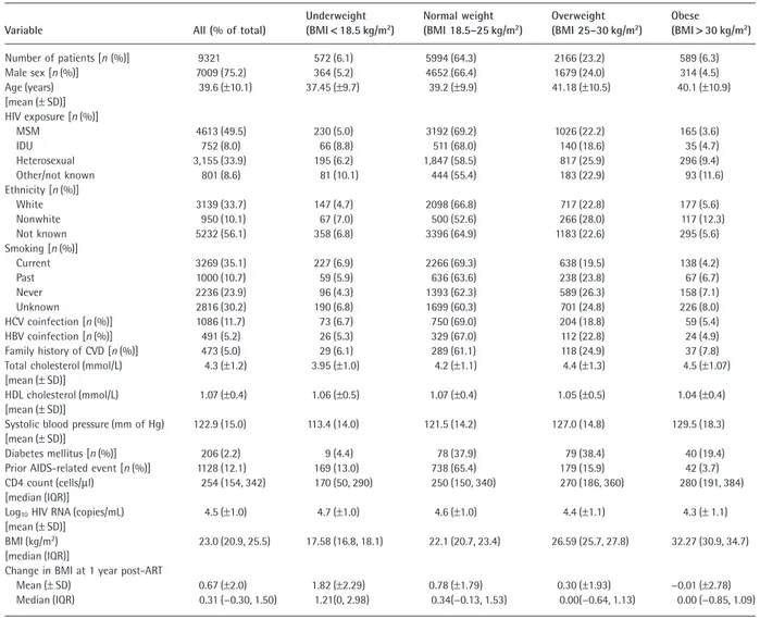
Table 1 provides characteristics at the time of ART ini-tiation for patients included in the CVD analysis, overall and by pre-ART BMI category. The majority were of normal
Table 1 Patient characteristics and incidence rates by pre-antiretroviral therapy (ART) body mass index (BMI) category for patients included in the
cardiovascular disease (CVD) analysis
Variable All (% of total)
Underweight (BMI< 18.5 kg/m2) Normal weight (BMI 18.5–25 kg/m2) Overweight (BMI 25–30 kg/m2) Obese (BMI> 30 kg/m2) Number of patients [n (%)] 9321 572 (6.1) 5994 (64.3) 2166 (23.2) 589 (6.3) Male sex [n (%)] 7009 (75.2) 364 (5.2) 4652 (66.4) 1679 (24.0) 314 (4.5) Age (years) [mean (± SD)] 39.6 (±10.1) 37.45 (±9.7) 39.2 (±9.9) 41.18 (±10.5) 40.1 (±10.9) HIV exposure [n (%)] MSM 4613 (49.5) 230 (5.0) 3192 (69.2) 1026 (22.2) 165 (3.6) IDU 752 (8.0) 66 (8.8) 511 (68.0) 140 (18.6) 35 (4.7) Heterosexual 3,155 (33.9) 195 (6.2) 1,847 (58.5) 817 (25.9) 296 (9.4) Other/not known 801 (8.6) 81 (10.1) 444 (55.4) 183 (22.9) 93 (11.6) Ethnicity [n (%)] White 3139 (33.7) 147 (4.7) 2098 (66.8) 717 (22.8) 177 (5.6) Nonwhite 950 (10.1) 67 (7.0) 500 (52.6) 266 (28.0) 117 (12.3) Not known 5232 (56.1) 358 (6.8) 3396 (64.9) 1183 (22.6) 295 (5.6) Smoking [n (%)] Current 3269 (35.1) 227 (6.9) 2266 (69.3) 638 (19.5) 138 (4.2) Past 1000 (10.7) 59 (5.9) 636 (63.6) 238 (23.8) 67 (6.7) Never 2236 (23.9) 96 (4.3) 1393 (62.3) 589 (26.3) 158 (7.1) Unknown 2816 (30.2) 190 (6.8) 1699 (60.3) 701 (24.8) 226 (8.0) HCV coinfection [n (%)] 1086 (11.7) 73 (6.7) 750 (69.0) 204 (18.8) 59 (5.4) HBV coinfection [n (%)] 491 (5.2) 26 (5.3) 329 (67.0) 112 (22.8) 24 (4.9) Family history of CVD [n (%)] 473 (5.0) 29 (6.1) 289 (61.1) 118 (24.9) 37 (7.8)
Total cholesterol (mmol/L) [mean (± SD)]
4.3 (±1.2) 3.95 (±1.0) 4.2 (±1.1) 4.4 (±1.3) 4.5 (±1.07)
HDL cholesterol (mmol/L) [mean (± SD)]
1.07 (±0.4) 1.06 (±0.5) 1.07 (±0.4) 1.05 (±0.5) 1.04 (±0.4)
Systolic blood pressure (mm of Hg) [mean (± SD)]
122.9 (15.0) 113.4 (14.0) 121.5 (14.2) 127.0 (14.8) 129.5 (18.3)
Diabetes mellitus [n (%)] 206 (2.2) 9 (4.4) 78 (37.9) 79 (38.4) 40 (19.4)
Prior AIDS-related event [n (%)] 1128 (12.1) 169 (13.0) 738 (65.4) 179 (15.9) 42 (3.7)
CD4 count (cells/μl) [median (IQR)]
254 (154, 342) 170 (50, 290) 250 (150, 340) 270 (186, 360) 280 (191, 384) Log10HIV RNA (copies/mL)
[mean (± SD)]
4.5 (±1.0) 4.7 (±1.0) 4.6 (±1.0) 4.4 (±1.1) 4.3 (± 1.1)
BMI (kg/m2)
[median (IQR)]
23.0 (20.9, 25.5) 17.58 (16.8, 18.1) 22.1 (20.7, 23.4) 26.59 (25.7, 27.8) 32.27 (30.9, 34.7) Change in BMI at 1 year post-ART
Mean (± SD) 0.67 (±2.0) 1.82 (±2.29) 0.78 (±1.79) 0.30 (±1.93) −0.01 (±2.78)
Median (IQR) 0.31 (−0.30, 1.50) 1.21(0, 2.98) 0.34(−0.13, 1.53) 0.00(−0.64, 1.13) 0.00 (−0.85, 1.09) HBV, hepatitis B virus [infection defined as HBV surface antigen (HBsAg) positive]; HCV, hepatitis C virus (infection defined as antibody positive); HDL, high-density lipoprotein; IDU, injecting drug use; IQR, interquartile range; MSM, men who have sex with men; SD, standard deviation.
weight, male and white, and they had a mean age of 39.6
years and a median CD4 count of 254 cells/μL. A large
proportion (> 40%) were reported to be current or ever
smokers. The median year of starting ART was 2006 (IQR 2003–2008). A total of 52.4% started on an NNRTI-based regimen (of whom 77.3% received efavirenz) and 41.5% on a PI-based regimen. A total of 49.9% received tenofovir, 32.4% received zidovudine and 9% received abacavir.
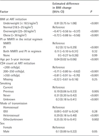
Factors associated with change in BMI at 1 year post-ART are shown in Table 2. Lower pre-post-ART BMI, male sex, use of PI (vs. NNRTI) in the initial regimen, older age, previous/never smoking (vs. current smoking) and higher CD4 count at ART initiation were associated with a smaller gain in BMI. The overall mean change in BMI at 1 year
post-ART was a gain of 0.67 kg/m2. Figure 1 shows the
mean change in BMI at years 1 to 5 post-ART by pre-ART BMI category using all of the available data. It appears that underweight people continued to gain BMI, those with a normal weight gained BMI which then remained relatively
stable or increased only slightly over time, while those with a high BMI experienced a more stable BMI or a slight weight loss over time.
Change in BMI at 1 year post ART initiation and CVD outcome
By the end of follow-up, there were 97 CVD events (includ-ing 46 MIs, 33 strokes and 18 invasive vascular procedures) at a rate of 2.21/1000 person-years [95% confidence inter-val (CI) 1.76–2.68/1000 person-years]. The median overall follow-up time was 5.3 years (IQR 3.8–7.5 years), which was similar within each pre-ART BMI category. Table 3 shows rates of CVD by quartiles of change in BMI overall and by each pre-ART BMI category. The overall rate tended to increase with increasing pre-ART BMI (Table 3).
The relationship between change in BMI and the risk of CVD outcomes, shown in Table 3 and Figure 2, signifi-cantly differed by pre-ART BMI category (P for interaction
between pre-ART BMI category and BMI change:< 0.05 in
all models). In the models adjusted for only demographic factors (including pre-ART BMI), the incidence rate ratio (IRR) (95% CI) per unit gain in BMI in those with normal pre-ART BMI was 1.20 (1.07 to 1.34), which was only slightly attenuated in fully adjusted models accounting for known CVD risk factors. Categorizing pre-ART BMI into quartiles (Fig. 2b) or further dividing the normal weight category (Table S1) suggested that individuals with pre-ART BMI in the middle range (middle two quartiles or normal weight, high) experienced an increase in the risk of CVD events per unit gain in BMI. Those with low pre-ART
BMI [< 20.95 (quartiles) or < 18.5 (alternative categories)]
may have slightly lowered their risk of CVD per unit gain
in BMI, while those with high pre-ART BMI [> 25.5
(quartiles) or> 30 (alternative categories)] did not
experi-ence a significant change in their risk of CVD events per unit change in BMI.
Change in BMI at 1 year post ART initiation and diabetes mellitus
Patient selection and characteristics were similar to those for CVD outcome (not shown). A total of 125 DM events occurred in 9193 eligible individuals (43 278 person-years), at the rate of 2.89/1000 person-years (95% CI 2.40–3.44/ 1000 person-years). The rates per 1000 person-years (95% CI) by pre-ART BMI category were as follows: underweight, 2.04 (0.76–4.53); normal weight, 2.01 (1.51–2.59); over-weight, 4.05 (2.88–5.54); and obese, 9.97 (6.32–14.96).
The incidence rates (95% CI) by quartiles of change in BMI in all patients were as follows: first quartile, 2.11
(1.32–3.20); second quartile, 3.22 (2.20–4.54); third
Table 2 Multivariable model for factors associated with body
mass index (BMI) change at 1 year post-antiretroviral therapy (ART) initiation
Factor
Estimated difference in BMI change
(95% CI) P
BMI at ART initiation
Underweight (< 18.5 kg/m2) 0.91 (0.75 to 1.08) <0.001
Normal (18.5–25 kg/m2) Reference
Overweight (25–30 kg/m2) −0.47 (−0.56 to −0.37) <0.001
Obese (> 30 kg/m2) −0.72 (−0.88 to −0.56) <0.001
PI or NNRTI in the initial regimen
NNRTI Reference
PI 0.2 (0.12 to 0.29) <0.001
Both NNRTI and PI in regimen 0.11 (−0.10 to 0.31) 0.32
None 0 (−0.27 to 0.26) 0.99
Age per 5-year increase 0.04 (0.02 to 0.06) <0.001 CD4 count at ART initiation
<200 cells/μL Reference 200–350 cells/μL −0.71 (−0.80 to −0.62) <0.001 >350 cells/μL −0.81 (−0.91 to −0.70) <0.001 Missing −0.22 (−0.61 to 0.16) 0.25 Smoking Current Reference Previous 0.19 (0.06 to 0.33) 0.005 Never 0.31 (0.20 to 0.42) <0.001 Unknown 0.3 (0.18 to 0.41) <0.001 Mode of transmission Homosexual Reference IDU 0.09 (−0.07 to 0.24) 0.28 Heterosexual 0.29 (0.18 to 0.40) <0.001 Other/unknown 0.25 (0.10 to 0.41) 0.002 Sex Female Reference Male 0.1 (0.00 to 0.22) 0.05
The model was adjusted for clinical cohort.
CI, confidence interval; IDU, injecting drug use; NNRTI, nonnucleoside reverse transcriptase inhibitor; PI, protease inhibitor.
quartile, 2.10 (1.32–3.12); and fourth quartile, 4.05 (3.00– 5.37). Figure 3 shows results from adjusted analyses for the DM outcome, overall and by pre-ART BMI. A unit gain in BMI was associated with an IRR (95% CI) of 1.13 (1.05– 1.23) in the models adjusted for demographical factors and pre-ART BMI. This relationship was only mildly attenuated in the fully adjusted models accounting for known risk factors for DM. The IRR per unit gain in BMI tended to only slightly vary by the pre-ART BMI (P value for
heterogene-ity in IRR across BMI categories:> 0.05 in all models).
Results were not sensitive to alternative categorization of BMI (Table S2).
Sensitivity analyses
Results were robust to all of the sensitivity analyses for both the outcomes. Excluding injecting drug users (n = 752) made the trend of increasing rates (per 1000 person-years) (95% CI) of CVD with quartile of BMI change in the normal pre-ART BMI category more apparent: first quartile, 0.83 (0.27–1.93); second quartile, 1.94 (1.00– 3.39); third quartile, 2.09 (1.14–3.50); and fourth quartile, 3.32 (2.12–4.94). The IRR per unit gain in BMI for the normal pre-ART BMI category was 1.23 (1.09 to 1.38) (Table S3 shows results for the fully adjusted model). Results for DM outcome did not change substantially (not shown).
When the per cent change in BMI was modelled, a 1% increase in BMI was associated with an IRR (95% CI) of CVD as: 1.04 (95% CI 1.01–1.08) and 1.06 (95% CI 1.02– 1.11) in the middle two quartiles of the pre-ART BMI, respectively (P = 0.01 for interaction between pre-ART BMI and BMI change in fully adjusted models). Finally, for diabetes outcome, a 1% increase in BMI in fully adjusted models was associated with an IRR (95% CI) of 1.03 (1.01– 1.04) (P = 0.009), regardless of pre-ART BMI. Results were also robust to further adjustment for blood glucose at study entry (data not shown).
Discussion
The relationship between short-term gain in BMI after ART initiation and long-term subsequent risk of CVD outcomes varied by the pre-ART BMI. Those with pre-ART BMI in the normal or middle two quartile categories experienced an increase of approximately 18–20% in the risk of CVD per unit gain in BMI. Those at the lower extreme possibly benefited while those in the upper extremes did not experi-ence any appreciable change in their already higher risk. For the DM outcome, each unit gain in BMI was associated with a 12% increase in risk regardless of pre-ART BMI.
Table 3 Rates (cases/1 000 per son-year s) of car diovascular disease (CVD) by quartile of change in body mass index (BMI) and pr e-antir etr ovir al ther apy (ART ) BMI category Quartile of change in BMI at 1 year post-ART BMI at ART initiation Underweight Normal weight Overweight Obese Over all Median change in BMI n (event s)/per son-year s; rate (95% CI) Median change in BMI n (event s)/per son-year s; rate (95% CI) Median change in BMI n (event s)/per son-year s; rate (95% CI) Median change in BMI n (event s)/per son-year s; rate (95% CI) Median change in BMI n (event s)/per son-year s; rate (95% CI) 1 0 1/657; 1.52 (0.04–8.48) −0.75 8/6723; 1.19 (0.5 1–2.34) −1.54 8/2524; 3.17 (1.37–6.24) −2.20 2/135 1; 1.48 (0.04–8.25) −0.97 22/1 0576; 2.09 (1.30 –3.14) 2 0.62 1/680; 1.47 (0.04–8.2 1) 0 13/6736; 1.93 (1.0 2–3.30) 0 6/252 1; 2.38 (0.88–5.20) 0 4/15 15; 2.64 (0.32–9.55) 0 2 1/1 009 1; 2.09 (1.29–3.18) 3 2.00 2/7 14; 2.80 (0.34–1 0.1 1) 0.89 12/73 17; 1.64 (0.85–2.85) 0.58 4/2260; 1.77 (0.48–4.54) 0.48 4/545; 7.34 (1.5 1–2 1.5) 0.78 16/1 1256; 1.42 (0.8 1 –2.30) 4 4.84 1/847; 1.18 (0.03–6.59) 2.67 28/7778; 3.60 (2.37–5.16) 2.16 6/2655;2.26 (0.83–4.9 1) 2.44 1/667; 1.50 (0.04–8.3 1) 2.65 38/12059; 3.15 (2.22– 4.32) Over all 1.2 1 5/2890; 1.73 (0.56–4.03) 0.33 6 1/28638; 2.13 (1.63–2.73) 0 24/9959; 2.4 1 (1.55–3.59) 0 11/3957;2.78 (1.12–5.74) 0.3 1 97/43982; 2.20 (1.78–2.69) BMI was measur ed in kg/m 2. Change in BMI = BMI at 1 year post-ART initiation minus pr e-ART BMI. Rates ar e in cases/1 000 per son-year s.
These associations did not disappear after adjusting for previously identified key risk factors for each outcome.
Our findings are consistent with a previous report from the VA cohort for the DM outcome, and further add to evidence of the importance of the gain in BMI after ART initiation for the long-term risk of CVD. To put our findings in clinical context, a 40-year-old man with a BMI of
24 kg/m2who is a current smoker, is ART-naïve, and has a
systolic blood pressure of 130 mm/Hg and total/HDL cho-lesterol of 6/1 mmol/L, respectively, will have a 2.5% risk
of CVD in the next 5 years (according to the D:A:D risk calculator [12]). A gain of 1 unit of BMI would increase this risk to 3%, an increase of 20% in relative terms. Thus, the total rise in absolute risk may not be large depending on the background risk. However, given the expected timing of BMI change around ART initiation, these observations may help to better strategize risk factor management in this high-risk population.
ART initiation, leading to viral suppression and CD4 count gain, often results in weight gain. Possible mecha-Fig. 2 Adjusted incidence rate ratio (IRR) for cardiovascular disease (CVD) per unit gain in body mass index (BMI) at 1 year post antiretroviral therapy
(ART) initiation overall and by pre-ART BMI. Pre-ART BMI was categorized using the World Health Organization (WHO) classification (a) or quartiles (b). Diamond: models were adjusted for pre-ART BMI category, cohort, sex, mode of transmission, family history of CVD, CD4 count at ART initiation and age and smoking status as measured at analysis entry. P for effect modification: (a) 0.02; (b) 0.004. Triangle: models were additionally adjusted for total cholesterol, high-density lipoprotein (HDL) cholesterol, systolic blood pressure, diabetes, current use of abacavir, and cumulative use of indinavir and lopinavir/ritonavir as measured at analysis entry. P for effect modification: (a) 0.04; (b) 0.01. Circle: models were adjusted for all of the above variables (including specific ART agents mentioned above) as well as calendar year in a time-updated fashion where possible. P for effect modification: (a) 0.04; (b) 0.01. In (a), the IRRs (95% confidence intervals) for the pre-ART BMI in the final model, with normal weight as the reference category, were as follows: underweight, 1.53 (0.49–4.72); overweight, 1.07 (0.64–1.80); and obese, 1.10 (0.48–2.54).
nisms include a reduction in metabolic requirements as well as inflammation in those with suppressed viral load, a return to good health and a resumption of “normal” life-styles and, possibly, the effects of individual ART classes or agents. In our exploratory analysis, receipt of a drug from the PI class was associated with a greater gain in weight compared with receipt of the NNRTI class. This was also observed in the 1917 cohort but not in the Swiss cohort [1,4].
The mechanistic link between this weight gain and out-comes is also unclear. As seen in our exploratory analysis, this gain in BMI appeared to be sustained over time (con-sistent with the 1917 cohort as well as the Swiss cohort) [1,4], especially in those who were underweight or of normal weight at baseline, and this may contribute to their CVD and DM risk [15]. Adjustments for factors potentially on the causal pathway (such as lipids and blood pressure) resulted in only a mild attenuation of the IRRs, suggesting that the observed association is unlikely to be fully explained by BMI-induced changes in these markers. Also, in the VA cohort, the risk of DM per unit gain in BMI in positive individuals was double the risk in HIV-negative matched controls with a similar gain in BMI [7]. While ART initiation is known to reduce inflammation, one recent study suggested that those initiating ART at normal or higher BMI and subsequently gaining weight do not experience a reduction in inflammation (or even have an increase in inflammation), suggesting a role of ongoing
inflammation related to weight gain [16]. Future studies should investigate how these risk factors interact and evolve over time to impact the risk of CVD and DM in this high-risk population.
While the finding that weight gain in underweight indi-viduals had a minimal association with CVD outcomes was expected, the finding that weight gain in those with high pre-ART BMI also seemed to lack an association with CVD outcome was unexpected. This finding should be interpreted with caution. First, all our analyses were adjusted for pre-ART BMI and, indeed, CVD rates were higher in those with greater pre-ART BMI. These individuals were also at a higher risk of DM, which is an important risk factor for CVD. The actual gain in BMI over time in those with high pre-ART BMI was minimal, with an average loss in BMI, in the obese group. Also, relatively few individuals had a high BMI, thereby limiting our power to detect associations or effect modifications. It is also possible that those with high BMI received prompter medical attention when required, and those events were more likely to be prevented. Finally, BMI may not accurately capture body fat [11]. Waist-to-hip ratio, if available, might be a better indicator of central obesity and could complement the information provided by BMI [11]. Unfortunately, waist-to-hip ratio is often difficult to measure, and was not available in our study.
The strengths of our study include a large heterogeneous multi-country cohort, with well-validated (through estab-lished protocols) outcomes. Also, we were able to account Fig. 3 Adjusted incidence rate ratio (IRR) for diabetes mellitus (DM) per unit gain in body mass index (BMI) at 1 year post-antiretroviral therapy (ART)
initiation overall and by pre-ART BMI. Diamond: the model was adjusted for pre-ART BMI category, cohort, sex, mode of transmission, CD4 count at ART initiation and age as measured at analysis entry. P for effect modification: 0.57. Triangle: the model was additionally adjusted for triglycerides, high-density lipoprotein (HDL) cholesterol, systolic blood pressure and lipodystrophy as measured at analysis entry. P for effect modification: 0.65. Circle: the model was adjusted for all of the above variables as well as calendar year in a time-updated fashion where possible. P for effect modification: 0.83. The IRRs (95% confidence intervals) for the pre-ART BMI in the final model, with normal weight as the reference category, were as follows: underweight, 0.96 (0.41–2.26); overweight, 1.70 (1.12–2.59); and obese, 4.19 (2.49–7.03).
for several key potential confounders in a time-updated fashion, including smoking and the use of specific ART agents. Further, this analysis included data from an ongoing modern cohort initiating ART with contemporary regimens at a relatively high CD4 count, with only a handful starting with thymidine analogues. This makes our findings more generalizable to the modern ART era.
The study has a few limitations. First, the cohort was a selected sample nested in a large cohort, which may limit generalizability. Secondly, many lifestyle factors, such as diet and exercise habits, were unavailable. Thirdly, although the D:A:D study follows a rigorous outcome vali-dation process, it is possible that some CVD events, in particular MIs, could in fact be secondary events. However, secondary MIs are known to be largely associated with injecting drug use [14,17]. In our study, when we excluded individuals exposed to HIV through injecting drug use, we found that the association between BMI gain and CVD was stronger. Finally, we could not analyse specific CVD events, mainly because of limited power.
In conclusion, we found that the short-term gain in BMI post ART initiation could be associated with the increased risk of CVD, but only in those with normal or in middle two quartiles of pre-ART BMI. The short-term gain in BMI post ART initiation was also associated with increased risk of DM, regardless of pre-ART BMI. Clinicians need to be aware that, while traditionally thought to be a good prognostic factor in people with HIV infection, excess weight gain post ART initiation could be detrimental to future cardiometabolic health. Also, given that this weight gain is expected to occur in a window after ART initiation, it could provide clinicians with an opportunity to consider interventions to pre-empt the risk of these events. Finally, the role of sustained weight gain over time in terms of cardiometabolic outcomes needs further exploration. Ultimately, to answer the question of whether weight-control interventions in the post-ART ini-tiation period truly decrease the risk of outcomes, a randomized trial will be needed.
Author contributions
ACA conceived the idea, performed the statistical analysis and wrote the first draft of the manuscript. AM, PR, LR, AP, SW, CS, CS, AdM and RW provided critical input at all stages of the project. MGL supervised the overall conduct of this project and provided critical input at all stages of the study.
Acknowledgements
The writing committee would like to acknowledge the D:A:D study participants and the D:A:D study Steering Committee (see Appendix).
Conflicts of interest: There are no conflicts of interest
to report. See the “Funding” section for full financial disclosure.
Funding: This work was supported by the Highly Active
Antiretroviral Therapy Oversight Committee (HAART-OC), a collaborative committee with representation from academic institutions, the European Agency for the Evaluation of Medicinal Products, the United States Food and Drug Administration, the patient community, and all pharmaceu-tical companies with licensed anti-HIV drugs in the Euro-pean Union: AbbVie, Boehringer Ingelheim, Bristol-Myers Squibb, Gilead Sciences, ViiV Healthcare, Merck, Pfizer, F. Hoffman-LaRoche and Janssen Pharmaceuticals. It was also supported by a grant (grant number CURE/97-46486) from the Health Insurance Fund Council, Amstelveen, the Neth-erlands, to the AIDS Therapy Evaluation Project Netherlands (ATHENA) and by a grant from the Agence Nationale de Recherches sur le SIDA (grant number Action Coordonnée no. 7, Cohortes) to the Aquitaine Cohort. The Australian HIV Observational Database (AHOD) is funded as part of the Asia Pacific HIV Observational Database, a programme of The Foundation for AIDS Research, amfAR, and is supported in part by a grant from the US National Institutes of Health’s National Institute of Allergy and Infectious Diseases (NIAID) (grant number U01-AI069907) and by unconditional grants from Merck Sharp & Dohme, Gilead Sciences,
Bristol-Myers Squibb, Boehringer Ingelheim, Roche, Pfizer,
GlaxoSmithKline, and Janssen Pharmaceuticals. The Kirby Institute is funded by The Australian Government Depart-ment of Health and Ageing, and is affiliated with the Faculty of Medicine, The University of New South Wales. The work was also supported by grants from the Fondo de Investigación Sanitaria (grant number FIS 99/0887) and Fundación para la Investigación y la Prevención del SIDA en Espanã (grant number FIPSE 3171/00) to the Barcelona Antiretroviral Surveillance Study (BASS); grants from the National Institute of Allergy and Infectious Diseases,
National Institutes of Health (grant numbers
5U01AI042170-10 and 5U01AI046362-03) to the Terry Beirn Community Programs for Clinical Research on AIDS (CPCRA); grants from the BIOMED 1 (grant number CT94-1637) and BIOMED 2 (grant number CT97-2713) pro-grammes and the fifth framework programme (grant number QLK2-2000-00773) of the European Commission and grants from Bristol-Myers Squibb, GlaxoSmithKline, Boehringer Ingelheim, and Roche to the EuroSIDA study; by unrestricted educational grants from AbbVie, Bristol-Myers Squibb, Gilead Sciences, GlaxoSmithKline, Pfizer, and Janssen Pharmaceuticals to the Italian Cohort Naive to Antiretrovirals (The ICONA Foundation); and by a grant from the Swiss National Science Foundation (grant #148522) to the Swiss HIV Cohort Study (SHCS).
Appendix: D:A:D study participating cohorts
AquitaineFrance
Community Programs for Clinical Research on AIDS (CPCRA) USA USA
Nice Cohort France
AIDS Therapy Evaluation in the Netherlands (ATHENA)
EuroSIDA Swiss HIV
cohort study (SHCS)
The Netherlands Europe Switzerland
AHOD
Australian HIV Observational Database HIV-BIVUS Sweden St. Pierre Brussels Cohort Belgium BASS Spain
The ICONA Foundation Italian cohort on naive antiretrovirals (ICONA)
D:A:D Steering Committee: names marked with *, Chair with #.
Members of the D:A:D Steering Committee from the Oversight Committee: B. Powderly*, N. Shortman*, C. Moecklinghoff *, G. Reilly*, X. Franquet*
D:A:D central coordination: L. Ryom, C. I. Hatleberg, C. A. Sabin*, D. Kamara, C. Smith, A. Phillips*, A. Mocroft, A. Bojesen, J. Nielsen, D. Raben, J. D. Lundgren#
D:A:D data managers: R. Salbøl Brandt (coordinator), M. Rickenbach, I. Fanti, E. Krum, M. Hillebregt, S Geffard, A. Sundström, M. Delforge, E. Fontas, F. Torres, H. McManus, S. Wright, J. Kjær.
Verification of endpoints: A. Sjøl (CVD primary end-point), P. Meidahl (oncology, new endend-point), J. Helweg-Larsen (haematology, new endpoint), J. Schmidt Iversen (nephrology, new endpoint).
Kidney working group: L. Ryom, A. Mocroft, O. Kirk*, P. Reiss*, M. Ross, C. A. Fux, P. Morlat, O. Moranne, A. M. Kesselring, D. A. Kamara, C. Smith, J. D. Lundgren#
Mortality working group: C. Smith, L. Ryom, A. Phil-lips*, R. Weber*, P. Morlat, C. Pradier*, P. Reiss*, N. Friis-Møller, J. Kowalska, J. D. Lundgren#
Cancer working group: C. Sabin*, M. Law*, A. d’Arminio Monforte*, F. Dabis*, M. Bruyand, P. Reiss*, C. Smith, D. A. Kamara, M. Bower, G. Fätkenheuer, A. Donald, A. Grulich, L. Ryom, J. D. Lundgren#
The members of the 11 cohorts are as follows: ATHENA (AIDS Therapy Evaluation Project Netherlands).
Central coordination: P. Reiss*, S. Zaheri, M. Hillebregt, L. Gras.
Participating physicians (Site coordinating physicians): Academisch Medisch Centrum bij de Universiteit van Amsterdam, Amsterdam: Prof. Dr J. M. Prins, Prof. Dr T. W. Kuijpers, Dr H. J. Scherpbier, Dr J. T. M. van der Meer, Dr F. W. M. N. Wit, Dr M. H. Godfried, Prof. Dr P. Reiss*, Prof. Dr T. van der Poll, Dr F. J. B. Nellen, Prof. Dr J. M. A. Lange, Dr S. E. Geerlings, Dr M. van Vugt, Dr D. Pajkrt,
Drs J. C. Bos, Drs M. van der Valk, Drs M. L. Grijsen, Dr W. J. Wiersinga, Dr A. Goorhuis, Dr J. W. R. Hovius. Academisch Ziekenhuis Maastricht, Maastricht: Dr S. Lowe, Dr A. Oude Lashof, Dr D. Posthouwer. Catharina-ziekenhuis, Eindhoven: Drs M. J. H. Pronk, Dr H. S. M. Ammerlaan. Erasmus Medisch Centrum, Rotterdam: Dr M. E. van der Ende, Dr T. E. M. S. de Vries-Sluijs, Dr C. A. M. Schurink, Dr J. L. Nouwen, Dr A. Verbon, Drs B. J. A. Rijnders, Dr E. C. M. van Gorp, Drs M. van der Feltz. Erasmus Medisch Centrum–Sophia, Rotterdam: Dr G. J. A. Driessen, Dr A. M. C. van Rossum. Flevoziekenhuis. Almere: Dr J. Branger. HagaZiekenhuis, Den Haag: Dr E. F. Schippers, Dr C. van Nieuwkoop, Drs E. P. van Elzakker. Isala Klinieken, Zwolle: Dr P. H. P. Groeneveld, Drs J. W. Bouwhuis. Kennemer Gasthuis: Drs R. Soetekouw, Prof. Dr R. W. ten Kate. Leids Universitair Medisch Centrum, Leiden: Dr F. P. Kroon, Prof. Dr J. T. van Dissel, Dr S. M. Arend, Dr M. G. J. de Boer, Drs H. Jolink, Dr H. J. M. ter Vollaard, Drs M. P. Bauer. Maasstadziekenhuis, Rotter-dam: Dr J. G. den Hollander, Dr K. Pogany. Medisch Centrum Alkmaar, Alkmaar: Drs G. van Twillert, Drs W. Kortmann, Dr J. W. T. Cohen Stuart, Dr B. M. W. Diederen. Medisch Centrum Haaglanden, Den Haag: Dr E. M. S. Leyten, Dr L. B. S. Gelinck. Medisch Spectrum Twente, Enschede: Drs G. J. Kootstra, Drs C. E. Delsing. Onze Lieve Vrouwe Gasthuis, Amsterdam: Prof. Dr K. Brinkman, Dr W. L. Blok, Dr P. H. J. Frissen, Drs W. E. M. Schouten, Drs G. E. L. van den Berk. Sint Elisabeth Ziekenhuis, Tilburg: Dr M. E. E. van Kasteren, Dr A. E. Brouwer. Sint Lucas Andreas Ziekenhuis, Amsterdam: Dr J. Veenstra, Dr K. D. Lettinga. Slotervaartziekenhuis, Amsterdam: Dr J. W. Mulder, Drs S. M. E. Vrouenraets, Dr F. N. Lauw. Stichting Medisch Centrum Jan van Goyen, Amsterdam: Drs A. van Eeden, Dr D. W. M. Verhagen. Universitair Medisch Centrum Groningen, Groningen: Drs H. G. Sprenger, Drs R. Doedens, Dr E. H. Scholvinck, Dr S. van Assen, Dr W. F.
W. Bierman. Universitair Medisch Centrum Sint
Radboud, Nijmegen: Dr P. P. Koopmans, Dr M. Keuter, Dr A. J. A. M. van der Ven, Dr H. J. M. ter Hofstede, Dr A. S. M. Dofferhoff, Dr A Warris, Dr R. van Crevel. Universitair Medisch Centrum Utrecht, Utrecht: Prof. Dr A. I. M. Hoepelman, Dr T. Mudrikova, Dr M. M. E. Schneider, Dr P. M. Ellerbroek, Dr J. J. Oosterheert, Dr J. E. Arends, Dr M. W. M. Wassenberg, Dr R. E. Barth. Vrije Universiteit Amsterdam, Amsterdam: Dr M. A. van Agtmael, Dr R. M. Perenboom, Drs F. A. P. Claessen, Dr M. Bomers, Dr E. J. G. Peters. Wilhelmina Kinderziekenhuis, Utrecht: Dr S. P. M. Geelen, Dr T. F. W. Wolfs, Dr L. J. Bont. Ziekenhuis Rijnstate, Arnhem: Dr C. Richter, Dr J. P. van der Berg, Dr E. H. Gisolf. Admiraal De Ruyter Ziekenhuis, Vlissingen: Drs M. van den Berge, Drs A. Stegeman. Medisch Centrum Leeuwarden, Leeuwarden: Dr M. G. A. van Vonderen, Drs
D. P. F. van Houte. Medisch Centrum Zuiderzee, Lelystad: Dr S. Weijer, Dr R. el Moussaoui. Sint Elisabeth Hospitaal, Willemstad – Curaçao: Dr C. Winkel, Drs F. Muskiet, Drs Durand, Drs R. Voigt.
Aquitaine Cohort (France).
Principal investigator: Pr F. Dabis*. Scientific commit-tee: Prs F. Bonnet, F. Dabis*, M. Dupon, G. Chêne, D. Breilh, H. Fleury, D. Malvy, P. Mercié, I. Pellegrin, P. Morlat, D. Neau, JL. Pellegrin; Drs S. Bouchet, V. Gaborieau, D. Lacoste, S. Tchamgoué, R. Thiébaut.
Composition of the GECSA: Epidemiology and biostatistics: Prs G. Chêne, F. Dabis, R. Thiébaut, Drs M. Bruyand, S. Lawson-Ayayi, L. Wittkop.
Clinical and biological hospital units: Bordeaux Uni-versity Hospital: Pr P. Morlat (Pr F. Bonnet, Drs N. Bernard, M. Hessamfar, D. Lacoste, M. A. Vandenhende); Pr M. Dupon (Drs F. A. Dauchy, H. Dutronc), Pr M. Longy-Boursier (Pr P. Mercié, Drs P. Duffau, J. Roger Schmeltz), Pr D. Malvy (Drs T. Pistone, MC Receveur), Pr D. Neau (Drs C. Cazanave, A. Ochoa, MO. Vareil), Pr JL. Pellegrin (Pr JF. Viallard, Drs C. Greib, E. Lazaro); Pr H. Fleury (Pr ME. Lafon, Drs S. Reigadas, P. Trimoulet); Pr D. Breilh; Pr M. Molimard (Drs S. Bouchet, K. Titier); Pr JF. Moreau (Dr I. Pellegrin); Drs F. Haramburu, G. Miremont-Salamé. Arcachon Hospital: Dr A. Dupont. Dax Hospital: Dr Y. Gerard (Drs L. Caunègre, K. André). Bayonne Hos-pital: Dr F. Bonnal (Drs S. Farbos, MC. Gemain). Libourne Hospital: Dr J. Ceccaldi (Dr S. Tchamgoué). Mont-de-Marsan Hospital: Dr S. De Witte (Dr C. Courtault). Pau Hospital: Drs E. Monlun (Dr V. Gaborieau).Périgueux Hospital: Dr P. Lataste (Dr JP. Meraud). Villeneuve-sur-Lot Hospital: Dr I. Chossat.
Permanent team: MJ. Blaizeau, M. Bruyand, V. Conte, M. Decoin, J. Delaune, S. Delveaux, F. Diarra, C. D’Ivernois, A. Frosch, S. Geffard, C. Hannapier, S. Lawson-Ayayi, E. Lenaud, O. Leleux, F. Le Marec, J. Leray, I. Louis, G. Palmer, A. Pougetoux, X. Sicard, D. Touchard B. Uwamaliya-Nziyumvira.
AHOD (Australian HIV Observational Database,
Australia).
Central coordination: M. Law*, K. Petoumenos, H. McManus, S. Wright, C. Bendall (Sydney, New South Wales).
Participating physicians (city, state): R. Moore, S. Edwards, J. Hoy, K. Watson, N. Roth, J. Nicholson (Mel-bourne, Victoria); M Bloch, T. Franic, D. Baker, R. Vale, A. Carr, D. Cooper (Sydney, New South Wales); J. Chuah, M. Ngieng (Gold Coast, Queensland), D. Nolan, J. Skett (Perth, Western Australia).
BASS (Spain).
Central coordination: G. Calvo*, F. Torres, S. Mateu (Barcelona).
Participating physicians (city): P. Domingo, M. A. Sambeat, J. Gatell, E. Del Cacho, J. Cadafalch, M. Fuster (Barcelona); C. Codina, G. Sirera, A. Vaqué (Badalona).
The Brussels St Pierre Cohort (Belgium): Coordina-tion: S. De Wit*, N. Clumeck, M. Delforge, C. Necsoi. Participating physicians: N. Clumeck, S. De Wit*, AF Gennotte, M. Gerard, K. Kabeya, D. Konopnicki, A. Libois, C. Martin, M. C. Payen, P. Semaille, Y. Van Laethem.
CPCRA (USA).
Central coordination: J. Neaton, G. Bartsch, W. M. El-Sadr*, E. Krum, G. Thompson, D. Wentworth.
Participating physicians (city, state): R. Luskin-Hawk (Chicago, Illinois); E. Telzak (Bronx, New York); W. M. El-Sadr (Harlem, New York); D. I. Abrams (San Francisco, California); D. Cohn (Denver, Colorado); N. Markowitz (Detroit, Michigan); R. Arduino (Houston, Texas); D. Mushatt (New Orleans, Louisiana); G. Friedland (New Haven, Connecticut); G. Perez (Newark, New Jersey); E. Tedaldi (Philadelphia, Pennsylvania); E. Fisher (Richmond, Virginia); F. Gordin (Washington, DC); L. R. Crane (Detroit, Michigan); J. Sampson (Portland, Oregon); J. Baxter (Camden, New Jersey).
EuroSIDA (multinational) Coordinating Centre: J Lundgren*#, O Kirk*, A Mocroft, A Cozzi-Lepri, D Grint, D Podlekareva, J Kjær, L Peters, J Reekie, J Kowalska, J Tverland, A H Fischer, J Nielsen.
Participating countries and physicians
Argentina: (M Losso), C Elias, Hospital JM Ramos Mejia, Buenos Aires.
Austria: (N Vetter), Pulmologisches Zentrum der Stadt Wien, Vienna; R Zangerle, Medical University Innsbruck, Innsbruck.
Belarus: (I Karpov), A Vassilenko, Belarus State Medical University, Minsk, VM Mitsura, Gomel State Medical
Uni-versity, Gomel; O Suetnov, Regional AIDS Centre,
Svetlogorsk.
Belgium: (N Clumeck), S De Wit*, M Delforge, Saint-Pierre Hospital, Brussels; R Colebunders, Institute of Tropi-cal Medicine, Antwerp; L Vandekerckhove, University Ziekenhuis Gent, Gent.
Bosnia-Herzegovina: (V Hadziosmanovic), Klinicki Centar Univerziteta Sarajevo, Sarajevo.
Bulgaria: (K Kostov), Infectious Diseases Hospital, Sofia. Croatia: (J Begovac), University Hospital of Infectious Diseases, Zagreb.
Czech Republic: (L Machala), D Jilich, Faculty Hospital Bulovka, Prague; D Sedlacek, Charles University Hospital, Plzen.
Denmark: (J Nielsen), G Kronborg,T Benfield, M Larsen, Hvidovre Hospital, Copenhagen; J Gerstoft, T Katzenstein, A-B E Hansen, P Skinhøj, Rigshospitalet, Copenhagen; C
Pedersen, Odense University Hospital, Odense; L Ostergaard, Skejby Hospital, Aarhus.
Estonia: (K Zilmer), West-Tallinn Central Hospital,
Tallinn; Jelena Smidt, Nakkusosakond Siseklinik,
Kohtla-Järve.
Finland: (M Ristola), Helsinki University Central Hospi-tal, Helsinki.
France: (C Katlama), Hôpital de la Pitié-Salpétière, Paris; J-P Viard, Hôpital Necker-Enfants Malades, Paris; P-M Girard, Hospital Saint-Antoine, Paris; JM Livrozet, Hôpital Edouard Herriot, Lyon; P Vanhems, University Claude Bernard, Lyon; C Pradier, Hôpital de l’Archet, Nice; F Dabis*, D Neau, Unité INSERM, Bordeaux.
Germany: (J Rockstroh), Universitäts Klinik Bonn; R Schmidt, Medizinische Hochschule Hannover; J van Lunzen, O Degen, University Medical Center
Hamburg-Eppendorf, Infectious Diseases Unit, Hamburg; HJ
Stellbrink, IPM Study Center, Hamburg; S Staszewski, JW
Goethe University Hospital, Frankfurt; M Bickel,
Medizinische Poliklinik, Munich; G. Fätkenheuer,
Universität Köln, Cologne.
Greece: (J Kosmidis), P Gargalianos, G Xylomenos, J Perdios, Athens General Hospital; G Panos, A Filandras, E Karabatsaki, 1st IKA Hospital; H Sambatakou, Ippokration Genereal Hospital, Athens.
Hungary: (D Banhegyi), Szent Lásló Hospital, Budapest. Ireland: (F Mulcahy), St. James’s Hospital, Dublin. Israel: (I Yust), D Turner, M Burke, Ichilov Hospital, Tel Aviv; S Pollack, G Hassoun, Rambam Medical Center, Haifa; S Maayan, Hadassah University Hospital, Jerusalem. Italy: (S Vella), Istituto Superiore di Sanità, Rome; R Esposito, I Mazeu, C Mussini, Università Modena, Modena; C Arici, Ospedale Riuniti, Bergamo; R Pristera, Ospedale Generale Regionale, Bolzano; F Mazzotta, A Gabbuti, Ospedale S Maria Annunziata, Firenze; V Vullo, M Lichtner, University di Roma la Sapienza, Rome; A
Chirianni, E Montesarchio, M Gargiulo, Presidio
Ospedaliero AD Cotugno, Monaldi Hospital, Napoli; G Antonucci, A Testa, P Narciso, C Vlassi, M Zaccarelli, Istituto Nazionale Malattie Infettive Lazzaro Spallanzani, Rome; A Lazzarin, A Castagna, N Gianotti, Ospedale San Raffaele, Milan; M Galli, A Ridolfo, Osp. L. Sacco, Milan; A d’Arminio Monforte, Istituto Di Clinica Malattie Infettive e Tropicale, Milan.
Latvia: (B Rozentale), I Zeltina, Infectology Centre of Latvia, Riga.
Lithuania: (S Chaplinskas), Lithuanian AIDS Centre, Vilnius.
Luxembourg: (R Hemmer), T Staub, Centre Hospitalier, Luxembourg.
Netherlands: (P Reiss*), Academisch Medisch Centrum bij de Universiteit van Amsterdam, Amsterdam.
Norway: (V Ormaasen), A Maeland, J Bruun, Ullevål Hospital, Oslo.
Poland: (B Knysz) J Gasiorowski, Medical University, Wroclaw; A Horban, E Bakowska, Centrum Diagnostyki i Terapii AIDS, Warsaw; A Grzeszczuk, R Flisiak, Medical University, Bialystok; A Boron-Kaczmarska, M Pynka, M Parczewski, Medical Univesity, Szczecin; M Beniowski, E Mularska, Osrodek Diagnostyki i Terapii AIDS, Chorzow; H Trocha, Medical University, Gdansk; E Jablonowska, E Malolepsza, K Wojcik, Wojewodzki Szpital Specjalistyczny, Lodz.
Portugal: (F Antunes), M Doroana, L Caldeira, Hospital Santa Maria, Lisbon; K Mansinho, Hospital de Egas Moniz, Lisbon; F Maltez, Hospital Curry Cabral, Lisbon.
Romania: (D Duiculescu), Spitalul de Boli Infectioase si Tropicale: Dr Victor Babes, Bucarest.
Russia: (A Rakhmanova), Medical Academy Botkin Hos-pital, St Petersburg; N Zakharova, St Petersburg AIDS Centre, St Peterburg; S Buzunova, Novgorod Centre for AIDS, Novgorod.
Serbia: (D Jevtovic), The Institute for Infectious and Tropical Diseases, Belgrade.
Slovakia: (M Mokráš), D Staneková, Dérer Hospital, Bra-tislava.
Slovenia: (J Tomazic), University Clinical Centre Lju-bljana, Ljubljana.
Spain: (J González-Lahoz), V Soriano, P Labarga, J Medrano, Hospital Carlos III, Madrid; S Moreno, JM Rod-riguez, Hospital Ramon y Cajal, Madrid; B Clotet, A Jou, R Paredes, C Tural, J Puig, I Bravo, Hospital Germans Trias i Pujol, Badalona; JM Gatell, JM Miró, Hospital Clinic i Provincial, Barcelona; P Domingo, M Gutierrez, G Mateo, MA Sambeat, Hospital Sant Pau, Barcelona.
Sweden: (A Karlsson), Venhaelsan-Sodersjukhuset, Stockholm; L Flamholc, Malmö University Hospital, Malmö.
Switzerland: (B Ledergerber), R Weber*, University Hos-pital, Zürich; P Francioli, M Cavassini, Centre Hospitalier Universitaire Vaudois, Lausanne; B Hirschel, E Boffi, Hos-pital Cantonal Universitaire de Geneve, Geneve; H Furrer, Inselspital Bern, Bern; M Battegay, L Elzi, University Hos-pital Basel.
Ukraine: (E Kravchenko), N Chentsova, Kiev Centre for AIDS, Kiev; V Frolov, G Kutsyna, Luhansk State Medical University; Luhansk; S Servitskiy, Odessa Region AIDS Center, Odessa; M Krasnov, Kharkov State Medical Univer-sity, Kharkov.
United Kingdom: (S Barton), St. Stephen’s Clinic, Chelsea and Westminster Hospital, London; AM Johnson, D Mercey, Royal Free and University College London Medical School, London (University College Campus); A Phillips, MA Johnson, A Mocroft, Royal Free and University College
Medical School, London (Royal Free Campus); M Murphy, Medical College of Saint Bartholomew’s Hospital, London; J Weber, G Scullard, Imperial College School of Medicine at St. Mary’s, London; M Fisher, Royal Sussex County Hospital, Brighton; C Leen, Western General Hospital, Edinburgh.
HivBivus (Sweden).
Central coordination: L. Morfeldt*, G. Thulin, A. Sundström.
Participating physicians (city): B. Åkerlund (Huddinge); K. Koppel, A. Karlsson (Stockholm); L. Flamholc, C. Håkangård (Malmö).
The ICONA Foundation (Italy).
Board of directors: M. Moroni (Chair), G. Angarano, A. Antinori, O. Armignacco, A. d’Arminio Monforte*, F. Castelli, R. Cauda, G. Di Perri, M. Galli, R. Iardino, G. Ippolito, A. Lazzarin, C. F. Perno, F. von Schloesser, P. Viale. Scientific secretary: A d’Arminio Monforte*, A. Antinori, A. Castagna, F. Ceccherini-Silberstein, A. Cozzi-Lepri, E. Girardi, S. Lo Caputo, C. Mussini, M. Puoti.
ICONA Steering Committee: M. Andreoni, A. Ammassari, A. Antinori, A. d’Arminio Monforte, C. Balotta, P. Bonfanti, S. Bonora, M. Borderi, R. Capobianchi, A.
Castagna, F. Ceccherini-Silberstein, A. Cingolani, P.
Cinque, A. Cozzi-Lepri, A. De Luca, A. Di Biagio, E. Girardi, N. Gianotti, A. Gori, G. Guaraldi, G. Lapadula, M. Lichtner, S. Lo Caputo, G. Madeddu, F. Maggiolo, G. Marchetti, S. Marcotullio, L. Monno, C. Mussini, M. Puoti, E. Quiros Roldan, S. Rusconi.
Statistical and monitoring team: A. Cozzi-Lepri, P. Cicconi, I. Fanti, T. Formenti, L. Galli, P. Lorenzini.
Participating physicians and centres: A. Giacometti, A. Costantini (Ancona); G. Angarano, L. Monno, C. Santoro (Bari); F. Maggiolo, C. Suardi (Bergamo); P. Viale, E. Vanino, G. Verucchi (Bologna); F. Castelli, E. Quiros Roldan, C. Minardi (Brescia); T. Quirino, C. Abeli (Busto Arsizio); P. E. Manconi, P. Piano (Cagliari); J. Vecchiet, K. Falasca (Chieti); L. Sighinolfi, D. Segala (Ferrara); F. Mazzotta, S. Lo Caputo (Firenze); G. Cassola, G. Viscoli, A. Alessandrini, R. Piscopo, G. Mazzarello (Genova); C. Mastroianni, V. Belvisi (Latina); P. Bonfanti, I. Caramma (Lecco); A. P. Castelli (Macerata); M. Galli, A. Lazzarin, G. Rizzardini, M. Puoti, A. d’Arminio Monforte, A. L. Ridolfo, R. Piolini, A. Castagna, S. Salpietro, L. Carenzi, M. C. Moioli, P. Cicconi, G. Marchetti (Milano); C. Mussini, C. Puzzolante (Modena); A. Gori, G. Lapadula (Monza); N. Abrescia, A. Chirianni, M. G. Guida, M. Onofrio (Napoli); F. Baldelli, D. Francisci (Perugia); G. Parruti, T. Ursini (Pescara); G. Magnani, M. A. Ursitti (Reggio Emilia); R. Cauda, M. Andreoni, A. Antinori, V. Vullo, A. Cingolani, A. d’Avino, A. Ammassari, L. Gallo, E. Nicastri, R. Acinapura, M. Capozzi, R. Libertone, G. Tebano (Roma); A. Cattelan (Rovigo); M. S. Mura, G.
Madeddu (Sassari); P. Caramello, G. Di Perri, G. C. Orofino, S. Bonora, M. Sciandra (Torino); G. Pellizzer, V. Manfrin (Vicenza).
Nice HIV Cohort (France).
Central coordination: C. Pradier*, E. Fontas, C. Caissotti. Participating physicians: P. Dellamonica, E. Bernard, E. Cua, F. De Salvador-Guillouet, J. Durant, S. Ferrando, V. Mondain-Miton, A. Naqvi, I. Perbost, B. Prouvost-Keller, S. Pillet, P. Pugliese, V. Rahelinirina, P. M. Roger.
Clinical research assistant: K. Dollet. SHCS (Swiss HIV Cohort Study, Switzerland).
V Aubert, M Battegay, E Bernasconi, J Böni, HC Bucher, C Burton-Jeangros, A Calmy, M Cavassini, G Dollenmaier, M Egger, L Elzi, J Fehr, J Fellay, H Furrer (Chairman of the Clinical and Laboratory Committee), CA Fux, M Gorgievski, H Günthard (President of the SHCS), D Haerry (deputy of “Positive Council”), B Hasse, HH Hirsch, M Hoffmann, I Hösli, C Kahlert, L Kaiser, O Keiser, T Klimkait, R Kouyos, H Kovari, B Ledergerber, G Martinetti, B Martinez de Tejada, K Metzner, N Müller, D Nadal, D Nicca, G Pantaleo, A Rauch (Chairman of the Scientific Board), S Regenass, M Rickenbach (Head of Data Center), C Rudin (Chairman of the Mother & Child Substudy), F Schöni-Affolter, P Schmid, J Schüpbach, R Speck, P Tarr, A Telenti, A Trkola, P Vernazza, R Weber, S Yerly.
References
1 Tate T, Willig AL, Willig JH et al. HIV infection and obesity: where did all the wasting go? Antivir Ther 2012; 17: 1281–1289.
2 Taylor BS, Liang Y, Garduno LS et al. High risk of obesity and weight gain for HIV-infected uninsured minorities. J Acquir Immune Defic Syndr 2014; 65: e33–e40.
3 Keithley JK, Duloy AM, Swanson B, Zeller JM. HIV infection and obesity: a review of the evidence. J Assoc Nurses AIDS Care 2009; 20: 260–274.
4 Hasse B, Iff M, Ledergerber B et al. Obesity trends and body mass index changes after starting antiretroviral treatment: the Swiss HIV Cohort Study. Open Forum Infect Dis 2014; 1: ofu040.
5 Lakey W, Yang LY, Yancy W, Chow SC, Hicks C. Short communication: from wasting to obesity: initial antiretroviral therapy and weight gain in HIV-infected persons. AIDS Res Hum Retroviruses 2013; 29: 435–440. 6 Krishnan S, Schouten JT, Atkinson B et al. Changes in
metabolic syndrome status after initiation of antiretroviral therapy. J Acquir Immune Defic Syndr 2015; 68: 73–80. 7 Herrin M, Tate JP, Freiberg MS et al., eds. Risk of incident
diabetes associated with weight gain after cART initiation. 20th Conference on Retroviruses and Opportunistic Infections. Atlanta, GA 2013 [Abstract 804].
8 So-Armah KA, Chang J, Alcorn C et al. HIV infection, antiretroviral therapy initiation and longitudinal changes in biomarkers of organ function. Curr HIV Res 2014; 12: 50–59.
9 Friis-Moller N, Reiss P, Sabin C et al. Class of antiretroviral drugs and the risk of myocardial infarction. N Engl J Med 2007; 356: 1723–1735.
10 World Health Organization (WHO). BMI classification. 2012. Available at http://apps.who.int/bmi/index.jsp?
introPage=intro_3.html (accessed 17 April 2012).
11 Chrysant SG, Chrysant GS. New insights into the true nature of the obesity paradox and the lower cardiovascular risk. J Am Soc Hypertens 2013; 7: 85–94.
12 Friis-Møller N, Thiébaut R, Reiss P et al. Predicting the risk of cardiovascular disease in HIV-infected patients: the Data collection on Adverse Effects of Anti-HIV Drugs Study. Eur J Cardiovasc Prev Rehabil 2010; 17: 491–501.
13 Petoumenos K, Worm SW, Fontas E et al. Predicting the short-term risk of diabetes in HIV-positive patients: the Data Collection on Adverse Events of Anti-HIV Drugs (D:A:D) study. J Int AIDS Soc 2012; 15: 17426.
14 Crane HM, Heckbert SR, Drozd DR et al. Lessons learned from the design and implementation of myocardial infarction adjudication tailored for HIV clinical cohorts. Am J Epidemiol 2014; 179: 996–1005.
15 Vistisen D, Witte DR, Tabak AG et al. Patterns of obesity development before the diagnosis of type 2 diabetes: the Whitehall II cohort study. PLoS Med 2014; 11: e1001602.
16 Erlandson K, Campbell T, Gupte N et al. Obesity and Inflammation in Resource-Diverse Settings of ART Initiation. Conference on Retroviruses and Opportunistic Infections (CROI). Seattle, WA 2015 [Abstract 778].
17 Drozd DR, Nance RM, Delaney JAC et al. Lower CD4 count and higher viral load are associated with increased risk of myocardial infarction. Conference on Retroviruses and Opportunistic Infections. Boston, MA 2014 [Abstract 739].
Supporting information
Additional Supporting Information may be found in the online version of this article at the publisher’s web-site: Figure S1 Patient selection over time for the cardiovascular disease (CVD) outcome.
Table S1 Adjusted analyses for the relationship between body mass index (BMI) change at 1 year post-antiretroviral therapy (ART) initiation and risk of cardiovascular disease (CVD) outcomes.
Table S2 Adjusted analyses for the relationship between body mass index (BMI) change at 1 year post-antiretroviral therapy (ART) initiation and risk of diabetes mellitus. Table S3 Relationship between body mass index (BMI) change at 1 year post-antiretroviral therapy (ART) initia-tion and risk of cardiovascular disease (CVD) outcomes in those not known to be injecting drug users (IDUs) (n = 8569).