FIRST RECORD OF FOSSIL CYSTOPHORINAE (CARNIVORA, PHOCIDAE):
MIDDLE MIOCENE SEALS FROM THE NORTHERN PARATETHYS
IRINA A. KORETSKY & SULMAN J. RAHMAT
Received: April 12, 2013; accepted: June 25, 2013
Key words: Phocidae, Cystophorinae, pachyosteosclerosis, se-xual dimorphism, Miocene, Middle Sarmatian, Paratethys, Ukraine.
Abstract. Despite a long history of phocid studies, no fossil members of the Subfamily Cystophorinae have ever been described. New fossil material from the Middle Sarmatian (11.2-12.3 Ma) in the Paratethyan Basin of Ukraine allows emended diagnoses and redescrip-tions to help clarify phylogenetic relaredescrip-tionships within the Family Pho-cidae. After cladistic and morphological analyses of the material, a new genus (Pachyphoca) was erected, with two new species of extinct fossil true seals (Pachyphoca ukrainica and Pachyphoca chapskii), belonging to the Subfamily Cystophorinae. This new material shows exceptional pachyosteosclerotic bones, which is uncommon for the family as a whole. The new Miocene genus shares numerous characters with se-veral Recent species of Cystophora and Mirounga, providing the first opportunity to study sexual dimorphism of limb bones and mandibles in the Subfamily Cystophorinae. Sexual dimorphism in postcranial bones and mandibles in living members of Cystophorinae is more ob-vious than in other representatives of true seals. Examination of anato-mical traits demonstrated that both new species are more primitive and better adapted for terrestrial locomotion than any living representatives of Cystophorinae. The smaller Pachyphoca ukrainica is more adapted to terrestrial locomotion than its larger relative, P. chapskii. Phyloge-netic analysis suggests that seals with 10 incisors (Phocinae) are more primitive than those with 8 (Monachinae), and that Monachinae are more primitive than seals with 6 incisors (Cystophorinae). These fin-dings indicate that the Subfamily Cystophorinae includes not only elephant and hooded seals, but also the two new Middle Sarmatian pachyosteosclerotic seals.
Riassunto. Nonostante la lunga storia dello studio dei focidi, non era mai stato descritto un membro della Sottofamiglia Cystopho-rinae. Nuovo materiale fossile dal Sarmatiano Medio (11.2-12.3 Ma) del Bacino della Paratetide dell'Ucraina permette ora di emendare diagnosi e descrizioni in modo da aiutare a chiarire i rapporti filogenetici all'in-terno della famiglia Phocidae. In seguito all'analisi morfologica e cladi-stica del materiale eÁ stato istituito un nuovo genere (Pachyphoca), con due nuove specie di foche in senso stretto estinte (Pachyphoca ukrainica e Pachyphoca chapskii), appartenenti alla Sottofamiglia Cystophorinae. Questo nuovo materiale mostra ossa pachiosteosclerotiche eccezionali,
inusuali per l'intera famiglia. Il nuovo genere del Miocene condivide numerosi caratteri con diverse specie Recenti di Cystophora e Miroun-ga, ed offre la prima opportunitaÁ di studiare il dimorfismo sessuale delle ossa degli arti e delle mandibole nella Sottofamiglia Cystophorinae. Il dimorfismo sessuale nelle ossa postcraniali e nelle mandibole eÁ mag-giormente evidente nei membri attuali di Cystophorinae rispetto agli altri rappresentanti delle foche in senso stretto. L'analisi dei tratti ana-tomici ha dimostrato che entrambe le nuove specie sono piuÁ primitive e meglio adattate per la locomozione terrestre di qualsiasi altro rappre-sentante vivente di Cystophorinae. Pachyphoca ukrainica, piuÁ piccola, eÁ meglio adattata alla locomozione terrestre rispetto alla specie piuÁ gran-de P. chapskii. L'analisi filogenetica suggerisce che le foche con 10 inci-sivi (Phocinae) sono piuÁ primitive di quelle con 8 (Monachinae), e che i Monachinae sono piuÁ primitivi delle foche con 6 incisivi (Cystophori-nae). Questi ritrovamenti indicano che la Sottofamiglia Cystophorinae include non solo elefanti marini e foche dal cappuccio, ma anche le due nuove foche pachiosteosclerotiche del Sarmatiano Medio.
Introduction
The study of fossil seals of the northern Black Sea region (Fig. 1A) began in the mid 19th century with work by Eichwald (1850, 1853) and Nordmann (1860), and was continued by Andrusov (1893), Alek-seev (1924), Simionescu (1925), Macarovici and Oescu (1941), McLaren (1960), Kirpichnikov (1961), Koretsky (2001), and Koretsky and Grigorescu (2002). A diverse mix of true seals (Phocidae) has been reported in these studies, but no members of the Otariidae.
Despite the abundance and broad distribution of Phocidae, many problems persist in interpreting the systematics of this group, with the taxonomic history of Cystophora being especially confusing (Koretsky & Holec 2002). Gray (1866) and Trouessart (1897) divided Phocidae into three subfamilies: Phocinae, Monachinae
Laboratory of Evolutionary Biology, Department of Anatomy, College of Medicine, Howard University 520 W St. NW, Washington, DC 20059 (USA). E-mail: [email protected]; [email protected]
and Cystophorinae. While there has never been univer-sal acceptance of this classification, it was followed by many subsequent workers including Ognev (1935), Grasse (1955), Scheffer(1958), King (1964) and Chaps-kii (1974). Characters supporting the existence of three subfamilies mainly derive from analyses of cranial, den-tal and pelage morphology. In particular, a comprehen-sive analysis of suprageneric systematics is found in the studies of Chapskii (1955, 1961, 1971, 1974). He de-scribed diagnostic cranial characters (number of inci-sors, shape of maxilla, form of anterior palatal foramina, and some others) supporting the separation of true seals into three subfamilies, which he in turn divided into tribes and sub-tribes.
Therefore, seals with six incisors belong to the Subfamily Cystophorinae (Gill 1866), comprising the genera Cystophora Nilsson, 1820 (hooded seal) and Mirounga Gray, 1827 (elephant seal or sea elephant). This classification is based on several synapomorphies between Cystophora and Mirounga: 1) reduction of the
incisors to 2/1; 2) possession of an inflatable nasal sac; 3) non-differentiated shape (homodonty) of the postcanine teeth; 4) shape and direction of mastoid process; and 5) shape of the maxilla. Allen (1880), followed by Simpson (1945), created an additional subfamily, Lobodontinae, for southern seals that Chapskii and previous workers regarded as a tribe within Monachinae.
Major divergences from the three-subfamily scheme began with the work of King (1966) and Burns and Fay (1970). Chapskii (1974) also proved that King's (1966) hypothesis of reassigning the genus Cystophora from the Subfamily Cystophorinae into the Subfamily Phocinae and the genus Mirounga into the Subfamily Monachinae is untenable. Muizon (1982) accepted the systematics of King, and returned to dismissing the Sub-family Cystophorinae as a whole. The correctness of Chapskii's concept was corroborated by Robinette and Stains (1970) and Polly (2008) in theircomparative studies of the pinniped calcaneus, emphasizing that it is inadmissible to taxonomically separate the hooded seal
Fig. 1 - A) Paleogeographic sketch-map of the Eastern Paratethys for the duration of the Sarma-tian Sea (12.3 ± 9.36 Ma) with reduced salinity and strong endemism (map modified afterIvanov et al., 2007). B) Generalized geographical map of Ukraine showing the prin-cipal fossil localities of the genus Pachyphoca from Mid-dle Miocene (Sarmatian) of Ukraine mentioned in the text: 1) Khomutovo Village (47ë16'11.4''N 38ë09'36.1''E). 2) Gnylozubovo (47ë06'56.3''N 37ë31'65.8''E). 3) Zolotaya Balka (47ë22'46.9'' N 33ë58'23.5''E.). 4) Zheltokamenka (47ë41'34.2''N 33ë50'06.8''E). 5) Tarchankut (45ë20'46.9''N 32ë29'23.5''E). 6) Uzunlar (45ë15'80.1''N 36ë22'27.0''E). 7) Gritsev (49ë58'05.2''N 27ë10'03''E).
and the sea elephant. Later, this point of view was also supported by Anbinder (1980:76): ``... analytical me-thods of chromosome investigations actually do not per-mit the separation of genera Cystophora and Mirounga, and this contrasts with the concept of their separate taxonomic status and of inclusion of Cystophora in Phocinae''. However, recent molecular and karyological studies do not support this point of view and place Cystophora into the Subfamily Phocinae and Mirounga into the Subfamily Monachinae as separate clades (Bi-ninda-Emonds & Russell 1996; AÈrnasson et al. 2006; Fulton & Strobeck 2010). Even molecular biologists conclude the need fora re-evaluation of pinniped taxo-nomy (Higdon et. al. 2007). Ourmorphological data indicates that the new fossil seals from the Middle Mio-cene (Pachyphoca) have a combination of cystophorine and miroungini characters, re-opening the discussion once again about the subfamilial relationships of seals.
King (1964), Burns and Fay (1970), Carr and Per-ry (1994), and later AÈrnason et al. (2006) assigned Cys-tophora to the Tribe Cystophorini, while McKenna and Bell (1997) included Cystophora in the Tribe Phocini. Even today, the exact taxonomic relationships of seals within Phocidae are still controversial (Wyss 1994; Kor-etsky & Grigorescu 2002; KorKor-etsky & Holec 2002). Various scientists either separate phocids into: only one subfamily, Phocinae (Wyss 1988; McKenna & Bell 1997); two subfamilies, Phocinae and Monachinae (Burns & Fay 1970; Muizon 1982, 1992; King 1983, 1989; Wyss 1994; Perry et al. 1995; Bininda-Emonds & Russell 1996; Berta & Sumich 1999); or do not sepa-rate true seals into subfamilies at all (Sokolov 1979; Wozencraft 1989). Therefore, this study will continue to follow the ``classical'' classification of Schefferand Chapskii by placing Cystophora into the Subfamily Cys-tophorinae ± the six-incisor seals.
Because of the paucity of cranial remains, the study of fossil seals has been based mainly on the use of postcranial characters, specifically the morphology of individual dissociated bones (mainly the femurorhu-merus). Interpretation of postcranial elements is aided by analysis of specific ecological niches that are re-flected in bones of the postcranial skeleton and mandi-ble of Recent seals (Koretsky 2001). This has allowed separation of modern phocines into morphological groups based on characters from the most common dis-sociated elements (more than 1000 bones, mainly mandi-ble, humerus, and femur). In addition, other publica-tions show associated parts of seal skeletons (Muizon 1981; Koretsky 2001; Cozzoul 2001; Koretsky & Gri-gorescu 2002; Koretsky & Ray 2008) that also can be used as a foundation of alpha classification. This infor-mation was the basis to associate individual bones from the Middle Sarmatian of Ukraine into two taxonomical units. Another hurdle for finding cranial fossil seal
ma-terial (as for today, only 10 known fossil seal skulls have ever been found and described) is that the survival rate of fossil seal skulls is extremely low due to the paper-thin thickness of skull bones. A similarcondition ap-pears in otaridae, but fossil otaridae skull bones are much denser and have a higher survival rate.
Due to the great rarity and usually unsatisfactory preservation of postcranial, and especially cranial, fossil remains of seals, compared to terrestrial carnivores, this remains one of the least investigated and most compli-cated groups of large mammals. This study focused on describing previously unknown extinct representatives of Cystophorinae and clarifying their relationships with otherseals of the Family Phocidae and its foursub-families (Phocinae, Monachinae, Cystophorinae, and Devinophocinae).
Among Neogene marine mammals, the remains of phocine seals (in comparison with monachines and cystophorines) are relatively numerous in the Middle Sarmatian-Maeotian deposits of the Central and Eastern Paratethys (Fig. 1). Such remains are found in the Uk-raine, Moldavia, Romania, Kazakhstan, Slovakia, Aus-tria, Hungary, and even in Turkey (Eichwald 1850; Nordmann 1860; Alekseev 1924; Simionescu 1925; McLaren 1960; Grigorescu 1977; Grigorescu et al. 1986; Koretsky 1986, 1987a, b, 2001; Koretsky & Gri-gorescu 2002). In spite of the abundance and broad dis-tribution of true seals, many problems persist in inter-preting the systematics of these animals (Koretsky 1987a). Among the three ``traditional'' subfamilies of Phocidae (Phocinae, Monachinae, Cystophorinae) and the more recently described subfamily Devinophocinae (Koretsky & Holec 2002), fossil remains of Cystophori-nae have never been found, illustrated or described. The extensive investigations of fossil seals from the Middle Miocene deposits of Ukraine now allow a more accurate diagnosis of the Subfamily Cystophorinae, and comparisons between this taxon and other genera of the Family Phocidae can help determine the phylogenetic relations among the subfamilies.
This study will improve the classification and knowledge of the general morphology of true seals, with special emphasis on previously unknown fossil re-presentatives of the Subfamily Cystophorinae from the Middle Sarmatian (12.3-11.2 Ma) of the Eastern Para-tethys, notably in the northern Black Sea littoral region of the Ukraine. Taxonomic characters used for the clas-sification of Phocidae (Koretsky 2001; Koretsky & Gri-gorescu 2002; Koretsky & Ray 2008) will be analyzed in order to determine which subfamily our two new spe-cies belong in and the relationships they have with other true seals. Thus, the new fossil evidence presented in this study also can be used to determine the relation-ships within the Subfamily Cystophorinae, and pro-vides a unique opportunity to study sexual dimorphism
of the limb bones and mandibles in living members of this subfamily.
Abbreviations
Specimens from the following institutions and departments have been examined forthis manuscript:
AMNH, American Museum of Natural History, New York, USA.
NMNHU-P, National Museum of Natural History at the Na-tional Academy of Science of Ukraine, Kiev, Ukraine.
USNM, National Museum of Natural History, Smithsonian Institution, Washington, D.C., USA.
ZIN, Zoological Institute of the Russian Academy of Sciences, St. Petersburg, Russia.
In reference to bones:
L. ± left; R. - right, m. ± muscle.
Northern Black Sea marine mammal localities
The Mediterranean, Black, Caspian and Aral Seas are remnants of the ancient Tethys seaway which, du-ring much of Miocene time (23 to 5.2 Ma), extended continuously from Western Europe into Central Asia (Fig. 1A). The northern shore of this seaway extended across the area that is now southern Ukraine. Sediments deposited in, and on the shores of, this ancient seaway are rich in the remains of fossil mammals. Near the present northern shore of the Black Sea are thick sec-tions of predominantly marine strata, with several lo-calities (Tarchankut, Kerch, Odessa) having an abun-dance of fossil marine mammal remains, including pin-nipeds (seals) and cetaceans (whales, porpoises), as well as some continental deposits with land mammal remains (Pidoplichko 1956; Dubrovo & Kapelist 1979; Koretsky 1986, 1987).
Farther to the north, deposits are predominantly continental with remains of land mammals preserved at many localities (Sevastopol, Velikomichailovka, Beri-slav, Grebeniki, Augustovka, Cubanka). The several lo-calities (Gricev, Zolotaya Balka, Zheltokamenka, Tya-ginka; Fig. 1B) where land mammals and marine mam-mals are associated (Koretsky 2001) are especially im-portant.
At certain intervals during the Neogene, the fossil record shows that the land mammalian fauna of the northern continents was quite cosmopolitan, indicating periods when it was possible for land mammals to dis-perse freely between Europe and Asia as well as to North America via Beringia (Bernor et. al. 1990). Natu-rally, the dispersal pattern for marine mammals is very different, as fossil seals and whales of the Tethys could range across the North Atlantic to eastern North Ame-rican coastal waters (Koretsky & Barnes 2006; Koretsky & Ray 2008; Koretsky et al. 2012). Fossil seal taxa found in Paratethyan localities across Europe also have been discovered in deposits along the eastern seaboard of North America, with terrestrial mammalian association
at some localities. These associations give the potential for two cross-checking systems (one through land re-cords via Asia, and the other through marine rere-cords via the North Atlantic) of biochronological correlation be-tween Eurasia and North America (Koretsky et al. 2012).
The geographic ranges of some modern pinniped species are very large and often extend through several zoogeographical regions. Likewise, fossil species are widespread. True seals (Phocidae) are of particular zoo-geographic interest as their fossils are very numerous in Miocene coastal-marine faunas of the northern Black Sea littoral region. In the former Soviet Union, remains of these animals have been found in the Transcaucasus and Kazakhstan, with the most numerous finds from the northern littoral region of the Black Sea in the Uk-raine and Moldavia from the Middle Sarmatian (12.3 ± 11.2 Ma). In Western and Central Europe, remains of fossil seals are regularly found in France (Friant 1942, 1947; Ginsburg & Janvier 1975, 1999), The Netherlands (Springhorn 1978; Koretsky & Peters 2008; Koretsky et al. 2012), Belgium (Koretsky & Ray 2008), Denmark (Koretsky, in press), Austria (Zapfe 1937), Hungary (Koretsky 2003), Romania (Grigorescu 1977; Koretsky & Grigorescu 2002), Czech Republic (Holec et al. 1987; Holec & Sabol 1996; Holec et al. 1997), and Slovakia (Koretsky & Holec 2002). Isolated finds are known from Turkey (Koretsky, unpublished data), Italy (Tava-ni 1942) and Malta (Bianucci et. al. 2011). Fossil Mio-cene seals are also known in the eastern United States (Koretsky and Ray, 2008), with the most primitive being Leptophoca lenis True, 1906, from the Middle Miocene of Maryland and Virginia (Ray 1976; Koretsky 2001; Koretsky et al. 2012) and recently discovered in North Africa in Libya (Koretsky and Domning, in press).
Many marine and continental Neogene deposits have not been thoroughly studied. Particularly insuffi-cient are studies of Neogene marine deposits of the Ukraine, especially their stratigraphic correlation with continental deposits of adjacent and distant regions. This study will concentrate upon Miocene (23.8 ± 5.2 Ma) marine deposits in southwestern Ukraine, which are the most extensively represented, but least charac-terized geologically. The thick marine layers exposed in this region are, almost as a rule, not very rich in fossil animal remains other than mollusks. In similar-aged continental deposits of the Ukraine, some large Mio-cene hipparion faunas have been found, suggesting that southwestern Ukraine is an important location for pa-leozoological investigations.
Since Alekseev (1924), a significant fossil seal collection from Eastern Paratethys has accumulated in Kiev's National Museum of Natural History at the Na-tional Academy of Science. These fossils have been
re-ferred to as Phoca sp. (Pidoplichko 1956; Dubrovo & Kapelist 1979). However, this collection of very pa-chyosteosclerotic postcranial elements must be revised and ``redescribed'' in order to broaden general knowl-edge and understand the specific distribution of cysto-phorines and their relationship with other subfamilies. This revision will yield a much more precise and de-tailed diagnosis of the material and delineate the distri-bution of this taxon. The information presented below on geographical locations, their geological age,
collec-ukrainica chapskii
Maximal length 118.5 121.0
Anteroposterior diameter of cavitas glenoidalis 17.5; 18.0 19.0 Transverse diameter of cavitas glenoidalis 21.0; 21.0 30.0 Maximal thickness of sapula in its spine 21.0; 19.0 24.5
Width of cervix 21.5; 24.0 26.5
Transverse width of body 60.0 75.5
Pachyphoca Characters
Tab. 1 - Measurements (mm) of scapulae.
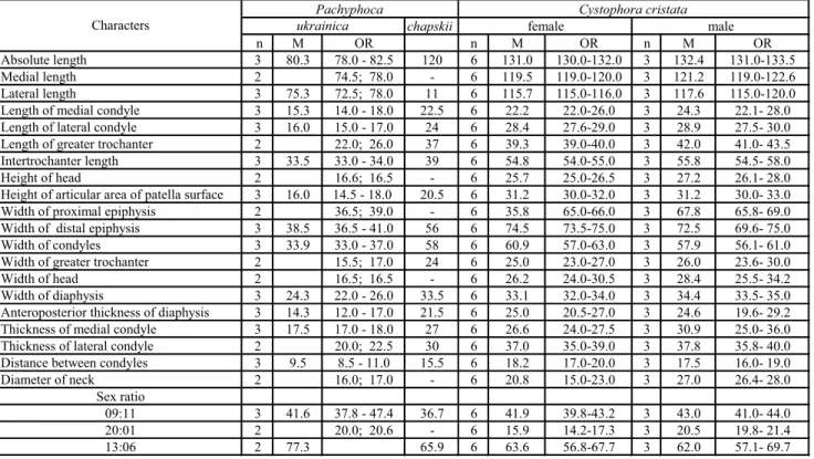
Fig. 2 - Rostral part of Recent seal maxillae in A) Dorsal views (modified afterChapskii, 1974). Labial views of Cysto-phora cristata mandibles: B) male (USNM 269130; re-versed) and C) female (USNM 188956).
Abbreviations: int, nasal pro-cess of intermaxilla; max, maxilla.
tors, and institutions where material is stored was com-piled using published data (Pidoplichko 1956; Dubrovo & Kapelist 1979).
Earlier, Koretsky (2001) showed that there are several localities (Gricev, Chomutovo, Zolotaya Balka, Zheltokamenka, and Tyaginka) in Ukraine where seal bones with pronounced pachyosteosclerosis are espe-cially noteworthy (Fig. 1B). These and others are as follows:
Zheltokamenka: Dnepropetrovsk region, Apos-tolovo district, Zheltokamenka village on Zeltenkaya River, limestone quarry 8-10 m deep; Middle Sarmatian (Bessarabian; 12.3 ± 11.2 Ma); expeditions of Pidoplich-ko 1938, 1940; Bezuglui 1953-54; and Semenov 2001; collection of NMNHU-P. Othermammalian remains, besides pinnipeds, such as Machairodus, Mastodon, Achtiaria and Cetotheium were found in this quarry in the 1940s. Some bones of the marine mammals (seals and whales) are very much rolled and abraded, which means that they were deposited in shallow water near the shore (shelf zone).
Khomutovo Village: Donetsk region, Novoazov district; limestone quarry 6-7 m deep; Upper Miocene (Pontian); collection of 1959; NMNHU-P.
Tarchankut: Crimea region, Chernomorsk dis-trict, Tarchankut Peninsula, 7 km south-east of village of Olenevka; Middle Sarmatian; collections of Antoniuk 1975-1986; and Koretsky 1983-1986; ZIN, NMNHU-P. Uzunlar: Crimea, Kerch Peninsula; Middle Sar-matian; collection 1952; NMNHU-P.
Zolotaya Balka: Cherson region, Novovorontso-vo district, right bank of the Dnieper River, south of city of Nikopol; limestone quarry north of village, upperlayer, 7 m deep, Middle Sarmatian; collection of Bezuglui 1952; NMNHU-P.
Gnylozubov: Donetsk region, Mariupol district, left bank of Kalmius River; village of Bobrinets; lime-stone quarry, Middle Sarmatian; collection of 1936; NMNHU-P.
Gritsev: Chmelnitsky region, Shepetovka district, 3 km west of village of Gritsev; karst deposits in lime-stone quarry on right bank of Chomora River; Middle
Sarmatian; collection of Semenov 1983-1985;
NMNHU-P.
Novovorontsovo: Cherson region, Novovoront-sovsky district; clay; Middle Miocene; collection of Be-zuglui 1952; NMNHU-P.
Nikolaev: Nikolaev region, vicinity of city of Ni-kolaev; Early Sarmatian; collection of 1935; NMNHU-P. The material was collected and gradually accumu-lated during many years (1932 ± 2003) of excavations carried out by expeditions of the Department of Paleon-tology of the NMNHU-P. The collection includes 58 individual postcranial bones representing the new genus
and two new species of the Subfamily Cystophorinae, all housed in the Department of Paleontology of the NMNHU-P.
Systematic Paleontology
Superfamily Phocoidea Smirnov, 1908 Family Phocidae Gray, 1825 Subfamily Cystophorinae Gray, 1866
Type Genus: Cystophora Nilsson, 1820; present distribution: Arctic and subarctic North Atlantic oceans; circumpolar in subantarc-tic region.
Distribution: Middle Miocene (Sarmatian) in Eastern Para-tethys; Recent in Arctic and Antarctic Oceans.
Emended Diagnosis: Large seals (length up to 5 m) with six incisors (I=2/1; in contrast to Monachinae with 8 incisors and Phocinae with 10 incisors); paroccipital process of skull poorly developed (in contrast to Devinophocinae); anteroposterior length of auditory bulla less than distance between bullae (in contrast to Phocinae and Devino-phocinae); infraorbital process present; interorbital space wide; inter-orbital width less than 30%, but equal to or greater than 25% of mastoid width (as in Devinophocinae); anterior palatal foramina oval (as in Devinophocinae) and shallow; preorbital part of maxilla with narrow concavity (similar to Lobodontini; in contrast to Monachinae, Phocinae, and Devinophocinae). Upper second incisors tend to enlarge rather than first incisors.
Mandibularchin prominence absent; alveoli of p4 biggerthan alveoli of m1; coronoid process very narrow and turned caudally, espe-cially in male; condyloid process not well marked; symphysis reaches posterior alveolus of p1; mandibular notch very narrow; retromandibu-larspace elongated (~3.5 cm in females and ~ 3 cm in males).
Middle of internal crest of humeral trochlea rises wave-like over coronoid fossa; widths of distal and proximal epiphyses almost equal. Medial and lateral femoral condyles almost equal in size; lesser trochanter present in males; minimum width of femoral shaft 1.4-1.9 times width of proximal epiphysis.
Included Genera: Cystophora Nilsson, 1820; Mirounga Gray, 1827.
Comparisons. The interorbital portion of the frontal bone in Cystophorinae (as in Devinophocinae), but in contrast to Phocinae, is widened (apomorphic or derived condition), even to a greater degree than in Monachinae. On the anterior part of the frontal bone, the origin of the temporal crest is present, in contrast to the other three subfamilies. The part of the maxilla (Fig. 2A) situated between the nasal meatus and orbits has a narrow concavity, as in Devinophocinae, and contrasts with Phocinae (with a short convexity) and Monachinae (with a long concavity). In contrast to the other three subfamilies, an interval is absent between the external auditory meatus and the anterior crest of the mandibu-lar fossa. The lacrimal bone has an antero-orbital pro-cess strongly protruding to a greater degree than in Monachinae. The convexity of the mastoid is not ro-bustly expressed and is directed cranio-caudally. The foramen ovale is ventrally covered by the presphenoid bone, and laterally by the postglenoid process. The
antero-inferior process of the jugal bone is located al-most at the level of the middle of the orbit, in contrast to the other subfamilies. The anterior palatal foramina are oval as in Devinophocinae and tend to disappear as in Monachinae.
The upper second incisors, in contrast to the other subfamilies, are much bigger than the first ones, and almost reach the size of the canines. The teeth are very small in comparison to the size of the mandible, p4 is bigger than m1, but the largest tooth is p3; P2, p2 are single-rooted; p2 ± m1 have fused roots, and the alveoli are shallow.
In Cystophorinae, the body of the mandible, and especially the ramus, are very unique, compared to the othersubfamilies. The mandibularbody is very thick, but not high. The ramus of the mandible is also very thick, wide, and high in contrast to the other subfamilies, as is the absence of the mandibularchin prominence. The condyloid process is especially short, narrow, and not pronounced. Although we believe that Cystophor-inae and Mirounga belong to the same taxon, some ecomorphological characteristics do differ, such as the Cystophorine coronoid process of the mandible being very sleek and turned caudally, the presence of a very narrow mandibular notch, and an elongated retroman-dibularspace, compared to those found in Mirounga.
The middle of the humeral crest of the trochlea (Fig. 3) in Cystophorinae is wave-like and elevated over the coronoid fossa, in contrast to that in Phocinae, which is at the level of the coronoid fossa, or in Mo-nachinae, where it is arch-like and concavely raised over the coronoid fossa.
The femoral condyles are almost equal in size (but less so than in Monachinae), in contrast to Phocinae. The minimum width of the diaphysis is almost twice as great as the width of the proximal epiphysis.
Discussion. Othertaxa have been suggested to be closely related to members of Cystophorinae. Zapfe (1937) proposed that the representatives of the genus Miophoca are undoubtedly ancestral to Cystophorinae. While Simpson (1945), Thenius (1950, 1952), King (1964), and McKenna and Bell (1997) assigned Miopho-ca vetusta to the Subfamily Monachinae, otherinvesti-gators (Ray 1977; Muizon 1982; Savage & Russell 1983) did not mention this taxon at all in theirreviews of the Tertiary seals of Europe. Thenius (1950, 1952), without any explanation, transferred Miophoca vetusta to an-othergenus, Pristiphoca. Later, Holec et al. (1987, 1996) supported this opinion with a proposal to reassign genus Miophoca to the Tribe Cystophorini. However, the distinctive morphological mandibular characters (Koretsky et al. in press), especially the similar degree of the condyloid angle (12ë-15ë), shows that Miophoca vetusta clearly belongs to the Subfamily Monachinae.
Sexual Dimorphism inCystophora cristata
In prior studies of true seals, sexual dimorphism was only examined on cranial material of Recent Pho-cinae (Ognev 1935; Chapskii 1952, 1967; Khuzin 1967). However, fossil remains of seals usually consist of iso-lated limb bones, mainly the femurand humerus. The lack of fossil cranial material makes the use of postcra-nial elements even more important in the proper classi-fication of specimens and in examining the sexual varia-bility (dimorphism) of various bones. Studies determin-ing ontogenetic and sexual variation based on postcra-nial elements of modern and fossil seals were done by Gadjiev 1982, Koretsky 1987a, Van Bree and Erdbrink 1987 and Koretsky 2001.
Fig. 3 - Tip of the arrow points to the middle of the humeral tro-chlearcrest; A) in Phocinae, at the level of the coronoid fossa; B) in Monachinae, arch-like elevated over the coronoid fossa; C) in Cysto-phorinae, wave-like raised overthe coronoid fossa.
Sexual dimorphism in the postcranial bones and mandibles of the Subfamily Cystophorinae (Fig. 4; Tabs 2, 6), especially in Mirounga, is more obvious than in other true seals. Prior to this study, there have never been any fossil remains found and described within Cy-stophorinae. The new fossil postcranial bones (58 invidual bones) examined in this study show obvious di-mensional and morphological diversity and fell into two size classes that do not fit previously described patterns of individual, ontogenetic, or sexual variations. Pre-viously, Koretsky (1987a, 2001) studied sexual di-morphism in the skull, mandible, humerus, and femur of Recent Phocinae and compared them to fossil mate-rial. The finds of fossil Cystophorinae provided a un-ique opportunity to study the sexual dimorphism in the limb bones and mandibles in extinct, as well as living, members of the Subfamily. The following sexual differ-ences were identified (similar differdiffer-ences can be ob-served in modern and fossil Phocinae).
Mandible (Figs 2B, C). The sexual dimorphism in Cystophora cristata is discernible not just in size but in the different proportions of the ramus and the body of the mandible. In addition, the male retromandibular space is even more elongated than in females, as well as in both sexes in other representatives of Phocidae, with the coronoid crest less inclined. The coronoid pro-cess in males is much lower, is turned caudally, and ends at almost the same level as the condyloid process (in contrast to other subfamilies). The angular process is more developed in females than in males; in contrast, the mandibularnotch in males is widerthan in females. The masseteric fossa in females is also deeper and better outlined than in males. The condyloid angle (the posi-tion of the condyloid process in relaposi-tion to the axis of the alveolar row) of 25 specimens were examined, with the average measurements being 53ë in females and 40ë
in males (see detailed explanation in Koretsky et al. in press).
Humerus (Figs 4A, B; Tab. 2). This bone in males of Cystophora is absolutely longer and more robust than in females, and the head is compressed in a dor-so-ventral direction, while in females it is larger and more spherical (contrary to Phocinae; Tab. 2). The ratio of the greatest width of the head to the height in the male of Cystophora cristata is 0.85; in the females it is 0.80. Despite the tallerbone in a male, the length of the deltoid crest is equal to that in females, but the distal part of the deltoid crest in a female ends as a narrow V, while in a male it is wider, U-like (Fig. 3A). The head of the humerus of the male C. cristata (Tab. 2) is bent caudally to a greater degree than that of females, indi-cating probable sexual dimorphism also. However, in the humerus of modern Phocinae this difference is not pronounced.
The fossa located medio-distal to the head seen in caudal aspect (between the lessertubercle and the head) is deeperin males (as in Phocinae). The enormous me-dial head of the triceps muscle arises from this fossa on the medial side of the neck of the humerus, and inserts onto the dorsal part of the olecranon of the ulna (Ho-well 1930; English 1977). The action of the triceps me-dialis muscle is to extend the elbow joint (Milleret al. 1964).
In females, although the deltoid crest is bigger in comparison to the rest of the bone (contrary to Phoci-nae), the deltoid tuberosity, which is part of the deltoid crest, is more developed (similar to Phocinae). The mus-culospiral groove, where the brachialis muscle origi-nates (Howell 1930; PieÂrard 1971; Tarasoff 1972; Ho-ward 1975; English 1977), is deeper in males. In fossils, this character is much less pronounced, evidently as a result of wear on the available material.
chapskii
n M OR n M OR n M OR
Absolute length 2 85.0; 89.0 - 4 148.2 143.0-153.3 3 153.1 152.0-154.0
Length of deltoid crest 2 55.0; 56.0 - 4 81.0 78.0-83.9 3 83.3 82.6-83.4
Height of head 4 20.8 19.5-23.0 - 4 30.4 28.9-31.5 3 30.7 28.7-34.5
Height of trochlea 3 15.0 14.0-16.0 21.0 4 29.5 28.0-31.0 3 27.8 27.7-28.0
Width of head 4 23.3 22.0-25.0 - 4 34.5 34.0-35.1 3 35.9 34.6-38.0
Width of deltoid crest 2 19.5; 20.0 - 4 36.8 31.0-41.5 3 45.5 44.1-50.0
Width of distal epiphysis 2 33.0; 34.5 - 4 57.3 55.0-60.0 3 57.2 56.7-63.0
Width of proximal epiphysis 2 29.0; 32.0 - 4 53.8 49.5-58.0 3 56.0 53.5-60.0 Width of trochlea below 3 18.0 17.0-19.0 - 4 31.3 30.6-32.0 3 33.1 30.6-38.0 Width of trochlea, frontal view 3 21.3 20.0-23.0 49.5 4 31.7 28.0-34.0 3 35.9 35.0-37.7 Transverse width of diaphysis 4 18.3 16.5-21.5 27.0 4 23.6 23.2-24.0 3 23.9 22.2-27.0 Thickness of proximal epiphysis 2 29.0; 35.0 - 4 49.1 44.7-53.5 3 53.9 53.7-68.0 Thickness of medial condyle 3 16.3 15.5-17.0 23.0 4 25.0 24.0-26.1 3 25.7 24.1-27.0 Thickness of lateral condyle 3 15.2 15.0-5.5 20.0 4 28.2 26.4-30.0 3 27.8 27.0-31.0 Diameter of diaphysis with deltoid crest 2 31.5; 33.0 - 4 53.8 53.0-55.0 3 58.4 53.2-66.0
Characters
Pachyphoca Cystophora cristata
ukrainica female male
In females, the medial epicondyle is more com-pressed and narrow in its lateral part (Howell 1930; English 1977). At the same time, the lateral epicondyle is shorter and narrower than in males, as in Phocinae. The entepicondylar foramen is always present in both sexes.
The shape of the humeral coronoid fossa of Cys-tophora depends on sex, with males having a sharp tri-angular form and females having a rounded-tritri-angular or semi-rounded form (Fig. 4A); its depth is somewhat greater in females.
Femur(Figs 4C, D; Tab. 6). The absolute length of this bone in males of C. cristata is less than in males (in contrast to Phocinae); however, the male fe-mur is more robust and wider. Moreover, in females of C. cristata the anteroposterior diameter of the diaphysis is greater than in males, but the least width of the diaphysis in females is located in the middle of the shaft, while in males it is shifted to the proximal por-tion of the shaft.
Sexual dimorphism in the structure of the diaphy-sis is associated with various degrees of development of the vastus intermedius and medialis muscles in cranial aspect (see PieÂrard 1971: 73), and of the m. adductor cranialis (= adductor anticus in Howell 1930) in caudal aspect. A detailed description of the actions of these muscles can be found in Koretsky (2001).
In males, the head of the femuris largerand the diaphysis is less compressed, while in females it is the reverse. The average width of the diaphysis is smaller in females, while the distal epiphysis is more developed in males. One unique and very primitive character of C. cristata is that the lessertrochanterin males is well developed, while in females it is not.
In contrast to Phocinae, the neck of the femur of C. cristata is shorter and wider in females. Therefore (as in Phocinae), the neck forms nearly a right angle with the long axis of the bone in females, but in males the angle is greater than 90ë.
The greater trochanter is wider and longer in males than in females. The distal part of the greater trochanter in females terminates more sharply or acutely (being V-shaped), while in males it is frequently rounded (U-shaped).
The gluteus medius and gluteus minimus muscles insert onto the cranial side of the greater trochanter, while the m. piriformis is attached to the caudal side. The attachment sites of these muscles are significantly more developed in males (Howell 1930; PieÂrard 1971; Howard 1975; Koretsky 2002), and consequently, the trochanter also is more developed in males.
Howell (1928, 1930), PieÂrard (1971), and Howard (1975) described the trochanteric fossa as a place of attachment forthe obturator internus and externus mus-cles, which share a common tendon of insertion with the superior and inferior gemelli muscles (Koretsky & Sanders 2002). The obturator externus and gemelli mus-cles are more developed in females than in males, resul-ting in a deeper and more closed trochanteric fossa (as in Phocinae).
In females of C. cristata, the plantarfossa above the lateral condyle is wider and deeper and is bordered by a very thin edge of bone. The plantar fossa itself is the place of origin of the plantaris and lateral head of the gastrocnemius muscles. Although the patellarsurface is larger in males, the sizes of the condyles are almost equal in both sexes. Fordetailed functional analysis of these muscles see Howard (1975) and Koretsky (2001). For determining the sex using the humerus and femurin the Subfamily Cystophorinae, the following characters can be used: 1) Humerus ± overall size; length of deltoid crest and width of its middle part; depth and shape of the coronoid fossa; depth of the fossa located caudal to the medial side of the neck of the humerus, distal to the lessertubercle (similarto Phocinae) and 2)
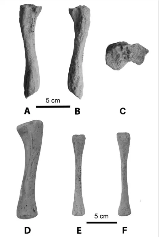
Fig. 4 - Sexual dimorphism of Recent Cystophora cristata. Hu-merus, cranial view of A) female (AMNH 184660, L.) and B) male (AMNH 184659, L.). Male femur(USNM 550411, R.) in C) cranial and D) caudal views.
Femur ± overall size; anteroposterior width (=dorso-ventral thickness) of diaphysis; length and thickness of neck; length and width of greater trochanter; anteropos-terior diameter of distal epiphysis; presence of lesser trochanter.
The most reliable characters for sex determination in Cystophorinae are: in males, the absence of the men-tal tubercle; the height of the first incisor almost equa-ling the dimensions of the canine; width of the distal epiphysis on the humerus; and presence of the lesser trochanter of the femur.
While the features noted above are characteristic for adult animals, they are not nearly as pronounced in young and subadult individuals. Overall, the established differences and variations of characteristics are fairly constant, allowing researchers to separate individual elements of the extremities according to sex.
Pachyphoca gen. n.
Type species: Pachyphoca ukrainica; Middle Sarmatian of Ukraine Etymology: Pachys, Greek, thick (referring to bone hypertro-phy); phoca, Latin, from the Greek phoke, seal (f.).
Diagnosis: Lesser tubercle of humerus small, round, and lo-cated distal to head and greater tubercle; head compressed craniocau-dally; intertubercular groove wide and shallow; medial epicondyle flat-tened, spreading from lower part of entepicondylar foramen; olecranon fossa deep and narrow.
Greater trochanter of femur slightly higher than head; trochan-teric fossa deep; intertrochantrochan-teric crest flat, wide, and thick, reaching lesser trochanter; head large, seated on distinct lip (shaped like mush-room) and wide neck; minimum width of diaphysis shifted proximally; body of femurswollen, thick, husky, heavy, and pachyosteosclerotic; condyles almost equal in size.
Scapularspine ends smoothly; infraspinous fossa deeperand widerthan supraspinous.
Medial surface of ulna flattened, not concave; olecranon short and thick; radial notch deep and long.
Ilium thick; iliac crest not averted, and excavated on its ventral surface as in other Monachinae and Cystophorinae; iliopectineal emi-nence flattened; fossa for m. gluteus medius shallow; auricular fossa deep; alar spine robust; iliac tuberosity and caudal dorsal iliac spine not very well developed; caudal dorsal ischial spine rounded and wide, not protruding; alar spine robust; acetabulum shallow.
Popliteal notch of tibia shallow and wide; tibial crest rounded in dorsomedial direction; tibial tuberosity flattened; muscular groove wide; grooves on distal end shallow and flattened.
All bones are thick, husky, and pachyosteosclerotic.
Included species: In the Middle Sarmatian (12.3-11.2 Ma) of the Northern Black Sea littoral region, southern Ukraine, two new cysto-phorine species are recorded: Pachyphoca ukrainica and P. chapskii.
Range: Middle Miocene, Middle Sarmatian (Bessarabian Stage; 11.2-12.3 Ma), the Northern Black Sea region, Ukraine, Eastern Para-tethys.
Comparisons. Pachyphoca has characters similar to other representatives of the Subfamily Cystophorinae such as: absence of the musculospiral groove of the hu-merus; the protruding femoral intertrochanteric crest extending more proximal than the head; the presence
of the lesser trochanter; almost equal-sized condyles. Otherspecific similarities to Cystophora include: cra-nio-caudal depression of the humeral head; short del-toid crest; enlargement of the deldel-toid crest in the mid-dle; and presence of the entepicondylar foramen.
Similarities to Mirounga are: shallow and wide intertubercular groove of humerus; shallow, oval coro-noid fossa extending to the same level as medial epicon-dyle; noticeable femoral intertrochanteric line; very deep intercondylar groove.
In addition, Pachyphoca differs from these two genera of the Subfamily Cystophorinae by: much smal-lersize; position and shape of the lessertubercle of the humerus; presence of a distinct lip between the femoral head and the neck; and deeper trochanteric fossa. Spe-cifically, Pachyphoca differs from Cystophora by the reverse relationship between lateral and medial epicon-dyles and from Mirounga by: the presence of the ente-picondylar foramen of the humerus; shorter deltoid crest; protruding lesser trochanter of the femur; higher position of the greater trochanter; shorter femoral neck and larger head compared to the size of the bone.
Pachyphoca ukrainicagen. n sp. n. Figs 5-10, Tabs 1-8
Etymology: ukrainica, from Latinized form of Ukraine. Holotype: Right humerus, NMNHU-P 64-701, Middle Sarma-tian.
Type Locality: Khomutovo Village, Donetsk region, Novoazov district, Ukraine.
Range: Middle Sarmatian (Bessarabian Stage, 11.2-12.3 Ma) deposits of the Northern Black Sea Region, Ukraine.
Diagnosis: Maximal enlargement of humeral deltoid crest lo-cated in middle; deltoid crest shorter than of bone length; lateral epicondyle shorter and lower than medial (in contrast to P. chapskii), does not reach proximal part of medial epicondyle; medial epicondyle extending more proximal than coronoid fossa; entepicondylar foramen located above coronoid fossa; coronoid fossa oval.
Proximal part of femoral greater trochanter wider than distal; trochanteric fossa narrow; lesser trochanter well developed; head mas-sive, seated on distinct lip on wide neck; supracondylar fossa deep, wide, rectangular.
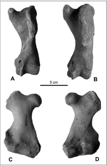
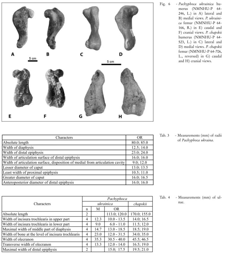
Referred Specimens: In addition to the holotype, the following specimens were found in Ukraine: humeri: NMNHU-P 64-246 (L.), Uzunlar; 64-703 (L.), 64-713 (L.), Zolotaya Balka; scapulae: NMNHU-P, 64-477 (L.), 64-702 (R.), Zolotaya Balka; radii: NMNHU-P 64-481 (L., immature), 64-482 (L., immature), Zolotaya Balka; ulnae: NMNHU-P 705 (L.), Zolotaya Balka; 383 (R.), 64-710 (R., possibly from the same individual as holotype), Khomutovo; 711, (R., from the same individual as rib 711, not described), 712 (R.), Zheltokamenka; innominata: NMNHU-P 479 (L.), 64-348 (L.), Zolotaya Balka; femora: NMNHU-P 64-354(R.), Zolotaya Balka; NMNHU-P 64-166 (R.), Gnylozubov; 64-471 (L.), Zolotaya Balka; tibiae and fibulae: NMNHU-P 64-472 (R., proximal fragment), 64-473 (L.), 64-478 (L., proximal fragment), Zolotaya Balka; vertebra: NMNHU-P 64-704 (thoracic), Zolotaya Balka.
Description. Scapula (Figs 5A-C; Tab. 1): The scapularspine ends smoothly and does not reach the
vertebral border, which is partially broken in available specimens. The acromion is not developed and does not reach the ventral angle. In the cervical region, the infra-articular tuberosity shows as a long ridge connected with a thick and distinct muscularline on the infraspi-nous fossa. The coracoid process and scapular tuberos-ity are not developed either. The infraspinous fossa is deeperand widerthan the supraspinous. The distal bor-der of the supraspinous fossa ends as a straight line. In the infraspinous fossa the caudal angle forms a thin, round edge at the vertebral border. The glenoid fossa is deep with a thin mushroom-like lip. The complete bone is very thick at the lateral border and becomes much thinner at the vertebral border.
Humerus (Figs 6A, B; Tab. 2): The intertubercular groove is shallow and wide. The widest portion of the deltoid crest is located in the middle; the crest extends about half the length of the bone, and smoothly des-cends to the condyles as a flat, almost invisible ridge. The deltoid tuberosity is small and located at the middle of the deltoid crest proximal to the middle of the dia-physis. The lesser tubercle is weakly developed, round, and located considerably inferior to the greater tubercle, just distal to the head, which is compressed craniocaud-ally. The musculospiral groove is absent. The lateral epicondyle is shorter and lower than the medial, and does not reach the distal part of the deltoid crest. The medial epicondyle is flattened, spreading from the lower part of the entepicondylar foramen, extending higher
than the coronoid fossa. The entepicondylar foramen is located above the coronoid fossa, with a laterally-located wide wall. The coronoid fossa is shallow, oval, extends further proximally than the lateral epicondyle, and ends at the same level as the medial epicondyle. The olecranon fossa is deep and narrow. The complete bone is extremely husky with a very pachyosclerotic condi-tion.
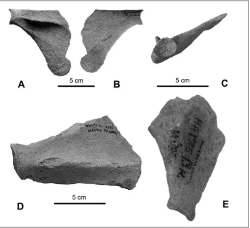
Radius (Figs 7A, B; Tab. 3): The radial tuberosity is very large, wide, flat, and does not protrude laterally. The groove for all tendons including the tendon of the m. abductor pollicis longus is wide and shallow, whereas the ridge for the m. extensor digitorum communis is only shallow. Both available bones belong to subadult animals.
Ulna (Figs 7C-E; Tab. 4): The medial surface of the bone is flattened and not concave; the olecranon is short and thick, gradually connecting to the proximal half of the bone. On the lateral surface, there is barely any visible prominence of the rugosity for brachialis muscle insertion. Rather than a fossa for origin of the m. abductor pollicis longus, a protuberance is present. Caudal to the articular surface is another deep, wide, and short groove; this groove is surrounded by a circu-lar (sharp) crest. On the bone's radial aspect, the coro-noid process only slightly protrudes forward over the radial notch; the radial notch is deep and long. The interosseous crest is swollen, forming a prominence that protrudes only slightly. The head is not preserved.
Fig. 5 - Scapula of Pachyphoca ukrai-nica (NMNHU-P, 64-477, L.) in A, dorsal B) ventral and C, glenoid views. Scapula of P. chapskii (NMNHU-P 64-707) in D) dorsal and E) ventral views.
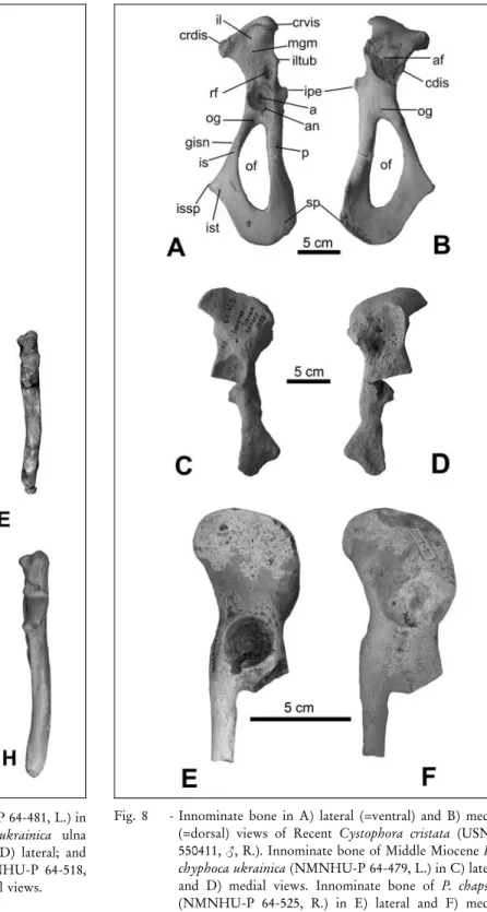
Innominate (Figs 8C, D; Tab. 5): As in other Monachinae and Cystophorinae, the ilium is thick and the iliac crest is not averted and not excavated on its ventral surface. The iliopectineal eminence is small, si-tuated higher than the proximal border of the acetabular fossa. The greater ischial notch is slightly concave, al-most straight. A shallow depression (fossa) for the m. gluteus medius is located on the medial aspect of the ilial wing; on its lateral aspect is a deep and narrow auricular
fossa fororigin of the m. psoas major. The alarspine does not protrude very far. The iliopectineal eminence is absent, as is the pectineal tubercle. The edges of the acetabular fossa are not raised above the plane surface of the bone; the acetabulum is circular with a slightly marked cotyloid notch. The ischium is thick, wide and robust; the ischial spine is large, rounded and well de-veloped forattachment of the m. biceps femoris. The pubis is not preserved.
Characters OR
Absolute length 80.0; 85.0
Width of diaphysis 12.5; 14.0
Width of distal epiphysis 23.0; 24.0
Width of articulation surface of distal epiphysis 16.0; 16.0 Width of articulation surface, disposition of medial from articulation cavity 9.0; 12.0
Lesser diameter of caput 13.0; 13.5
Least width of proximal epiphysis 10.5; 11.0
Greater diameter of caput 16.0; 16.5
Anteroposterior diameter of distal epiphysis 16.0; 16.0
Tab. 3 - Measurements (mm) of radii of Pachyphoca ukraina.
chapskii
n M OR
Absolute length 2 113.0; 120.0 170.0; 155.0
Width of incisura trochlearis in upper part 4 12.3 10.0 - 13.5 14.0; 16.5 Width of incisura trochlearis in lower part 4 9.0 6.0 - 11.0 11.5; 12.0 Maximal width of middle part of diaphysis 4 14.7 13.0 - 18.5 18.5; 19.0 Width of bone at the level of incisura trochlearis 4 23.0 12.0 - 31.5 34.0; 35.0
Width of olecranon 4 35.3 30.5 - 40.0 45.5; 46.5
Transverse width of olecranon 4 13.3 12.0 - 14.0 16.5; 19.0 Maximal width of distal epiphysis 2 15.0; 17.5 19.5; 21.0
ukrainica Pachyphoca
Characters
Tab. 4 - Measurements (mm) of ul-nae.
Fig. 6 - Pachyphoca ukrainica hu-merus (NMNHU-P 64-246, L.) in A) lateral and B) medial views. P. ukraini-ca femur(NMNHU-P 64-166, R.) in E) caudal and F) cranial views. P. chapskii humerus (NMNHU-P 64-523, L.) in C) lateral and D) medial views. P. chapskii femur(NMNHU-P 64-706, L., reversed) in G) caudal and H) cranial views.
Femur (Fig. 6E, F; Tab. 6): The greater trochanter extends proximally slightly higher than the head; its proximal part is somewhat wider than the distal. The trochanteric fossa is deep, narrow and open proximally, reaching the distal half of the greater trochanter. The flat, wide and thick intertrochanteric crest reaches the lesser trochanter, which serves for insertion of the m. iliopsoas. The lesser trochanter is very visible, thick, and not separated from the crest; the lesser trochanter reaches below the distal border of the greater trochanter and ends at the same level as the distal border of the
head (orslightly higherthan the head). The femoral head is large, relative to the bone's mass, and seated on a very short, thick, and wide neck; between the head and neck is a distinct lip. The smallest width of the
Fig. 7 - Pachyphoca ukrainica radius (NMNHU-P 64-481, L.) in A) dorsal and B) ventral views. P. ukrainica ulna (NMNHU-P 64-710, R.) in C) medial; D) lateral; and E) cranial views. P. chapskii ulna (NMNHU-P 64-518, R.) in F) medial; G) lateral; and H) cranial views.
Fig. 8 - Innominate bone in A) lateral (=ventral) and B) medial (=dorsal) views of Recent Cystophora cristata (USNM 550411, <, R.). Innominate bone of Middle Miocene Pa-chyphoca ukrainica (NMNHU-P 64-479, L.) in C) lateral and D) medial views. Innominate bone of P. chapskii (NMNHU-P 64-525, R.) in E) lateral and F) medial views. Abbreviations: a, acetabulum; af, auricular fossa; an, acetabular notch; cdis, caudal dorsal iliac spine; crdis, cranial dorsal iliac spine; crvis, cranial ventral iliac spine; gisn, greater ischial sciatic notch; il, ilium; iltub, iliac tu-berosity; ipe, iliopectineal eminence; is, ischium; issp, is-chial spine; ist, isis-chial tuberosity; mgm, m. gluteus med-ius; of, obturator foramen; og, obturator groove; p, pubis; rf, m. rectus femoris attachment; sp, symphysis pubis.
diaphysis is shifted toward the proximal end of the bone. The supracondylar fossa is located above the medial condyle and is deep, wide, and rectangular; the condyles are almost equal in size.
Tibia (Figs. 9A-C; Tab. 7): The two condyles are weakly concave in their centers, shortened and oval. The intercondyloid eminences are weak and only slightly raised above the two lateral, well-developed borders of the condyles. The popliteal notch is shallow and wide, but well marked. The tibial crest is rounded in a dorsomedial direction. On the ventral side of the tibia, the tibial tuberosity is flattened and not well marked. The muscular groove is flat and wide; the grooves on the distal end are shallow and flattened.
Discussion. The innominate bone of Pachyphoca ukrainica has several very primitive characteristics, such as:
* The femoral lesser trochanter is present, for in-sertion of the m. iliopsoas (which flexes and laterally rotates the femur, tilts the pelvis forward, flexes the
thigh upon the pelvis, and adducts the femur; Koretsky & Sanders 2002).
* The greater ischial notch of the os coxa is almost straight.
* A deep and narrow auricular fossa is present for insertion of the m. psoas major (a flexorof the hip joint). * The ischial spine is large, rounded, and well developed forattachment of the m. biceps femoris (an extensorof the hip joint).
Therefore, it can be concluded that this animal was well adapted for terrestrial locomotion due to the presence of these muscular attachments.
Pachyphoca chapskiigen. n. sp. n. Figs 5-10, Tabs 1, 2, 4-9
Etymology: Named in honorof the late K. K. Chapskii (of the Zoological Institute, St. Petersburg, Russia) in recognition of his nu-merous contributions to the study of fossil and modern true seals.
Holotype: Left femur, NMNHU-P 64-706, Middle Sarmatian. Type Locality: Zheltokamenka, village on Zheltenkaya River, Dnepropetrovsk region, Apostolovo district, Ukraine.
Range: Middle Sarmatian (Bessarabian Stage, 11.2-12.3 Ma) deposit of the Northern Black Sea Region, Ukraine.
Diagnosis: Medial epicondyle does not reach coronoid fossa (in contrast to P. ukrainica); coronoid fossa wide, not well outlined, does not reach proximal end of lateral epicondyle.
Greater trochanter of femur is oblique; its distal part is wider than the proximal; trochanteric fossa is wide and reaches middle of greater trochanter; lesser trochanter is wide, not separated from inter-trochanteric crest; condyles flat, small, widely spaced; instead of plantar fossa, plantar eminence present; supracondylar fossa has thick protu-berance. P. ukrainica P. chapskii OR 52.5 70.5 61.0 - 45.0 53.0 60.0; 67.0 44.0 11.0; 13.5 14.0 117.6 133.0 1 : 3 Ratio Characters
Length from center of acetabulum to iliac crest Length of pubic from center of acetabulum Width of level of iliac crest
Width of pubic Thickness of pubic
Tab. 5 - Measurements (mm) of innominate of Pachyphoca.
chapskii
n M OR n M OR n M OR
Absolute length 3 80.3 78.0 - 82.5 120 6 131.0 130.0-132.0 3 132.4 131.0-133.5 Medial length 2 74.5; 78.0 - 6 119.5 119.0-120.0 3 121.2 119.0-122.6 Lateral length 3 75.3 72.5; 78.0 11 6 115.7 115.0-116.0 3 117.6 115.0-120.0 Length of medial condyle 3 15.3 14.0 - 18.0 22.5 6 22.2 22.0-26.0 3 24.3 22.1- 28.0 Length of lateral condyle 3 16.0 15.0 - 17.0 24 6 28.4 27.6-29.0 3 28.9 27.5- 30.0 Length of greater trochanter 2 22.0; 26.0 37 6 39.3 39.0-40.0 3 42.0 41.0- 43.5 Intertrochanter length 3 33.5 33.0 - 34.0 39 6 54.8 54.0-55.0 3 55.8 54.5- 58.0
Height of head 2 16.6; 16.5 - 6 25.7 25.0-26.5 3 27.2 26.1- 28.0
Height of articular area of patella surface 3 16.0 14.5 - 18.0 20.5 6 31.2 30.0-32.0 3 31.2 30.0- 33.0 Width of proximal epiphysis 2 36.5; 39.0 - 6 35.8 65.0-66.0 3 67.8 65.8- 69.0 Width of distal epiphysis 3 38.5 36.5 - 41.0 56 6 74.5 73.5-75.0 3 72.5 69.6- 75.0
Width of condyles 3 33.9 33.0 - 37.0 58 6 60.9 57.0-63.0 3 57.9 56.1- 61.0
Width of greater trochanter 2 15.5; 17.0 24 6 25.0 23.0-27.0 3 26.0 23.6- 30.0
Width of head 2 16.5; 16.5 - 6 26.2 24.0-30.5 3 28.4 25.5- 34.2
Width of diaphysis 3 24.3 22.0 - 26.0 33.5 6 33.1 32.0-34.0 3 34.4 33.5- 35.0 Anteroposterior thickness of diaphysis 3 14.3 12.0 - 17.0 21.5 6 25.0 20.5-27.0 3 24.6 19.6- 29.2 Thickness of medial condyle 3 17.5 17.0 - 18.0 27 6 26.6 24.0-27.5 3 30.9 25.0- 36.0 Thickness of lateral condyle 2 20.0; 22.5 30 6 37.0 35.0-39.0 3 37.8 35.8- 40.0 Distance between condyles 3 9.5 8.5 - 11.0 15.5 6 18.2 17.0-20.0 3 17.5 16.0- 19.0
Diameter of neck 2 16.0; 17.0 - 6 20.8 15.0-23.0 3 27.0 26.4- 28.0 Sex ratio 09:11 3 41.6 37.8 - 47.4 36.7 6 41.9 39.8-43.2 3 43.0 41.0- 44.0 20:01 2 20.0; 20.6 - 6 15.9 14.2-17.3 3 20.5 19.8- 21.4 13:06 2 77.3 65.9 6 63.6 56.8-67.7 3 62.0 57.1- 69.7 Characters ukrainica Cystophora cristata Pachyphoca female male
Referred Specimens: In addition to the holotype, the following specimens were found in Ukraine: humeri: NMNHU-P 64-708 (R., distal end), 64-523 (L.), Zheltokamenka; scapula: NMNHU-P, 64-707 (L.), Zheltokamenka; ulnae: NMNHU-P 64-518 (R.), 64-519 (R.), Zheltokamenka; innominate: NMNHU-P 64-525 (R.), Zheltokamen-ka; tibia: NMNH-P 64-520 (L., immature), ZheltokamenZheltokamen-ka; fibula:
NMNHU-P 521 (L., immature, from the same individual as 64-520), Zheltokamenka.; vertebra: NMNHU-P 64-528 (lumbar), Zhelto-kamenka; sacrum: NMNHU-P 64-527, ZheltoZhelto-kamenka; IMtc 2nd phalange: NMNHU-P 64-517, Zheltokamenka.
Description. Scapula (Figs 5D, E; Tab. 1): The scapular spine ends smoothly, reaching the vertebral border as a small, almost flat ridge; the vertebral border is broken on the available specimen. The acromion is present as a small hook, ending at the same level and height as the scapularspine.
In the cervical region, the infra-articular tuberos-ity is present as a thick, wide protuberance. The mus-cular line on the infraspinous fossa is pronounced as a
Fig. 9 - Pachyphoca ukrainica (NMNHU-P 64-473, L.) tibia in A) caudal; B) cra-nial and C) proximal views. D) P. chapskii tibia (NMNH-P 64-520, L., immature) in caudal view. P. chapskii fibula (NMNHU-P 64-521, L., immature, from the same individual as 64-520) in E) caudal and F) cranial views.
chapskii
n M OR
Width of proximal epiphysis 3 33.2 32.0 - 35.0 40.0 Height of proximal epiphysis 3 20.0 18.0 - 22.0 27.0 Width of distal epiphysis 2 20.5; 21.5 31.5
Characters
Pachyphoca ukrainica
long, very well developed ridge (bigger and stronger than in P. ukrainica). The infraspinous fossa is deep and wider than the supraspinous fossa. In contrast to P. ukrainica, the coracoid process is thick, short, and wide.
The distal border of the supraspinous fossa ends as a straight line (as in P. ukrainica); the caudal angle is not preserved. The glenoid fossa is shallow (deep in P. ukrainica), without a pronounced lip, and without a border (in contrast to P. ukrainica).
The complete bone is very thick, heavy, and much biggerand thickerthan in P. ukrainica.
Humerus (Figs 6C, D; Tab. 2): The lateral epicon-dyle is much higherand widerthan the medial. The middle part of the trochlea is raised wave-like over the coronoid fossa. The small, oval entepicondylar fora-men has a very wide bridge over the medial epicondyle, and extends lowerthan both the coronoid fossa and the lateral epicondyle.
The medial epicondyle is flattened, spreading from the lower part of the entepicondylar foramen, and it does not reach the coronoid fossa. The medial epicondyle is lower than the coronoid fossa; the coro-noid fossa is shallow, wide and not well defined (=out-lined); it extends far proximally, but it does not reach the proximal end of the lateral epicondyle.
The olecranon fossa is deep, long, and narrow, seated transversely to the bone axis. Situated on the anterior-medial side of the humerus is a thick protru-sion, located medial to the trochlea and adjacent to the olecranon fossa. The bone is big, strong, and heavy like a stone.
Ulna (Figs 7F-H; Tab. 4): The medial surface is flattened and not concave (as in P. ukrainica). The ole-cranon is short, thick, and forms a hook at the caudal end. The rugosity for the brachialis muscle insertion has a very well developed prominence. On the lateral sur-face, the fossa forthe m. abductor pollicis longus is pre-sent. Another narrow and long groove is located caudal to the articular surface; this groove is not surrounded by a sharp crest, but very smoothly transitions into the diaphysis, reaching the ulnar protuberance (in contrast to the ulna of P. ukrainica). The trochlear notch is flat-tened and not visible on the radial aspect of the bone. The coronoid process greatly protrudes forward, at al-most 90ë, over the radial notch; the radial notch is deep and long. The anconeal process is very large, wide, and protruding. The ulnar tuberosity, situated on the middle of the diaphysis, reaches the rugosity for the tendon. The big, long, and wide interosseous crest is located on the middle of the diaphysis. The head and styloid process are preserved.
Innominate (Figs 8E, F; Tab. 5): The ilium is thick and flattened, with the iliac crest slightly averted and not excavated on its ventral surface as in other
Mona-chinae and Cystophorinae (similar to P. ukrainica). The iliopectineal eminence is wide and not pronounced; the pectineal line is hardly visible. On the medial aspect of the ilium is a shallow depression (compared to size of the bone) forthe insertion of the m. gluteus medius (this is much smallerand shallowerthan the same fossa of P. ukrainica). On the lateral aspect of the ilial wing is located a deep and wide fossa forinsertion of the m. psoas major. The alar spine is rounded, robust, and turned posteriorly. The edges of the acetabulum are raised high above the plane surface of the bone (in con-trast to P. ukrainica); the acetabulum is conical (a pri-mitive character), with a deep and well-marked cotyloid notch. Above the acetabulum, there is a deep and nar-row depression for the insertion of the m. rectus femoris (in contrast to P. ukrainica, where there is a big, flat, and wide depression). The pubis is rounded.
Femur (Figs 6G, H; Tab. 6): The greater trochan-ter is very obliquely oriented along the bone's axis; its distal part is wider than the proximal and ends in a narrow V-shape. The trochanteric fossa is deep and wide, reaches the middle of the greater trochanter's length, and has a shallow proximal border. The flat, wide, and thick intertrochanteric crest reaches the weakly developed lessertrochanter, which is wide and not separated from the intertrochanteric crest. The con-dyles are flat and small, compared to the size of the bone, and very widely spaced. Considerable swelling of the bone is seen at the location of the plantarfossa. The supracondylar fossa is not pronounced, but does have a thick protuberance. The smallest width of the diaphysis is shifted toward the proximal end of the bone. The condyles are almost equal in size.
Tibia and fibula (Figs 9D-F; Tab. 7): The two tibial condyles are shortened and oval; the tibial crest is well marked and developed. The intercondyloid emi-nence is not developed, only slightly raised above the two lateral condyles, with hardly noticeable borders. The popliteal notch is shallow and wide, but not well marked. The tibial crest is well pronounced and rounded in the dorsomedial direction. On the ventral side of the tibia, the tibial tuberosity is flattened, wide, triangular, and extends along the axis of the bone. The muscular groove is deep and wide; grooves on the distal end are shallow and flattened. The fibula is without condyles, but has a well-developed interosseous crest. Both bones probably belong to the same individual an-imal.
Vertebra (Figs 10D-F; Tab. 8): The body of the lumbar vertebra is flattened in the dorso-ventral direc-tion and heart-shaped; the vertebral foramen is oval in shape. The spinous process is very short and wide, while the transverse processes are directed cranio-ventrally. The caudal articular process is very short and rounded; the mammillary process is thick, wide, and short. The
cranial articular surface is rounded, like a short protu-berance.
Sacrum (Figs 10G-I; Tab. 9): The sacrum consists of 4 fused vertebrae as in other cystophorines (with 125 mm absolute length and 72 mm wide). The sacral pro-montory is almost flat and not pronounced. However, the ala is thick and the anterior surface of the first cen-trum is lower than the wings of the sacrum (as in mir-oungini and monachines, but in contrast to cystophor-ines and phoccystophor-ines, where the ala is on the same level as the centrum; Antoniuk 1979). The maximum width across the wings is 57.6 % of the length of the sacrum, which according to Antoniuk (1979) is a miroungine character, as the percentage in cystophorines should be less than 40%. On S2 are located thick, wide, and well-defined transverse processes. The median sacral crest, which is formed by the spinous processes, is fused between S1 and S2. All spinous processes become progressively smaller from S1 to S4, with immense, thick bases. Dorsal sacral foramina are smaller than ventral.
According to the sexually dimorphic characters of sacral bones described by Gadjiev (1982), this particular bone should belong to an adult male because its first ventral foramen is rounded and the lateral wall is very thick. The bone is partially rolled, but it is very distinc-tive and unusual. The body of the sacrum is short, wide, and very pachyosteosclerotic.
Cladistic Analysis
The data matrix for the 48 included characters of Phocidae is shown in Tab. 10. In this matrix, 0 desig-nates the most primitive state among the taxa studied; 1-2, derived states; ?, unknown or missing data. Some characters have the opposite polarity to that of Berta and Wyss (1994) and Burns and Fay (1970), while some characters have the same polarity as that of Chapskii (1974). Fora detailed discussion of polarities of the characters, see Koretsky and Grigorescu (2002).
Skull
1. Mastoid process: (0) prominence lateral to auditory bulla not strongly pronounced; (1) pronounced.
2. Mastoid process: (1) narrow (width of the process less than length of process itself); (0) wide (Chapskii 1974:301; in contrast to Berta and Wyss 1994:48).
3. Maxilla: (0) has very pronounced convexity anterior to the orbits; (1) short concavity; (2) long concavity (Chapskii 1974; in con-trast to Berta and Wyss 1994:46).
4. Anterior palatal foramina: (0) faintly marked (Burns and Fay 1970:72); (1) oval and shallow; (2) round and deep.
5. Interorbital space: (0) less than 25.0% of width of skull across mastoids; (1) less than 30.0%, but equal to orgreaterthan 25.0% of width of skull across mastoids; (2) equal to or greater than 30.0% of width of skull across mastoids (Burns and Fay 1970:370; Chapskii 1974:299).
6. Jugular process: (0) well developed; (1) poorly developed. 7. Rostrum: (0) elongate; (1) short, compared with skull. 8. Diameter of infraorbital foramen: (0) less than diameter of alveolus of maxillary canine; (1) approximately equal to diameter of Fig. 10 - Thoracic vertebra of
Pachy-phoca ukrainica (NMNHU -P 64-704) in A) lateral and B) dorsal views. Lumbar vertebra of P. chapskii (NMNHU-P 64-528) cau-dal; and E) lateral views. Sa-crum of P. chapskii (NMNHU-P 64-527) in F) dorsal; G) ventral; and H) cranial views.
alveolus of maxillary canine; (2) greater than diameter of alveolus of maxillary canine.
9. Anteroposterior length of auditory bullae: (0) greater than distance between them; (1) less than distance between them; (2) about equal to distance between them (unordered character) (Burns and Fay 1970:382; Cahpskii 1974:300).
Mandible
10. Symphyseal part: (0) continues at least to the middle of the alveolus of p3; (1) reaches only the alveolus of p2; (2) reaches only the alveolus of p1.
11. Lateral outline of symphyseal region: (0) square, symphysis thin; (1) rounded, symphysis thick; (2) straight, symphysis thick.
12. Chin prominence: (0) pronounced; (1) absent or weakly outlined.
13. Chin prominence: (0) extends from the anterior or posterior alveolus of p2 to the posterior or anterior alveolus of p4; (1) extends from the anterior alveolus of p2 to anterior alveolus of p3; (2) extends from the anterior alveolus of p2 to posterior alveolus of m1 (unordered character).
14. Maximum height of body of mandible: (0) situated between p2 and p3; (1) situated in the middle or at the posterior portion of p2; (2) situated between alveoli of p4-m1 orposteriorto alveolus of m1 (unordered character) (Korestky and Ray 1994).
15. Diastemata and tooth alveoli: (0) alveoli small, with equal diastemata; (1) alveoli round and large, with equal diastemata between them; (2) alveoli shallow, and diastemata unequal (unordered charac-ter).
16. Alveoli of p4 and m1: (0) alveoli similarin size; (1) alveolus of p4 smallerthan alveoli of m1; (2) alveolus of p4 largerthan the alveolus of m1 (unordered character).
17. Retromandibular space: (0) elongated; (1) short. Teeth
18. Numberof incisors: (0) 3/2; (1) 2/2; (2) 2/1 (Chapskii 1974: 289; in contrast to Burns and Fay 1970: 380).
19. Roots of postcanine teeth (P,p 2 ± P,p 4): (0) one root, divided partially at the base; (1) two (Berta and Wyss 1994: 51).
20. Crowns of postcanine teeth: (0) single-cusped; (1) multi-cusped (reversal to primitive condition) (Berta and Wyss 1994: 51).
21. Relative dimensions of postcanine teeth to the size and massivity of the skull: (0) large; (1) small.
22. Relative dimensions of canine to the size of the skull: (0) large; (1) small.
23. Basal cingulum of postcanine teeth: (0) well developed; (1) not developed.
24. Number of additional cusps of premolars: (0) two; (2) more than two.
25. Premolars: (0) seated parallel to axis of tooth row; (1) seated obliquely.
26. Upper incisors: (0) arranged in curved line; (1) arranged in straight line.
27. Upper incisors: (0) third larger than second (Phocinae); (1) second larger than first (Monachinae); (2) incisors equal in size to canine (Cystophorinae) (unordered character). *Phocinae have 10 in-cisors (3 upper); Monachinae have 8 inin-cisors (2 upper); and Cystophor-inae have 6 incisors (1 upper).
Humerus
28. Lesser tubercle: (0) pronounced; (1) not pronounced (in contrast to Berta and Wyss 1994: 52).
29. Trochlear crest: (0) raised in a wave-like shape over coro-noid fossa; (1) raised arch-like over corocoro-noid fossa; (2) not separated from coronoid fossa by a distinct lip.
30. Lesser tubercle and head: (0) equal in height or tubercle insignificantly higher than head; (1) tubercle very much higher than head; (2) tubercle lower than head (unordered character).
31. Lesser tubercle: (0) rounded; (1) extended along the bone's axis; (2) oval.
32. Head: (0) medio-laterally compressed; (1) round; (2) flat-tened proximo-distally.
33. Deltoid crest: (0) maximum enlargement is in its proximal part; (1) neither part noticeably enlarged; (2) maximal enlargement is in its middle part.
34. Deltoid crest: (0) shorter than one-half length of the bone, confined to the proximal half of the bone; (1) longer than one-half length of the bone but not reaching coronoid fossa; (2) reaches coro-noid fossa (in contrast to Berta and Wyss 1994: 52).
35. Coronoid fossa: (0) deep; (1) shallow.
36. Head and trochlea: (0) head wider than trochlea; (1) head almost equal in width to trochlea; (2) trochlea wider than head (in contrast to Berta and Wyss 1994: 53).
Femur
37. Lesser trochanter: (0) present; (1) absent (Berta and Wyss 1994: 54).
38. Condyles: (0) different in size; (1) similar in size. 39. Epiphyses: (0) distal epiphysis widerthan proximal by one-fourth to one-fifth; (1) widths of proximal and distal epiphyses about equal; (2) proximal epiphysis wider than distal one.
40. Shaft: (0) minimum width less than orabout equal to thirds width of proximal epiphysis; (1) minimum width more than two-thirds width of proximal epiphysis.
41. Intertrochanteric crest: (0) well developed; (1) absent or poorly developed.
42. Intertrochanteric crest: (0) reaches lower than head; (1) short, ends on same level as distal edge of head or fovea capitis.
43. Head: (0) round; (1) flattened in proximo-distal direction; (2) compressed in medio-lateral direction.
44. Intercondylar area: (0) narrow, deep; (1) wide, shallow. 45. Greater trochanter: (0) maximum width in its middle part; (1) maximum width in its proximal part (Koretsky 1987).
46. Head and greater trochanter: (0) both reach same level; (1) greater trochanter higher than head; (2) head higher than greater tro-chanter.
47. Neck: (0) long, slender; (1) short, wide.
48. Shaft: (0) minimum width in its proximal part; (1) minimum width in its middle part.
The analysis of the phocid taxa used 48 unweighted characters and the Bootstrap analysis. Fig. 11 shows the resulting single most
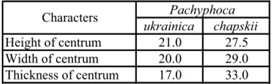
ukrainica chapskii Height of centrum 21.0 27.5 Width of centrum 20.0 29.0 Thickness of centrum 17.0 33.0 Pachyphoca Characters
Tab. 8 - Measurements (mm) of lumbar vertebrae.
Characters Pachyphoca chapskii
Absolute length 125.0 Hight of centrum 25.0 Width of centrum 25.0 Thickness of centrum 25.0 Thickness of 4-th vertebra 19.0 Length of ala 46.0 Width of ala 23.5