Structural Plasticity Induced by Ketamine
in Human Dopaminergic Neurons
as Mechanism Relevant for
Treatment-Resistant Depression
Ginetta Collo
1, Laura Cavalleri
1, and Emilio Merlo Pich
2,3Abstract
The mechanisms underlying the antidepressant effects of ketamine in treatment-resistant depression are only partially understood. Reactivation of neural plasticity in prefrontal cortex has been considered critical in mediating the effects of standard antidepressants, but in treatment-resistant depression patients with severe anhedonia, other components of the affected brain circuits, for example, the dopamine system, could be involved. In a recent article in Molecular Psychiatry, we showed that ketamine induces neural plasticity in human and mouse dopaminergic neurons. Human dopaminergic neurons were differentiated from inducible pluripotent stem cells for over 60 days. Mimicking the pharmacokinetic exposures occurring in treatment-resistant depression subjects, cultures were incubated with either ketamine at 0.1 and 1 mM for 1 h or with its active metabolite (2R,6R)-hydroxynorketamine at 0.1 and 0.5 mM for up to 6 h. Three days after dosing, we observed a concentration-dependent increase in dendritic arborization and soma size. These effects were mediated by the activation of a-amino-3-hydroxy-5-methyl-4-isoxazolepropionic acid receptor that triggered the pathways of mammalian target of rapamycin and extracellular signal-regulated kinase via the engagement of brain-derived neurotrophic factor sig-naling, as previously described in rodent prefrontal cortex. Interestingly, we found that neural plasticity induced by ketamine requires functionally intact dopamine D3 receptors. These data are in keeping with our recent observation that plasticity can be induced in human dopaminergic neurons by the D3 receptor-preferential agonist pramipexole, whose effect as augmen-tation treatment in treatment-resistant depression has been reported. Overall, the evidence of pharmacologic response in human inducible pluripotent stem cell-derived neurons could provide complementary information to those provided by circuit-based imaging when assessing the potential response to a given augmentation treatment.
Keywords
inducible pluripotent stem cells, a-amino-3-hydroxy-5-methyl-4-isoxazolepropionic acid receptor, brain-derived neurotrophic factor, mammalian target of rapamycin, extracellular signal-regulated kinase, D3 receptor
Received 2 March 2019; Accepted 18 March 2019
Commentary on: Cavalleri L, Merlo Pich E, Millan MJ, Chiamulera C, Kunath T, Spano PF, Collo G. Ketamine enhances structural plasticity in mouse-mesencephalic and human iPSC-derived dopaminergic neurons via AMPAR-driven BDNF and mTOR signaling. Mol Psychiatry 2018; 23: 812–823.
Introduction
Treatment-resistant depression (TRD) is among the most important unmet need in Psychiatry. TRD patients are
1
Department of Molecular and Translational Medicine, University of Brescia, Brescia, Italy
2
Neuroscience Therapeutic Area Unit, Takeda Pharmaceuticals International, Zurich, Switzerland
3
The Division of Brain Science, Imperial College London, London, UK
Corresponding author:
Ginetta Collo, Department of Molecular and Translational Medicine, University of Brescia, Viale Europa 11, 25123 Brescia, Italy. Email: [email protected]
Creative Commons Non Commercial CC BY-NC: This article is distributed under the terms of the Creative Commons Attribution-Non-Commercial 4.0 License (http://www.creativecommons.org/licenses/by-nc/4.0/) which permits non-commercial use, reproduction and distri-bution of the work without further permission provided the original work is attributed as specified on the SAGE and Open Access pages
(https://us.sagepub.com/en-us/nam/open-access-at-sage).
Volume 3: 1–6 !The Author(s) 2019 Article reuse guidelines: sagepub.com/journals-permissions DOI: 10.1177/2470547019842545 journals.sagepub.com/home/css
commonly defined as subjects that do not respond to at least two standard selective serotonin reuptake inhibitors (SSRI) or tricyclic antidepressant (TCA) treatments; they represent about 20% to 30% of all patients with major depressive disorder (MDD) diagnosis.1 Generally, TRD patients become eligible for adjunctive treatments, consist-ing of antipsychotics, dopaminergic (DA) agonists, behav-ioral therapies, transcranial magnetic stimulation, vagus nerve stimulation, and electroconvulsive treatment (ECT). In TRD, the efficacy of augmentation treatments is variable, leaving a large percentage of subjects with a partial or unsatisfactory therapeutic response. One hypothesis is that different TRD individuals respond dif-ferently to different augmentation therapies depending on which component of the underlying brain circuits is mostly affected. This paradigm requires a characterization of defective circuits from the neurobiological standpoint showing evidences of structural, neurochemical, and cellu-lar response to effective augmentation treatment.
While the understanding of the neuroimaging-defined brain circuits in mood disorders is constantly improving,2 only the recent introduction of inducible pluripotent stem cell (iPSC)-derived neurons technologies3,4 is beginning to elucidate the human cellular neurobiology of these brain circuits.
Defective Neural Plasticity as Hallmark
for TRD and Chronic Stress
The neuropathology of TRD has been poorly understood until recently. A significant breakthrough came from the clinical evidence that exposure to ketamine5 produced rapid and persistent antidepressant effects in a significant portion of TRD subjects.6,7 Its enantiomer S-ketamine, administrated intranasally, showed rapid antidepressant and antisuicidal effects8and was just approved by food and drug administration (FDA). Experimental evidences suggest that one ketamine metabolite observed in vivo few hours after ketamine administration, (2 R,6 R)-hydroxynorketamine (HNK),5 is also involved in the antidepressant effects. All these agents indirectly engage the neurotransmission mediated by a-amino-3-hydroxy-5-methyl-4-isoxazolepropionic acid receptors (AMPARs) and the brain-derived neurotrophic factor (BDNF)-TrkB signaling, activating the pathway of the mammalian target of rapamycin (mTOR) and of the extracellular signal-regulated kinase (ERK), both critical molecular drivers of neural plasticity.9,10 Accordingly, ketamine and S-ketamine produced these effects by blocking upstream N-methyl-D-aspartate receptors, while HNK by acting on the glutamatergic synapse. These results are of relevance: they suggest that the triggering of neural plasticity is a common denominator for producing antidepressant effects; they imply that defective neural plasticity is a critical cellular pathogenic mechanism of
the depressive episode, and they support a glutamater-gic-specific targeting of the glutamate-sensitive compo-nent of circuits involved in depression.11 Defective neural plasticity in frontocortical and hippocampal cir-cuits associated to reduced BDNF levels was observed in rodents following chronic stress.12 These observations partially parallel those in TRD/MDD patients, including reduced hippocampal volume, hypometabolism in pre-frontal cortex, and reduced postmortem levels of BDNF and mTOR.11,13
Reversibility of Neural Plasticity by
Treatment in Specific Brain Circuits
The evidence of reversibility of the defective status of neural plasticity associated to depression and chronic stress has been considered a biomarker indicative of treat-ment efficacy. This tenet is reinforced by preclinical studies showing that chronic treatments with SSRIs and TCAs are capable of reverting defective neural plasticity induced by chronic stress in rodent acting via glutamatergic fronto-cortical circuits.12,14 In human, neuroimaging evidence supports a normalization of the prefrontal cortex dysfunc-tion in those MDD/TRD subject that responds to any antidepressant treatment.15,16Renormalization of hippo-campal structural deficit was observed in TRD patients following augmentation therapies such as vagus nerve stimulation17or ECT.18Reduced DA neurotransmission in ventral striatum, amygdala, and hippocampus asso-ciated to anhedonia was observed in MDD/TRD patients and rodents exposed to chronic stress.19,20 Interestingly, pharmacologic augmentation was observed with aripipra-zole and pramipexole, two agents that enhance the func-tioning of the limbic DA system by targeting the presynaptic DA D2/D3 receptors.21,22 Pramipexole was also reported to enhance response to TRD patients who did not respond to ECT.23Consistent functional engage-ment of the mesencephalic DA system was also observed with ketamine,19,24suggesting a role for this mechanism in its therapeutic antidepressant effects. These findings sug-gest a possible DA-specific, circuit-based segregation of the pharmacologic effects in TRD patients. However, stu-dies aimed to differentiate TRD patients based on this approach are not available yet, being the direct evidence of plasticity-related pharmacologic effects in human DA neurons only recently obtained.
Human iPSC-Derived Neurons as
Translational Model for Defective Brain
Circuits of TRD Patients
Since inception, the discipline of Neuropharmacology has developed behavioral and cellular models to study neuroactive drugs with the goal to understand their mechanism of action (MoA). Animal models have been
Figure 2. Schematic representation of a proposed translational approach implementing iPSC-derived neurons to assess neural plasticity induced by pharmacological agents potentially active as augmentation antidepressant treatment. In the example (a) a MDD/TRD subject is profiled with neuroimaging for neural circuit involved in depression; (b) a particularly defective DA system is identified; (c) human iPSCs are differentiated to reproduce the neuron phenotype of the circuits involved, in this case DA neurons; (d) iPSC-derived DA neurons are exposed to pharmacologic agents; increase in neural plasticity is obtained with ketamine; (e) ketamine treatment is prescribed, resulting in a neuroimaging and clinical resolution of the episode, with normalization of the state-dependent DA circuit dysfunction. AC: anterior cingulate; DA: dopaminergic; iPSC: inducible pluripotent stem cell; TRD: treatment-resistant depression.
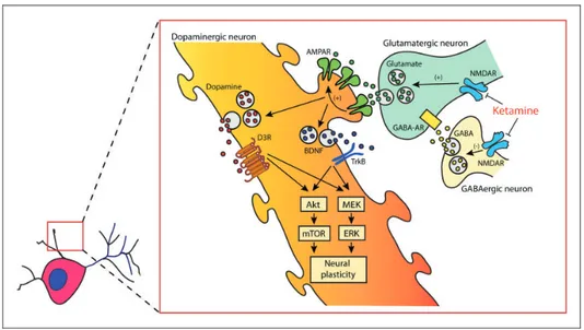
Figure 1. Cartoon representing the putative mechanism of action of ketamine and the molecular signalings involved in determining structural plasticity of dopaminergic neurons in vitro. Akt: thymoma viral proto-oncogene; AMPAR: a-amino-3-hydroxy-5-methyl-4-iso-xazolepropionic acid receptor; BDNF: brain-derived neurotrophic factor; D3R: dopamine D3 receptor; ERK: extracellular signal-regulated kinase; GABA: g-aminobutyric acid; GABA-AR: type A g-aminobutyric acid receptor; MEK: mitogen-activated protein kinase;
critical for drug development for decades and still they are. However, when used for precise translation into humans, this paradigm may falter. This problem has been particularly relevant for psychiatric disorders, with the failure to translate in effective treatments of several dozens of chemical entities whose preclinical profile was otherwise suggestive of efficacy. Translation failure can be, at least in part, due to subtle but substantial differ-ences in the molecular make-up of human cells versus rodent cells or versus immortalized cell cultures. The recent introduction of human iPSCs has allowed to study in vitro the neurobiology of human neurons with various phenotypes, including the glutamatergic pyram-idal neurons of the cortex and the DA neurons of the mesencephalon, adding a new paradigm for the develop-ment of novel treatdevelop-ments.4Differences and similarities in the MoA of a drug can be now analyzed in standardized cultures of human neurons or organoids.3,4 As discussed earlier, when assessing the MoA of a pharmacologic agent with putative antidepressant effects, one of the cel-lular processes expected is neural plasticity.11
In a recent article published in Molecular Psychiatry,25 we showed that ketamine and HNK activate plasticity in both human and mouse DA neurons, effects that required an active neurotransmission at the dopamine D3 autore-ceptor. We found that ketamine activates BDNF, ERK, and mTOR pathways, showing a molecular signature in DA neurons that overlaps the signature of ketamine in prefrontal cortex.9,11 Methodologically, we differentiated human DA neurons from iPSCs for 60 to 80 days. The stabilized cultures consisted of a mixture of differentiated DA neurons (30%–35%), GABAergic neurons (20%– 25%), and glutamatergic neurons (35%–40%).25,26 For pharmacologic testing, cultures were exposed to either 1 mM ketamine for 1 h or 0.5 mM HNK for up to 6 h, so to mimic the pharmacokinetic exposures occurring in TRD subjects that received a subanesthetic infusion of keta-mine.26,27 Structural neural plasticity in DA neurons was measured as changes in dendrite and soma morphology three days after exposure, a time considered of relevance in modeling the antidepressant effects of single exposures to ketamine in patients.6We observed that structural plasti-city at three days was dependent on the activation of both BDNF-TrkB-ERK and PI3K-Akt-mTOR pathways and related to a rapidly induced (within minutes) phosphoryl-ation cascade in the mTOR-p70S6K that persisted for 1 h. These phenomena were driven by an increased AMPAR neurotransmission, as suggested by the blockade of both rapidly induced phosphorylation and structural plasticity produced by pretreatment with the AMPAR antagonists NBQX and GYKI 52466.25,26 Increased levels of expres-sion of AMPAR subunits GluR1 and GluR2 were observed in the dendrites and soma of DA neurons, respectively, supporting the hypothesis of a ketamine-induced prolonged upregulated AMPAR-dependent
function.28 Immunoneutralization of extracellular BDNF and blockade of TrkB-dependent ERK signaling also pre-vented ketamine-induced structural plasticity, confirming the critical role of both mTOR and ERK signaling path-ways. Finally, we also showed that selective D3R antagon-ist SB277011-A (but not the D1R antagonantagon-ist SCH23390) blocked the neural plasticity induced by ketamine and pramipexole, supporting the necessity of a significant autoreceptor-mediated DA tone.25,26,29 These results are summarized in a cartoon representing the mechanism of structural plasticity in the dendrites of DA neurons (Figure 1). Intriguingly, these findings are suggestive for a possible augmented efficacy of cotreatments with ketamine and pramipexole (or other DA D3R preferential DA agon-ists); this augmented efficacy could be more important in those TRD subjects with a defective DA component in the limbic circuits involved in depression and, possibly, a rele-vant symptomatic anhedonia.
Conclusion and Implications
In this commentary, we presented data supporting the pos-sible translational utility of human iPSC-derived DA neu-rons in modeling the cellular MoA of ketamine, HNK, and pramipexole. Our study suggests that ketamine and HNK effects on neural plasticity are not restricted to the frontocortical and hippocampal component of the brain circuits involved in depression but extends to other cir-cuits, namely, the DA system. Overall, these data together with the recent published data involving the lateral habe-nula circuit30provide further support to the multiplicity of substrates that can be targeted to ameliorate depression in MDD/TRD patients. Advanced neuroimaging will be soon able to cluster subgroups of TRD patients for defect-ive components of the neural circuits involved in depres-sion.2This approach, coupled with the in vitro modeling of these circuits using neurons differentiated from hiPSCs (Figure 2), could contribute to the progress toward evi-dence-based precision treatment.
Authors’ Contribution
G.C., E.M.P., and L.C. wrote the manuscript. G.C., E.M.P., and L.C. designed and produced the figures. All authors approved the final version of the manuscript.
Declaration of Conflicting Interests
The author(s) declared the following potential conflicts of inter-est with respect to the research, authorship, and/or publication of this article: Emilio Merlo Pich is a full-time employee of Takeda Pharmaceuticals International, Zurich, Switzerland.
Funding
The author(s) disclosed receipt of the following financial sup-port for the research, authorship, and/or publication of this article: This research was supported by Grant from Ministry
of Education, University and Research (MIUR) ex 60%, University of Brescia to G.C.
ORCID iD
Ginetta Collo http://orcid.org/0000-0003-1888-5049
References
1. Brown S, Rittenbach K, Cheung S, McKean G, MacMaster FP, et al. Current and common definitions of treatment-resistant depression: findings from a systematic review and qualitative interviews [published online ahead of print
February 14, 2019]. Can J Psychiatry doi:10.1177/
0706743719828965
2. Williams LM. Precision psychiatry: a neural circuit tax-onomy for depression and anxiety. Lancet Psychiatry 2016; 3: 472–480.
3. Quadrato G, Arlotta P. Present and future of modeling human brain development in 3D organoids. Curr Opin Cell Biol2017; 49: 47–52.
4. Shi Y, Inoue H, Wu JC, Yamanaka S. Induced pluripotent stem cell technology: a decade of progress. Nat Rev Drug Discov2017; 16: 115–130.
5. Zanos P, Moaddel R, Morris PJ, Riggs LM, Highland, et al. Ketamine and ketamine metabolite pharmacology: insights into therapeutic mechanisms. Pharmacol Rev 2018; 70: 621–660.
6. Zarate CA Jr, Singh JB, Carlson PJ, Brutsche NE, Ameli R, et al. A randomized trial of an N-methyl-D-aspartate antagonist in treatment-resistant major depression. Arch Gen Psychiatry2006; 63: 856–864.
7. Serafini G, Howland RH, Rovedi F, Girardi P, Amore M. The role of ketamine in treatment-resistant depression: a systematic review. Curr Neuropharmacol 2014; 12: 444–461.
8. Molero P, Ramos-Quiroga JA, Martin-Santos R, Calvo-Sa´nchez E, Gutie´rrez-Rojas L, et al. Antidepressant efficacy and tolerability of ketamine and esketamine: a critical review. CNS Drugs 2018; 32: 411–420.
9. Li N, Lee B, Liu RJ, Banasr M, Dwyer JM, et al. mTOR-dependent synapse formation underlies the rapid anti-depressant effects of NMDA antagonists. Science 2010; 329: 959–964.
10. Yang C, Ren Q, Qu Y, Zhang JC, Ma M, et al. Mechanistic target of rapamycin-independent antidepressant effects of
(R)-Ketamine in a social defeat stress model. Biol
Psychiatry2018; 83: 18–28.
11. Duman RS, Aghajanian GK, Sanacora G, Krystal JH. Synaptic plasticity and depression: new insights from stress and rapid-acting antidepressants. Nat Med 2016; 22: 238–249.
12. Castre´n E, Antila H. Neuronal plasticity and neurotrophic factors in drug responses. Mol Psychiatry 2017; 22: 1085–1095.
13. Jernigan CS, Goswami DB, Austin MC, Iyo AH, Chandran A, et al. The mTOR signaling pathway in the prefrontal cortex is compromised in major depressive disorder. Prog
Neuropsychopharmacol Biol Psychiatry 2011; 35:
1774–1779.
14. Bessa JM, Ferreira D, Melo I, Marques F, Cerqueira JJ, et al. The mood-improving actions of antidepressants do not depend on neurogenesis but are associated with neur-onal remodeling. Mol Psychiatry 2009; 14: 764–777. 15. Abdallah CG, Averill CL, Salas R, Averill LA, Baldwin
PR, et al. Prefrontal connectivity and glutamate transmis-sion: relevance to depression pathophysiology and
keta-mine treatment. Biol Psychiatry Cogn Neurosci
Neuroimaging2017; 2: 566–574.
16. Meyer BM, Rabl U, Huemer J, Bartova L, Kalcher K, et al. Prefrontal networks dynamically related to recovery from major depressive disorder: a longitudinal pharmacological fMRI study. Transl Psychiatry 2019; 9: 64.
17. Perini GI, Toffanin T, Pigato G, Ferri G, Follador H, et al. Hippocampal gray volumes increase in treatment-resistant depression responding to vagus nerve stimulation. J ECT 2017; 33: 160–166.
18. Gryglewski G, Baldinger-Melich P, Seiger R, Godbersen GM, Michenthaler P, et al. Structural changes in amygdala nuclei, hippocampal subfields and cortical thickness follow-ing electroconvulsive therapy in treatment-resistant depres-sion: longitudinal analysis. Br J Psychiatry 2018; 16: 1–9. 19. Belujon P, Grace AA. Dopamine system dysregulation in
major depressive disorders. Int J Neuropsychopharmacol 2017; 20: 1036–1046.
20. Hamilton JP, Sacchet MD, Hjørnevik T, Chin FT, Shen B, et al. Striatal dopamine deficits predict reductions in striatal functional connectivity in major depression: a concurrent 11C-raclopride positron emission tomography and func-tional magnetic resonance imaging investigation. Transl Psychiatry2018; 8: 264.
21. Cusin C, Iovieno N, Iosifescu DV, Nierenberg AA, Fava M, et al. A randomized, double-blind, placebo-controlled trial of pramipexole augmentation in treatment-resistant major depressive disorder. J Clin Psychiatry 2013; 74: e636–e641.
22. Conway CR, Chibnall JT, Cumming P, Mintun MA, Gebara MA, et al. Antidepressant response to aripiprazole augmentation associated with enhanced F-DOPA utiliza-tion in striatum: a preliminary PET study. Psychiatry Res 2014; 221: 231–239.
23. Gauthier C, Souaiby L, Advenier-Iakovlev E, Gaillard R.
Pramipexole and electroconvulsive therapy in
treatment-resistant depression. Clin Neuropharmacol 2017; 40: 264–267.
24. Kokkinou M, Ashok AH, Howes OD. The effects of keta-mine on dopaketa-minergic function: meta-analysis and review of the implications for neuropsychiatric disorders. Mol Psychiatry2018; 23: 59–69.
25. Cavalleri L, Merlo Pich E, Millan MJ, Chiamulera C, Kunath T, et al. Ketamine enhances structural plasticity in mouse mesencephalic and human iPSC-derived dopamin-ergic neurons via AMPAR-driven BDNF and mTOR sig-naling. Mol Psychiatry 2018; 23: 812–823.
26. Collo G, Cavalleri, Chiamulera C, Merlo Pich E. (2R,6R)-hydroxynorketamine promotes dendrite outgrowth in human iPSC-derived neurons via AMPA receptor with timing and exposure compatible with human
pharmacokin-etics of ketamine infusion. Neuroreport 2018; 29:
27. Zhao X, Venkata SL, Moaddel R, Luckenbaugh DA, Brutsche NE, et al. Simultaneous population pharmacoki-netic modelling of ketamine and three major metabolites in patients with treatment-resistant bipolar depression. Br J Clin Pharmacol2012; 74: 304–314.
28. Collo G, Cavalleri L, Chiamulera C, Merlo Pich E.
Ketamine increases the expression of GluR1 and
GluR2 a-amino-3-hydroxy-5-methy-4-isoxazole propionate
receptor subunits in human dopaminergic neurons
differentiated from induced pluripotent stem cells.
Neuroreport2019; 30: 207–212.
29. Collo G, Cavalleri L, Bono F, Mora C, Fedele S, et al. Ropinirole and pramipexole promote structural plasticity in human iPSC derived dopaminergic neurons via BDNF and mTOR signaling. Neural Plast 2018; 2018: 4196961. 30. Yang Y, Cui Y, Sang K, Dong Y, Ni Z, et al. Ketamine
blocks bursting in the lateral habenula to rapidly relieve depression. Nature 2018; 554: 317–322.