Targeted next-generation sequencing
provides novel clues for associated epilepsy
and cardiac conduction disorder/SUDEP
Monica Coll1*, Pasquale Striano2, Carles Ferrer-Costa3, Oscar Campuzano1,4,5, Jesu´s Mate´s1, Bernat del Olmo1, Anna Iglesias1, Alexandra Pe´rez-Serra1,5, Irene Mademont1, Ferran Pico´1, Antonio Oliva6‡, Ramon Brugada1,4,5,7‡1 Cardiovascular Genetics Center, IDIBGI, Dr. Trueta University Hospital, Parc Hospitalari Martı´ i Julià,
Edifici, Salt (Spain), 2 Pediatric Neurology and Muscular Diseases Unit, Department of Neurosciences, Rehabilitation, Ophthalmology, Genetics, Maternal and Child Health, University of Genoa, "G. Gaslini" Institute, Genova (Italy), 3 Gendiag SL, Barcelona (Spain), 4 Department of Medical Sciences, School of medicine, University of Girona, Girona (Spain), 5 Centro de Investigacio´n Biome´dica en Red de
Enfermedades Cardiovasculares (CIBERCV), Madrid (Spain), 6 Institute of Public Health, Section of Legal Medicine, Catholic University, Rome (Italy), 7 Cardiac Genetics Unit, Cardiology Service, Hospital Josep Trueta, Girona (Spain)
‡ These authors are co-senior authors on this work. *[email protected]
Abstract
Sudden unexpected death in epilepsy is an unpredicted condition in patients with a diagno-sis of epilepsy, and autopsy does not conclusively identify cause of death. Although the pathophysiological mechanisms that underlie this entity remain unknown, the fact that epi-lepsy can affect cardiac function is not surprising. The genetic factors involving ion channels co-expressed in the heart and brain and other candidate genes have been previously described. In the present study, 20 epilepsy patients with personal or family history of heart rhythm disturbance/cardiac arrhythmias/sudden death were sequenced using a custom re-sequencing panel. Twenty-six relatives were genetically analysed to ascertain the family segregation in ten individuals. Four subjects revealed variants with positive genotype-phe-notype segregation: four missense variants in the CDKL5, CNTNAP2, GRIN2A and
ADGRV1 genes and one copy number variant in KCNQ1. The potential pathogenic role of
variants in new candidate genes will need further studies in larger cohorts, and the evalua-tion of the potential pathogenic role in the cardio-cerebral mechanisms requires in vivo/in vitro studies. In addition to family segregation, evaluation of the potential pathogenic roles of these variants in cardio-cerebral mechanisms by in vivo/in vitro studies should also be per-formed. The potential pathogenic role of variants in new candidate genes will need further studies in larger cohorts.
Introduction
Sudden unexpected death (SUDEP) is the sudden, unexpected, witnessed or unwitnessed, non-traumatic, and non-drowning death in patients with epilepsy, with or without evidence of
a1111111111 a1111111111 a1111111111 a1111111111 a1111111111 OPEN ACCESS
Citation: Coll M, Striano P, Ferrer-Costa C,
Campuzano O, Mate´s J, del Olmo B, et al. (2017) Targeted next-generation sequencing provides novel clues for associated epilepsy and cardiac conduction disorder/SUDEP. PLoS ONE 12(12): e0189618.https://doi.org/10.1371/journal. pone.0189618
Editor: Tomohiko Ai, Indiana University, UNITED
STATES
Received: September 15, 2017 Accepted: November 29, 2017 Published: December 19, 2017
Copyright: © 2017 Coll et al. This is an open access
article distributed under the terms of theCreative Commons Attribution License, which permits unrestricted use, distribution, and reproduction in any medium, provided the original author and source are credited.
Data Availability Statement: All relevant data are
within the paper and its Supporting Information files.
Funding: The authors received no specific funding
for this work. The funder provided support in the form of salaries for authors [C.F-C], but did not have any additional role in the study design, data collection and analysis, decision to publish, or preparation of the manuscript. The specific roles of
a seizure, with exclusion of documented status epilepticus, and no conclusive cause of death after a comprehensive medico-legal autopsy[1]. SUDEP is the most common cause of death in individuals with epilepsy and accounts for 7.5–17% of all deaths[2]. Nevertheless, the incidence of SUDEP varies widely due to the heterogeneity of the studies (type of epilepsy, size of studied cohort, definition of SUDEP and how the cause of death was determined)[3]. The risk of SUDEP is higher in males than in females; however, other risk factors, such as frequency of tonic-clonic seizures, early age of onset of epilepsy and polytherapy with antiepileptic drugs, have been identified as risk factors[1,4]. The pathophysiological mechanisms of SUDEP are likely multifactorial, but it has been suggested that a specific genetic background may predis-pose these patients to potentially fatal cardiorespiratory dysfunctions[3]. Indeed, seizures may induce a variety of transient cardiac effects, which include arrhythmias, asystole and other heart abnormalities, some of which are potentially lethal[3]. As pathogenic abnormalities in genes co-expressed in the heart and in the brain may eventually result in SUDEP, several stud-ies have described pathogenic alterations in genes encoding ion channels in patients with con-comitant heart conduction disorder and SUDEP[5,6].
The advent of massive parallel DNA sequencing platforms, referred to as next-generation sequencing (NGS) technology, has revolutionized the field of medical genomics, enabling fast and cost-effective generation of genetic data. We previously performed a genetic study in a large cohort of individuals with epilepsy and cardiac conduction disorders and showed puta-tive pathogenic disease-causing alterations in genes encoding cardiac ion channels in approxi-mately 25% of unrelated individuals [7]. However, since SUDEP likely involves some
combination of abnormal brain and cardiac physiology, certain additional gene categories, e.g., encoding for proteins with a key function of cell excitability, could be suitable candidates to explain the physiopathology of SUDEP [8].
To test this hypothesis, we used applied NGS analysis to a follow-up cohort of patients with epilepsy associated with cardiac conduction disorder/SUDEP.
Results
Genetic findings
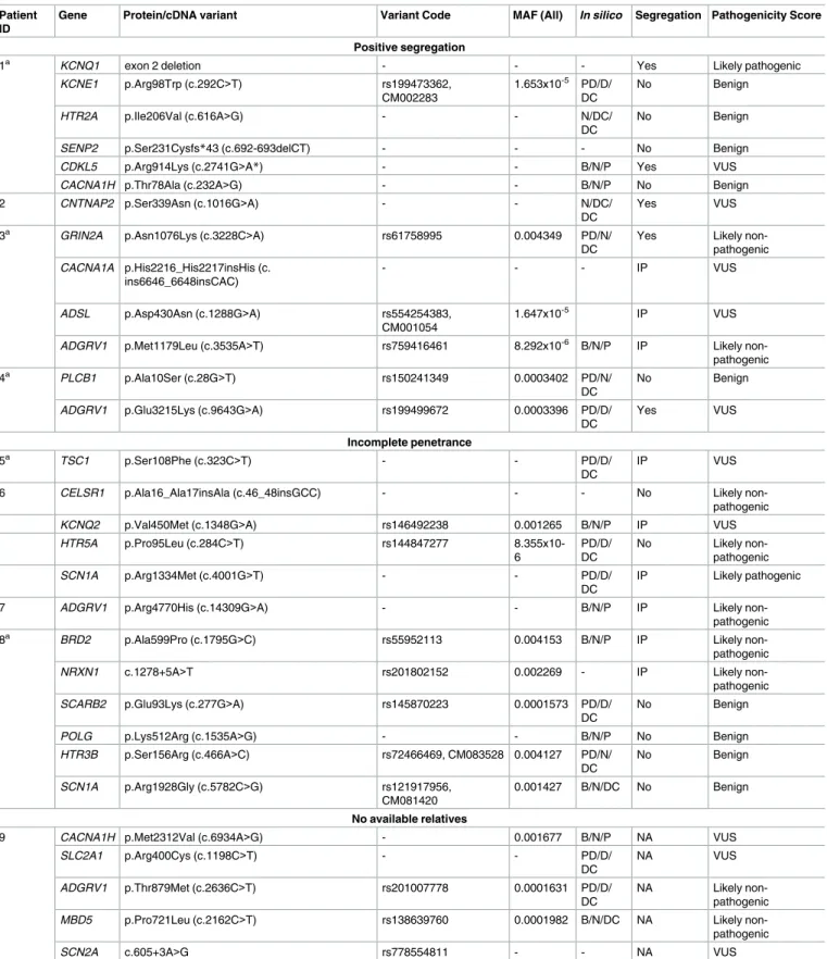
Nineteen out of the 20 recruited patients carried 56 rare genetic variants in 37 genes. Among these genetic anomalies, only four variants, i.e.,KCNQ1, KCNE1, SCN1A, and CACNA1C, were previously associated with SUDEP or other disorders associated with sudden cardiac death (SCD). We also identified 2 CNVs (Fig 1), 45 missense variants, 6 indels and 3 intronic variants (Table 1).
After the pathogenicity score classification, 3 variants were classified as likely pathogenic, 20 variants were classified as unknown significance, 21 variants were classified as likely non-pathogenic and 12 variants were classified as benign variants for no segregation within the family.
Segregation studies were performed in 10 out of 19 SUDEP cases, and a total of 26 relatives were genetically analysed to ascertain familiar segregation. Four cases showed at least one vari-ant with positive segregation (ID#1–4), four cases with varivari-ants with incomplete penetrance pattern (ID#5–8) and two cases with negative segregation (ID#18–19). Four subjects, previ-ously investigated with inconclusive results[9], showed positive segregation (individuals ID#1, ID#3 and ID#4) or an incomplete pattern of inheritance (ID#5).
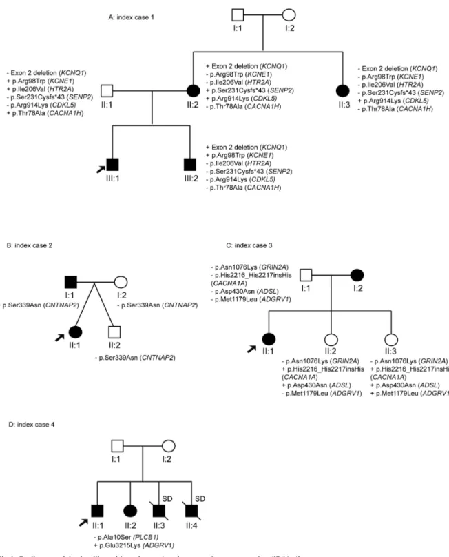
Genetic variants inKCNQ1 and CDKL5 genes were identified in ID#1, who had family members affected by epilepsy and/or long QT syndrome (Fig 2A). While the exon 2 deletion inKCNQ1 (Fig 1) showed complete segregation with the LQTS, it is unlikely that theCDKL5 variant was causative of the observed epilepsy, as mutations related to this gene typically occur these authors are articulated in the ’author
contributions’ section.
Competing interests: The authors have declared
that no competing interests exist. This does not alter our adherence to PLOS ONE policies on sharing data and materials.
de novo and are associated with early-onset, severe, drug-refractory epileptic encephalopathy [10].
The variant in theCNTNAP2 gene identified in individual ID# 2 is a novel change classified as a variant of unknown significance. Segregation studies showed that the non-affected sister (II:2) and their parents (I:1 and I:2) did not carry this variant (Fig 2B). Heterozygous muta-tions inCNTNAP2 have been identified in patients with a range of complex phenotypes, including intellectual disability, autism and schizophrenia [11,12]. However, heterozygous CNTNAP2 mutations are also found in the normal population, and it is likely that this variant alone does not fully explain the patient’s phenotype.
Patient #3 is a 24-year-old woman with family history (individual I:2) of SUDEP. However, her mother was not available for genetic analysis. Segregation analysis in the non-affected sis-ters (individuals II:2, II:3) and the father (subject I:1) was inconclusive (Fig 2C).
Individual ID#4 is a 52-year-old man with family history of epilepsy (sister) and sudden death (his brothers). The variant identified inADGRV1 is shared by the affected sister (II:2), but DNA for the deceased brothers was unavailable (Fig 2D).
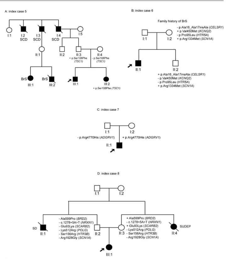
Individual ID#5 is a 15-year-old boy carrying a novel variant in theTSC1 gene. This variant was shared by his healthy father (II:3) and affected sister (III:2) with epileptic encephalopathy. There were antecedents of SCD from the paternal lineage, and the father’s cousins (III:1 and III:2) were affected by Brugada syndrome (Fig 3A).
Another case with IP was ID#6, a 9-year-old boy affected by SUDEP and Brugada syn-drome. The non-affected mother (I:2) had family history of Brugada syndrome and showed the variants inKCNQ2 and SCN1A genes. The variant in the SCN1A gene was also present in the non-asymptomatic sister (II:2) (Fig 3B).
ID#7 is a 16-year-old boy affected by focal epilepsy and LQTS and carrying a rare genetic variant inADGRV1 that was harboured by his healthy mother (I:2) (Fig 3C).
Finally, ID#8 is a 21-year-old girl who suffered focal epilepsy and SUDEP. She carried six rare genetic variants inBRD2, NRXN1, SCARB2, POLG, HTR3B, and SCN1A. The brother’s
Fig 1. A: Deletion of exon 2 of the KCNQ1 gene in ID#1. B: Duplication of exon 4 of the CNTNAP2 gene in ID#15.
Table 1. Rare genetic variants identified in our cohort.
Patient ID
Gene Protein/cDNA variant Variant Code MAF (All) In silico Segregation Pathogenicity Score
Positive segregation
1a KCNQ1 exon 2 deletion - - - Yes Likely pathogenic
KCNE1 p.Arg98Trp (c.292C>T) rs199473362, CM002283
1.653x10⁻⁵ PD/D/
DC
No Benign
HTR2A p.Ile206Val (c.616A>G) - - N/DC/ DC
No Benign
SENP2 p.Ser231Cysfs*43 (c.692-693delCT) - - - No Benign
CDKL5 p.Arg914Lys (c.2741G>A*) - - B/N/P Yes VUS
CACNA1H p.Thr78Ala (c.232A>G) - - B/N/P No Benign 2 CNTNAP2 p.Ser339Asn (c.1016G>A) - - N/DC/
DC
Yes VUS
3a GRIN2A p.Asn1076Lys (c.3228C>A) rs61758995 0.004349 PD/N/
DC
Yes Likely
non-pathogenic
CACNA1A p.His2216_His2217insHis (c.
ins6646_6648insCAC)
- - - IP VUS
ADSL p.Asp430Asn (c.1288G>A) rs554254383,
CM001054
1.647x10⁻⁵ IP VUS
ADGRV1 p.Met1179Leu (c.3535A>T) rs759416461 8.292x10⁻⁶ B/N/P IP Likely
non-pathogenic 4a PLCB1 p.Ala10Ser (c.28G>T) rs150241349 0.0003402 PD/N/
DC
No Benign
ADGRV1 p.Glu3215Lys (c.9643G>A) rs199499672 0.0003396 PD/D/ DC Yes VUS Incomplete penetrance 5a TSC1 p.Ser108Phe (c.323C>T) - - PD/D/ DC IP VUS
6 CELSR1 p.Ala16_Ala17insAla (c.46_48insGCC) - - - No Likely non-pathogenic
KCNQ2 p.Val450Met (c.1348G>A) rs146492238 0.001265 B/N/P IP VUS
HTR5A p.Pro95Leu (c.284C>T) rs144847277 8.355x10⁻⁶ PD/D/ DC No Likely non-pathogenic SCN1A p.Arg1334Met (c.4001G>T) - - PD/D/ DC IP Likely pathogenic
7 ADGRV1 p.Arg4770His (c.14309G>A) - - B/N/P IP Likely non-pathogenic
8a BRD2 p.Ala599Pro (c.1795G>C) rs55952113 0.004153 B/N/P IP Likely
non-pathogenic
NRXN1 c.1278+5A>T rs201802152 0.002269 - IP Likely non-pathogenic
SCARB2 p.Glu93Lys (c.277G>A) rs145870223 0.0001573 PD/D/ DC
No Benign
POLG p.Lys512Arg (c.1535A>G) - - B/N/P No Benign
HTR3B p.Ser156Arg (c.466A>C) rs72466469, CM083528 0.004127 PD/N/ DC No Benign SCN1A p.Arg1928Gly (c.5782C>G) rs121917956, CM081420 0.001427 B/N/DC No Benign No available relatives
9 CACNA1H p.Met2312Val (c.6934A>G) - 0.001677 B/N/P NA VUS
SLC2A1 p.Arg400Cys (c.1198C>T) - - PD/D/ DC NA VUS ADGRV1 p.Thr879Met (c.2636C>T) rs201007778 0.0001631 PD/D/ DC NA Likely non-pathogenic
MBD5 p.Pro721Leu (c.2162C>T) rs138639760 0.0001982 B/N/DC NA Likely
non-pathogenic
SCN2A c.605+3A>G rs778554811 - - NA VUS
Table 1. (Continued)
Patient ID
Gene Protein/cDNA variant Variant Code MAF (All) In silico Segregation Pathogenicity Score
10 CACNA1C p.Val1707Ile (c.5119G>A) rs147896322 - PD/N/ DC
NA VUS
HTR2A p.Ala447Val (c.1340C>T) rs6308, CM115490 0.001484 B/N/P NA Likely non-pathogenic
PNKP c.1029+2T>C rs199919568 0.0006509 - NA Likely
non-pathogenic
CACNA1I p.Asp2040Tyr (c.6118G>T) rs771105041 0.004771 PD/D/P NA Likely non-pathogenic 11 HCN2 c.2143_2145delCCG rs527536363 - - NA VUS CACNA1H p.Ala1570Val (c.4709C>T) rs558718048 5.014x10⁻⁵ PD/D/ DC NA VUS
12 ADGRV1 p.Pro4181Leu (c.12542C>T) rs200957385 - B/D/DC NA Likely non-pathogenic KCNQ3 p.Pro574Ser (c.1720C>T) rs74582884, CM083709 0.001995 PD/N/ DC NA Likely pathogenic 13 CACNA1A p.2319Gln_2320GlninsGln (c.6955_6957insCAG) - - - NA VUS BRD2 p.Asn194Lys (c.582C>G) rs201572264 7.753x10⁻⁵ B/N/DC NA Likely non-pathogenic ADGRV1 p.Val754Ala (c.2261T>C) rs374609813 - PD/N/ DC NA Likely non-pathogenic HTR3B p.Gln334Glu (c.1001G>A) rs199833563 8.238x10⁻⁶ B/N/P NA Likely non-pathogenic 14 CACNA1A p.Gln2326_Gln2331del (c.6976_6993del18) rs765169827 - - NA VUS
SCARB2 p.Val149Met (c.445G>A) rs147159813 0.002053 B/N/P NA Likely non-pathogenic
CNTNAP2 exon 4 duplication - - - NA VUS
15 CACNA1I p.Arg1448Gln (c.4343G>A) rs201480163 9.944x10⁻⁵
PD/D/ DC
NA VUS
16 GRIN2B p.Lys1090Arg (c.3269A>G) - - PD/D/ DC
NA VUS
HTR7 p.Lys445Asn (c.1335G>C) rs138700544 - B/N/P NA Likely
non-pathogenic CHRNB2 p.Phe472Ser (c.1415T>C) rs200855905 4.118x10⁻⁵ PD/N/ DC NA Likely non-pathogenic 17 ADGRV1 p.Glu2897Asp (c.8691A>C) rs201586455 0.004302 PD/N/
DC NA Likely non-pathogenic No segregation 18 TBC1D24 p.Glu94Lys (c.280G>A) - - PD/D/ DC No Benign
ADSL p.Asp215Asn (c.643G>A) - - PD/D/
DC
No Benign
KCNMA1 p.Val792Ile (c.2374G>A) rs142770262 - B/N/DC No Benign 19 CACNA1H p.Ala2208Val (c.6623C>T) rs777217625 - B/N/P No VUS
EFHC1 p.Arg42Pro (c.125G>C) rs773598517 - B/N/DC No VUS
ATP1A2 p.Arg3His (c.8G>A) rs781687346 8.12x10⁻⁵ PD/N/P No Likely non-pathogenic
NA: DNA not available from relatives. IP: Incomplete penetrance. CM: Human gene variation database code. MAF: Minor allele frequency in the ExAG (Last consulted May 2016). In silico predictors were PolyPhen2, Provean and Mutation Taster. N: Neutral; D: Deleterious; P: Polymorphism; DC: Disease causing; B: Benign; PD: Possibly damaging; VUS: Variant of uncertain significance.
*Variant detected in homozygous state.
aSUDEP cases sequenced in a previous custom resequencing panel.
father (II:1) died from sudden death and the mother’s sister (II:4) experienced SUDEP. Her healthy mother (II:3) carried the variants in theBRD2 and SCARB2 genes. Thus, segregation analysis in this family was also inconclusive (Fig 3D).
Fig 2. Pedigrees of the families with variants showing complete segregation (ID#1–4).
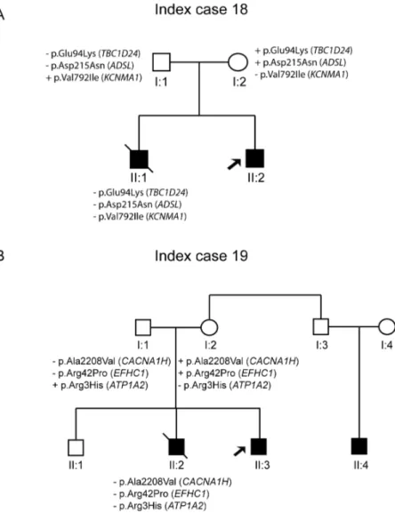
There were two index cases with rare genetic variants not showing segregation with the pathology. ID#19 carried three variants in theADSL, TBC1D24, and KCNMA1 genes, but his dead brother (II:1) did not harbour any of these variants (Fig 4A). Patient ID#20 is a
12-year-Fig 3. Pedigrees of the families with variants showing an incomplete pattern of inheritance (ID#5–8).
old boy with focal epilepsy, and her brother (II:1) died of SUDEP while asleep at age 11 years. The maternal uncle has a 9-year-old child (II:3) who suffered a single seizure. Three rare genetic variants were identified in the index case, but only two of these variants (EFHC1 and CACNA1H) were derived from the maternal lineage. However, these variants were absent in the deceased brother (II:2) (Fig 4B).
Discussion
Sudden unexpected death is a catastrophic complication of human epilepsy with an incidence of 6.3–9.3 per 1,000 person years in epilepsy patients entering surgery programmes [1]. Despite recent advances, both cellular mechanisms and genetic bases remain largely unknown.
A leading hypothesis suggests a dysfunction of excitability that could underlie both epilepsy and cardiac arrhythmias, leading to death [13]. Recently, there has been increased interest in the potential association between epilepsy channelopathies and cardiac arrhythmias [5–7]. NGS is a fast and cost-effective method for the simultaneous detection of single nucleotide var-iants (SNVs) and small insertions or deletions (indels). Various studies have employed massive
Fig 4. Pedigree of the family with variants that did not show segregation (ID#18–19).
sequencing technology to identify new potential genes responsible for SUDEP[9,14]. Olson et al. identified several CNVs with causative roles in some cases of paediatric epilepsy, suggest-ing that these types of variations may potentially be associated with SUDEP cases[15]. We per-formed a comprehensive genetic analysis using NGS technology to further explore the role of already known and novel candidate genes associated with epilepsy/SUDEP.
The first index case (ID#1) evidenced the coexistence of both LQTS and epilepsy. The exon 2 deletion showed positive segregation with the LQTS and the mutation in theCDKL5 gene with the epilepsy in this family. Deletions inKCNQ1 exons were previously reported by NGS in individuals with LQTS [16]. TheCDKL5 gene acts as a kinase, and one of its target proteins is MeCP2, which plays important roles in the function of nerve cells (neurons) and other brain cells and in the maintenance of connections (synapses) between neurons. Pathogenic variants in theCDKL5 gene have been associated with a disorder known as X-linked infantile spasm syndrome, which is characterized by recurrent seizures called infantile spasms that begin in the first year of life and with intellectual disability; however, it is not clear how mutations in this gene can result in this phenotype [10].
In ID#2 we detected a novel rare variant in theCNTNAP2 gene (contactin associated pro-tein-like 2). Pathogenic alterations in this gene were associated with cortical dysplasia focal epilepsy syndrome, a rare disorder resulting in epileptic seizures, language regression, intellec-tual disability, hyperactivity and autism in nearly two-thirds of the patients[11]. However, vari-ants in this gene have been found in control populations, whereas homozygous varivari-ants are rare in patient populations and have not been found in any unaffected individuals[12]. Although the father was affected by Brugada syndrome (BrS) and epilepsy and carried this var-iant, the absence of this complex phenotype and the lack of expression of theCNTNAP2 gene in the heart complicate the association between this variant and SUDEP.
Individual ID#3 carried four rare genetic variants, and only variants with positive segrega-tion were detected in theGRIN2A gene, although pathogenic score classified as probably no-pathogenic variant. Mutations in this gene have been associated with focal epilepsies as the phenotype of our index case.
The variant with positive segregation in the ID#4 was identified in theADGRV1 gene also known asGPR98 (MASS1 gene). The encoded protein contains a 7-transmembrane receptor domain, binds calcium and was expressed in the central nervous system. Pathogenic alter-ations in this gene have been associated with recurrent febrile seizures and afebrile seizures [17].
The variant identified in the ID#5 was inTSC1 gene, which encodes for hamartin, a protein that interacts with tuberin and helps to control cell growth and size through the mTOR path-way[18]. Mutations in this gene are responsible for Tuberous sclerosis complex (TSC) and focal cortical dysplasia associated with drug-resistant epilepsy. Although the proband does not have a phenotype compatible with TSC, the role of hamartin deserves additional investigation due to its high expression in heart muscle [19].
TheKCNQ2 variant identified in individual ID#6 and the non-affected mother belongs to a large family of genes encoding potassium channels associated with benign neonatal seizures [20], but the role of this variant in the BrS phenotype of this family remains unclear. Con-versely,SCN1A is a good gene candidate associated with a large range of epileptic phenotypes as well as cardiac dysfunction [21].
The variant identified in theADGRV1 gene of ID#7 is present in the non-affected mother, and no family history of epilepsy or LQTS is known. Mutations in this gene have been associ-ated with Usher’s syndrome and febrile seizures familiar 4 (FEB4) and was not expressed in the heart, indicating that this variant was more related to epilepsy than to LQTS in this family
[22]. Genetic testing of the main and associated genes related to LQTS would be the next strat-egy to identify the genetic cause of the disease in this patient.
The last genes with IP with the diseases were detected in ID#8. The non-affected mother carried variants in theBRD2 and SCARB2 genes, which could explain the SUDEP in her sister. Mutations in both genes have been associated with myoclonic epilepsy[23,24].SCARB2 gene encodes for LIMP-2 protein, which is found in the heart, specifically in the intercalated discs, although its precise function is not clear[22].
Pathogenic variants in the genes identified in the present cohort were previously associated with syndromes involving intellectual disabilities with episodes of seizures difficulty the possi-ble relation with SUDEP. However, ion channel genes, such as calcium (GRIN2A), potassium (KCNQ2) or sodium (SCN1A) channels, may represent interesting candidate genes that under-lie SUDEP, as the expression and regulation of these genes contributes to the central control of cardiac and respiratory function.
Conclusion
Rare genetic alterations in genes encoding brain/heart ion channels are responsible for epi-lepsy associated with cardiac conduction disorder/SUDEP. NGS technology enables the comprehensive cost-effectives genetic analysis of all current known genes as well as candi-date genes. Overall, we identified 5 rare genetic variants that segregated within the pedigree and explain the cardiac conduction disorder/SUDEP. Only one variant was a previously described gene, revealing four new potential genes associated with this entity. The new cus-tom re-sequencing panel enabled the identification of potential pathogenic variants in three out of four negative SUDEP cases in a previous study. Genetic data should be interpreted by a group of experts, and translation into clinical and forensic fields should be done with caution.
Methods
Patients
The present study is a follow-up study in patients with clinical and EEG features consistent with a diagnosis of non-lesional (MRI-negative) focal or generalized epilepsy and a personal or family history of heart rhythm disturbances/cardiac arrhythmias/sudden death (Table 2).
DNA samples of relatives were obtained when available to ascertain the segregation of each genetic variant. All patients received at least one surface electrocardiogram (EKG), which was evaluated by an expert cardiologist. Vague histories of heart rhythm disturbances in family members not clearly known by the proband or with any available clinical/instrumental data were excluded.
Each proband was genetically evaluated by target re-sequencing of exonic and intron-exon boundary regions. Clinical data and DNA from available relatives were obtained for testing in segregation studies of the rare genetic variants. The four SUDEP cases included in this cohort were genetically negative in a previous published SUDEP study [9].
All individuals and their family members signed a written informed consent to participate in the present study in accordance with the guidelines of the International Review Board of Catholic University. The study was approved through the Ethics Committee of Catholic Uni-versity (Rome, Italy) in accordance with the principles outlined in the Declaration of Helsinki. The Full name of Ethic Committee is “Ethics Commitee, Università Cattolica del Sacro Cuore Largo Agostino Gemelli 8, 00168 Roma, Italy. The individuals in the present study provided written informed consent (as outlined in PLOS consent form) to publish these case details.
DNA samples
Genomic DNA was extracted from whole blood or saliva with Chemagic MSM I, and the con-centration was checked by fluorometry (Qubit, Life Technologies). DNA integrity was assessed on a 0.8% agarose gel.
NGS studies
We designed a custom resequencing panel, which included 122 genes associated with epilepsy as well as the main genes associated with cardiac channelopathies (S1 Table). The final size was 487.874 kbp of encoding regions and UTR boundaries. We targeted one isoform for each gene and only coding exons plus 10 bp inside intronic regions. Exons were obtained from ensembl site[25] version 81 in GRCh38 and translated to hg19. Bait design was achieved using an in-house algorithm and submitted to Agilent SureSelect Design Web. The chip design was achieved using variable tailing bait and variable multiplicity bait to obtain homogeneous cover-age and optimize sample load. The present bait design algorithm considers the GC content of the bait itself, and the target and surrounding region of the targeted gene. This algorithm also considers multimapping of the bait regions and uses, when available, previous NGS results.
Genomic DNA was fragmented by sonication using the Bioruptor (Diagenode). The 122 genes were enriched using the SureSelect Custom Target Enrichment System Kit (Agilent Technologies, Santa Clara, CA USA) according to the manufacture’s instructions for the "Sure-Select Target Enrichment System for Illumina Paired-End Sequencing version B.1” (Sure"Sure-Select XT Custom 0–499 kbp library, Agilent Technologies, Inc.). The paired-end sequencing process was developed on a MiSeq System (Illumina) using 2x76 bp reads length.
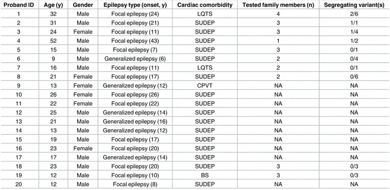
Table 2. Clinical data for the investigated cohort.
Proband ID Age (y) Gender Epilepsy type (onset, y) Cardiac comorbidity Tested family members (n) Segregating variant(s)
1 32 Male Focal epilepsy (24) LQTS 4 2/6
2 31 Male Focal epilepsy (21) SUDEP 3 1/1
3 24 Female Focal epilepsy (11) SUDEP 3 1/4
4 52 Male Focal epilepsy (43) SUDEP 1 1/2
5 15 Male Focal epilepsy (7) SUDEP 3 0/1
6 9 Male Generalized epilepsy (6) SUDEP 2 0/4
7 16 Male Focal epilepsy (11) LQTS 2 0/1
8 21 Female Focal epilepsy (17) SUDEP 2 0/6
9 13 Female Generalized epilepsy (12) CPVT NA NA
10 26 Female Focal epilepsy (26) SUDEP NA NA
11 22 Female Focal epilepsy (22) SUDEP NA NA
12 25 Male Generalized epilepsy (14) SUDEP NA NA
13 21 Male Generalized epilepsy (16) SUDEP NA NA
14 13 Male Generalized epilepsy (12) SUDEP NA NA
15 19 Male Focal epilepsy (17) SUDEP NA NA
16 23 Female Focal epilepsy (20) SUDEP NA NA
17 17 Male Generalized epilepsy (14) SUDEP NA NA
18 23 Male Focal epilepsy (20) SUDEP 3 0/3
19 12 Male Focal epilepsy (10) BS 3 0/3
20 12 Male Focal epilepsy (8) SUDEP NA NA
NA: DNA not available from relatives. Familial individual tested excluding the proband. Variant/s segregated indicates the number of variants showing a complete segregation/total of the variants identified in the proband.
Data analysis
NGS analysis was submitted to the GendiCall Pipeline (Gendicall Software from FerrerIn-code), which cleans up and trims fastq files from sequencer and then Gendicall maps using either GEM[26] or BWA mapper[27]. Subsequently, bam was generated and sorted, and dupli-cates were removed with Picard (http://picard.sourceforge.net). Variant call for SNVs and Small Indels were performed using Samtools (v.1.3.1)[28] and internal Gendicall Caller. Anno-tation of called variants was based on several sources, and for population data, we used dbSNP [29], 1000 Genomes Project[30], Exome Variants Server (EVS)[31] and Exome Aggregation Consortium (ExAC) Cambridge, MA (URL:http://exac.broadinstitute.org). The protein pre-dictors used in the present study were Polyphen2[32], Provean[33] and Mutation Taster[34]. Finally, we also used the splicing predictors MaxEntScan[35], FSPLICE[36], GeneSplicer[37] and NNsplice[38].
Validation and bioinformatics analysis
The variants with MAF (minor allele frequency) lower than 0.1 (<1%) were assessed in locus-specific databases that include dbSNP[29], Ensembl genome browser[25], the 1000 Genomes Project[39], EVS (Exome Variant Server, NHLBI GO Exome Sequencing Project (ESP), Seat-tle, WA (URL:http://evs.gs.washington.edu/EVS/) [date (July, 2017) accessed] and ExAC[40]. Rare single nucleotide variants (SNPs) and insertions/deletions (indels) were confirmed by conventional Sanger sequencing, and copy number variants (CNVs) were confirmed by a mul-tiplex ligation-dependent probe amplification (MLPA) technique.
Rare genetic variants and sequence regions with coverage lower than 30X were validated by the Sanger method. First, polymerase chain reaction (PCR) was performed, and the product was purified by ExoSAP-IT (USB Corporation, Cleveland, OH, USA) and directly sequenced by the dideoxy chain-termination method in ABI Prism Big Dye1 Terminator v3.1 Cycle Sequencing Kit (Applied Biosystems, USA). Sequencing was processed in a 3130XL Genetic Analyzer (Applied Biosystems) and analysed by SeqScape Software v2.5 (Life Technologies), comparing the obtained results with the reference sequence from hg19. Protein numbering reflects the translation initiator methionine as +1.
The CNVs detected by NGS were confirmed by MLPA (MRC-Holland, Amsterdam, Neth-erlands) with the corresponding probe mix SALSA MLPA: SALSA MLPA P114 Long-QT probe mix and SALSA MLPA P297 Microdeletion Syndromes-2 probe mix. The MLPA DNA detection and quantification were performed according to the manufacturer’s protocol (MRC-Holland, Amsterdam, Netherlands). After the multiplex PCR reaction, electrophoresis was performed using the ABI3130xl Genetic Analyzer (Applied Biosystems, CA, USA), and the data were analysed with Coffalyser.net software (MRC-Holland). Significantly (30%) decreased or increased signals in the patient sample relative to controls were considered as deletions or duplications, respectively. Familial segregation of CNVs was also performed using MLPA. Variant frequencies (minor allele frequency, MAF>1%) were obtained from the ExAC collection (http://exac.broadinstitute.org/).
A pathogenicity score was assigned to each variant for classification into the following cate-gories: pathogenic, likely pathogenic, variant of unknown significance, likely non-pathogenic and benign, as described[41]. In silico analysis of the pathogenicity of rare nonsynonymous variants was performed by Polyphen2, Provean and MutationTaster [42–44].
Limitations
The present study has primary limitations. First, the lack of family segregation impedes the proper classification of the potential pathogenic role of each variant, and consequently,
translation into clinical and forensic practice should be implemented with caution. In addi-tion, functional in vivo/in vitro studies should also be performed to unravel the cellular mecha-nisms involved in SUDEP. We cannot exclude that these patients carry a genetic alteration in other genes that were not included in the present genetic panel. In these negative cases, and if the relatives are available, whole-exome sequencing studies could be a proper approach. Finally, additional studies in large cohorts should be performed to corroborate these results and identify new genetic alterations; however, the low incidence of SUDEP complicates sample collection.
Supporting information
S1 Table. List of genes included in the custom resequencing panel and the associated dis-ease/s.
(DOCX)
Acknowledgments
The authors received no specific funding for this work. The authors would like to thank the network of Genetic Commission of Italian League Against Epilepsy (LICE).
Author Contributions
Conceptualization: Carles Ferrer-Costa, Antonio Oliva. Data curation: Monica Coll, Pasquale Striano.
Formal analysis: Monica Coll, Jesu´s Mate´s, Bernat del Olmo. Funding acquisition: Oscar Campuzano, Antonio Oliva.
Investigation: Monica Coll, Alexandra Pe´rez-Serra, Irene Mademont, Ferran Pico´. Methodology: Monica Coll, Alexandra Pe´rez-Serra, Irene Mademont, Ferran Pico´. Project administration: Anna Iglesias, Ramon Brugada.
Software: Carles Ferrer-Costa, Jesu´s Mate´s, Bernat del Olmo.
Supervision: Pasquale Striano, Oscar Campuzano, Antonio Oliva, Ramon Brugada. Validation: Carles Ferrer-Costa, Ramon Brugada.
Visualization: Ramon Brugada. Writing – original draft: Monica Coll.
Writing – review & editing: Pasquale Striano, Oscar Campuzano, Antonio Oliva, Ramon Brugada.
References
1. Nashef L. Sudden unexpected death in epilepsy: terminology and definitions. Epilepsia. 1997; 38(11 Suppl):S6–8. Epub 1997/11/01.https://doi.org/10.1111/j.1528-1157.1997.tb06130.xPMID:19909329.
2. Terra VC. Sudden unexpected death in epilepsy: From the lab to the clinic setting. Epilsepy & Behavior. 2012.
3. Dlouhy BJ, Gehlbach BK, Richerson GB. Sudden unexpected death in epilepsy: basic mechanisms and clinical implications for prevention. Journal of neurology, neurosurgery, and psychiatry. 2016; 87 (4):402–13. Epub 2016/03/17.https://doi.org/10.1136/jnnp-2013-307442PMID:26979537.
4. Shorvon S, Tomson T. Sudden unexpected death in epilepsy. Lancet. 2011; 378(9808):2028–38. Epub 2011/07/09.https://doi.org/10.1016/S0140-6736(11)60176-1PMID:21737136.
5. Partemi S, Cestele S, Pezzella M, Campuzano O, Paravidino R, Pascali VL, et al. Loss-of-function KCNH2 mutation in a family with long QT syndrome, epilepsy, and sudden death. Epilepsia. 2013; 54 (8):e112–6. Epub 2013/08/01.https://doi.org/10.1111/epi.12259PMID:23899126.
6. Parisi P, Oliva A, Coll Vidal M, Partemi S, Campuzano O, Iglesias A, et al. Coexistence of epilepsy and Brugada syndrome in a family with SCN5A mutation. Epilepsy research. 2013; 105(3):415–8. Epub 2013/03/30.https://doi.org/10.1016/j.eplepsyres.2013.02.024PMID:23538271.
7. Partemi S, Vidal MC, Striano P, Campuzano O, Allegue C, Pezzella M, et al. Genetic and forensic impli-cations in epilepsy and cardiac arrhythmias: a case series. International journal of legal medicine. 2014. Epub 2014/08/15.https://doi.org/10.1007/s00414-014-1063-4PMID:25119684.
8. Glasscock E. Genomic biomarkers of SUDEP in brain and heart. Epilepsy & behavior: E&B. 2013. Epub 2013/10/22.https://doi.org/10.1016/j.yebeh.2013.09.019PMID:24139807.
9. Coll M, Allegue C, Partemi S, Mates J, Del Olmo B, Campuzano O, et al. Genetic investigation of sud-den unexpected death in epilepsy cohort by panel target resequencing. Int J Legal Med. 2016; 130 (2):331–9. Epub 2015/10/02.https://doi.org/10.1007/s00414-015-1269-0PMID:26423924.
10. https://ghr.nlm.nih.gov. Genetics Home ReferenceNational Library of Medicine (US). National Library of Medicine (US). 2016 June 07.
11. Strauss KA, Puffenberger EG, Huentelman MJ, Gottlieb S, Dobrin SE, Parod JM, et al. Recessive symptomatic focal epilepsy and mutant contactin-associated protein-like 2. N Engl J Med. 2006; 354 (13):1370–7. Epub 2006/03/31.https://doi.org/10.1056/NEJMoa052773PMID:16571880.
12. Rodenas-Cuadrado P, Pietrafusa N, Francavilla T, La Neve A, Striano P, Vernes SC. Characterisation of CASPR2 deficiency disorder—a syndrome involving autism, epilepsy and language impairment. BMC medical genetics. 2016; 17:8. Epub 2016/02/05.https://doi.org/10.1186/s12881-016-0272-8 PMID:26843181; PubMed Central PMCID: PMC4739328.
13. Devinsky O, Hesdorffer DC, Thurman DJ, Lhatoo S, Richerson G. Sudden unexpected death in epi-lepsy: epidemiology, mechanisms, and prevention. Lancet neurology. 2016; 15(10):1075–88. Epub 2016/08/30.https://doi.org/10.1016/S1474-4422(16)30158-2PMID:27571159.
14. Hata Y, Yoshida K, Kinoshita K, Nishida N. Epilepsy-Related Sudden Unexpected Death: Targeted Molecular Analysis of Inherited Heart Disease Genes using Next-Generation DNA Sequencing. Brain Pathol. 2016. Epub 2016/05/03.https://doi.org/10.1111/bpa.12390PMID:27135274.
15. Olson H, Shen Y, Avallone J, Sheidley BR, Pinsky R, Bergin AM, et al. Copy number variation plays an important role in clinical epilepsy. Ann Neurol. 2014; 75(6):943–58. Epub 2014/05/09.https://doi.org/10. 1002/ana.24178PMID:24811917; PubMed Central PMCID: PMC4487364.
16. Campuzano O, Sarquella-Brugada G, Mademont-Soler I, Allegue C, Cesar S, Ferrer-Costa C, et al. Identification of Genetic Alterations, as Causative Genetic Defects in Long QT Syndrome, Using Next Generation Sequencing Technology. PloS one. 2014; 9(12):e114894. Epub 2014/12/11.https://doi.org/ 10.1371/journal.pone.0114894PMID:25494010; PubMed Central PMCID: PMC4262446.
17. Nakayama J, Fu YH, Clark AM, Nakahara S, Hamano K, Iwasaki N, et al. A nonsense mutation of the MASS1 gene in a family with febrile and afebrile seizures. Annals of neurology. 2002; 52(5):654–7. Epub 2002/10/29.https://doi.org/10.1002/ana.10347PMID:12402266.
18. Striano P, Zara F. Genetics: mutations in mTOR pathway linked to megalencephaly syndromes. Nature reviews Neurology. 2012; 8(10):542–4. Epub 2012/08/22.https://doi.org/10.1038/nrneurol.2012.178 PMID:22907262.
19. The Genotype-Tissue Expression (GTEx) project. Nature genetics. 2013; 45(6):580–5. Epub 2013/05/ 30.https://doi.org/10.1038/ng.2653PMID:23715323; PubMed Central PMCID: PMC4010069.
20. Hortiguela M, Fernandez-Marmiesse A, Cantarin V, Gouveia S, Garcia-Penas JJ, Fons C, et al. Clinical and genetic features of 13 Spanish patients with KCNQ2 mutations. Journal of human genetics. 2017; 62(2):185–9. Epub 2016/08/19.https://doi.org/10.1038/jhg.2016.104PMID:27535030.
21. Camfield P, Camfield C. Febrile seizures and genetic epilepsy with febrile seizures plus (GEFS+). Epi-leptic disorders: international epilepsy journal with videotape. 2015; 17(2):124–33. Epub 2015/04/29. https://doi.org/10.1684/epd.2015.0737PMID:25917466.
22. Library GHRIBMT. Home page: National Library of Medicine (US). 2013 Sep 16
23. Dibbens LM, Michelucci R, Gambardella A, Andermann F, Rubboli G, Bayly MA, et al. SCARB2 muta-tions in progressive myoclonus epilepsy (PME) without renal failure. Annals of neurology. 2009; 66 (4):532–6. Epub 2009/10/23.https://doi.org/10.1002/ana.21765PMID:19847901.
24. Delgado-Escueta AV, Koeleman BP, Bailey JN, Medina MT, Duron RM. The quest for juvenile myo-clonic epilepsy genes. Epilepsy & behavior: E&B. 2013; 28 Suppl 1:S52–7. Epub 2013/06/14.https:// doi.org/10.1016/j.yebeh.2012.06.033PMID:23756480.
25. Kersey PJ, Staines DM, Lawson D, Kulesha E, Derwent P, Humphrey JC, et al. Ensembl Genomes: an integrative resource for genome-scale data from non-vertebrate species. Nucleic acids research. 2012; 40(Database issue):D91–7. Epub 2011/11/10.https://doi.org/10.1093/nar/gkr895PMID:22067447; PubMed Central PMCID: PMC3245118.
26. Napolitano C, Priori SG, Bloise R. Catecholaminergic Polymorphic Ventricular Tachycardia. In: Pagon RA, Adam MP, Ardinger HH, Wallace SE, Amemiya A, Bean LJH, et al., editors. GeneReviews(R). Seattle (WA)1993.
27. Li H, Durbin R. Fast and accurate long-read alignment with Burrows-Wheeler transform. Bioinformatics. 2010; 26(5):589–95. Epub 2010/01/19.https://doi.org/10.1093/bioinformatics/btp698PMID:20080505; PubMed Central PMCID: PMC2828108.
28. Lodola F, Morone D, Denegri M, Bongianino R, Nakahama H, Rutigliano L, et al. Adeno-associated virus-mediated CASQ2 delivery rescues phenotypic alterations in a patient-specific model of recessive catecholaminergic polymorphic ventricular tachycardia. Cell death & disease. 2016; 7(10):e2393. Epub 2016/10/07.https://doi.org/10.1038/cddis.2016.304PMID:27711080; PubMed Central PMCID: PMC5133973.
29. Sherry ST, Ward MH, Kholodov M, Baker J, Phan L, Smigielski EM, et al. dbSNP: the NCBI database of genetic variation. Nucleic acids research. 2001; 29(1):308–11. Epub 2000/01/11. PMID:11125122; PubMed Central PMCID: PMC29783.
30. Abecasis GR, Auton A, Brooks LD, DePristo MA, Durbin RM, Handsaker RE, et al. An integrated map of genetic variation from 1,092 human genomes. Nature. 2012; 491(7422):56–65. Epub 2012/11/07. https://doi.org/10.1038/nature11632PMID:23128226; PubMed Central PMCID: PMC3498066.
31. Exome Variant Server NGESPE, Seattle, WA.
32. Roston TM, Guo W, Krahn AD, Wang R, Van Petegem F, Sanatani S, et al. A novel RYR2 loss-of-func-tion mutaloss-of-func-tion (I4855M) is associated with left ventricular non-compacloss-of-func-tion and atypical catecholaminer-gic polymorphic ventricular tachycardia. Journal of electrocardiology. 2017; 50(2):227–33. Epub 2016/ 09/21.https://doi.org/10.1016/j.jelectrocard.2016.09.006PMID:27646203.
33. Ackerman MJ, Priori SG, Dubin AM, Kowey P, Linker NJ, Slotwiner D, et al. Beta-blocker therapy for long QT syndrome and catecholaminergic polymorphic ventricular tachycardia: Are all beta-blockers equivalent? Heart rhythm: the official journal of the Heart Rhythm Society. 2017; 14(1):e41–e4. Epub 2016/09/24.https://doi.org/10.1016/j.hrthm.2016.09.012PMID:27659101.
34. Willis BC, Pandit SV, Ponce-Balbuena D, Zarzoso M, Guerrero-Serna G, Limbu B, et al. Constitutive Intracellular Na+ Excess in Purkinje Cells Promotes Arrhythmogenesis at Lower Levels of Stress Than Ventricular Myocytes From Mice With Catecholaminergic Polymorphic Ventricular Tachycardia. Circu-lation. 2016; 133(24):2348–59. Epub 2016/05/14.https://doi.org/10.1161/CIRCULATIONAHA.116. 021936PMID:27169737; PubMed Central PMCID: PMC4902321.
35. Yeo G, Burge CB. Maximum entropy modeling of short sequence motifs with applications to RNA splic-ing signals. Journal of computational biology: a journal of computational molecular cell biology. 2004; 11 (2–3):377–94. Epub 2004/08/03.https://doi.org/10.1089/1066527041410418PMID:15285897.
36. Desmet FO, Hamroun D, Lalande M, Collod-Beroud G, Claustres M, Beroud C. Human Splicing Finder: an online bioinformatics tool to predict splicing signals. Nucleic acids research. 2009; 37(9):e67. Epub 2009/04/03.https://doi.org/10.1093/nar/gkp215PMID:19339519; PubMed Central PMCID: PMC2685110.
37. Pertea M, Lin X, Salzberg SL. GeneSplicer: a new computational method for splice site prediction. Nucleic acids research. 2001; 29(5):1185–90. Epub 2001/02/27. PMID:11222768; PubMed Central PMCID: PMC29713.
38. Reese MG, Eeckman FH, Kulp D, Haussler D. Improved splice site detection in Genie. Journal of computational biology: a journal of computational molecular cell biology. 1997; 4(3):311–23. Epub 1997/10/01.https://doi.org/10.1089/cmb.1997.4.311PMID:9278062.
39. Auton A, Brooks LD, Durbin RM, Garrison EP, Kang HM, Korbel JO, et al. A global reference for human genetic variation. Nature. 2015; 526(7571):68–74. Epub 2015/10/04.https://doi.org/10.1038/ nature15393PMID:26432245; PubMed Central PMCID: PMC4750478.
40. Lek M, Karczewski KJ, Minikel EV, Samocha KE, Banks E, Fennell T, et al. Analysis of protein-coding genetic variation in 60,706 humans. Nature. 2016; 536(7616):285–91. Epub 2016/08/19.https://doi.org/ 10.1038/nature19057PMID:27535533; PubMed Central PMCID: PMC5018207.
41. Campuzano O, Allegue, C., Ferna´ndez, A., Iglesias, A., Brugada, R. Determining the pathogenicity of genetic variants associated with cardiac channelopathies. Scientific reports. 2015;IN PRESS. 10.1038.
42. Choi Y, Sims GE, Murphy S, Miller JR, Chan AP. Predicting the functional effect of amino acid substitu-tions and indels. PloS one. 2012; 7(10):e46688.https://doi.org/10.1371/journal.pone.0046688PMID: 23056405; PubMed Central PMCID: PMC3466303.
43. Adzhubei IA, Schmidt S, Peshkin L, Ramensky VE, Gerasimova A, Bork P, et al. A method and server for predicting damaging missense mutations. Nature methods. 2010; 7(4):248–9.https://doi.org/10. 1038/nmeth0410-248PMID:20354512; PubMed Central PMCID: PMC2855889.
44. Schwarz JM, Rodelsperger C, Schuelke M, Seelow D. MutationTaster evaluates disease-causing potential of sequence alterations. Nature methods. 2010; 7(8):575–6.https://doi.org/10.1038/ nmeth0810-575PMID:20676075.