UNIVERSITY OF MOLISE
CAMPOBASSO
PhD School in Translational and Clinical Medicine
XXXII Cycle
Department of Medicine and Health Science
S.S.D. MED/50
Final Dissertation
Tutor:
Prof. Daniela CARNEVALE Dept. of Angio-Cardio-Neurology IRCCS Neuromed
"Sapienza" University of Rome �,;_J.r:µ 8,�ajL ______ _
Coordinator:
Prof. Marco SARCHIAPONE
Dept. of Medicine and Health Science University of Molise
"Investigating the relation of candidate genetic 1narkers with Parkinson Disease,
related endophenotypes and L-Dopa induced dyskinesia
in an Italian cohort"
PhD Student:
Dr. Alfonsina TIROZZI
Matr.
157764 I Supervisor: Dr Teresa ESPOSITO Institute of Genetics and Biophysics CNR, NaplesSupervisor:
Dr Alessandro GIALLUISI Dept. of Epidemiology and Prevention IRCCS Neuromed
INDEX
Abstract (Italian version)..………..……….………..1
Abstract (English version)...……….….…....3
Chapter 1 INTRODUCTION………...5 Parkinson Disease (PD).……….………...6 Genetics of PD.………....………...7 SNCA.………..………....………...…...10 PD endophenotypes.………..………....………13 Dyskinesias.………..………....……….………..…….….14
Hypothesized mechanisms of LIDs……….………..15
Risk factors of LIDs……….………..16
Genetics of LIDs……….………...18
Dopamine receptors…………...……….………...18
Other receptors…………...……….………...19
Enzymes involved in dopamine metabolism.……….………...20
Dopamine transporters.………..……….………..21
Other pathways and PD genes.……….………...…..21
Chapter 2 AIMS.……….……….23
Chapter 3 MATERIAL AND METHODS.……….………...25
PD patients cohort.……….………...26
Phenotypic assessment of PD cases.……….………...28
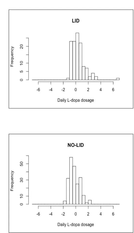
Levodopa (L-Dopa) dosage calculations.……….………...29
Genotyping……….………...31
DNA extraction……….………...…...31
Genotyping of SNCA variant rs356219………...………...31
Genotyping of SNCA D4S3481 variant……….………...32
Case-control genetic association tests………...………...33
Genetic association with continuous PD endophenotypes………...…………34
Quality Control and elaboration of continuous traits………..…...…………34
Association tests with continuous PD traits………...……….37
Survival analyses on LID onset………...………..38
Testing basic assumptions of Cox proportional hazards (PH) models………...39
Cox PH models………...41
Investigating genetic basis of LIDs at an exome-wide level...42
Whole Exome Sequencing (WES): protocol and quality control...42
Identification of rare mutations with potential risk/protective effect on LID onset...43
Chapter 4 RESULTS...46
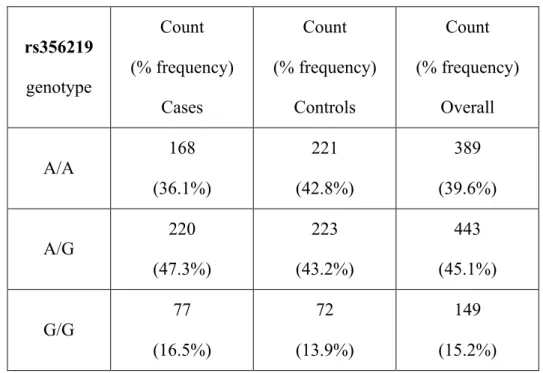
Genotype and allele frequencies of the two candidate variants...47
SNCA polymorphisms and PD risk...48
SNCA polymorphisms and continuous PD endophenotypes...49
Survival analyses on LID onset...55
Exploratory Cox PH Models using non-genetic exposures...55
SNCA polymorphisms and incident LID risk...62
Identification of rare mutations with potential effects on LID onset...68
Chapter 5 DISCUSSION...69
Case-control analyses and associations with continuous PD endophenotypes...70
Analysis of L-Dopa induced dyskinesia (LID) risk...71
Strengths and Limitations...73
Conclusions and future perspectives...75
Appendices...77
Bibliography...120
1
ABSTRACT
I
NTRODUZIONE: il Morbo di Parkinson è una patologia neurodegenerativa caratterizzata da numerosi sintomi motori e non motori, che di solito vengono stimati attraverso diverse scale. La formazione di aggregati tossici della proteina α-sinucleina (codificata dal gene SNCA) è stata proposta come uno dei principali meccanismi molecolari alla base del Parkinson, e sembra che tale meccanismo dipenda anche dai livelli di espressione del gene SNCA. L'attuale trattamento è solo sintomatico e la Levodopa (L-Dopa) rimane il farmaco migliore, nonostante crei, in alcuni casi, gravi effetti collaterali, come movimenti involontari chiamati discinesie (indotte da L-Dopa, o LID). Poiché il Parkinson mostra un'estrema eterogeneità genetica, che è anche influenzata dal background genetico di ciascun soggetto, sono stati condotti studi sulle popolazioni di differenti etnie, in particolare per le varianti di suscettibilità più studiate. Tuttavia, pochi studi si sono focalizzati su fenotipi di tipo continuo correlati al Parkinson quali scale di sintomi neurologici, cognitivi e clinici, anche noti come endofenotipi. Analogamente, la genetica delle LID è in gran parte poco chiara e solo alcune varianti sono state testate in relazione al loro rischio incidente.O
BIETTIVI EM
ETODI: Abbiamo studiato due varianti di suscettibilità del gene SNCA - rs356219 e D4S3481 - associate al livello di espressione del gene e al rischio Parkinson, in una coorte italiana di 472 pazienti e 518 controlli. Prima abbiamo testato la potenziale influenza di queste varianti sul rischio prevalente, attraverso test di associazione caso-controllo aggiustati per sesso. Quindi abbiamo testato, nei soggetti affetti, associazioni con scale motorie (UPDRS), cognitive (MoCA) e non motorie (NMS), e con l'età di insorgenza della patologia (AAO), che ne rappresentano un altro importante endofenotipo. Infine, abbiamo testato l'influenza di rs356219 e D4S3481 sul rischio di insorgenza di LID, attraverso regressioni di Cox (follow-up totale 17.434 persone-mese, tempo di follow-up mediano 49 mesi). Queste analisi sono state aggiustate tenendo in considerazione diverse covariate quali età, sesso, terapia con L-Dopa (stato ON/OFF e dosaggio) e ulteriori scale di stadiazione della malattia.R
ISULTATI: Abbiamo riscontrato due associazioni nominalmente significative del microsatellite D4S3481, una per l’allele 261 con una minore età di insorgenza della malattia (β (SE) = -2.02 (1.00); p = 0.045) - trend non confermato per l’allele di rischio putativo 263 - e l’altra con il rischio incidente di LID, in cui i portatori dell'allele 263 mostrano un rischio ridotto di complicanze motorie (HR [CI] = 0,56 [0,32; 0,98], p = 0,04). Tali associazioni non risultano significative dopo correzione per test multipli. Non sono state osservate altre associazioni significative per nessuno dei modelli genetici alternativi testati.2
D
ISCUSSIONE: Per la prima volta viene riportata un’associazione della variante D4S3481-261 bp con una minore età di insorgenza della malattia, e un effetto protettivo della nota variante di rischio D4S3481-263 bp contro l’insorgenza di LID, con i portatori dell’allele 263 che mostrano la metà del rischio rispetto ai non portatori. Nonostante l’assenza di una significatività statistica dopo correzione per test multipli, questo risultato potrebbe avere un impatto importante nella gestione del trattamento del PD e pertanto è necessario confermarlo in coorti Parkinson indipendenti di grandi dimensioni.3
ABSTRACT
B
ACKGROUND: Parkinson’s disease (PD) is a neurodegenerative disorder characterized by several motor and non-motor symptoms, which are usually evaluated trough different scales (see below). Toxic aggregates of α-synuclein (encoded by the SNCA gene) have been proposed as one of the main molecular mechanisms at the basis of PD, which seem to depend also on the levels of expression of the gene. Although current PD treatment is only symptomatic, Levodopa (L-Dopa) remains the therapeutic gold standard for PD, which however creates in some cases severe side effects like involuntary movements called L-Dopa induced Dyskinesias (LIDs). Since PD shows an extreme genetic heterogeneity, which is also influenced by different genetic backgrounds, population-specific studies are warranted, also for known PD susceptibility variants. Similarly, the genetic of LIDs is largely unclear, and only a few variants in candidate PD genes have been tested with relation to LID risk.O
BJECTIVE &M
ETHODS: Here, we investigated two candidate SNCA susceptibility variants - rs356219 and D4S3481 - which have been linked with the level of expression of the gene and have been consistently associated with PD risk, in an Italian cohort (472 patients and 518 controls). First we tested the potential influence of these variants on PD prevalent risk, through crude case-control association tests adjusted for sex. Then we tested, within PD cases, associations with scales assessing motor (UPDRS), cognitive (MoCA) and non-motor symptoms (NMS), and on PD age-at-onset (AAO), which represent powerful PD endophenotypes. Finally, we tested the influence of rs356219 and D4S3481 on the incident risk of LIDs, through multivariable Cox PH regressions (total follow-up 17,434 person-months, median follow-up time 49 months). These analyses were adjusted for an extended panel of covariates which may influence the outcome, including age, sex, L-Dopa therapy (status and dosage), and additional PD staging scales, where appropriate.R
ESULTS: We observed a nominally significant association of D4S3481 with incident risk of LIDs, where carriers of the 263 (putative risk) allele showed a decreased risk (HR [CI] = 0.56 [0.32; 0.98], p = 0.04) of motor complications. Another nominally significant association was observed with AAO for D4S3481-261 bp allele vs 259 bp allele carriers (β (SE) = -2.02 (1.00); p = 0.045) in a pseudo-additive model, where however we did not observe any evidence of association for 263 vs 259 bp allele carriers. Both these associations did not survive correction for multiple testing. No other significant associations were observed for any of the alternative genetic models tested, neither in the case-control test nor in the analysis of continuous PD endophenotypes.4
D
ISCUSSION: Here we report for the first time an association of D4S3481-261 bp variant with earlier age at PD onset, and a protective effect of the known PD risk D4S3481-263 bp variant against motor side-effects of L-Dopa treatment, with 263 carriers showing half the risk of non-carriers. Since this aspect has never been investigated before for D4S3481 and we observed only a nominally significant association, further studies in large independent PD cohorts are warranted to clarify the potential influence of this marker on LID susceptibility.5
Chapter 1
6
Parkinson Disease (PD)
Idiopathic Parkinson’s Disease (PD) is the second most common neurodegenerative disorder after Alzheimer’s disease (1). Despite almost 200 years since James Parkinson first described the disease, the exact mechanisms underlying this condition remain unclear (2).
PD is a progressive disorder characterized by dopaminergic cell degeneration in the substantia nigra pars compacta and is associated with intracytoplasmic Lewy body inclusions (3).
PD affects about 1% of people above 60 years of age and 4% of adults over 80 years (4), with increased prevalence in advancing age (5).
PD is characterized by several motor symptoms such as resting tremor, rigidity, bradykinesia and postural instability (6), and also non-motor symptoms (NMSs) such as depression, dementia, rapid eye movement, sleep behaviour disorder and anosmia, among others (7). Motor symptoms result from the degeneration of dopaminergic neurons in the midbrain substantia nigra, whereas NMSs are thought to result from the dysfunction of the serotonergic, cholinergic, and catecholaminergic systems (8). Based on these clinical and pathological findings, PD is recognized as a disease involving multiple systems and neurotransmitters (9).
In spite of the increasing knowledge of PD mechanisms, so far only symptomatic treatments have been discovered, either through pharmacological therapy or electrostimulation (7). Among pharmacological treatments, the most used active principle is Levodopa (L-Dopa; l-3,4-dihydroxyphenylalanine), a metabolic precursor of dopamine which is considered a gold standard in the field. L-Dopa is actively absorbed in the upper small intestine, and transported across the intestinal mucosa and blood–brain barrier (BBB). Once absorbed, it is converted into dopamine by aromatic amino acid decarboxylase (AADC) and metabolized to 3-O-methyldopa (3-OMD) by catechol-O-methyltransferase (COMT). Inhibitors of AADC (carbidopa or benserazide) and COMT are co-administered with L-Dopa to suppress the peripheral degradation of dopamine. This is done in order to reduce the exogenous dose of L-Dopa by maximizing the amount of the substance transported across the BBB, and to reduce adverse effects of peripheral dopamine, such as nausea and hypotension (10). Unfortunately, long-term L-Dopa treatment and over-dosage cause important side effects like L-Dopa induced Dyskinesias (LIDs). This motor complication - characterized by involuntary movements throughout the body - represent an important source of disability and notably worsens patients’ quality of life.
7 Since this dissertation mainly focuses on investigating the genetic underpinnings of PD, of related neurological and clinical endophenotypes, and of side effects of its pharmacological treatment (i.e LIDs), we briefly review these aspects below.
Genetics of PD
PD tends to recur in families and is moderately heritable, with about 60% of its variance being explained by genetic factors (11), and is characterized by a complex architecture, with a number of genetic and environmental factors influencing susceptibility to the disease (12). It shows an extreme genetic heterogeneity, with 10% of PD cases having Mendelian inheritance (13,14). The genes which have been most robustly implicated in Mendelian forms of PD include:
SNCA (4q22.1; α-synuclein), encoding α-synuclein, a neuronal protein that plays several roles in
synaptic activity, such as regulation of synaptic vesicle, trafficking and subsequent neurotransmitter release. Mutations have emerged as a rare, but important cause of PD with high penetrance (15). Since most of this dissertation focuses on the analysis of SNCA variants, this gene is reviewed more in details below.
LRRK2 (12q12; Leucine Rich Repeat Kinase 2), encoding a leucine rich repeat kinase 2 containing
multiple functional domains. LRRK2 has been implicated in several autosomal dominant forms of PD, where several mutations have been identified (reviewed (16)), which make a large contribution towards both sporadic and familial forms of PD (17). Different studies have repeatedly shown linkage of PD risk to LRRK2, and a meta-analysis indicated LRRK2 as one of the most important genomic loci influencing PD risk (18).
PARK2 (Parkin, 6q26), encodes Parkin, an E3 ubiquitin ligase protein, but the mechanism of its
pathogenicity remains unclear. Point mutations in this gene are mostly transmitted from common founders (19). These mutations are involved in development of Parkinson’s disease probably by a loss-of-function mechanism (20). Patients with Parkinson’s disease and Parkin mutations have a mean age at onset of 32 years in the Caucasian population (21). Hence, Parkin mutations are the most common cause of early-onset Parkinson’s disease, occurring in up to 50% of those with age at onset under 25 years (and only 3%–7% in those with age at onset 30–45 years) (15).
ATP13A2 (Cation-transporting ATPase 13A2, 1p36), encodes an ATPase that plays a role in
intracellular cation homeostasis and in the maintenance of neuronal integrity (22). It is required for a proper lysosomal and mitochondrial maintenance (23,24), where it regulates the autophagy-lysosome pathway through the control of SYT11 expression, both at the transcriptional and at the
8 post-translational levels (25). Mutations in ATP13A2 cause also autosomal recessive parkinsonism with a complex phenotype (15).
PINK1 (Serine/threonine-protein kinase, 1p36.12) codes for a serin/threonine kinase localized to
the mitochondria. Mutations in PINK1 are a rare cause of early-onset PD, accounting only for 2%– 4% of early-onset cases in Caucasian populations (26,27) and 4%–9% in Asian populations (28,29). The penetrance for homozygous and compound heterozygous mutation carriers seems to be 100% but the specific mechanism of pathogenicity in PD is unclear and require further investigations (15).
DJ-1 (Protein/nucleic acid deglycase DJ-1, 1p36.23), also known as PARK7 since it encodes
Parkinson disease protein 7. Mutations in DJ-1 cause autosomal recessive PD. Its product inhibits the aggregation of α-synuclein via its chaperone activity, (30,31) acting as a redox-sensitive chaperone protein and as a sensor for oxidative stress (15).
VPS35 (VPS35 endosomal protein sorting factor-like, 16q11.2) this gene belongs to a group of
vacuolar protein sorting (VPS) genes. The encoded protein is a component of a large multimeric complex, termed the retromer complex, involved in retrograde transport of proteins from endosomes to the trans-Golgi network. Mutations in this gene cause an autosomal dominant, adult-onset form of the disorder. It is phenotypically similar to idiopathic PD (32).
DNAJC13 (DnaJ heat shock protein family (Hsp40) member C13, 3q22.1) is involved in
membrane trafficking through early endosomes. In fact, it is implicated in the transport and recycling of transferrin and in the transport and degradation of endosomal growth factors from early endosome to late endosome (33). A novel mutation in this gene (p.Asn855Ser) was found to segregate with PD (34).
GBA (Glucosylceramidase Beta, 1q22) (35) encodes the lysosomal glucocerebrosidase enzyme,
which cleaves the β-glucosyl. Proposed gain-of-function mechanisms include facilitation of α-synuclein accumulation perhaps loss-of-function mechanisms include substrate accumulation (35).
These genes are extensively reviewed in (13,36,37). Although other chromosomal loci - including
PARK3, PARK10, PARK11 and others (13) - have been identified, and these regions might contain
further genes for typical, late-onset PD (13), we do not review them here since these have been not robustly implicated in PD as the candidate loci mentioned above. In these and other genes, rare mutations with both dominant (12,14) or recessive inheritance modes (38,39) have been identified, often through genome-wide linkage studies followed by targeted genotyping (e.g. 14) or, more
9 recently, through Next Generation Sequencing (NGS) studies (e.g. (40,41)). In addition to rare mutations, also common susceptibility variants like Single Nucleotide Polymorphisms (SNPs) have been detected within these genes, e.g. in LRRK2 and SNCA (13). However, the genetic variants identified so far – be they common or rare - explain only a minor part of PD heritability (34), and for a large majority of PD cases the genetic diagnosis remains unresolved. The issue of missing heritability has been tackled through different approaches, including Genome Wide Association Scans (GWAS) to identify common variants with moderate/weak effect sizes on PD susceptibility (e.g. (42)), and NGS (mostly Whole Exome Sequencing) studies to identify rare causative mutations (e.g. (12,14,43–47)). Moreover, the genetic architecture and the mutational spectrum of PD can vary based on the ethnic and genetic background of the population (46,48) hence population-specific genetic studies are warranted (as in (43,46)).
Large-scale genomic studies carried out so far have scarcely investigated inter-individual variation in PD endophenotypes like neurological scales (12,14,42–47,49).
A GWAS study of age-at-onset in 25,568 PD cases reported two genome-wide significant associations within SNCA and TMEM175 (50), while other preliminary GWAS of cognitive performance and motor symptoms progression are ongoing (51,52). Other SNP-based genomic studies tested associations of Polygenic Risk Scores (PRS) for PD with alpha-synuclein levels in the cerebrospinal fluid, age-at-onset of the disease, motor/cognitive symptoms and PD status (as reviewed in (53), detecting significant associations with PD risk (54), earlier PD onset (54,55), and faster motor and cognitive decline (56). With regard to large scale Next Generation Sequencing (NGS) studies, several Whole Exome Sequencing (WES) but no Whole Genome Sequencing (WGS) studies have been carried out so far on PD (12,14,18,44–47,57).These mostly focused on PD case-control analysis, but failed to find robust statistical evidence of association, probably due to the small sample size - compared to the huge genetic heterogeneity of the disease - and to the difficulty in recruiting proper neurological controls (i.e. people free of disease at an advanced age). Among these, our group attempted to identify genetic variants associated with continuous scales associated with PD (or PD endophenotypes, see below), assessing motor, cognitive and non-motor PD symptoms, but found no statistically significant associations (57). On the other hand, association with specific scales related to PD has been more often tested for genetic variants in candidate PD susceptibility genes. Loss-of-function GBA mutations have been associated with a distinct cognitive profile characterized by greater impairment in working memory/executive function and visuospatial abilities in PD patients (58). PD cases carrying variants in PARK16 - another gene implicated in PD (59) - exhibited greater motor progression
10 after 5 years of disease compared with non-carriers, based on assessment through Hoehn & Yahr (HY) staging scale, UPDRS motor score and UPDRS sub-scores (see below for details on these scales) (60). The common variant rs356182 in SNCA has been associated with a more tremor-predominant phenotype and predicted a slower rate of motor progression (61), while rs11931074 showed an association with worse motor symptoms (62). PD patients carrying rare variants in the
APP, PSEN1, PSEN2, and GRN genes exhibit lower cognitive tests scores than non-carriers,
regardless of age at PD diagnosis, age at evaluation, APOE status or recruitment site (63).
One of the most investigate genes in relation to PD endophenotypes is by far SNCA, the first PD locus identified (64). Since this dissertation focuses on the investigation of SNCA variants, we review below this gene and its implication in PD.
SNCA
SNCA (4q22.1) was the first gene identified as associated with idiopathic PD (38,64). Linkage
analysis study of a large Italian kindred with autosomal dominant PD form revealed a locus at 4q22.1-q22.3 associated to the disease (64). This was further refined through the identification of a causative mutation in the SNCA gene (Ala53Thr), in the same Italian pedigree and in three unrelated dominant families of Greek origin (65). Since then, several studies have examined SNCA in relation to PD risk and its endophenotypes (reviewed in (66)).
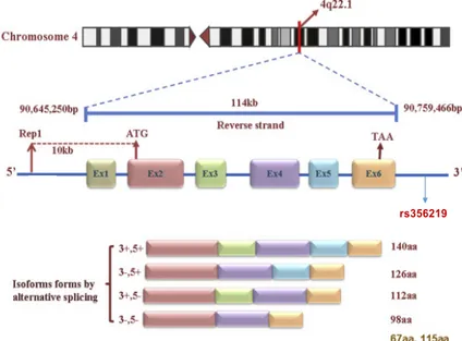
SNCA (Figure 1.1) gene encodes for alpha-synuclein (α-syn) protein, a member of the synuclein
family, which also includes beta- and gamma-synuclein. Synucleins are abundantly expressed in the brain, and alpha- and beta-synuclein inhibit phospholipase D2 selectively (67). α-syn plays a fundamental role in the molecular pathogenesis of PD, forming toxic oligomers and aggregates within neurons (68), acting in a prion-like manner. These aggregations ultimately result in Lewy bodies, which represent the histopathological hallmark lesions of PD (69). Similarly, SNCA has been implicated in another neurological disorder highly comorbid with PD, with a partly shared etiopathological mechanism, namely Dementia with Lewy Bodies (70). α-syn peptides are also a major component of amyloid plaques in the brains of patients with Alzheimer's Disease (71). In physiological conditions, neuronal α-syn protein plays several roles in synaptic activity, such as regulation of synaptic vesicle trafficking and subsequent neurotransmitter release (72). It also participates as a monomer in synaptic vesicle exocytosis by enhancing vesicle priming, fusion and dilation of exocytotic fusion pores (73). Mechanistically, α-syn acts by increasing local Ca2+ release from micro-domains, which is essential for the enhancement of ATP-induced exocytosis (73). It also acts as a molecular chaperone in its multimeric membrane-bound state, assisting in
11 the folding of synaptic fusion components called SNAREs (Soluble NSF Attachment Protein Receptors) at presynaptic plasma membrane, in conjunction with cysteine string protein-alpha (74). This chaperone activity is important to sustain normal SNARE-complex assembly during aging (74). SNCA plays also a role in the regulation of the dopamine neurotransmission in association with the dopamine transporter (DAT1) and thereby modulating its activity (73). α−synuclein is currently seen as one of the most promising targets of disease-modifying therapies for PD (37), which is why investigating in detail the genetic risk/protection conferred by its genetic variants is more and more important. Studies carried out so far supported an influence of polymorphisms in multiple regions of SNCA gene, such as the promoter (5’) region (REP1-SNCA), 3′ end (e.g., rs11931074 and rs356219), 3′ untranslated regions (e.g., rs356165), and introns (e.g., rs7684318, rs894278, and rs276990) (as reviewed in (75)). Among these variants, increasing attention have received specific variants which have been reported to alter SNCA gene expression levels (75–78), which is considered one of the main mechanisms through which α-syn causes PD (79–82).
One of these variants is represented by D4S3481 (commonly known as REP1), a complex polymorphic microsatellite (dinucleotide) repeat located 10 kb upstream of the translation start site of SNCA (83). A microsatellite is a tract of repetitive DNA in which certain DNA motifs (ranging in length from one to ten base pairs) are repeated, typically 5 to 50 times. Several small studies have suggested that certain alleles of a dinucleotide repeat sequence (REP1) of the SNCA promoter might be associated with the risk of developing PD (84). SNCA-REP1 is essentially triallelic (259, 261, and 263 bp in length) (85). The 259/259 bp genotype has been associated with a decreased levels of expression of α-syn in the blood, compared to genotypes 261/261, 259/261, and 259/263 (78). A down-regulation effect of the 259 bp variant on SNCA gene expression has been supported also by functional analyses (86–89).
A meta-analysis of association studies showed higher frequency of 263 bp allele in cases compared to controls (90). Conversely, the 259 bp allele was found to be associated with a decreased risk of PD, while no effect was observed for the 261 bp allele (85). These alleles have been also associated with continuous PD-related traits, although not always consistently. PD patients carrying at least one 263bp allele in SNCA-REP1 exhibited four-fold higher odds of fast disease progression compared to non-carriers (91), and the 263 allele was also associated with a worse cognitive outcome in PD. (85). Conversely, REP1-259 allele was also associated with the development of worse motor outcomes (92). As opposed to these lines of evidence, other studies have reported an association of REP1-263 allele with better motor and cognitive outcomes or no association, as in
12 Ritz et al, 2012. In the latter study, authors observed no association between SNCA-REP1-259 allele and motor symptom progression under a dominant genetic model, although the risk was in the expected (‘‘protective’’) direction (91).
Another variant which has been robustly associated with changes in the level of expression of
SNCA is the single-nucleotide polymorphism (SNP) rs356219, which lays in the 3′ region of the
gene (Figure 1.1). This SNP is probably the most investigated common variant in SNCA, and it stands out as a consistent risk factor for PD in several studies (as reviewed in (75)). Moreover, this variant has showed a significant effect on SNCA mRNA levels in the substantia nigra and in the cerebellum (78), and was shown to affect also the blood plasma levels of α-syn (93). This evidence is in line with independent transcriptomic analyses which revealed a positive association between the count of the rs356219-G allele and the level of expression of specific SNCA isoforms, assessed through quantitative Polymerase Chain Reaction (qPCR), RNA-sequencing (RNA-seq) and cap analysis of gene expression (CAGE-seq) in postmortem frontal cortex tissues of neurologically healthy subjects (94).
In a two-tiered analysis of 1,956 patients with PD and 2,112 controls on 15 candidate SNPs within
SNCA, rs356219 showed the most significant association among all variants tested, which was
larger than and independent of the REP1 marker (95). Author suggested that this effect on increased PD susceptibility might by mediated by an upregulation of SNCA expression in a dose-dependent manner (95). In a later meta-analysis of 18 PD case-control observational studies focused on rs356219, (86,96–98), a significant association with PD risk was found in Caucasian populations, showing an increased risk by ∼26% and ∼38% in the dominant and recessive models, respectively (96). This finding was later supported in a PD case-control GWAS, where rs356219 was detected as a genome-wide significant hit (99), and in candidate variant studies of different genetic ancestries, such as Chinese Han (100) and South-Americans (96). In the latter study, rs356219-G allele was associated with an increased risk for cognitive impairment in Brazilian PD patients (96). Of note, rs356219 was also shown to significantly contribute to other PD continuous endophenotypes, such as an earlier age at onset of the disease (101).
13
Figure 1.1: SNCA gene structure and protein isoforms generated by alternative splicing.
Modified by: The link between the SNCA gene and parkinsonism Wei Xu et al. 2015
PD endophenotypes
Endophenotypes are measurable components (e.g., neurophysiological, biochemical,
neuroanatomical, cognitive or neuropsychological) that exist between the behavioural symptoms of a disease and a distal genotype (102).
The purpose of the endophenotype concept is to divide symptoms and signs of a disease into more stable phenotypes with a clear genetic connection. The rationale at its basis is that a smaller number of genes will be associated with a less complex phenotype than a complex disorder, increasing the power to detect genetic associations with the endophenotype and, indirectly, with the disease of interest.
An ideal endophenotype should meet the following criteria:
• association with the disease in the population; • heritability;
• primary state-independence (i.e. it should be seen in individuals with and without the active
illness/diagnosis).
• co-segregation with the disease within families;
14
• familial clustering (i.e. it should present in both patients and their unaffected relatives at a
higher rate than in the general population or, alternatively, should show intermediate values between probands and the general population, in case of continuous traits).
In PD, several continuous phenotypes (also known as “traits”) can be considered as endophenotypes, e.g. motor symptoms, cognitive performance, depression, and age-at-onset. Currently, these components are notably under investigated in relation with PD patients’ genetic profile (57). For specific PD candidate genes like SNCA, the influence of common genetic variants within or close to the gene is related to different aspects of PD phenotypic spectrum, as we briefly reviewed above. More in general, the study of common variants may provide valuable insights into the mechanisms underlying heterogeneity in PD (103).
Dyskinesias
Dyskinesias represent “Abnormal involuntary movements attributed to pathologic state of one or
more parts of the striate body and characterized by insuppressible, stereotyped, automatic movements that cease only during sleep.” (The American Heritage® Stedman's Medical
Dictionary). Schoenecker recorded the first clear description of clinical dyskinesia in 1957. The term “tardive dyskinesia” was coined to indicate abnormal movements induced by neuroleptics (104). In PD, dyskinesia was recognized with the advent of L-Dopa, and since then “levodopa-induced dyskinesias” (LIDs) (Figure 1.2) has become one of the major clinical limitations of the long-term treatment of PD. By the late 1970s, several classifications of LIDs have been proposed based on the type of movements, the timing of L-Dopa dosage and combinations of the two factors:
1. Peak-dose dyskinesia: dyskinesia noted at the peak clinical benefit of L-Dopa;
2. Dystonia-improvement-dystonia (105), or diphasic dyskinesia (106): appearing at beginning and at the end of each L-Dopa dose;
3. OFF dystonia: dystonia occurring early in the morning, when the effect of previous night’s dose of L-Dopa had completely worn off.
15
Figure1.2: Changes in motor response associated with chronic levodopa treatment.
Levodopa-induced motor complications. Here we report a schematic illustration of the gradual shortening of the duration of a beneficial motor response to L-Dopa treatment (wearing off), and the appearance of dyskinesias as this time range (“on” time) shortens. Image courtesy of Harrison's Neurology in Clinical Medicine, 3rd Edition. C. Warren Olanow Image Anthony H.V. Schapira
LIDs comprise a variety of phenomena, the most common of which are chorea, choreo-athetosis, and dystonia. Chorea is the most common form of LID and it is most commonly associated with peak dose dyskinesia (107). Dystonia is the second most common form of LID, while ballism is characterized by abnormal choreic movements of the proximal parts of the limbs causing flinging movements, which can be unilateral or bilateral (107). Myoclonus, a sudden brief shock-like involuntary movement, is rarely classified as a part of LIDs (107). Other LID movements include respiratory dyskinesia (108,109), ocular dyskinesia (110), restlessness/hyperactivity, akathisia and enhanced tremor (107). The rate of LID development ranges between 3 and 94% among PD patients, depending on different factors which mainly include PD age-at-onset, disease duration, severity, and duration of L-Dopa therapy (3) (see Risk factors of LIDs subsection below for details).
Hypothesized mechanisms of LIDs
The aetiology of LIDs is largely unknown yet. With the reduction of dopamine in PD patients, it is believed that hypersensitization of the dopamine receptor contributes to the development of LIDs (111). The short half-life of L-Dopa and pulsatile release of dopamine, once the buffering capacity of dopamine transporter has been lost, is considered to be one of the major mechanism
16 generating LIDs (112). The use of extended-release carbidopa/levodopa and continuous intrajejunal infusion of carbidopa/levodopa intestinal gel has been reported to improve motor symptoms and motor fluctuations, without aggravating dyskinesia when compared to standard L-Dopa (113–115). Recently, it has been reported that carbidopa/levodopa intestinal gel infusion cause dyskinesias, including diphasic dyskinesia (116).
Compared to L-Dopa, dopamine agonists cause less dyskinesia, given that they have longer half-life (117–119). The use of a dopamine agonist in early stage PD patients to delay the use of L-Dopa is considered to be clinically effective, and to successfully postpone the occurrence of LIDs (120).
Risk factors of LIDs
In addition to the use of L-dopa rather than other dopamine agonists for the treatment of PD (see Sharma et al., 2010 for a review (121)), many other risk factors have been associated with the onset of LIDs. Some of them are modifiable, like L-Dopa dose and body weight, while others are non-modifiable, like age, sex, PD age at onset, duration of disease, clinical subtype, disease progression, disease severity, and genetic factors (which we review in the next subsection) (121). PD age at onset (AAO) represents one of the main risk factors for dyskinesias (122). The younger is AAO, the more likely is the development of LIDs (123–125). A 5-year follow-up study of PD patients showed a prevalence of LIDs up to 50% at age 40–59, and 16% after 70 years (126). Another study found that after 5 years of L-Dopa treatment, the rates of dyskinesia in patients with PD onset at 40–49, 50–59, 60–69, and 70–79 years were 70%, 42%, 33%, and 24%, respectively (123). Furthermore, patients with AAO < 40 years (young-onset PD) had a higher incidence of LIDs than those with late-onset PD (AAO ≥ 50 years) (125). In line with this evidence, patients with longer duration of disease - which is connected with AAO - are more likely to develop LIDs (127). Of note, age per se has been detected as a risk factor in a single cross-sectional study, which reported a positive correlation between patients’ age and time from onset to development of motor complications (128).
Sex represents another important risk factor for LIDs, with women showing greater incidence of dyskinesias than men (124,129). Moreover, women develop dyskinesias earlier in relation to time of L-Dopa administration, compared to men (130). This may be due to the fact that women have less “genetic protection” related to lower expression of dopamine receptor DRD2, which seems to exert a protective role against dyskinesia in men (131). An alternative explanation may be the
17 higher bio-availability of L-dopa in women, due to their lower body weight (121). Of note, the higher LID risk conferred by sex was not confirmed in another study (96), and in a multivariate analysis including additional risk factors (AAO and L-Dopa dosage) (132).
As mentioned above, low body weight and a resulting higher bioavailability of L-Dopa is a prominent risk factor for LIDs (124,132,133), which may be also easily explainable from a biological point of view. Indeed, several studies have proposed that the increased risk of motor complications in PD patients with lower body weight may be due to elevated peripheral L-Dopa levels in these patients ((133); Group 1996).
Clinical subtypes of PD are also an important risk factor for LIDs. An observational study of 144 L-Dopa-treated patients showed that the tremor dominant subgroup had lower rates of dyskinesia (29%) compared to the bradykinesia dominant subgroup (69%) (Friedman 1985). Similarly, in another study, resting tremor subtype was associated with lower risk of developing LIDs than other initial manifestations (134). Of note, resting tremor subtype is considered to be independent on all the other known risk factors for LIDs, for the occurrence of motor complications (135).
In PD patients in the early stage of the disease (HY score 1), the time from the beginning of L-Dopa treatment to the occurrence of dyskinesias was 66 months, while, in “stage 3” patients (i.e. with HY score 3), it was only 24 months (136). Similarly, a recent analysis of Chinese PD patients revealed a positive association of prevalent LID risk with low UPDRS-III and high HY scores in ON-state (i.e. under L-Dopa treatment), which indicated severity of motor symptoms and progression of the disease, in addition to early AAO, long disease duration, female sex, and high L-Dopa equivalent dose (137). Of note, the emergence of dyskinesia had no association with the initiation time of L-Dopa (137). A community-based study of L-Dopa-related motor complications in PD found that the overall dose of L-Dopa was the most important predictor of motor fluctuation, with the dose and treatment having the strongest impact on LID prevalence (138). The recommended initial dose - less than 400 mg per day - helps to reduce the risk of motor complications (124,139). These studies suggest that L-Dopa dosage may be more important than the duration of treatment.
In other words, the higher the dose, the greater the risk of dyskinesia (134).
In addition to classical risk factors, more recently functional imaging has also been used to find predictors of LIDs (reviewed in (140)). E.g., a research showed that pre-synaptic dopamine deficiency assessed through PET scanning in 127 drug-naive de novo patients with PD predicted the risk of LIDs (141). Given the focus of the present dissertation, here we do not extensively review these works.
18
Genetics of LIDs
The development of LIDs reflects a profound reorganization of the neural circuit and balance between different pathways in the basal ganglia (142). LIDs are determined in part by genetic factors with multiple polymorphisms in various candidate genes. PD patients show a remarkable heterogeneity in their response to L-Dopa and this likely suggests that there is a certain genetic predisposition. However, if and how the inherited predisposition to PD affects the development of LIDs is currently an unanswered and largely under-investigated issue, both in candidate gene studies and in genome or exome-wide studies with no a priori hypotheses. We briefly review below the different genes which have been studied in relation to LID onset, and the genetic influences identified so far.
Dopamine receptors
Dopamine exerts its physiological action through the activation of dopamine receptors (DRD1– DRD5), which can be divided into D1-like receptors (DRD1 and DRD5), and D2-like receptors (DRD2, DRD3, and DRD4) (143). Normally, dopamine triggers an excitatory response on direct pathways through D1-like receptors, and an inhibitory response on the indirect pathway through D2-like receptors (144). In PD, which is characterized by the loss of dopamine, usually underactivity of direct pathways and hyperactivity of indirect pathways is observed (142). The DRD2 gene (dopamine receptor D2, 11q22-23) is one of the most investigated with reference to LID risk (see below). It encodes for a transmembrane G protein coupled receptor which activates intracellular signalling by the inhibition of cAMP synthesis (145). Oliveri et al were the first to study an intronic short tandem repeat (CAn-STR) with four common alleles (13, 14, 15, and 16
CA repeats) in this gene, reporting a higher frequency of the 13 and 14 alleles in non-dyskinetic compared to dyskinetic PD patients (146). Another study reported a similar protective effect in males but not in female PD patients (129). Strong et al, found that the 14 allele and/or the 14/15 genotype was a risk factor for dyskinesia, in partial contrast with the above mentioned studies (147). The impact of another polymorphism in the DRD2/ANKK1 locus, rs1800497 (or Taq1A, coding for Glu713Lys change in the protein) has been found to influence the risk of developing ‘wearing off’ motor fluctuations in PD (148). An influence of other variants in the DRD2/ANKK1 region - including 141CIns/Del (rs1799732), rs2283265, rs1076560, C957T (rs6277), rs1800497 and rs2734849 - on LIDs was also reported in a recent study (1). Similarly, Kusters et al found three DRD2-haploblocks to be associated with dyskinesia in about 60% of the studied patients carrying one to three risk haplotypes (149). After combining “risk haplotypes” into a DRD2
19 genotypic risk score, they observed this was associated with an increased risk of dyskinesias and with their severity (149).
The DRD3 gene (dopamine receptor D3, 3q13.3) - encoding for a receptor with an activity mediated by G proteins which inhibit adenylyl cyclase - has been reported to be overexpressed in experimental primate animal models presenting with LIDs (150). The overexpression was in accordance with the severity of LIDs and was prominent in the D1 expressing neurons (151). Similarly, the DRD3 rs6280-A allele, encoding a p.S9G substitution which confers a high binding affinity to dopamine, has been associated with tardive dyskinesia, that can be attributed to dopamine-receptor hypersensitivity (152–154). According to this, rs6280 was associated with the presence of diphasic dyskinesia (i.e. taking place at the beginning and/or end of dose), after adjusting for gender, age at PD onset, Hoehn & Yahr stage and duration of L-Dopa treatment, (155). A recent study supported this association, with patients carrying the rs6280-A allele showing an increased risk of LIDs (111).
Some studies have explored the possible role of DRD1 (dopamine receptor D1) variants on LIDs development in PD, but the reported results were not consistent (111,149). Notably, a growing body of biochemical and biophysical studies show that dopamine receptors can form homomeric and heteromeric complexes (156), hence it may be hypothesized that synergistic interactions between different receptors may induce LID in PD (157).
Other receptors
The adenosine A2A receptor (Adora2A) gene (22q11.23), encodes a receptor binding to G proteins which is highly expressed in the striatum of the brain, where it indirectly competes with DRD2, regulating neurotransmission (158). A recent study showed that Adora2A receptors are highly expressed in the basal ganglia of PD patients (159), especially in the striatum of PD patients who had developed dyskinesia (160).
Adora2A polymorphisms located in intron 1 of the gene - like rs2298383 and rs3761422 - were recently associated with LID events in PD patients (161). Previously, an association of the rs2298383 polymorphism with LID risk was revealed (161), although this association warrants further investigations (161).
It has been suggested that some of the changes in opioid transmission are directly implicated in LIDs (162). Opioids are co-transmitters in both the direct and the indirect basal ganglia pathways, where they regulate dopamine function, and basal ganglia have one of the highest levels of
20 endogenous opioids and opioid peptide receptors in the brain (163). Importantly, a Positron Emission Tomography (PET)-scan study revealed that PD dyskinetic patients had lower opioid binding in striatum and thalamus (162). Among opioid receptors, µ (mu) receptors received the main attention with reference to LIDs. Indeed, in the human mu opioid receptor (MOR) gene, the SNP rs1799971 has been associated with earlier development of dyskinesia in L-Dopa–treated PD patients (147), and has been found to increase binding affinity and functional activity of the endogenous opioid peptide, endorphin (164). Interestingly, receptor-specific opioid antagonists used in primate models have also been observed to affect LIDs (165,166).
N-methyl-D-aspartate ionotropic glutamate receptor (NMDAR) is a ligand-gated ion channel that responds to the neurotransmitters glutamate and NMDA. Dyskinesia, partly involves also changes in glutamatergic receptors in the striatum (142). This hypothesis is supported by evidence that amantadine - a NMDA receptor antagonist widely used in PD patients - reduces LIDs (142,167). The predominant inhibitory mechanism results from the increasing rate of channel closed states (ref). Interestingly, susceptibility to LIDs was recently associated with two GRIN2A (glutamate
ionotropic receptor NMDA type subunit 2A) variants, rs7192557 and rs8057394, which had been
previously associated with the age of dyskinesia onset in Huntington's Disease, suggesting that these movement complications may arise from the same neuronal pathways (79).
Enzymes involved in dopamine metabolism
Catechol-O-methyltransferase (COMT, 22q11.21) is an enzyme that inactivates catechols and degrades catecholamine neurotransmitters, including dopamine (168). It is implicated in the metabolism of L-Dopa, producing 3-O-methyldopa (3-OMD), which antagonizes L-Dopa’s therapeutic action. COMT inhibitors, e.g. Tolcapone and Entacapone, reduce the conversion of L-Dopa to 3-OMD and thus improve its bioavailability in the brain (169,170). A common polymorphism in exon 4 of the COMT gene, rs4680, causes a Valine to Methionine substitution in the protein (Val108/158Met, depending on the COMT isoform). This results in altered activity of the enzyme: high activity in Val/Val, intermediate activity in Val/Met, and low activity in Met/Met genotype. Patients with the Met/Met (i.e. rs4680-A/A) genotype have been documented to experience more frequently severe dyskinesias and other motor fluctuations (171), and especially LIDs (172). Moreover, the doses of L-Dopa treatment for PD patients have previously been found to be influenced by specific COMT haplotypes (173). However, other studies have failed to confirm these associations (174–176).
21 including, among others, norepinephrine, dopamine, and serotonin. Two distinct forms of the enzyme exist, encoded by MAOA (monoamine oxidase A; Xp11.3) and MAOB (monoamine
oxidase B; Xp11.3). A recent study found that patients carrying MAOB rs1799836A allele and
-AA genotype suffered more frequently from LIDs (172), but no other studies have supported these associations (177).
Dopamine transporters
DAT (dopamine transporter; 5p.15.32) encodes a product which is fundamental for transporting
dopamine across the plasma membrane. According to Sossi et al, greater DAT levels are directly associated with lower dopamine turnover and lower changes in synaptic dopamine concentration in PD patients (178). In this gene, a statistically significant association between the C allele of the intronic SNP rs393795 and longer time to LID has been found, which was hypothesized to be due to an altered rate of dopamine reuptake in the synapse (179). Furthermore, the nine copy allele of the 40-bp Variable Number Tandem Repeat (VNTR) polymorphism rs28363170 significantly predicted the occurrence of dyskinesia in a retrospective study on L-Dopa treated PD patients (180).
Other pathways and PD genes
The human BDNF (brain-derived neurotrophic factor; 11p14.1) gene encodes a precursor protein, proBDNF, which is then cleaved to the mature 14-kDa form (mBDNF) by protease tissue plasminogen activator (tPA)-mediated activation of plasmin (181). BDNF exerts multiple biological functions in the central nervous system, and its expression is decreased in PD (182). PD patients with Val66Met polymorphism (rs6265) in the 5’-pro-BDNF sequence had a significantly higher risk of developing dyskinesias earlier in the course of treatment with dopaminergic agents (183). A recent study has also found an association of the minor (A) allele with dyskinesia risk after dopaminergic treatment (184). Recently, the possible role of BDNF in levodopa motor complications was also highlighted in experimental animal models. E.g., rats that over-expressed BDNF were more prone to develop LIDs (157).
The leucine-rich repeat kinase 2 (LRRK2; 12q12) is one of the genes most robustly implicated in PD aetiology (17), and has been also associated with LID onset in some studies. In a North African cohort, the prevalence of LIDs was significantly higher in carriers of the known PD-causative mutation G2019S, compared to non-carriers (185). However, a study in the Israeli population did
22 not replicate this association (186). A recent study showed that LRRK2 phosphorylation levels directly correlate with LID onset, and inhibition of LRRK2 induced a significant increase in the dyskinetic score in L-DOPA treated parkinsonian rat animal models (187).
As LRRK2, also SNCA has been robustly implicated in PD aetiology and progression (188), but has so far been mostly neglected with regard to motor complications connected to the treatment of the disease, in spite of some interesting findings. A heterozygous autosomal dominant point mutation in SNCA (c.158C>A; p.A53E in transcripts NM_000345.3, NM_001146054.1, NC_000004.11) was revealed in two Finnish PD patients, a mother and her daughter, characterized by severe bradykinesia, very little tremor and early onset of LIDs (189). No cognitive decline or dysautonomic features have emerged in these patients during more than 5 years of follow-up. In a recent study, C. elegans model overexpressing human α-synuclein was exposed to L-Dopa in continuous and alternating fashions (190). Chronic exposure to the drug led to hyperactivity of the animal model without meaningful increase in motor activity, and to an increase in peripheral clustering and expression of dopamine receptors in motor neurons. Both of these changes were significantly higher in alternating, compared to continuous, exposure to L-Dopa (190). More recently, Corrado et al (149) investigated the influence of the D4S3481-263 bp allele on the incident risk of LIDs, in an longitudinal cohort of Italian PD patients, reporting no significant differences between 263 allele carriers vs non carriers. These lines of evidence warrant further investigation of SNCA influence on LID onset, in addition to PD risk and endophenotypes.
23
Chapter 2 AIMS
24 To sum up, most of the reported heritability of Parkinson Disease is largely unknown, and its genetic bases remain unclear. This is likely due to the notable genetic heterogeneity of the disease, and to the relatively low power of genetic studies carried out so far. Moreover, PD endophenotypes, such as scales assessing motor, cognitive and other non-motor symptoms, have been largely under-investigated, due to the difficulties to collect PD cohorts with complete and detailed phenotypic assessment. Using such continuous scales to investigate PD genetics may provide powerful tools to identify PD susceptibility variants. Similarly, the genetic of LIDs is largely unclear, with different single variant associations reported, which have not been replicated yet. Therefore, further studies in independent cohorts are needed to clarify the genetic underpinnings of PD, its endophenotypes and genetic influences on side effects of L-Dopa therapy. To investigate these aspects, we adopted a multi-faceted and comprehensive approach (resumed below).
First, we investigated in an Italian PD cohort (N=470) collected at IRCCS Neuromed, the SNCA gene in order to:
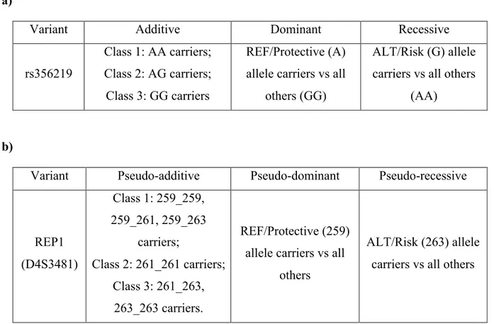
1. clarify the role of two of the most investigated PD susceptibility variants which have also been associated with the level of expression of SNCA - namely rs356219 and D4S3481 - in the genetic susceptibility to PD, through case-control associations tests;
2. test the potential influence of these variants on PD scales assessing motor, cognitive and non-motor symptoms, as well as on PD age-at-onset (which represent powerful PD endophenotypes), through genetic association analyses;
3. determine whether SNCA affects also susceptibility to L-Dopa induced dyskinesia, by testing genetic associations of rs356219 and D4S3481 with the incident risk of LIDs in survival analyses.
Then, to identify rare variants with a potential risk/protective effect on LIDs occurrence in response to low/high L-Dopa daily dosages, we performed a variant prioritization bioinformatics pipeline in a subset of 114 PD patients which underwent Whole Exome Sequencing (WES) analyses.
25
Chapter 3
26
PD patients cohort
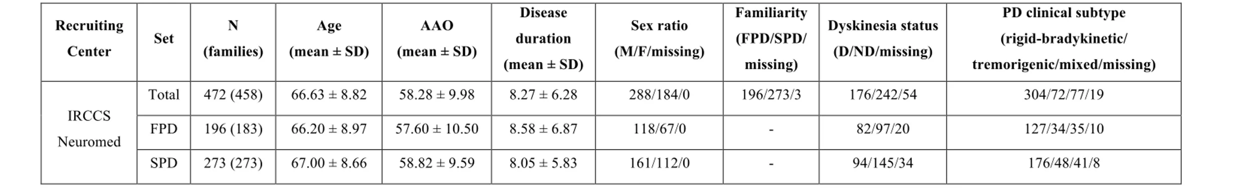
472 PD patients (288 males; 196 familiar cases; mean (SD) age of 66.6 (8.8) years) were recruited at the Parkinson Centre of the specialized clinics IRCCS Neuromed, Pozzilli, Italy, between June 2015 and December 2017 (57). All the cases involved in the study (hereafter called Neuromed cohort) were diagnosed with PD by a qualified neurologist, according to published diagnostic criteria (appendix 1), which included rigidity, postural instability, resting tremor and positive response to levodopa treatment(191). Where diagnosis was uncertain, dopaminergic loss observed through neuroimaging techniques (PETscan or DaTscan) was used to confirm PD diagnosis. PD patients underwent a detailed phenotypic assessment, which included neurological examination and evaluation of non-motor domains (see below). Information about family history, demographic characteristics, anamnesis, pharmacological therapy and side effects was also collected. Mean (Standard Deviation) age and age at diagnosis were 66.6 (8.8) and 58.3 (10.0) years, respectively. Among these patients, 114 samples - including 42 familiar cases and 70 males - underwent Whole Exome Sequencing analysis (mean (SD) age and age at diagnosis 65.08 (8.83) and 55.89 (9.98), respectively).
A summary description of the whole Neuromed PD cohort and of the sequenced subset is reported in Table 3.1a, b.
The project was approved by the ethical committee of IRCCS Neuromed, Pozzilli, and written informed consent was obtained from all the participating subject.
27
Table 3.1: Description of a) the full Neuromed PD Cohort and b) the subset of 114 sequenced PD cases. a) Recruiting Center Set N (families) Age (mean ± SD) AAO (mean ± SD) Disease duration (mean ± SD) Sex ratio (M/F/missing) Familiarity (FPD/SPD/ missing) Dyskinesia status (D/ND/missing) PD clinical subtype (rigid-bradykinetic/ tremorigenic/mixed/missing) IRCCS Neuromed Total 472 (458) 66.63 ± 8.82 58.28 ± 9.98 8.27 ± 6.28 288/184/0 196/273/3 176/242/54 304/72/77/19 FPD 196 (183) 66.20 ± 8.97 57.60 ± 10.50 8.58 ± 6.87 118/67/0 - 82/97/20 127/34/35/10 SPD 273 (273) 67.00 ± 8.66 58.82 ± 9.59 8.05 ± 5.83 161/112/0 - 94/145/34 176/48/41/8 b) Recruiting Center Set N (families) Age (mean ± SD) AAO (mean ± SD) Disease duration (mean ± SD) Sex ratio (M/F/missing) Familiarity (FPD/SPD/ missing) Dyskinesia status (D/ND/missing) PD clinical subtype (rigid-bradykinetic/ tremorigenic/mixed/missing) IRCCS Neuromed Total 114 (110) 65.08 ± 8.83 55.89 ± 9.98 9.22 ± 5.41 70/44/0 72/42/0 50/51/13 57/24/26/6 FPD 42 (38) 63.31 ± 8.39 53.68 ± 10.57 9.75 ± 6.54 25/17/0 - 20/16/6 21/7/11/3 SPD 72 (72) 66.13 ± 8.98 57.16 ± 9.46 8.91 ± 4.67 45/27/0 - 30/35/7 36/17/15/3
28
Phenotypic assessment of PD cases
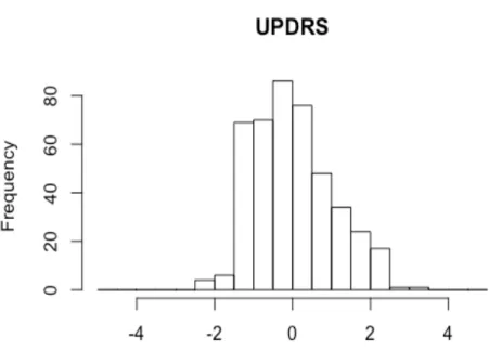
Phenotypic assessment of PD cases recruited has been recently described in a recent paper by our group (57). The Movement Disorder Society revised version of the Unified Parkinson’s Disease Rating Scale Part III (18 items, maximum score 72; hereafter called UPDRS) (48) was used to assess clinical motor symptoms. These included language, facial expressions, tremor, rigidity, agility in movements, stability, gait and bradykinesia. Cognitive abilities were tested through an Italian validated version of the Montreal Cognitive Assessment (MoCA) (12). Cognitive domains assessed include short-term memory (5 points); visuospatial abilities via clock drawing (3 points), and a cube copy task (1 point); executive functioning via an adaptation of Trail Making Test Part B (1 point), phonemic fluency (1 point), and verbal abstraction (2 points); attention, concentration, and working memory via target detection (1 point), serial subtraction (2 points), digits forward and backward (1 point each); language via confrontation naming with low-familiarity animals (3 points), and repetition of complex sentences (2 points); and orientation to time and place (6 points). The total score was given by the sum of these domains, then divided by the maximum score obtainable (30). If one or more domains could not be tested (e.g. visuospatial tasks, due to unavailability of optical devices), the corresponding score was subtracted from the maximum total score. Non motor symptoms were assessed through an Italian validated version of Non Motor Symptoms Scale (NMS) for Parkinson Disease (14). This scale tests 9 items, including cardiovascular domain, sleep/fatigue, mood/cognition, perceptual problems/hallucinations, attention/memory, gastrointestinal, urinary, sexual function, and ability to taste or smell. For each item, both severity and frequency of symptoms is measured, so that the scale accounts for both aspects. This scale is available in (14) and in Appendix 2. Here, the sleep domain was slightly modified by adding a further question on the occurrence of vivid dreams. This question was treated as all the others, i.e. the severity of impairment was scored from 0 (no symptoms) to 3 (severe impairment), and the frequency of impairment was scored from 0 (less than once a week) to 4 (daily impairment), then the total score of the sub-item was computed as the product of severity by frequency, and added to the scores of the other sub-items. For this reason, and due to the high missing rate of sub-items in the sexual domain, we computed the NMS total score as the sum of all the answered items, divided by the maximum total score obtainable. This produced a continuous score ranging between 0 and 1 (hereafter called NMS). Age-at-onset (AAO) information was also collected at the time of recruitment, since it has been reported as an endophenotype that influence the clinical course of pathology.
29 more powerful tools to investigate its genetic underpinnings, they are considered good PD endophenotypes, and were therefore investigated in this thesis.
Levodopa (L-Dopa) dosage calculations
During the visit, the neurologist verified if the patient manifested LID and registered the therapeutic protocol followed by patients before the control, as well as drug prescriptions for the period to come. All of these informations were recorded at each visit in a proprietary software system (Novamed©), so that they can be rescued at any time for usage in any epidemiological research project involving these patients.
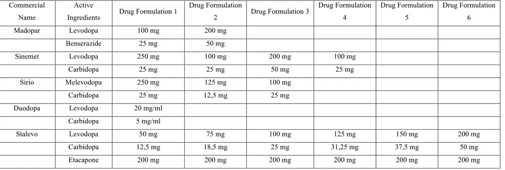
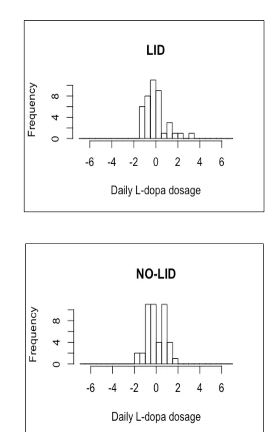
For each patient, the daily L-Dopa dose was calculated by summing the total quantity contained in all drug formulations which were taken during the day. Table 3.2 reported all the drug formulations used by PD patients of the Neuromed cohort. Only L-Dopa dosages were summed to obtain the total amount of active ingredient taken during the day.
Daily Levodopa dosage was computed as follows: !"#$%$&' )* %+" = )*- ∗
/
-0/
1 2$3)4 $2 %35*
Where forms of drug indicate either tables or cassettes of the prescribed drugs and mg indicate the amount of L-Dopa contained in each form.
30
Table 3.2: Pharmaceutical forms of L-Dopa in the PD Neuromed cohort.
Commercial Name
Active
Ingredients Drug Formulation 1
Drug Formulation 2 Drug Formulation 3 Drug Formulation 4 Drug Formulation 5 Drug Formulation 6 Madopar Levodopa 100 mg 200 mg Benserazide 25 mg 50 mg Sinemet Levodopa 250 mg 100 mg 200 mg 100 mg Carbidopa 25 mg 25 mg 50 mg 25 mg Sirio Melevodopa 250 mg 125 mg 100 mg Carbidopa 25 mg 12,5 mg 25 mg Duodopa Levodopa 20 mg/ml Carbidopa 5 mg/ml Stalevo Levodopa 50 mg 75 mg 100 mg 125 mg 150 mg 200 mg Carbidopa 12,5 mg 18,5 mg 25 mg 31,25 mg 37,5 mg 50 mg Etacapone 200 mg 200 mg 200 mg 200 mg 200 mg 200 mg
31
Genotyping
DNA extraction
Genomic DNA was isolated from peripheral blood lymphocytes by Blood and Cell Culture DNA Midi Kit (QIAGEN, Hilden, Germany) according to manufacturer protocol, which included the following steps:
1. Prepare blood samples using PBS, adjust volume to 10 ml.
2. Equilibrate a QIAGEN Genomic-tip 500/G with10 ml of Buffer QBT, and allow the QIAGEN Genomic-tip to empty by gravity flow.
3. Vortex the sample for 10 s at maximum speed and apply it to the equilibrated QIAGEN Genomic-tip. Allow it to enter the resin by gravity flow.
4. Wash the QIAGEN Genomic-tip with 2 x 15 ml of Buffer QC. 5. Elute the genomic DNA with 1 x 15 ml of Buffer QF.
6. Add 10.5 ml (0.7 volumes) room-temperature (15–25°C) isopropanol to the eluted DNA. Precipitate the DNA and resuspend in 0.1–2 ml of a suitable buffer (e.g., TE buffer, pH 8.0, or 10 mM Tris·Cl, pH 8.5). Precipitate the DNA by inverting the tube 10 to 20 times, and by centrifuging immediately at >5000 x g for at least 15 min at 4°C. Carefully remove the supernatant. Wash the centrifuged DNA pellet with 4 ml of cold 70% ethanol. Vortex briefly and centrifuge at >5000 x g for 10 min at 4°C. Carefully remove the supernatant without disturbing the pellet. Air-dry for 5–10 min, and resuspend the DNA in buffer. 7. Dissolve the DNA overnight on a shaker or at 55°C for 1–2 h. Resuspend the DNA pellet
by rinsing the walls to recover the DNA. Pipette the DNA up and down to promote resuspension should be avoided.
Genotyping of SNCA variant rs356219
The SNP rs356219 (hg19 coordinates chr4:90637601; A/G; allelic frequencies ̴ 49/51%) – lying in the 3’ untranslated region (3’UTR) of the SNCA gene (4q22.1) and previously associated with its circulating levels of expression (75–78,86–89)– was genotyped using TaqMan® custom assays (Bio-Rad, USA), according to the manufacturer’s protocol, and analysed in a Bio-Rad® CFX96TM Real Time PCR detection system. About 10–50 ng of DNA were amplified with 5 µL of 2X TaqMan Universal PCR master mix, 0.5 µL of 40X primer and TaqMan probe dye mix. Cycling
32 conditions were 3 min at 95 °C, followed by 40 cycles of 15 s at 95 °C and 30 s at 60 °C. Genotyping was performed on 470 PD cases for which DNA samples were available at the time of genetic analyses. Along with patients, 518 controls were genotyped for the purpose of case-control association analyses, which included:
• 122 non-consanguineous family members (mean (SD) age 62.9 (9.1) years; 44 males) with no neurological signs or symptoms of PD at the time of recruitment.
• 338 unscreened controls (pseudo-controls) belonging to the general Italian population, collected at the Institute of Genetics and Biophysics of the National Research Council in Naples for the purpose of other genetic studies (122 males; age information not available); • 58 neurological controls selected from the Moli-sani study – a large population-based cohort study of citizens from the Molise-region (192) - which showed no signs/symptoms, nor took any specific drug for neurodegenerative disorders (mean (SD) age 77 (5.4) years; 13 men).
We performed a general quality control (QC) of genotyped samples, in PLINK v1.9 (193). The SNP analysed showed a very good call rate (>98%, 17 samples with missing genotype) and was in Hardy Weinberg Equilibrium (HWE, p=0.62), suggesting the good quality of genotyping.
Genotyping of SNCA D4S3481 variant
The SNCA microsatellite D4S3481 (hereafter called Rep1) was analysed in the 469 PD patients of the Neuromed cohort, as well as in 518 general population controls (see above), as described in Maraganore et al, 2006 and in the following studies. Briefly, the region was amplified through Polymerase Chain Reaction (PCR) from genomic DNA, using the following primer pairs: Fam5′-CCTGGCATATTTGATTGCAA-3′ and 5′-GACTGGCCCAAGATTAACCA-3′. PCR reactions (25 µl final volume) containing 2 mmol/L MgCl2, 0.5 mol/L of each primer, 200 mol/L dNTPs, 1 unit of Taq polymerase (Life Technologies) and approximately 20 ng of genomic DNA. Thermal cycling was performed with an initial denaturation of 180 seconds at 94°C, followed by 35 cycles of 30 seconds at 94°C, 30 seconds at melting temperature (MT), 30 seconds at 72°C, and a terminal extension of 10 min at 72°C. PCR products were then diluted 1:10 and resolved by capillary electrophoresis on an ABI-3130XL DNA Analyzer (Applied Biosystem, Foster City, CA, USA), using GeenScan-500 ROX (Applied Biosystem) as molecular weight marker. Allelic sizes were assessed using the GeneMapper® Software Version 4.0 SNPlex™ (Applied Biosystem, Foster City, CA, USA). This method allows to determine the length of dinucleotide repeats at the