http://journals.tubitak.gov.tr/veterinary/ © TÜBİTAK
doi:10.3906/vet-1210-47
Carbon dioxide laser-assisted staphylectomy in brachycephalic and nonbrachycephalic dogs
Adolfo Maria TAMBELLA*, Cecilia VULLO, Fabrizio DINI, Angela PALUMBO PICCIONELLO, Anna Rita ATTILI
School of Veterinary Medical Sciences, University of Camerino, Matelica, Italy
1. Introduction
The elongated soft palate (ESP) is the most common upper airway anomaly in brachycephalic dogs. However, it can occasionally be revealed in mesocephalic and dolichocephalic breeds. The soft palate is considered pathologic when it extends for more than 1–3 mm beyond the tip of the epiglottis, interfering with the normal patency of the airways during the breathing phases (1–4). ESP may also be accompanied by other primary anatomical abnormalities, including stenotic nares and hypoplastic trachea (4–6). Secondary problems can include everted laryngeal saccules and laryngeal collapse (5,7–9). Less common sequelae may be gastrointestinal disease, hiatal hernia, tracheal collapse, and epiglottic entrapment (4,10,11). The upper respiratory obstructive condition is usually increased during exercise or excitement by the consequent edema and inflammation. Clinical presentation may usually be progressive and can include snoring, coughing, respiratory wheezing/stridor, inspiratory dyspnea, reduced exercise tolerance, and, in more severe cases, cyanosis, collapse, syncope, and death (3,4,12,13).
Nonbrachycephalic dogs may present one or, less commonly, more components of airway obstruction. Despite clinical signs of airway obstruction and turbulent airflow, these dogs should have a low incidence of secondary
laryngeal collapse (8). An ESP may be an important cause of reduced exercise tolerance in nonbrachycephalic breeds. Nevertheless, the scientific literature considering this pathology in nonbrachycephalic dogs is undoubtedly far from completion.
Conservative treatments are merely palliative (3,4). Dogs with clinical signs of airways obstruction secondary to ESP can require surgery to relieve the obstructive signs (4,14). Surgical resection of the redundant portion of the soft palate (staphylectomy) is the preferred procedure (12,15), although effective palate shortening using folded flap palatoplasty has also been described (16). Traditional staphylectomy is performed by sharply excising a portion of the soft palate and apposing the oral and nasal mucosa with a monofilament absorbable suture, the “cut and oversew” technique, as described by Bright and Wheaton (17) and Davidson et al. (14). Monopolar electrocautery (18), bipolar sealing devices (9,19), and harmonic scalpels (20) have also been used to perform the resection. Clark and Sinibaldi demonstrated that carbon dioxide (CO2) laser is a suitable tool for soft palate resection (21). CO2 laser-assisted uvulopalatoplasty is also considered to be an effective treatment for nonapneic snoring in human medicine (22). Davidson et al. documented similar clinical outcomes between the CO2 laser and the traditional method, but found significantly shorter surgical times with the CO2
Abstract: Elongated soft palate is a common upper airway anomaly in brachycephalic dogs. The scientific literature considering this
pathology in nonbrachycephalic dogs is limited. The aim of this study was to estimate the importance of the elongated soft palate in nonbrachycephalic dogs (n = 10), carrying out, at the same time, a comparison with selected brachycephalic dogs (n = 19), on the effect of carbon dioxide laser staphylectomy. The selected population had only elongated soft palates and no other disorders. The surgery was rapid and uncomplicated; no suture material was used. The comprehensive recovery was significant (P < 0.0001): 68.4% of brachycephalic dogs and 100% of nonbrachycephalic dogs had complete recovery. The incomplete recovery in older brachycephalic dogs matched a more advanced pathologic phase. This study suggests that, in the case of high respiratory tract alteration and poor exercise tolerance, it is imperative to consider in differential diagnosis the elongated soft palate in nonbrachycephalic breeds, too; their clinical signs are less severe and frequently underestimated. The surgical treatment has a favorable prognosis when the only alteration is elongated soft palate. The carbon dioxide laser has been confirmed as an useful option for staphylectomy in dogs.
Key words: Elongated soft palate, laser surgery, brachycephalic, nonbrachycephalic, dog
Received: 25.10.2012 Accepted: 10.04.2013 Published Online: 13.11.2013 Printed: 06.12.2013
laser (14). In a recent study comparing CO2 laser, diode laser, and monopolar electrocautery techniques, Dunié-Mérigot et al. considered the CO2 laser as the most suitable for the soft palate resection (18). According to the existing literature, during CO2 laser emission, the beam instantly vaporizes the molecules of water in the target cells, causing limited thermal damage in the tissues in and around the point of application (0.1 mm) (23–25). Complications associated with staphylectomy are uncommon. They can include pharyngeal edema and inflammation during the postoperative period, nasal regurgitation as result of excessive soft palate resection, and aspiration pneumonia (3,5,17).
The aim of the present study was to estimate the clinical significance of ESP in nonbrachycephalic dogs. The hypothesis under study is that ESP could be a cause of airway alteration and reduced exercise tolerance in nonbrachycephalic breeds. At the same time, the authors wanted to assess the feasibility and the effectiveness of the CO2 laser staphylectomy technique in the treatment of ESP and to compare the effect of surgery between brachycephalic (B) dogs and nonbrachycephalic (NB) dogs.
2. Materials and methods 2.1. Inclusion criteria
In the study, only those dogs showing respiratory clinical signs attributable to ESP were considered. The subjects with other abnormalities (e.g., stenotic nares or hypoplastic trachea) or with clear secondary alterations (e.g., everted laryngeal saccules or laryngeal collapse) and dogs that had previous upper airway surgery were categorically excluded, thereby eliminating any variable that would potentially affect the results.
2.2. Preoperative assessment
Each dog had a complete physical examination; prior to anesthesia, a complete blood cell count, serum biochemical analysis, and an electrocardiographical evaluation were performed. Direct and endoscopic evaluations of the nasal, oral, pharyngeal, and laryngeal cavities, as well
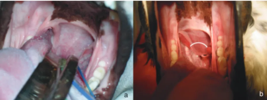
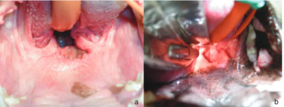
as radiographic assessments of the neck and the thorax, were performed under general anesthesia with the aim of excluding the most common pathologies in the respiratory airways. The detection of a soft palate extension beyond its articulation with the epiglottis for more than 5 mm (Figure 1a) allowed for the diagnosis of ESP and led to the indication for corrective surgery. To carry out an unbiased study, the surgeon was blinded to the observer that undertook the clinical assessment before surgery (T0) and during the follow-up.
2.3. Surgical procedure and laser equipment
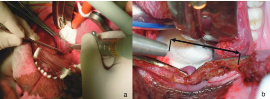
The anesthesia procedure was performed by a premedication through intramuscular administration of medetomidine (10 µg/kg) and tramadol (2 mg/kg), followed by induction with propofol and prompt tracheal catheterization; therefore, maintenance with gaseous mixture of isoflurane in oxygen was provided. Every patient was placed in dorsal recumbency with the mouth open and the tongue pulled out and was shifted by a shovel spatula and/or by a traditional laryngoscope to guarantee suitable exposure of the oropharynx. The caudal border of the soft palate was grasped with thumb forceps and pulled rostrally. With the tongue in a relaxed position, the surgeon visually estimated the amount of tissues to be removed, as described by Berger and Eeg (26). The goal was to adapt the soft palate to the shape of the epiglottis and just barely make contact with it. The suitable landmark for resection was identified as beginning from the point at which the extremity of the epiglottis grazed the soft palate. It lies approximately at the level of an imaginary curved line between the caudal poles of the tonsils (Figure 1b). To delimit the line of amputation, 2 curved hemostatic forceps were applied across the soft palate at the level of the caudal edge of the tonsillar crypts. They were then kept in place during the phase of resection (Figure 2). One or more wet gauzes were placed behind the soft palate and around the endotracheal tube to absorb the laser energy, prevent inadvertent lasing of tissues in the back of the oropharynx, and avoid combustion, as recommended by Davidson et al. (14) and Berger and Eeg (26).
Figure 1. Direct evaluation of soft palate (a) with the identification of the line of
To perform the staphylectomy, a laser technique was used. It differed slightly from the technique described for the first time by Clark and Sinibaldi (21). Particularly, a carbon dioxide laser surgical system was used (Shanghai Wonderful Opto-Electrics Tech. Co., Ltd; model ML015-CA). It is a microprocessor-controlled instrument based on a sealed-off circuit providing up to 15 W of output power on the tissues. The wavelength is 10,600 nm. The active medium is a mixture of CO2, helium, and nitrogen. The focal spot diameter is 0.4 mm at lens focal distance of 100 mm.
A pilot beam represented by a red diode laser (wavelength: 635 nm) allows the directing of the CO2 laser beam at preestablished points. The CO2 laser beam and diode laser beam are combined and guided coaxially into the 7-joint articulated arm delivery system. A footswitch is used to control laser output.
To perform the soft palate resection, the instrument was employed at the optimal focal area of the beam, in super pulse wave (SP) mode, with an intensity of 9 ± 2 W. The SP mode emission provides bursts of laser energy at high peaks for very brief periods of time, up to 0.02 s, with long periods without emission. The surgeon performed an arch-shaped incision beginning at the lateral margin of the soft palate and vaporizing the tissue in a path heading to the controlateral side (Figure 3). The laser beam was therefore
slowly passed across the nasopharyngeal, muscular, and oropharyngeal layers of the soft palate until resection was completed.
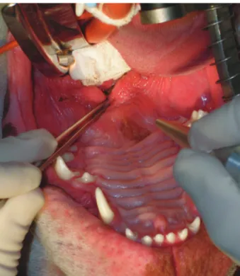
After the resection of the proper portion of the soft palate, keeping on the boundary forceps, the laser was set to a lower power (3–4 W), in continuous wave (CW) mode and with a defocalized spot. In CW mode, the laser energy is continually released through an open shutter during activation. The laser beam defocusing was merely performed by increasing the distance between the laser tip and the target tissue: the goal is to decrease the power density by increasing the spot size. After the desired tissue was excised, the boundary forceps were removed and the tissue edges were examined for carbonization, hemorrhage, adhesion, and proper length. A further defocusing was performed when necessary (Figure 4). The duration of each surgery was recorded.
2.4. Postoperative care and follow-up
Following surgery, water was withheld for 12 h and food was withheld for 24 h; dogs were then fed with soft canned food. In every subject, a single dose of dexamethasone sodium phosphate (1 mg/kg) was administered on the day of the surgery to control postoperative swelling. The dogs were hospitalized under strict observation for at least 24 h and subsequently underwent a clinical follow-up after 7 days (T1), 1 month (T2), and 6 months (T3) from surgery.
Figure 2. Application of the boundary forceps: B-dog (a) and NB-dog (b) in dorsal
recumbency.
Six months after surgery, in every dog, an endoscopy of the airways was performed to evaluate the condition and the possible alterations.
2.5. Clinical score assessment
To give details about the severity of symptomatology and to render easy the comparison with the clinical outcome, a clinical score was assessed for each dog at T0, T1, T2, and T3.
The scoring method for clinical assessment was based on a 6-point scale:
●○○○○= Snoring (frequent or manifested only after intense physical activity), reduction of exercise tolerance and physical performances;
●●○○○= Recurrent snoring with light severity of dyspnea showed during exercise or under particular environmental conditions, excitement, stress;
●●●○○= Snoring, episodes of light severity of dyspnea at rest;
●●●●○= Snoring, permanence of medium severity of dyspnea at rest;
●●●●●= Snoring, serious dyspnea, hypoxia, cyanosis, syncope;
○○○○○= Healthiness, complete recovery.
2.6. Statistical analysis
In relation to the breed, the population was divided into 2 groups: the brachycephalic group (group B) and nonbrachycephalic group (group NB). Clinical scoring data were analyzed at each time point and were compared by breed, age (≤12 months versus >12 months), and sex.
All data were analyzed using statistical software (STATA Software, Version 5, College Station, TX, USA). Statistical significance for all frequency comparisons of qualitative variables was evaluated by Fisher’s exact test. Ordinal variables were summarized using median, mean, and standard deviation and were compared using the Wilcoxon rank sum test. P-values below 0.05 were considered statistically significant.
3. Results
A total of 51 dogs had surgery to correct ESPs at the Veterinary Teaching Hospital of the University of Camerino, Italy, from January 2010 to December 2011. Twenty-two B-dogs were preventively excluded from the study because of the above-mentioned criteria; on the same basis, no NB-dogs were excluded. The present study involved 29 selected dogs showing a respiratory alteration attributable to the stenosis of upper airways caused by the elongation of the soft palate. These patients were aged between 3 and 24 months. Nineteen of them (65.5%) belonged to brachycephalic breeds and 10 (34.5%) belonged to nonbrachycephalic breeds. The population data are shown in Table 1.
Physical examination findings were normal in each dog, and results of biochemical analysis prior to anesthesia were within reference ranges. No anesthetic complications occurred. Patients treated with laser did not manifest remarkable signs of swelling postoperatively; intraoperatory bleeding was totally avoided, resulting in better surgeon’s visualization and reduction of the surgery time (mean: 5 min and 20 s; range: 4 min and 15 s to 7 min and 10 s). It was not necessary to perform, in any case, a tracheotomy. The suture was not necessary, nor did any foreign body inflammatory reaction occur during the postoperative time. Following staphylectomy, direct examination confirmed that the rim of the soft palate was always just above the epiglottis (Figure 5).
All patients showed clear improvements of clinical condition after surgery, with an evident decrease in both respiratory effort and inspiratory stridor. The comprehensive differential clinical score (DCS) before and after surgery (T0–T1) was, on average, 2.17 ± 0.89 levels, with a median 2, and there were improvements of 4 levels in 6.90% of dogs, 3 levels in 27.59%, 2 levels in 41.38%, and 1 level in 24.14%. The comprehensive DCS T0–T2 (mean: 2.07 ± 0.96; median: 2) and T0–T3 (mean: 2.13 ± 0.95; median: 2) did not differ significantly from T0–T1 (P > 0.05).
Twenty-three subjects (79.31%, n = 29) no longer manifested remarkable clinical signs (P < 0.0001), while, in the remaining population (6 dogs out of 29), it was possible to point out a lowering DCS of 3 levels in 6.90%, 2 levels in 10.34%, and 1 level in 3.45% of dogs. The recovery
was complete and durable in all the NB-dogs (100%, n = 10) (P < 0.0001), with a mean DCS of 1.6 ± 0.69 levels and median 1.5 (the same for T0–T1, T0–T2, and T0–T3). The remission was complete for 13 B-dogs (68.4% of the B-population, n = 19), with a statistically significant difference for recovery (P < 0.0001). Only 6 B-patients (31.6% of the B-dogs, 20.7% of the whole population)
showed merely partial improvements. The mean DCS T0– T1 in B-dogs was 2.47 ± 0.84 levels, with a median of 2. In B-dogs, the DCS during the remaining follow-up (T0–T2: mean of 2.32 ± 1.00, median of 2; T0–T3: mean of 2.42 ± 0.96, median of 2) did not differ significantly from T0–T1 (P > 0.05). A significant difference between B-dogs and NB-dogs was identified for remission of symptoms (P < 0.05),
Table 1. Population: Groups B and NB.
Group Case no. Breed Sex Age (months)
Group B 1 Pug F 3 2 Pug M 5 3 Pug M 15 4 Pug M 8 5 Boxer F 9 6 Boxer M 12 7 Boxer F 5 8 Rottweiler F 8 9 Rottweiler F 6 10 Rottweiler M 10 11 French bulldog M 4 12 French bulldog M 18 13 English bulldog F 5 14 English bulldog M 6 15 American bulldog M 24 16 Dogue de Bordeaux M 12 17 Bullmastiff M 20 18 Pekinese M 9
19 Cavalier King Charles spaniel F 10 Group NB 1 English setter M 18 2 English setter F 14 3 Brittany spaniel M 12 4 Pointer F 24 5 Segugio Italiano F 9 6 Labrador retriever M 11
7 German shepherd dog M 18
8 Mix-breed F 20
9 Mix-breed M 12
10 Mix-breed F 10
Group B: brachycephalic dogs. F: female [n = 7 (36.84%)]; M: male [n = 12 (63.16%)]. Mean age ± SD: 9.95 ± 5.75 months [≤12 months: n = 15 (78.95%); >12 months: n = 4 (21.05%)].
Group NB: nonbrachycephalic dogs. F: female [n = 5 (50%)]; M: male [n = 5 (50%)]. Mean age ± SD: 14.8 ± 4.94 months [≤12 months: n = 5 (50%); >12 months: n = 5 (50%)].
with a higher recovery rate in group NB. On the contrary, considering the lowering of the clinical score, there was a better improvement in group B (R = 73; P < 0.01). None of them manifested any complications during and after surgery. Results of surgical treatment with clinical scoring recorded at admission (T0) and after surgery (T1, T2, T3) are summarized in Table 2.
In dogs 12 months old or less, a complete recovery was observed in 90% with a mean DCS of 2.40 ± 0.88 and
median of 2; in dogs older than 12 months there was a recovery of 55.56% with a mean DCS of 1.67 ± 0.71 and a median of 2. Therefore, considering the whole population, the staphylectomy was more effective for recovery in younger dogs (P < 0.05). Considering group B, it is possible to point out a more significant difference in effectiveness of surgery between dogs aged ≤12 months (86.67% recovery) and dogs aged >12 months (0% recovery) (P < 0.001). The mean DCS in B-dogs of ≤12 months was 2.60 ± 0.83 with a
Figure 5. Examination of the soft palate after surgery: B-dog (a) and NB-dog (b).
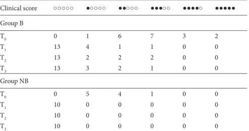
Table 2. Clinical score assessment before and after surgery in group B and in group NB.
Clinical score ○○○○○ ●○○○○ ●●○○○ ●●●○○ ●●●●○ ●●●●● Group B T0 0 1 6 7 3 2 T1 13 4 1 1 0 0 T2 13 2 2 2 0 0 T3 13 3 2 1 0 0 Group NB T0 0 5 4 1 0 0 T1 10 0 0 0 0 0 T2 10 0 0 0 0 0 T3 10 0 0 0 0 0
Number of dogs included in each point scale and classified by group: before surgery (T0), 1 week after surgery (T1), 1 month after surgery (T2), and 6 months after surgery (T3). Clinical score legends are given in Section 2.5.
Group B (brachycephalic dogs, n = 19): mean score = 2.95 ± 1.08, median = 3 (T0); mean score = 0.47 ± 0.84, median = 0 (T1); mean score = 0.63 ± 1.06, median = 0 (T2); mean score = 0.53 ± 0.90, median = 0 (T3).
Group NB (nonbrachycephalic dogs, n = 10): mean score before surgery (T0) = 1.60 ± 0.69, median = 1.5; mean score after surgery (T1, T2, T3) = 0 ± 0, median = 0.
median of 3; it was 2.00 ± 0.82 with a median of 2 in B-dogs of >12 months. The mean DCS in NB-dogs of ≤12 months was 1.80 ± 0.84, median 2; it was 1.40 ± 0.55, median 1, in NB-dogs of >12 months. There was no significant difference between the 2 classes of age for effectiveness of surgery considering the lowering of the clinical score in the whole population (R = 119; P > 0.05), in group NB (R = 24; P > 0.05), or in group B (R = 38; P > 0.05).
Considering the distribution of population per sex, it is possible to point out a recovery of 100% in female dogs and 64.71% in male dogs, with a significant difference for recovery between males and females (P < 0.05). In group B, there was a significant difference for recovery (P < 0.05) with complete clinical remission in all the females and in half of the group of males. However, the comprehensive mean clinical score assessed before surgery was 1.83 ± 0.83, median 2, in females; it was 2.94 ± 1.14, median 3, in males. Additionally, 29.41% of male dogs were assessed at scores of 4 or 5, while no female dogs were assessed at scores of 4 or 5. Moreover, in group NB the mean DCS was 1.2 ± 0.45 with a median of 1 in females, and it was 2.0 ± 0.71 with a median of 2 in males. In group B, the mean DCS was 2.29 ± 0.76, median 2, in females, and 2.58 ± 0.90, median 2.5, in males. There was no significant difference between the 2 classes of sex for the improvement of the clinical score in the whole population (R = 173; P > 0.05), in group NB (R = 23; P > 0.05), or in group B (R = 63; P > 0.05).
Six months after surgery, endoscopic evaluation allowed the highlighting, in all the subjects, of the good condition of the site of operation with complete healing of the surgical lesion and no findings of further problems in the airways (Figure 6).
4. Discussion
The overall outcome of laser surgery in the present study was good. Dogs with moderate to severe ESP benefited from surgical correction. Their clinical status was improved following surgery. Life-threatening signs ceased or were greatly reduced in all cases. In all cases, postoperative signs were considered less severe than preoperative signs. The
number of animals with dyspnea decreased substantially after surgery and no animals were reported with cyanosis episodes or pneumonia.
The occurrence of a significant difference in the mean DCS before and after surgery may be considered a good index of effectiveness of the surgery, both in B- and NB-dogs. On the other hand, the absence of significant difference in the mean DCS during postoperative follow-up demonstrates that the clinical improvement obtained with surgery is durable over time.
The surgery was more effective in dogs aged 1 year or younger in comparison to older dogs, and especially in group B. Snoring and other symptoms did not resolve completely in every B-dog. It seems likely that the whole upper respiratory tract in brachycephalic animals is maldeveloped and turbulent air flow may well still occur despite resolution of some clinically apparent primary abnormalities, as reported in other studies (8). We have to consider that B-dogs older than 1 year showed a more serious clinical condition and more advanced pathological phases at admission. The more significant improvement in DCS showed by group B, in seeming contrast with the higher recovery rate in group NB, is the mathematical demonstration of this. This fact allows the confirming and emphasizing of the importance of precocity in diagnosis and therapy for this pathology (4,27).
At first appraisal, it appeared that female dogs experienced a better outcome in comparison with male dogs. However, the comparison of average DCS between the 2 classes of sex was not significant in the whole population, neither in group B nor in group NB. Therefore, we conclude that the difference in outcome was apparent: the main reason was based on the more serious clinical condition showed by male dogs at admission, as we can observe by the clinical scoring assessed before surgery.
The results of this study support the hypothesis that ESP is not limited to the B-breeds, where it is often considered as a part of brachycephalic airway obstruction syndrome. ESP, more often as a single pathology, can be also found in NB-breeds, even if they usually show less serious clinical signs. Actually, the clinical importance of ESP in NB-dogs is still underestimated. The excellent results obtained in
this study in NB-dogs leads to the necessity of paying close attention to the importance of including ESP in the differential diagnosis in these breeds, too. ESP must be sought not only in cases of frank alteration in the higher respiratory tract, but also and especially in dogs with poor exercise tolerance with or without snoring. This may be considered a marked change from the commonly accepted beliefs about ESP.
The laser surgical technique allowed performance of the staphylectomies and had satisfactory outcomes both in the B-dogs and in the NB-dogs, without complications during or after surgery. Postoperative swelling was almost entirely prevented. Nevertheless, it commonly happens when the ablation is performed by electrocautery, as was described by Hedlund (4). In comparison to the traditional surgical technique of “cut and oversew”, intraoperative bleeding was totally avoided, with reduction of the surgery time by more than half. The times required for the whole soft palate resection using the CO2 laser in this study were similar to or lower than times reported by other authors (9,14,18). Dorsal recumbency, in comparison to sternal, allowed a good visualization of the deep portion of the oral cavity, a more comfortable position of the surgeon, and a better preservation of the endotracheal tube from the laser beam. Using the laser source in SP mode allowed the surgeon to obtain clear-cut palatal incisions without remarkable thermal effects on the surrounding tissues. However, it had a negligibly slower action in comparison to the CW mode. During resection it is very important to remain cautious to direct the beam perpendicularly to the
target tissue to achieve the greatest laser surgical precision. Tangential beams may have a less efficient vaporization and could cause thermal necrosis, postoperative discomfort, and bleeding (26). The laser beam at reduced power, in CW mode, and with defocalized spot, applied at the edge of resection and together with the action of the boundary forceps, contributed to achieving total cohesion among the 2 mucous layers of the soft palate. The defocusing of the beam on the ablation surface gave rise to the collagen contraction; consequently, the introflection of the dorsal and ventral mucous layers allowed the surgeon to avoid the suture of the soft palate. During the resection, the forceps application facilitated the rostral bending of the soft palate (around 90°); moreover, after the ablation phase, it avoided the risk of skidding of the nose-pharyngeal mucosal layer from the oral-pharyngeal one; the forceps, at the same time, contributed to a temporary hemostasis and, consequently, a surgery without bleeding.
The present study suggests that ESP can be diagnosed and surgically treated in NB-dogs, in spite of the lower frequency and the less severe clinical manifestations than in B-dogs. Early surgical correction of ESP in dogs is associated with a favorable prognosis. Performing staphylectomy, the CO2 laser technique proved to be very practical and extremely effective in brachycephalic as well as in nonbrachycephalic breeds, contributing very well to the achievement of a satisfactory clinical outcome. These bases allow for the confirmation of the CO2 laser as an effective alternative to the traditional methods employed for surgery of the soft palate in dogs.
References
1. Bright RM. Surgery of elongated soft palate (staphylectomy). In: Bojrab MJ, Ellison GW, Slocum B. Current Techniques in Small Animal Surgery. 4th ed. Baltimore, MD, USA: Williams and Wilkins; 1998. pp. 143–144.
2. Bjorling D, McAnulty J, Swainson S. Surgically treatable upper respiratory disorders. Vet Clin North Am Small Anim Pract 2000; 30: 1227–1251.
3. Riecks TW, Birchard SJ, Stephens JA. Surgical correction of brachycephalic syndrome in dogs: 62 cases (1991–2004). J Am Vet Med Assoc 2007; 230: 1324–1328.
4. Hedlund CS. Chirurgia delle prime vie respiratorie. In: Fossum TW, editor. Chirurgia dei piccoli animali. 3rd ed. Milan, Italy: Elsevier-Masson Edizioni; 2008. pp. 834–891 (book chapter in Italian).
5. Wykes PM. Brachycephalic airway obstructive syndrome. Probl Vet Med 1991; 3: 188–197.
6. Coyne BE, Fingland RB. Hypoplasia of the trachea in dogs: 103 cases (1974-1990). J Am Vet Med Assoc 1992; 201: 768–772. 7. Amis TC, O’Neill N, Wheatley JR, van der Touw T, di Somma
E, Brancatisano A. Soft palate muscle responses to negative upper airway pressure. J Appl Physiol 1999; 86: 523–530.
8. Torrez CV, Hunt GB. Results of surgical correction of abnormalities associated with brachycephalic airway obstruction syndrome in dogs in Australia. J Small Anim Pract 2006; 47: 150–154.
9. Brdecka D, Rawlings C, Howerth E, Cornell K, Stiffler K. A histopathological comparison of two techniques for soft palate resection in normal dogs. J Am Anim Hosp Assoc 2007; 43: 39–44.
10. Hardie EM, Ramirez O 3rd, Clary EM, Kornegay JN, Correa MT, Feimster RA, Robertson ER. Abnormalities of the thoracic bellows: stress fractures of the ribs and hiatal hernia. J Vet Intern Med 1998; 12: 279–287.
11. Poncet CM, Dupré GP, Freiche VG, Bouvy BM. Long-term results of upper respiratory syndrome surgery and gastrointestinal tract medical treatment in 51 brachycephalic dogs. J Small Anim Pract 2006; 47: 137–142.
12. Knecht CD. Upper airway obstruction in brachycephalic dogs. Compend Contin Educ Vet 1979; 1: 25–30.
13. Hendricks JC. Brachycephalic airway syndrome. Vet Clin North Am Small Anim Pract 1992; 22: 1145–1153.
14. Davidson EB, Davis MS, Campbell GA, Williamson KK, Payton ME, Healey TS, Bartels KE. Evaluation of carbon dioxide laser and conventional incisional techniques for resection of soft palates in brachycephalic dogs. J Am Vet Med Assoc 2001; 219: 776–781.
15. Harvey CE. Upper airway obstruction surgery II: soft palate resection in brachycephalic dogs. J Am Anim Hosp Assoc 1982; 18: 538–544.
16. Findji L, Dupré G. Folded flap palatoplasty for treatment of elongated soft palates in 55 dogs. Eur J Companion Anim Pract 2009; 19: 125–132.
17. Bright RM, Wheaton LG. A modified surgical technique for elongated soft palate in dogs. J Am Anim Hosp Assoc 1983; 19: 288–292.
18. Dunié-Mérigot A, Bouvy B, Poncet C. Comparative use of CO2 laser, diode laser and monopolar electrocautery for resection of the soft palate in dogs with brachycephalic airway obstructive syndrome. Vet Rec 2010; 167: 700–704.
19. Brdecka D, Rawlings C, Perry AC, Anderson JR. Use of an electrothermal, feedback-controlled, bipolar sealing device for resection of the elongated portion of the soft palate in dogs with obstructive upper airway disease. J Am Vet Med Assoc 2008; 233: 1265–1269.
20. Michelsen J. Use of the harmonic scalpel for soft palate resection in dogs: a series of three cases. Aust Vet J 2011; 89: 511–514.
21. Clark GN, Sinibaldi KR. Use of a carbon dioxide laser for treatment of elongated soft palate in dogs. J Am Vet Med Assoc 1994; 204: 1779–1781.
22. Lauretano AM, Khosla RK, Richardson G, Matheson J, Weiss JW, Graham C, Fried MP. Efficacy of laser-assisted uvulopalatoplasty. Lasers Surg Med 1997; 21: 109–116. 23. Durante EJ, Kriek NP. Clinical and histological comparison of
tissue damage and healing following incisions with the CO2 -laser and stainless steel surgical blade in dogs. J S Afr Vet Assoc 1993; 64: 116–120.
24. Wilder-Smith P, Arrastia AM, Liaw LH, Berns M. Incision properties and thermal effects of three CO2 lasers in soft tissue.
Oral Surg Oral Med Oral Pathol Oral Radiol Endod 1995; 79: 685–691.
25. Courey MS, Fomin D, Smith T, Huang S, Sanders D, Reinisch L. Histologic and physiologic effects of electrocautery, CO2
laser, and radiofrequency injury in the porcine soft palate. Laryngoscope 1999; 109: 1316–1319.
26. Berger N, Eeg PH. Veterinary Laser Surgery – A Practical Guide. 1st ed. Ames, IA, USA: Blackwell Publishing Professional; 2006.
27. Pink JJ, Doyle RS, Hughes JM, Tobin E, Bellenger CR. Laryngeal collapse in seven brachycephalic puppies. J Small Anim Pract 2006; 47: 131–135.