Contents lists available atScienceDirect
Journal of Pediatric Surgery Case Reports
journal homepage:www.elsevier.com/locate/epscRecurrent intussusception caused by submucosal, heterotopic gastric
mucosa in the terminal ileum
Dacia Di Renzo
a,∗, Riccardo Rizzo
a, Mario Fusillo
a, Laura Travascio
b, Antonello Persico
a,
Pierluigi Lelli Chiesa
aaPediatric Surgery Unit,“G. d’Annunzio” University of Chieti, “Spirito Santo” Hospital of Pescara, Italy bNuclear Medicine Unit,“Spirito Santo” Hospital of Pescara, Italy
A R T I C L E I N F O
Keywords:
Heterotopic gastric mucosa Intussusception
99mTc pertechnetate scan
A B S T R A C T
Isolated, heterotopic gastric mucosa (HGM) can be a rare, pathologic leading point for intestinal intussusception in pediatric age. Affected children often experience recurrent episodes of intussusception, sometimes with delay in diagnosis and more than one surgery, because of difficulty in clear diagnosis and identification of HGM. Intraoperativefindings of isolated HGM, as reported in literature, are visible or palpable lesions, protruding into the intestinal lumen. We describe the case of a 4 years old boy with recurrent intussusception, caused by a very small islet of ileal, isolated HGM, entirely developing in the submucosal layer and with a normal overlying mucosa. The difficulties in its diagnosis and treatment are described and the role of99mTc pertechnetate scan and ultrasound are discussed.
1. Introduction
Isolated, heterotopic gastric mucosa (HGM) is a focus of mature gastric tissue in a location outside the stomach, not associated with congenital anomalies such as Meckel's diverticulum or intestinal du-plications. Isolated HGM is rarely found in the ileum and even more rarely it acts as pathologic leading point (PLP) for intestinal in-tussusception. As a matter of fact, only few reports of intussusception over isolated HGM are present in literature [1–11]: these cases are characterized by difficulty in preoperative diagnosis and in in-traoperative identification of a clear PLP. Indeed, affected children often experience recurrent episodes of intussusception, sometimes with delay in diagnosis and more than one surgery. In all the previously reported cases the intraoperative findings of isolated HGM include polypoid or nodular masses protruding into the intestinal lumen, ser-osal irregularity or coarse, rugose mucosa, developing over an island of HGM [2,4,5,7,9,11]. These lesions, even if with a certain difficulty, can be manually palpated or visualized on the intestinal mucosa after en-terotomy or during endoscopy [3,4,8]. However definitive diagnosis typically occurs postoperatively, with histopathologic examination of a surgical specimen.
Differently from previous reports, we describe the case of boy with
recurrent intussusception, caused by a very small islet of ileal, isolated HGM, developing only in the submucosal layer and with a normal overlying mucosa. The extreme difficulty in its identification, diagnosis and treatment is discussed.
2. Case report
A 4-years-old boy was transferred from the Pediatric Unit of another hospital for suspected intussusception. The patient had a 48 h history of colicky abdominal pain, associated to somnolence. He presented two previous analogous episodes, when he was 6 months and 2 years old, with spontaneous resolution and without a definite diagnosis.
At admission, his general conditions were poor. On physical ex-amination he had abdominal tenderness, with a palpable mass in the left hypochondrium. We performed an abdominal ultrasound (US) which confirmed ileo-colic intussusception and a tubular image with a small, anechoic cystic area inside was seen into the intussusceptum, posing the suspicion of a PLP such as a Meckel's diverticulum, a polyp or a duplication. As usually performed at our operative unit to treat intussusception [12], an US-guided saline enema was performed twice, but only partial reduction of the intussusception was achieved, up to the right colon. Therefore, the boy was taken to the operating room,
https://doi.org/10.1016/j.epsc.2018.12.017
Received 30 November 2018; Received in revised form 18 December 2018; Accepted 19 December 2018 Abbreviations: HGM, heterotopic gastric mucosa; PLP, pathologic leading point; US, ultrasound
∗Corresponding author. Pediatric Surgery,“G. d’Annunzio” University of Chieti and “Spirito Santo” Hospital of Pescara, Via Fonte Romana n.8, 65124, Pescara, Italy.
E-mail address:[email protected](D. Di Renzo).
Journal of Pediatric Surgery Case Reports 42 (2019) 38–41
Available online 26 December 2018
2213-5766/ © 2018 The Authors. Published by Elsevier Inc. This is an open access article under the CC BY-NC-ND license (http://creativecommons.org/licenses/BY-NC-ND/4.0/).
where a residual ileo-colic intussusception was confirmed and complete manual reduction was achieved. A careful inspection of the ileum showed no evidence of macroscopic PLP and a meticulous palpation did not reveal any mass. Post-operative course was uneventful.
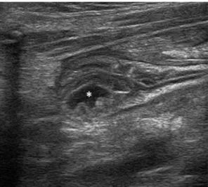
Ten days after hospital discharge the patient presented again for intermittent abdominal pain and alimentary vomit, with spontaneous resolution in few hours. An US showed no actual signs of intussuscep-tion but in the last centimeters of ileum, which presented increased wall thickness, a small round cystic formation or a small amount of anechoic fluid was noted, probably in the ileum wall [Fig. 1], posing again a suspicion of a PLP that could have caused an intussusception. However, an US conducted 24 h later could not show the pathologic image again. Considering this clinical scenario, we suspected HGM, thus decided to perform a 99mTc pertechnetate scan. 99mTc pertechnetate scan showed a focal area of contrast-enhancement in the right iliac fossa, simultaneously with the activity of the gastric mucosa [Fig. 2], sug-gestive for HGM.
An elective laparoscopy was performed the day after, with trans-umbilical externalization of the distal ileum which was interested by numerous adhesions, probably caused by previous recurrent episodes of intussusception. After an accurate lysis of adhesions of the small bowel, meticulous inspection and palpation of the ileum could not confirm for
sure the presence of macroscopic areas of HGM or other abnormalities. An US-guided enterotomy on the last 5 cm of ileum did not disclose any lesion suggestive for duplication or isolated HGM. Based on 99mTc pertechnetate scan and US images, we decided to resect the terminal ileum, preserving the ileo-cecal valve, and an ileo-ileal anastomosis was performed. Post-operative period was held without complications. Histology of the resected specimen showed a very small islet of HGM, 9 mm in diameter, located only in the submucosal layer, covered by a normal ileal mucosa [Fig. 3]. It was located at 2 cm from the distal end of resected ileum.
Clinical and ultrasonographic follow up was uneventful, and the patient kept in good health, with no further episodes of abdominal symptoms. Eight months after surgery, a99mTc pertechnetate scan was performed, negative for residual HGM [Fig. 4].
3. Discussion
Intussusception is the most common cause of acute bowel obstruc-tion in infants and it typically occurs in thefirst 3 years of life, with a peak incidence from 5 to 9 months [13,14]. About 95% of all in-tussusceptions in the typical age group are due to hyperplasia of bowel wall lymphoid tissue in the distal ileum, acting as a lead point, while the incidence of a PLP ranges from 2% to 12%. This rate is reported to increase up to 60% in children over 5 years of age. The recurrence of intussusceptions following either a surgical or nonsurgical reduction enhances the probability of congenital morphological abnormalities. Fig. 1. Ultrasound showing in the terminal ileum a small anechoic image
(as-terisk), probably in the intestinal wall.
Fig. 2.99mTc pertechnetate scan showing a focal area of intense abnormal tracer accumulation in the right iliac fossa (arrow), simultaneously with the gastric mucosa uptake, suggestive for functioning HGM.
Fig. 3. Small nodule of HGM recapitulating the normal gastric, oxyntic mucosal architecture, lying deep in the submucosa; two gland-like structures are present (arrows) (EE, 2.5×); in the box: gland-like architecture in the submucosal HGM nodule and normal intestinal epithelial cells in the overlying mucosa (EE, 4×).
Fig. 4.99mTc pertechnetate scan showing no abnormal tracer accumulation.
D. Di Renzo et al. Journal of Pediatric Surgery Case Reports 42 (2019) 38–41
PLP include Meckel's diverticulum, polyps, intestinal duplications, carcinoid tumors, submucosal hemorrhage resulting from Schönlein-Henoch purpura, lymphomas, intestinal malignant melanoma, foreign bodies, ectopic pancreas or HGM [14].
HGM may be present anywhere in the gastrointestinal tract from the mouth to the anus and can be found also in the airways, umbilicus, urinary bladder, and even in the scrotum. It is commonly found in congenital abnormalities such as Meckel's diverticulum and gastro-intestinal duplications [4]. Apart from congenital anomalies, it is rarely found as isolated HGM in the small intestine distal to the ligament of Treitz [1] and even more rarely it acts as PLP for intussusception. HGM is reported to cause intussusception, gastrointestinal bleeding, per-foration and obstruction [1–11,15–22].
HGM was described by Schmidt as early as 1805 [1]. Poindecker reported thefirst case of isolated HGM in 1912 [1]. Since then, less than 30 pediatric cases of HGM, located in the small intestine beyond the ligament of Treitz and not associated with morphological abnormal-ities, have been reported. Affected patients were mostly males, with a wide range of age (few months to 17 years old) and the most frequent presentation was intussusception, followed by perforation and bleeding. Most of these patients who underwent surgery for in-tussusceptions were older than the usual age of inin-tussusceptions pre-sentation [8].
The majority of the HGM cases are intraoperative discoveries in patients who had undergone open reductions at least twice, for re-current intussusceptions [2,4,7,9]. The reported macroscopic in-traoperative findings vary from a polypoid mass protruding into the intestinal lumen to a serosal irregularity (coarse, rugose mucosa) [2,4,5,7–9,11]. Indeed, in the small intestine, congenital gastric het-erotopias are well-organized structures (small nodules or polyps) re-capitulating the normal oxyntic mucosal architecture: tightly packed oxyntic glands composed of chief and parietal cells, lying deep in the mucosa or submucosa, while the intestinal epithelial cells of the over-lying surface are replaced by gastric foveolar-type mucosal epithelium, with or without erosion [23]. Ours is thefirst reported case of isolated HGM with only submucosal extent, with a completely normal overlying mucosa, which made very difficult its identification.
Although intraoperative diagnosis can be made by inspection and palpation of the intestine, determination of the affected segment may be quite difficult in some cases [3,8]. An enterotomy during a lapar-otomy may be a choice in case of palpable lesions [3,8]. In addition, preoperative or intraoperative endoscopy can facilitate the detection of such an abnormality, if the site of the lesion is accessible with endo-scopic methods [4,8,9]. New methods such as video capsule endoscopy may be beneficial for the diagnosis of HGM in the small intestine, if the lesion is visible into the intestinal lumen. We think that, in our patient, the HGM nodule, although very small and not protruding into the in-testinal lumen, could present periodic secretions that let it able to act as a leading point for intussusception. Only during these secretive phases, it could be seen at US: probably the anechoic cystic image seen at US, was due to HGM secretions trapped in the intestinal wall, because of the submucosal location. While 99mTc pertechnetate scan suggested the approximate location of HGM in the right iliac fossa, the US, that we perform by ourselves in our operative unit, gave us a more precise lo-calization of the lesion in the last segment of ileum. This was why we decided to resect the last centimeters of distal ileum, also because its walls brought signs of chronic inflammation.
99mTc pertechnetate scintigraphy is a defined diagnostic method for the evaluation of HGM [24]. The uptake and secretion of the99mTc pertechnetate by tubular glands of the gastric mucosa are often useful to localize foci of HGM, especially in a nonduplicated small bowel distal to the ligament of Treitz, where localization by endoscopy is difficult [18]. Although a positive scan is not specific to distinguish the exact location and size of HGM, it can help to detect the abnormal area and support the decision for surgery [8,24,25]. On the other hand, tofind the exact location of the lesion during surgery,99mTc scanning with a
handheld gamma probe could also be practical [4,18,26] and it has been reported by some authors [26]. Unfortunately, a handheld gamma probe was not available in our hospital at the moment of surgery. Anyway, based on US and scintigraphic images, we could resect the right segment of ileum, involved by the HGM.
4. Conclusion
In conclusion, although HGM of the small intestine is a rare clinical condition, in situations of recurrent intussusception without clear evi-dence of PLP it should be suspected.99mTc pertechnetate scintigraphy is thefirst examination to perform in order to pose the diagnosis. An US, performed by an experienced operator, can identify the lesion with more accuracy, too. Localization of the lesion during surgery is however demanding, especially if HGM is located exclusively in the submucosal layer, with a normal overlying mucosa, as in our case.
Declarations of interests None.
Consent
Consent to publish the case report was not obtained. This report does not contain any personal information that could lead to the identification of the patient.
Funding
This research did not receive any specific grant from funding agencies in the public, commercial, or not-for-profit sectors.
Author contribution
All authors attest that they meet the current ICMJE criteria for Authorship.
Financial disclosures
The following authors have nofinancial disclosures: DDR, RR, MF, LT, AP, PLC.
Acknowledgements
Assistance provided by Alessia Casoria, MD (Pathology Unit, Spirito Santo Hospital of Pescara, Italy) for histologic examination of surgical specimens and for providing the pertinent images, was greatly appre-ciated.
References
[1] Anand P, Singh S, Sarin N. Intussusception caused by heterotopic gastric mucosa in small intestine: a case report. J Med Case Rep 2017;11(1):258–61.
[2] Bertin P. Ileo-ileal intussusception over an islet of heterotopic gastric mucosa without Meckel's diverticulum. Chir Pediatr 1981;22(1):7–11.
[3] Doberneck RC, Deane WM, Antoine JE. Ectopic gastric mucosa in the ileum: a cause of intussusception. J Pediatr Surg 1976;11(1):99–100.
[4] Elemen L, Oz F, Erdogan E. Heterotopic gastric mucosa leading to recurrent in-tussusceptions: report of a case. Surg Today 2009;39(5):444–7.
[5] Erez I, Kovalivker M, Lew S, et al. Ectopic gastric mucosa in a polyp causing ileo-ileal intussusception: a case report of a three-month-old baby. Eur J Pediatr Surg 1991;1(2):118–20.
[6] Galligan ML, Ulich T, Lewin KJ. Heterotopic gastric mucosa in the jejunum causing intussusception. Arch Pathol Lab Med 1983;107(6):335–6.
[7] Justrabo E, Michiels R, Nivelon JL, et al. Unusual cause of intestinal invagination: congenital polypoid-form gastric heterotopia. Study of a case and review of the literature. Arch Fr Pediatr 1984;41(6):391–4.
[8] Sari S, Dalgic B, Demirogullari B, et al. Recurrent intussusception and gastro-intestinal bleeding secondary to gastric heterotopia of the small intestine. J Pediatr Gastroenterol Nutr 2007 Apr;44(4):494–7.
D. Di Renzo et al. Journal of Pediatric Surgery Case Reports 42 (2019) 38–41
[9] Turck D, Bonnevalle M, Gottrand F, et al. Intraoperative endoscopic diagnosis of heterotopic gastric mucosa in the ileum causing recurrent acute intussusception. J Pediatr Gastroenterol Nutr 1990;11(2):275–8.
[10] Yadav K, Nayar PM, Patel RV. Recurrent atypical intussusception caused by ileal gastric ectopia simulating Hirschsprung's disease. Indian Pediatr 1986;23(5):382–5. [11] Zoller M, Löw S, Müller G. Differential diagnosis of acute appendicitis and ileus.
Invagination due to polypoid heterotopic gastric mucosa in the ileum without Meckel diverticulum. Chirurg 2003;74(10):958–60.
[12] Di Renzo D, Colangelo M, Lauriti G, et al. Ultrasound-guided Hartmann's solution enema:first-choice procedure for reducing idiopathic intussusception. Radiol Med 2012;117(4):679–89.
[13] Columbani PM, Scholz S. Intussusception. In: Coran AG, Adzik NS, Krummel TM, editors. Pediatric surgery. Philadelphia: Saunders-Elsevier; 2012. p. 1093–110. [14] Ignacio RC, Fallat ME. Intussusception. In: Holcomb GW, Murphy JP, editors.
Ashcraft's pediatric surgery. Philadelphia: Saunders-Elsevier; 2010. p. 508–16. [15] Briggs FL, Moore JP. Heterotopic gastric mucosa of the small bowel with perforated
ulcer. Am Surg 1979;45(6):413–7.
[16] Chandrakamol B. Gastric heterotopia in the ileum causing hemorrhage. J Pediatr Surg 1978;13(6):484–7.
[17] Hammers YA, Kelly DR, Muensterer OJJ, et al. Giant polypoid gastric heterotopia with ectopic thyroid tissue: unusual cause of jejuno-jejunal intussusception. J Pediatr Gastroenterol Nutr 2007;45(4):484–7.
[18] Jimenez JC, Emil S, Steinmetz B, et al. Recurrent gastrointestinal tract bleeding
secondary to jejunal gastric heterotopia. J Pediatr Surg 2005;40(10):1654–7. [19] Lambert MP, Heller DS, Bethel C. Extensive gastric heterotopia of the small intestine
resulting in massive gastrointestinal bleeding, bowel perforation, and death: report of a case and review of the literature. Pediatr Dev Pathol 2000;3(3):277–80. [20] Murray FE, Lombard M, Dervan P, et al. Bleeding from multifocal heterotopic
gastric mucosa in the colon controlled by an H2 antagonist. Gut 1988;29(6):848–51.
[21] Nawaz K, Graham DY, Fechner RE, et al. Gastric heterotopia in the ileum with ulceration and chronic bleeding. Gastroenterology 1974;66(1):113–7. [22] Soule EH, Hallenbeck GA. Polypoid gastric heterotopia of the jejunum and ileum
causing subacute intestinal obstruction. Surg Gynecol Obstet 1959;108(3):282–8. [23] Yantiss RK, Antonioli DA. Polyps of the small intestine. In: Odze RD, Goldblum JR,
editors. Surgical pathology of the gastrointestinal tract, liver, biliary tract, and pancreas. Philadelphia: Saunders-Elsevier; 2009. p. 449–52.
[24] Emamian SA, Shalaby-Rana E, Majd M. The spectrum of heterotopic gastric mucosa in children detected by Tc-99m pertechnetate scintigraphy. Clin Nucl Med 2001;26(6):529–35.
[25] Heinrichs VM, Kemper MJ, Burdelski M, et al. Disseminated islands of gastric mucosa in jejunum and ileum detected by technetium-99m-pertechnetate scinti-graphy. J Nucl Med 1997;38(5):818–20.
[26] Bueno RC, Hardman JM, Shim WK. Intraoperative localization of ectopic gastric mucosa in the nonduplicated intestinal lumen with technetium 99m pertechnetate scanning. J Pediatr Surg 2001;36(11):1720–1.
D. Di Renzo et al. Journal of Pediatric Surgery Case Reports 42 (2019) 38–41