Received: 13 June 2014
Accepted for publication: 7 October 2015
UDC 636.09:616-006.6:599.744.212:59:069.029
METASTASIZING OVARIAN CARCINOMA IN AN EURASIAN
BROWN BEAR (Ursus arctos arctos): A CASE REPORT
Giacomo Rossi1, Fulvio Laus1*, Andrea Piccinini1, Renato Piccinini2, Fabrizio Pasquinelli2, Raffaello Gambi2, Emanuele Paggi1, Beniamino Tesei1
School of Biosciences and Veterinary Medicine, University of Camerino, Via Circonvallazione 93-95, 62024, Matelica (MC), Parco Zoo Falconara Marittima, Via Castello di Barcaglione 19, 60015, Falconara Marittima (AN) Italy
*Corresponding author, E-mail: [email protected]
Summary: A case of ovarian carcinoma, never previously reported in bear is described. A 37-year-old, nulliparous, female Eurasian brown bear hosted at the Falconara Parco Zoo in Italy, showed neurological clinical signs including bilateral blindness and signs of hemiparesis involving both limbs of the left side.
A therapy based on fluid, dexamethasone sodium phosphate, ranitidine, ceftriaxone, propentofylline, and a vitamin B complex administration was started after the onset of symptoms. After about a week of therapy the bear was able to stand up and walk, partially recovered the vision and ate regularly. Despite this initial improvement, three weeks after the clinical onset the bear died. At necropsy a large tumourous mass involving the left ovary and spread of tumour metastases to the regional lymph node and brain has been found. Based on the typical histological and immunohistochemical features of neoplastic cells, this tumor was diagnosed as papillary to solid serous type ovarian carcinoma. Because of the scattered distribution pattern of neoplastic nodules, the involvement of the brain and lumbo-aortic lymph node was considered to be metastatic. Only few reports of neoplasms in Ursidae can be found in scientific literature and these include lymphosarcoma, osteoma, osteosarcomas, chondrosarcoma, squamous cell, biliary, thyroid, mammary, and hepatocellular carcinomas. According to these results, the presence of tumor should be considered in bears with neurological signs.
Keywords: brain; brown bear; metastases; ovary; tumor
Case description
Only few reports of neoplasms in Ursidae can be found in scientific literature (1, 2, 3, 4, 5, 6, 7, 8, 9, 10, 11, 12, 13, 14) and none of this refer about ovarian tumor.
A 37-year-old nulliparous female Eurasian brown bear (Ursus arctos arctos) 87 Kg in weight, hosted at the Falconara Parco Zoo, in Italy, was referred because found in lateral recumbency.
The bear was borne in the Zoo of Verona, Italy, and moved in the Falconara Parco Zoo shortly after bird. The area that housed the bear was made of concrete, divided into different rooms, and equipped with windows of armored glass. The floor was in concrete and enriched with straw, leaves and dead branches. The animal was feed once a day with fruits and vegetables and once a week beef or chicken meat was administered. No other bears were hosted in the zoo and no previous health problems were recorded. Deworming were performed once a year with ivermectine.
The general conditions allowed to approach the bear without a dangerous situation occurs. In effect, the bear did not react to the approach of the people but she tried some slight reaction (feeble attempts to bite), when was touched. The bear resulted to be slightly dehydrated, appeared bilaterally blind at evaluation of the “response to the threat” and had signs of hemiparesis
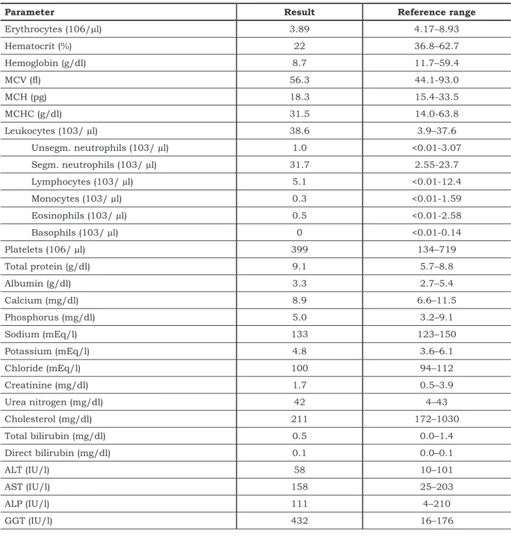
Parameter Result Reference range
Erythrocytes (106/µl) 3.89 4.17–8.93 Hematocrit (%) 22 36.8–62.7 Hemoglobin (g/dl) 8.7 11.7–59.4 MCV (fl) 56.3 44.1-93.0 MCH (pg) 18.3 15.4-33.5 MCHC (g/dl) 31.5 14.0-63.8 Leukocytes (103/ µl) 38.6 3.9–37.6 Unsegm. neutrophils (103/ µl) 1.0 <0.01-3.07 Segm. neutrophils (103/ µl) 31.7 2.55-23.7 Lymphocytes (103/ µl) 5.1 <0.01-12.4 Monocytes (103/ µl) 0.3 <0.01-1.59 Eosinophils (103/ µl) 0.5 <0.01-2.58 Basophils (103/ µl) 0 <0.01-0.14 Platelets (106/ µl) 399 134–719 Total protein (g/dl) 9.1 5.7–8.8 Albumin (g/dl) 3.3 2.7–5.4 Calcium (mg/dl) 8.9 6.6–11.5 Phosphorus (mg/dl) 5.0 3.2–9.1 Sodium (mEq/l) 133 123–150 Potassium (mEq/l) 4.8 3.6–6.1 Chloride (mEq/l) 100 94–112 Creatinine (mg/dl) 1.7 0.5–3.9 Urea nitrogen (mg/dl) 42 4–43 Cholesterol (mg/dl) 211 172–1030 Total bilirubin (mg/dl) 0.5 0.0–1.4 Direct bilirubin (mg/dl) 0.1 0.0–0.1 ALT (IU/l) 58 10–101 AST (IU/l) 158 25–203 ALP (IU/l) 111 4–210 GGT (IU/l) 432 16–176
Table 1: Results of the hemato-biochemical analysis. Reference ranges are from Ramsay 2003 (15), except for
leukocytes, white subcellular population (16) and GGT (17)
involving both limbs of the left side. Anyway, deep and superficial sensitivities were preserved. Widespread tremors and nystagmus were also present. The bear appeared conscious and was able to eat. A venous sample of blood was collected from the jugular vein to analyse the hematologic parameters reported in table 1.
Number of erythrocytes, haematocrit and haemoglobin resulted to be decreased causing a moderate status of anemia. The number of leukocytes was increased with most of them represented by segmented neutrophils. Values for total protein were slightly increased while GGT (gamma-glutamyl transpeptidase) resulted to have a fourfold increased activity.
Based on the clinical suspicious of central nervous system damage, a therapy including fluid administration (ringer lactate only the first day), dexamethasone sodium phosphate (4 mg/ Kg, IM, BID the first day and 2 mg/Kg IM, SID in the following days), ranitidine (6 mg/Kg PO BID; Ranitidina TEVA®, Teva Pharma Italy), ceftriaxone (1.5 g, IM, SID; ceftriaxone hexal®, Hexal Spa Italy), propentofylline (3 mg/Kg, PO, BID; Karsivan®, Intervet Italia Srl), and a vitamin B complex PO, SID (Stimulfos®, Teknopharma S.p.a.) has been started. Drugs to be given parenterally, were administered by dart using a blow pipe; PO therapy was administer with food. After about a week of therapy the bear was able to stand up and walk, partially recovered the vision and ate regularly. Nevertheless, three weeks after the clinical onset, she went back in decubitus and died after 12 hours.
Necropsy revealed an irregular aspect of left ovary, and old clots were noted throughout the pelvis. The right ovary appeared normal. On gross examination, a 20×15×8 centimeter and 435 g in weight left ovarian mass was evident. Grossly, the external surface was intact and smooth, and the cut surface was gray to yellow, predominantly solid and lobulated, with a few small areas of cystic change and hemorrhage.
A 2.5 cm-sized nodule occupying the lombo-aortic lymph nodes was also observed. Additionally brain metastases were noted, characterized by multiple lesions, typically at grey white junction, profuse perilesional edema, and relatively smooth margin.
Intimal calcification of the aortic arch and scattered petechiae on the mucous membrane of the urinary bladder were also observed.
Tissue samples from tumors and all organs were fixed in 10% neutrally-buffered formalin (pH 7.4), processed for paraffin embedding, sectioned at 4 μm and stained with haematoxylin and eosin (H&E) and used also for immunohistochemical characterization. On the histological examination, the ovarian tumor was composed of epithelioid to
spindle-shaped neoplastic cells that were arranged in closely packed hollow or solid tubules with a fibrous stroma, resulting in a lobular pattern. Diffuse solid areas, microcystic pattern with eosinophilic secretion, and focal characteristic sieve-like structure were also noticed (Fig. 1). The tumor cells showed mild nuclear atypia and frequent mitotic figures. Nuclear pleomorphism and tumor necrosis was also found. Histology of brain metastases revealed a sharp interface between brain parenchyma and metastatic tumor. The neoplasm had areas of follicular architecture and areas of solid nests (Fig. 1), with tumor nuclei that were round to oval in shape with prominent nucleoli and finely granular chromatin. The neoplastic cells had abundant eosinophilic cytoplasm and mild pleomorphism. Some features of papillary carcinoma were occasionally observed. The tumor was intermixed with areas of fibrosis with hemosiderin deposition and calcification. Focal necrosis and perivascular macrophages were also noted. The adjacent brain parenchyma contained hemosiderin deposits, vascular sclerosis, and reactive astrocytes. Finally, microscopy of neoplastic nodules in the lymph nodes showed a similar histological pattern, with neoplastic cells being arranged in diffusely proliferating sheet-like cellular nests. Neoplastic cells were separated by variable amounts of fibrous stroma, but they were invasive to the stroma and lymphatic vessels. In all these three sites of neoplastic development, the neoplastic cells were largely polygonal with round to oval-shaped nuclei and abundant eosinophilic cytoplasm and prominent nucleoli with indistinct cellular borders sometimes forming rosettes, papillae, and duct-like structures; mitotic figures were observed frequently. In the lombo-aortic lymph nodes, the lymphoid tissue was mostly replaced by neoplastic cells forming a large tumor mass. In the brain, a scattered distribution of neoplastic cell aggregation of various sizes was evident, as well as larger discrete nodules.
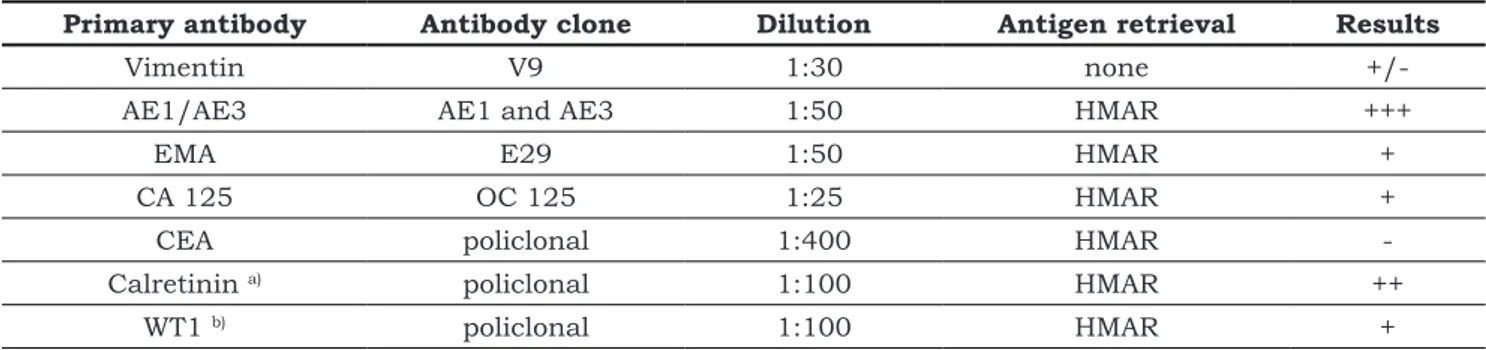
Sections from neoplastic tissues of the ovary, brain, and lombo-aortic lymph node were immunohistochemically stained by the avidin-biotin-peroxidase complex (ABC) procedure (Vectastain Elite ABC Kit; Vector Laboratories, Burlingame, CA, U.S.A.) and examined microscopically. Details of the specific primary antibodies used and the staining results for the neoplastic tissues are summarized in Table 2.
Figure 1: Low-power view of ovarian neoplastic tissue, characterized by epithelioid to spindle-shaped neoplastic cells that were arranged in closely packed hollow or solid tubules with a fibrous stroma (arrowhead); right lower insert: characteristic microcystic (sievelike) structure with eosinophilic secretion (arrow), interspersed in a diffuse solid area. (H&E, Bar = 250µm; insert = 100µm)
Primary antibody Antibody clone Dilution Antigen retrieval Results
Vimentin V9 1:30 none
+/-AE1/AE3 AE1 and AE3 1:50 HMAR +++
EMA E29 1:50 HMAR +
CA 125 OC 125 1:25 HMAR +
CEA policlonal 1:400 HMAR
-Calretinin a) policlonal 1:100 HMAR ++
WT1 b) policlonal 1:100 HMAR +
Table 2: Panel of antibodies utilized for tumour characterization
a) made by ZYMED; b) made by LifeSpan Bioscience, Inc., c)HMAR = Heat Mediated Antigen Retrieval
Deparaffinized sections were blocked for endogenous peroxidase in 0.3% H2O2 with methanol for 30 min. Incubation of sections with the primary antibody was performed at 4°C for 16 h, followed by incubation with the biotinylated secondary antibody for 30 min, and with avidin peroxidase conjugate for 30 min at room temperature. Sections were developed in 0.05% 3,3’-diaminobenzidine/H2O2 solution. As positive control for each immunoreactivity, an ovary serous carcinoma, and a colonic carcinoma belonging to dog were employed. Additionally, a normal ovary and brain tissues from a brown bear (archive material) were also used. Samples from canine serous ovarian carcinoma were used for confirmation of the immunoreactivity for epithelial membrane antigen (EMA), Wilms
Tumor 1 antigen (WT-1), and Cancer Antigen 125 (CA125). Neoplastic colonic tissue was used for confirmation of the immunoreactivity for Carcino-Embryonic Antigen (CEA), and EMA. Finally normal brown bear tissue from ovary and brain were used as positive control for cytokeratins and vimentin stain.
The utilized antibodies were concentrated antibodies made by DAKOCytomation, Denmark, ZYMED Laboratories Inc., San Francisco (for Calretinin polyclonal antibody), and LifeSpan Bioscience, Inc. (for WT-1 polyclonal antibody, LS-B4578). In order to confirm immunohistochemically the diagnosis of carcinoma of the ovary, all the specimens were initially marked with vimentin to control the primary processing of the analyzed case. First we chose a panel of antibodies to help
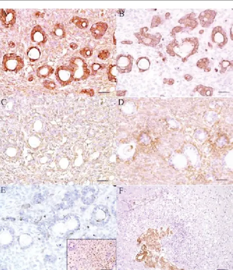
Figure 2: A) Diffuse and strong
positivity of tumor cells, especially pseudo-cysts forming cells, for A1-A3 citockeratins. B) Diffuse positivity restricted to pseudo-cysts forming cells for EMA. C) Calretinin diffuse and light to moderate positivity of all tumoral cells. D) Similar pattern of diffuse and light positivity, with the exception of the pseudo-cysts forming cells, for CA125 antigen. E) Substantial negativity of all neoplastic cells for CEA antigen. Note in right lower insert the strong and diffuse nuclear positivity of the neoplastic cells for WT-1 antigen.
F) Strong positivity of brain metastasis
for citockeratins A1-A3. Note the intense inflammatory reaction and edema around the neoplastic tissue. (IHC stain. Bar = 100 µm; insert and panel F = 250 µm)
establish the epithelial origin of the tumour. Thus, we opted for cytokeratin AE1/AE3, and EMA antibodies. Since there is no specific marker to indicate the ovarian origin of the tumor, the differentiation of the studied tumor from possible ovarian metastasis was realized through several associated immunohistochemical reactions. Thus, the anti-calretinin antibody was used in attempt to help, together with the CA125, CEA, and WT-1 antibodies, to eliminate a mesothelioma belonging to the peritoneal cavity. The CEA staining allowed the separation of this tumor from ovarian metastasis originating in the gastrointestinal tract. Finally, WT-1 staining was used in attempt to speculate the serous, non mucinic origin of the ovarian neoplasia. The immunostaining intensity was evaluated using a four-degree-system in accordance with the model of Wauters, 1995. Immunohistochemistry stain was notable and intense for pan-cytokeratin (Fig. 2A, and F), moderate and diffuse for calretinin (Fig. 2C), light to moderate and “periacinar” for EMA (Fig. 2B), and focal and nuclear for WT-1 (Fig. 2E, insert).
Not unexpected in such a high grade carcinoma, vimentin staining was positive in a minority of epithelial cells. Calretinin staining was focally strongly positive, also in brain and lymph node metastases. Additionally, neoplastic cells showed a moderate and focal stain for CA125 (Fig. 2D), resulting consistently negative for CEA antigen (Fig. 2E). In all the neoplastic samples (primary tumor and metastases) the inconspicuous and myxoid stroma resulted strongly positive for vimentin, as expected.. On the basis of the tumor architecture, and the immunohistochemical profile, the tumor was classified, according WHO guidelines (18), as ovarian carcinoma serous type, papillary to solid.
According to WHO classification, ovarian cancers are classified in three categories: epithelial tumors (arise from cells that line or cover the ovaries); germ cell tumors (originate from cells that are destined to form eggs within the ovaries); and sex cord-stromal cell tumors (begin in the connective cells that hold the ovaries together and produce female hormones) (18). Common epithelial tumors begin in the surface epithelium of
the ovaries are divided into serous, endometrioid, mucinous, and clear cell tumors subtypes. Unfortunately, extraovarian mesotheliomas, peritoneal carcinoma, or ovarian metastatic carcinomas belonging to gastrointestinal tract, represent some tumors that are adjacent to ovarian tissues and may be viewed as ovarian cancer, complicating the histological diagnosis.
In our case, the immunohistochemical analysis of primary tumor and metastases revealed a characteristic profile of the neoplasia. Anticalretinin antibody was used (presently considered to be the best marker for mesotheliomas) together with cytokeratins, EMA and CA125 antibodies (epithelial cells markers). The immunostaining for cytokeratin AE1/AE3 was of great importance in the determination of the epithelial origin of the tumour since, for this marker, primary neoplasia and metastases were positive at the level of tumour cells (Fig. 2A, and F). The positivity of the ovarian surface epithelium for calretinin, was very helpful since it represented the positive internal control. In our case, the light cytoplasmic and diffuse pattern of calretinin positivity in neoplastic cells, suggested an ovarian surface epithelial tumor (Fig. 2C). The immunostaining was frequently diffuse and the intensity of the reaction was strong positive. The EMA expression of neoplastic cells is tipical for poorly differentiated ovarian carcinomas (19), and this antibody emphasized acinar differentiation (Fig. 2B), often where it was not easily observed in haematoxylin and eosin preparations (20). Even if CA125 is not a specific marker of the ovarian origin of the tumor, some studies have shown a CA125 heterogeneous positivity in specimens of ovarian serous carcinomas (21). This type of heterogeneous staining, observed also in our case, was in favor of the ovarian origin (Fig. 2D). Since tumors with gastrointestinal origin are intense and diffusely positive for CEA (20), the negativity of this brown beer tumor supported the ovarian origin (Fig. 2E). Besides, when present, CEA expression is indicative of the mucinous type differentiation in ovarian tumors which are, at the same time, negative for CA125 (21).
Then, immunohistochemistry completed the present study of metastasizing ovarian tumor, allowing its separation from peritoneal mesotheliomas with ovarian extension and from ovarian metastasis of a primary gastrointestinal carcinoma.
The brain, along with the bone, liver, and lung, is one of the most common sites of metastasis of ovarian carcinomas. Weakness, depression, anorexia, unstable gait, alteration of the vision, slurred speech, dizziness or vertigo, head tilting are some of the neurological symptoms reported in human and animals with brain metastases (22, 23, 24) but, of course, they depend form the site of metastases implantation. Regarding clinical pathology, results were not indicative of specific diseases: many cancer patients have a mild normocytic, non-regenerative anemia associated with chronic disease. The increase in serum γ-glutamyltransferase was considered to be due to hepatic damage, but no metastases were found in the liver. Neutrophilia could be due to the reaction against the tumors and increasing in total protein could have been caused by slight dehydration.
In summary, we report here a case of malignant metastasizing ovarian tumor in a brown beer. Because of typical histological and immunohistochemical features of neoplastic cells, this tumor was diagnosed as papillary to solid serous type ovarian carcinoma. Because of the scattered distribution pattern of neoplastic nodules, the involvement of the brain and lombo-aortic lymph node was considered to be metastatic. According to these results, the presence of tumor should be considered in bears with neurological signs.
References
1. Moulton JE. Bile duct carcinomas in two bears. Cornell Vet 1961; 51: 285–93.
2. Blancquaert AM, Porter RE Jr, Bruyninckx WJ, Cambre RC. Lymphosarcoma with perfora-tion of the ileum in a grizzly bear. J Am Vet Med Assoc 1984; 185: 1433–5.
3. Gosselin SJ, Kramer LW. Extrahepatic bili-ary carcinoma in sloth bears. J Am Vet Med Assoc 1984; 185: 1314–6.
4. Miller RE, Boever WJ, Thornburg LP, Cur-tis-Velasco M. Hepatic neoplasia in two polar bears. J Am Vet Med Assoc 1985; 187: 1256–8.
5. Momotani E, Aoki H, Ishikawa Y, Yoshino T. Osteosarcoma in the maxilla of a brown bear (Ursus arctos). Vet Pathol 1988; 25: 527–9.
6. Hellmann J, Hofmeister R, Göltenboth R. The occurrence of tumors in large bears (Ursidae): a literature review and six case descriptions. Berl
Münch Tierärztl Wochenschr 1991; 104: 262–8. 7. Ponomar’kov VI, Khutorianskiĭ AA. A case of osteosarcoma in a white polar bear. Arkh Patol 1995; 57: 81–3.
8. Yoon BI, Lee JK, Kim JH, Shin NS, Kwon SW. Lymphosarcoma in a brown bear (Ursus arc-tos). J Vet Sci 2001; 2: 143–5.
9. Mylniczenko ND, Manharth AL, Clayton LA, Feinmehl R, Robbins M. Successful treatment of mandibular squamous cell carcinoma in a Malay-an sun bear (Helarctos malayMalay-anus). J Zoo Wildl Med 2005; 36: 346–8.
10. Rotstein DS, Govett P, Wolfe B. Larynge-al squamous cell carcinoma in a North American black bear (Ursus americanus). J Zoo Wildl Med 2005; 36: 543–5.
11. Ozyigit MO, Aytug N, Cihan H. Mandibular osteoma in a brown bear (Ursus arctos). In: 6th Scientific Meeting of the European Association of Zoo and Wildlife Veterinarians. Budapest : Hun-gary 2006: 91–3.
12. Nak D, Cangul IT, Nak Y, Cihan H, Celim-li N. Tubulopapillary mammary carcinoma in a brown bear (Ursus arctos). J Wildl Dis 2008; 44: 505–8.
13. Matsuda K, Qiu Y, Kawamura Y, Suzuki H, Takita Y. Hepatocellular carcinoma in a Hok-kaido brown bear (Ursus arctos yesoensis). J Vet Med Sci 2010; 72: 1213–6.
14. Murakami T, Kobayashi Y, Chiba S, et al. Humeral chondrosarcoma in a Hokkaido brown bear (Ursus arctos yesoensis). J Vet Med Sci 2012; 74: 1195–7.
15. Ramsay EC. Ursidae and Hyaenidae. In: Fowler ME, Miller RE. eds. Zoo and wild animal medicine. 5th ed. St. Louis : Saunders, 2003: 523–38.
16. Kusak J, Rafaj RB, Zvorc Z, Huber D, Forsek J. Effects of sex, age, body mass, and cap-turing method on hematologic values of brown bears in Croatia. J Wildl Dis 2005; 41: 843–7.
17. Bassart GD, Reidarson TH, Dierauf LA, Duffield DA. Clinical pathology. In: Dierauf LA, Gulland LA, eds. Handbook of marine mammal medicine. 2nd ed. Boca Raton : CRC Press, 2001: 383–436.
18. Kennedy PC, Cullen JM, Edwards JF. His-tological classification of tumors of the genital system of domestic animals. In: World Health Or-ganization international histological classification of tumors of domestic animals. Washington DC: Armed Force Institute of Pathology, 1998.
19. Seidman JD, Russel P, Kurman RJ. Sur-face epithelial tumours of the ovary. In: Kurman RJ, eds. Blaustein’s pathology of the female geni-tal tract. 5th ed. New York : Springer, 2002: 791– 904.
20. Hammond RH, Bates TD, Clarke DG, et al. The immunoperoxidase localization of tumour markers in ovarian cancer: the value of CEA, EMA, cytokeratin and DD9. Br J Obstet Gynecol 1991; 98: 73–83.
21. Neunteufel W, Breitenecker G. Tissue ex-pression of CA 125 in benign and malignant le-sions of ovary and fallopian tube, a comparison with CA 19-9 and CEA. Gynecol Oncol 1989; 32: 297–302.
22. Chiang YC, Qiu JT, Chang CL, et al. Brain metastases from epithelial ovarian carcinoma: evaluation of prognosis and managements - a Taiwanese Gynecologic Oncology Group (TGOG) study. Gynecol Oncol 2012; 125: 37–41.
23. Kim JH, Im KS, Kim NH, et al. Inflamma-tory mammary carcinoma with metastasis to the brain and distant organs in a spayed Shih Tzu dog. J Vet Diagn Invest 2011; 23: 1079–82.
24. Davis JL, Gilger BC, Spaulding K, et al. Nasal adenocarcinoma with diffuse metastases involving the orbit, cerebrum, and multiple cra-nial nerves in a horse. J Am Vet Med Assoc 2002; 221:1460–3.
METASTAZIRAJOČI KARCINOM JAJČNIKA PRI EVROAZIJSKEM RJAVEM MEDVEDU
(Ursus arctos arctos): KLINIČNI PRIMER
Povzetek: V članku je opisan primer karcinoma jajčnika pri rjavemu medvedu. Evroazijska rjava medvedka, stara 37 let, brez mladičev, iz živalskega vrta Falconara v Italiji, je kazala klinične znake nevroloških motenj, in sicer obojestransko slepoto ter znake delne levostranske pareze obeh okončin.
Po opaženih kliničnih znakih je bila medvedka zdravljena z deksametazonom, ranitidinom, ceftriaksonom, propentofilinom, vitamini kompleksa B in dodajanjem tekočine. Približno po enem tednu so znaki pareze popustili, tako da je medvedka znova lahko hodila, delno se ji je povrnil vid in pričela je jesti. Kljub temu začetnemu kliničnemu izboljšanju je medvedka poginila tri tedne po pojavu bolezenskih znakov. Med raztelesbo smo našli močno povečan in tumorozno spremenjen levi jajčnik ter metastaze v regionalnih bezgavkah in možganih. Glede na značilno histološko sliko in imunohistokemične značilnosti tumorskih celic smo tumor diagnosticirali kot papilarni do solidni serozni tip karcinoma jajčnika. Zaradi razpršenega vzorca razporeditve žarišč tumorskih celic v možganih ter v ledveno-aortnih bezgavkah smo le-ta opredelili kot metastaze. V literaturi je opisanih le nekaj primerov tumorjev pri različnih vrstah medvedov (Ursidae), poročajo o primerih limfosarkoma, osteoma, osteosarkoma, hondrosarkoma, ploščatoceličnega karcinoma ter karcinoma žolčevodov, ščitnice, mlečne žleze in jetrnih celic. Glede na naše rezultate je potrebno pri medvedih z nevrološkimi kliničnimi znaki pri diferencialni diagnostiki pomisliti tudi na tumorje.