Selection of our books indexed in the Book Citation Index in Web of Science™ Core Collection (BKCI)
Interested in publishing with us?
Contact [email protected]
Numbers displayed above are based on latest data collected. For more information visit www.intechopen.com Open access books available
Countries delivered to Contributors from top 500 universities
International authors and editors
Our authors are among the
most cited scientists
Downloads
We are IntechOpen,
the world’s leading publisher of
Open Access books
Built by scientists, for scientists
12.2%
116,000
120M
TOP 1%
154
Predator-Prey Interactions in Ciliated Protists
Federico Buonanno and Claudio Ortenzi
Additional information is available at the end of the chapter
© 2016 The Author(s). Licensee InTech. This chapter is distributed under the terms of the Creative Commons Attribution License (http://creativecommons.org/licenses/by/3.0), which permits unrestricted use, distribution, and reproduction in any medium, provided the original work is properly cited.
Federico Buonanno and Claudio Ortenzi
Additional information is available at the end of the chapter
Abstract
Protists appeared relatively early in evolution, about 1.8 billion years ago, soon after the first prokaryotic organisms. During this time period, most species developed a variety of behavioral, morphological, and physiological strategies intended to improve the ability to capture prey or to avoid predation. In this scenario, a key role was played by spe-cialized ejectable membrane-bound organelles called extrusomes, which are capable of discharging their content to the outside of the cell in response to various stimuli. The aim of this chapter is to describe the two main strategies adopted in ciliate predator-prey interactions: (a) the first is mediated by mechanical mechanisms and involves, for example, extrusomes called trichocysts and (b) the second is mediated by toxic secondary metabolites and involves different kinds of chemical extrusomes.
Keywords: protists, ciliates, extrusomes, secondary metabolites, chemical offense
1. Introduction
A common definition for predatory behavior describes it as the process through which one animal, the predator, captures and kills another animal, the prey, before eating it in part or entirely [1]; however, according to the opinion of a number of microbiologists and protis-tologists, this definition should be also extended to different organisms included in other life Kingdoms, with particular regard to microorganisms. Indeed, especially in the last 30 years, a lot of studies have been devoted to describing the predator-prey interactions among uni-cellular eukaryotic organisms like protists. Whittaker [2] originally defined protists as those “organisms which are unicellular or unicellular-colonial and which form no tissues,” and for this reason they must carry out at the cellular level all the basic functions which can be observed in multicellular eukaryotes. Among these functions, self-nonself recognition
© 2018 The Author(s). Licensee IntechOpen. This chapter is distributed under the terms of the Creative Commons Attribution License (http://creativecommons.org/licenses/by/3.0), which permits unrestricted use, distribution, and reproduction in any medium, provided the original work is properly cited.
mechanisms are represented by a large repertoire in protists and can trigger either autocrine or paracrine processes in some ciliates (see [3] for a review), together with the capability to detect prey (food) or predators in others. In this regard, it is known that protists have developed a variety of strategies of feeding behaviors especially in response to different envi-ronmental factors, together with a diverse kind of food available in micro-habitats. Figure 1 shows a general scheme of predator-prey interactions, where the predator recognizes the presence of the prey (step 1) and can attack it (step 2). On the other hand the prey recognizes the presence of the predator (step 1′) and it can organize its defense mechanisms (step 2′) [4]. This scheme should be considered functional for both animals and protists, and indeed several studies have shown that the food recognition and the offense-defense mechanisms adopted by some groups of protists can be compared, in terms of complexity and variability, with those observed in animals.
In this context, a common feeding mechanism found in heterotrophic protists is phagocy-tosis, a process which requires specific organelles for food assimilation and which occurs in three steps: food capture, phagosome formation, and food digestion [5]. Different techniques of phagocytosis have been described in various protists, where they have especially been investigated in ciliates [5–7]. Verni and Gualtieri [5] describe three main phagocytotic pro-cesses in ciliates: filter feeding, suctorial feeding, and raptorial feeding. The authors compare them to the strategies used in fishing, like netting, trapping, and harpooning. In filter-feeding ciliates, the food, represented by small organisms or edible debris of various types, was pushed into the ciliate buccal cavity by the rhythmical beats of the cilia located in its adoral apparatus. Suctorial-feeding ciliates are represented by sessile or sedentary species that for most of their lives remain attached to other organisms or various substrates, intercepting the food particles with their specialized tentacles. Finally, raptorial ciliates are able to directly catch other organisms using peculiar organelles to paralyze and/or kill their prey, generally called extrusomes.
2. Extrusomes, the specialized organelles for predator-prey
interaction
The term “extrusome” was proposed, for the first time, by Grell in 1973 for extrusive (eject-able) bodies, which occur widely in protists [8]. They are membrane-bound organelles usu-ally located in the cell cortex, attached to the cell membrane. They can display differences in structure and morphology, but they share the common characteristic of discharging their contents to the outside of the cell in response to mechanical or chemical stimuli. Remarkably, when the extrusomes are discharged, the cell remains intact and functional. Studies on extru-somes and related organelles have been reviewed by Hausmann [9], Dragesco [10], Kugrens et al. [11], Hausmann and Hülsmann [12], and Rosati and Modeo [13]. Typical examples of these organelles include toxicysts, trichocysts, mucocysts, cortical, or pigment granules in cili-ates and flagellcili-ates, haptocysts in suctorians, and kinetocysts in heliozoan actinopods. Some extrusomes are known to be related in predator-prey interactions, for example, to catch and kill the prey (such as toxicysts, haptocysts, kinetocysts, and some cortical granules), or used as defensive organelles (such as the trichocysts and various cortical or pigment granules), but the role of other kinds of extrusomes such as the mucocysts in Tetrahymena or the trichites in Strombidiidae [13] still remains obscure.
3. Offensive extrusomes
Offensive extrusomes generally possessed by raptorial protists and located usually at or near the feeding apparatus are discharged after contact with a possible prey, which is immobi-lized, damaged, or firmly bound to the predator. Among these, organelles, certainly the most widely studied, belong to the category of toxicysts (toxic extrusomes) and they play an essen-tial role in capturing and killing prey [7, 13]. Toxicysts are synthesized in Golgi or ER vesicles and are usually localized in the cell cortex attached to the cell membrane. Most of them are observed in species belonging to the class Litostomatea and subclass Haptoria, but they are also present in other predatory ciliates. They are usually positioned in a specific region of the cell, near the oral apparatus, and generally in the first portion which contacts the prey during the raptorial feeding [13]. Independently of the specific differences in the morphology of the cytostome, the toxicysts are always present in an appreciable number, for example, in the genera Didinium, Dileptus, Prorodon, Litonotus, Colpes, Homalozoon, and many others. In resting position, the toxicysts appear generally as rod-like elements (Figure 2), and could be discharged in milliseconds, if exposed to an appropriate stimulus such as contact with a prey (Figure 3) [7]. In this case, the tubules of the toxicysts are suddenly introduced into the cytoplasm of the prey’s body, like hypodermic needles, to inject the toxic material. Hausmann [7] reports essentially two ways by which the toxicysts may be discharged: in the first case, there is a fusion of the toxicyst’s membrane with the plasma membrane, followed by the tubule discharge via evagination; in the second, observed in certain ciliate species, a telescopic
discharge of the tubules was observed. During or near the end of the toxicysts’ discharge, the toxic secondary metabolites were secreted by the tubules. It is worth noting that this mecha-nism of discharging toxic substances shows the structural and functional similarities that can be found between the toxicysts in ciliated protists and nematocysts in Cnidaria, despite the substantial differences in size [7].
Figure 2. Transmission electron microscope (TEM) picture of the toxicysts in a dividing cell of a ciliate Didinium nasutum.
Scale bar = 1 μm. Original picture by R. Allen from http://www.cellimagelibrary.org/images/10010.
Figure 3. Predatory behavior of Coleps hirtus on Pseudokeronopsis erythrina. The predator attacks the prey with its toxicysts
In contrast with recent and less recent studies about the nature of the toxic secondary metab-olites used by ciliates in chemical defense, no exhaustive data are yet available about the composition of the toxins stored in the toxicysts of predatory ciliates. This is essentially due to the difficulty in separating the content of extrusomes from other molecules produced by the ciliate, in order to purify them at homogeneity for subsequent chemical and structural analyses.
To date the presence of acid phosphatase has been demonstrated in the toxicysts of Didinium
nasutum [14] and four other raptorial ciliates such as Enchelys mutans, Lacrymaria olor,
Homalozoon vermiculare, and Pseudoprorodon niveus [15]. It has been supposed that this enzyme,
generally present in lysosomes of animal cells, may probably be used by these ciliates to start the digestion of the prey.
3.1. The predatory behavior of Coleps hirtus
The complete analysis of the content of the toxicysts, together with observations of the preda-tory behavior, was also performed on another species, Coleps hirtus, a freshwater protostomatid ciliate. C. hirtus (40–65 × 20–35 μm) has an oral apparatus placed at the anterior end of the cell and its barrel-shaped body is covered by calcified armor arranged in plates. This ciliate is able to feed off bacteria, algae, flagellates, and ciliates, but it is also histophagous, that is, it feeds on living plant and animal tissue such as rotifers, crustaceans, and fish [16, 17]. Coleps is also reported to show a scavenger feeding on tissues of dead metazoans, such as Daphnia,
Diaphanosoma, and chironomid larvae [18], as well as toward dead ciliates and dead specimens
of its own species. Coleps is usually equipped with toxicysts used by the ciliate to assist its car-nivorous feeding, and its predatory behavior has recently been analyzed against another ciliate species used as prey, Euplotes aediculatus. Observations conducted on a mixture of predators and prey showed several contacts between the specimens of Colpes and Euplotes, but only after 5–10 min did interactions between the anterior section of a predator with a specimen of Euplotes become effective. This time was probably essential for prey detection and recognition, followed by prolonged contact between predator and prey, generally ending with the rapid backward swimming of the latter which separated the two organisms. When the attacks became numer-ous some individuals of Coleps remained attached to their prey (Figure 4), which decreased their swimming speed and gradually stopped swimming. After 20–30 min, the prey was frag-mented and eaten by several specimens of Coleps, and a similar predatory behavior was also observed using different ciliate species as prey [19]. On the contrary the toxicysts-deficient specimens of Colpes (Figure 5) obtained by means of the application of the cold-shock method capable of inducing an exclusive massive discharge of extrusomes in ciliates [20] appear unable to catch and kill their prey [19].
Unexpectedly, the analysis of the bioactive fraction of the toxicyst discharge of Coleps hirtus (performed by liquid chromatography-electro-spray-mass spectrometry and gas chromatog-raphy-mass spectrometry) showed the presence of a mixture of 19 saturated, monounsatu-rated and polyunsatumonounsatu-rated free fatty acids (FFAs) with the addition of a minor amount of a diterpenoid (phytanic acid) but did not reveal the presence of enzymes, as reported for other
predatory ciliates [19]. To date this is the only report on the presence of FFAs as toxic sub-stances in the extrusomes of ciliated protists, but the use of this class of compounds as toxins by Coleps is shared with at least 15 freshwater, 13 marine, and 6 brackish water potentially
Figure 4. Multiple attacks by different cells Coleps hirtus on a cell of Euplotes aediculatus. Micrograph extracted from a
film clips. Scale bar = 200 μm.
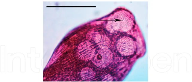
Figure 5. (A) The toxicysts in Coleps hirtus appear as rod-shaped organelles (arrow) in the oral basket of a cell. (B) The
photomicrograph shows the toxicysts discharged (arrow) into the medium, immediately after a cold-shock treatment. (C) No toxicysts are detected in a toxicyst-deprived cell. Photomicrographs of fixed specimens by protargol stain. Scale bar = 10 μm. Pictures from [19].
harmful microalgae, as well with some multicellular organisms. For example, a chemical defense by a mixture of FFAs was studied and demonstrated for the harmful microalga
Fibrocapsa japonica (Raphidophyceae) [21–23], and also in animals, a defensive strategy
medi-ated by FFAs was recently described for the fish Barbus barbus which adopted it to protect its eggs from predators [24].
Very little is known about the role and source of phytanic acid in ciliates, this being the addi-tional component detected in the toxicyst discharge of Coleps. Phytanic acid can be produced from the biodegradation of the side chain of chlorophyll [25], so one possible source arises from Coleps’ carnivorous feeding on photosynthetic microorganisms [19]. Some insects, such as the sumac flea beetle, accumulate chlorophyll-derived metabolites as a chemical deterrent in excrements [26]. Komen et al. [27] demonstrated the toxic effect of phytanic acid on human skin fibroblasts, where it impaired mitochondrial respiration through protonophoric action. Regarding the role of phytanic acid in Coleps, it is possible to hypothesize that it can be used as a weapon, deterrent, or, at least, it could be stored in toxicysts given its potential toxic activity. In addition, it is known that ciliates themselves are also able to synthesize a huge number of terpenoids [28, 29]. This is the case of Euplotes focardi [30] and Euplotes rariseta [31] where the production of new diterpenoids was demonstrated. Terpene compounds and FFAs may also act together to exert cytotoxic effects [19]. FFAs may serve as a matrix to deliver toxic compounds to prey or predators and also to create a perfect environment where toxic metabolites can exert their functions.
It has been demonstrated that the substances discharged from the toxicysts by Coleps are highly toxic for a number of ciliate species such as Euplotes aediculatus, Paramecium tetraurelia,
Spirostomum teres, and S. ambiguum or Oxytricha sp. [19], and their action mechanism appears
to be related to a necrotic process. The term necrosis refers to a rapid (unprogrammed) cell death, with plasmatic membrane rupture, often caused by external factors such as toxins. On the contrary, the apoptosis is programmed cell death characterized by nuclear condensation, cytoplasmic shrinkage, and disintegration of the cell into small, membrane-bounded frag-ments. As shown in Figures 6 and 7, the purified toxin from Coleps is able to induce rapid cell death in E. aediculatus and in S. ambiguum preceded by cell membrane fracture without any changes in the morphology of the macronucleus. An action mechanism of this type seems to be a “good choice” for Coleps as it induces paralysis and a very rapid death in the prey. Interestingly, the cells of Coleps can also be damaged if exposed, in vitro, to their own toxin discharge [19]. Nevertheless, this cannot occur in nature, because on the one hand, the toxins are stored in the toxicysts of the ciliate, thus avoiding autotoxicity and on the other hand, the accidental exposure of Coleps to the toxicyst discharge dissolved in the medium is also unlikely, due to the choice of the predator to directly inject the toxins into the prey [19]. In this context, it is worth remembering the peculiar predatory behavior of Coleps, which usually leads to the observation that the same prey undergoes multiple attacks by several raptorial specimens, a behavior also adopted against young larvae of zebrafish [17]. It is likely that this behavior has evolved to ensure a fast immobilization of the prey, that after simultaneous multiple attacks, it can easily accumulate lethal concentrations of toxins injected by numerous predators. Therefore, essentially for the “wolf-like” group hunting behavior of Coleps, the species that appeared relatively resistant to its toxicyst discharge may also be easily caught and killed.
3.2. Didinium nasutum, a specialized hunter
Differently to Coleps, other ciliate species have specialized in hunting and catching a few preferential prey. This is, for example, the case of Didinium nasutum that is capable of captur-ing and killcaptur-ing several species of Paramecium and few other ciliates. Generally, Paramecium species are able to defend themselves by means of mechanical extrusomes like trichocysts (that will be discussed later on this chapter) but Didinium seems to overcome the defense of Paramecium by means of a highly specialized combination of extrusomes. Present on the proboscis of Didinium are several units of two different kinds of extrusomes: toxicysts, as in other Litostomatea, and pexicysts, another specialized offensive extrusome observed only in this species [32]. These authors describe the discharge of pexicysts as the first response after the prey recognition [14], which is typically followed by the discharge of toxicysts. At the same time, the prey (generally a Paramecium) discharges its trichocysts which separate the two organisms, but the proboscis of Didinium remains attached to the prey by a tiny con-nection probably composed of a bundle of discharged pexicysts and toxicysts (Figure 8).
Figure 6. (A) Necrotic effects of the toxicyst discharge of Coleps hirtus on Euplotes aediculatus and (B) Spirostomum
Figure 7. (A, B) Effects of the toxicyst discharge of Coleps hirtus on the macronuclear morphology in specimens of
Euplotes aediculatus and (C, D) Spirostomum ambiguum. Cells were stained with acridine orange and ethidium bromide
and observed by fluorescent microscopy. Viable cells show intact, bright green nuclei, nonviable cells show red/orange nuclei. M = macronucleus, m = micronucleus. Scale bar = 100 μm. Pictures from [19].
Figure 8. Scanning electron microscope (SEM) picture on the predator-prey interaction between a cell of Didinium
nasutum and a cell of Paramecium multimicronucleatum. The bundle of toxicysts and pexicysts can be seen between
the two organisms (arrow). Magnification ×50. Original picture by G. Antipa from http://www.cellimagelibrary.org/ images/21991.
Subsequently, the Paramecium will be reached again and captured by the predator. In the light of this observation, the pexicysts seem to act most by a mechanical function (as harpoon-like organelles) rather than with a chemical offense. This assumption is supported by the fact that another species of predatory ciliate, Monodinium balbiani, which is morphologically similar and phylogenetically close to Didinium, but without the presence of the pexicysts on its pro-boscis, unlike the Didinium, is sensitive to the defense mechanism possessed by Paramecium, which is often able to avoid capture [33].
3.3. The peculiar tentacles of suctorians
In this context it is also relevant to mention the subclass Suctoria, represented by ciliates which become sessile during development and consequently lose the ciliary structure. Suctorians are able to feed on other protists and frequently on other ciliates by means of specialized tentacles. The distal ends of these tentacles are often equipped by peculiar extrusomes called haptocysts that are involved in prey capture. When a tentacle touches a possible prey, the dis-charge of haptocysts is able to penetrate the prey’s membrane, forming a connection between the predator and the prey and injecting the extrusome content into the latter, which also con-curs to the fusion of the membranes belonging to the two organisms [13, 34]. However, the fusion of the two membranes is not always immediate, for example, in Heliophrya erhardi, Spoon et al. [35] observed that many specimens of Paramecium contacting the tentacles of the suctorian escaped discharging trichocysts at the point of contact, suggesting that Paramecium is able to defend itself from the puncture of the haptocysts.
4. Defensive extrusomes
In addition to predatory behavior, ciliated protists have also evolved different defense strate-gies, many based on the discharge of extrusomes. Two different mechanisms involved in their defense behavior are essentially observed: the first is mediated by the mechanical actions of trichocysts as in Paramecium or Frontonia and the second is mediated by the toxic secondary metabolites of different kinds of chemical extrusomes.
4.1. The mechanical defense
Spindle trichocysts (or simply, trichocysts) are spindle-shaped organelles which discharge their content in the form of a thread. They are found in some ciliates and flagellates and are sometimes furnished with a specially constructed tip [9]. The best known and studied trichocysts are those in the genus Paramecium. Trichocysts in Paramecium are 3–4 μm long, carrot-shaped membrane-bounded organelles armed with a sharply pointed tip, and are present in thousands all over the cell surface, except at the oral apparatus (Figures 9 and 10). When paramecia are subjected to various stimuli, the membranes of the trichocysts and the cell membrane blend together, and the content of the extrusomes is immediately discharged to the outside of the cell, forming a spear-like structure in milliseconds (Figure 11) (see [13] for a review). Trichocyst discharge has therefore been extensively studied as a model system
of exocytosis [36] (see [37] for a review). Synthesis, processing, and sorting of component proteins in trichocysts are also studied as model systems of protein biosynthesis [36] for a review.
Figure 9. Scheme of the ciliary structure and the trichocysts of Paramecium. Picture from http://biodidac.bio.uottawa.ca,
redrafted by R. D’Arcangelo.
Figure 10. Membrane details of resting trichocysts under the freeze fracture. The trichocyst tip (tt) and body (tb) are
covered by the same membrane. The A-face of this membrane (A-tin) possesses randomly distributed particles whereas the B-face (B-tin) shows corresponding depressions. Scale bar = 1 μm. Picture from [9].
Maupas, one of the pioneers of protozoology, first proposed the defensive function of tricho-cysts in Paramecium in 1883, observing its morphological features and judging it as self-evident [38]; however, this point was questioned for years. The main controversy was due to the fact that
Paramecium species are easily preyed upon by Didinium in spite of massive trichocyst discharge
by paramecia. Pollack reported that Didinium preys on wild-type cells as easily as trichocyst-defective mutants in P. tetraurelia [39]. However, further studies have unequivocally indicated that trichocysts in Paramecium exert an effective defensive function against unicellular preda-tors, including the raptorial protists Dileptus margaritifer, Monodinium balbiani, Climacostomum
virens, Echinosphaerium akamae, and E. nuceofilum [33, 40–43]. In addition, a more recent paper
also analyzed the defensive function of trichocysts in P. tetraurelia against some microinverte-brate predators, such as a rotifer (Cephalodella sp.), an ostracod (Eucypris sp.), and a turbellar-ian flatworm (Stenostomum sphagnetorum) [44]. The results of this study show the success in the defensive function of trichocysts against the rotifer and the ostracod while the mechanism seems ineffective against the flatworm. The authors speculate that the efficiency of the defense by means of trichocysts depends essentially on the kind of prey-capture behavior displayed by the predators. In particular, the success of the defense mediated by trichocysts appears posi-tively related to the time that the predator requires to capture and manipulate the prey before ingestion. Consequently, and different from the turbellarian flatworm that directly swallows paramecia, predators such as the rotifer and the ostracod that, prior to ingesting paramecia, contact it with a ciliated corona or articulated appendices, give the prey sufficient time to acti-vate the trichocysts discharge that allows it to escape [44]. Essentially this looks like the same phenomenon observed during the interaction between Paramecium and the predatory ciliate
Dileptus margaritifer, that attempts to paralyze its prey with the toxicysts on its proboscis before
ingestion, thereby inducing an explosive extrusion of trichocysts by Paramecium, which then swims away [44]. In this regard, another interesting observation was made when Paramecium was placed in a cell-free fluid containing the toxic material derived from the toxicysts from
Dileptus [45] (Miyake A. personal communication); indeed after contact with this toxic solution,
Paramecium cells violently reacted by immediately discharging most of their trichocysts before
being killed. In this reaction, sometimes a single specimen (cell) of Paramecium was completely surrounded by its discharged trichocysts. When this occurred, the Paramecium survived long after other cells were killed, moving slowly in the narrow space in the capsule of discharged trichocysts. But when it happened that one of these encapsulated cells managed to squeeze out of the capsule, it was quickly killed. This observation suggests that discharged trichocysts of
Paramecium function as a barrier against the Dileptus toxins and hence the locally discharged
trichocysts in the Paramecium-Dileptus interaction function as an instant shield against Dileptus. To summarize, the mechanical defense by trichocysts and related extrusomes appear to be multiple, including quick physical displacement, the thrust into a predator, and protec-tion against the predator’s toxins, increasing the chance for the prey to survive and escape. However, especially in ciliates and flagellates, other kinds of extrusomes used for defense were found, ones that, unlike trichocysts, are capable of discharging toxic materials in response to predatory behavior.
4.2. The chemical defense
Pigment granules (also called pigmentocysts) and cortical granules are extrusive organelles
containing pigmented or colorless toxic material, respectively, and they were originally clas-sified as a special type of mucocysts [9]. Pigment and cortical granules are mainly present in heterotrich and karyorelictean ciliates, such as Blepharisma, Stentor, Loxodes, and Trachelonema, but they may also exist in other groups of ciliates. They are usually present in great numbers throughout the cell cortex, sometimes providing bright colors to their bearers. Examples are
Stentor coeruleus, whose coloration is due to the pigment called stentorin, and several red
spe-cies of Blepharisma, whose coloration is due to blepharismins, formerly overall called zoopur-purin by Giese [46]. The coloration of these common heterotrichs has long attracted attention and most studies on pigment granules have been carried out using S. coeruleus, and a few red species of Blepharisma. B. japonicum (Figure 12) is the best studied species of the genus
Blepharisma and it presents pigment granules usually in a size of 0.3–0.6 μm diameter, arranged
in stripes between the rows of cilia that confer a red-pink coloration to the ciliate (Figure 13). These granules have been shown to contain a mixture of five compounds called blepharismins that are multifunctional quinone derivatives structurally related to hypericin, a photodynamic toxin of Hypericum perforatum (St. John’s Wort), and stentorin, produced by the ciliate S.
coeru-leus [47, 48] (Figure 14). To date, two primary functions of blepharismins have been
demon-strated: light perception and defense function against predators [47–52]. With regard to light perception, B. japonicum shows a temporal backward swimming or rotating movement (step-up photophobic response) if exposed to a sudden increase in light intensity. The step-(step-up pho-tophobic response helps the cells avoid strongly illuminated regions and lethal damage due to the photodynamic action of blepharismins [53]. In addition to light perception, blepharismins were found to act as chemical weapons via their light-independent cytotoxic effect against predatory protozoans and methicillin-resistant Gram-positive bacteria [49, 50, 54]. A possible explanation for this cytotoxicity can be found in the capability of blepharismins to form cation-selective channels in planar phospholipid bilayers [51], a phenomenon also expected to occur in the cell membranes of microorganisms exposed to toxic concentrations of ciliate pigments.
The defensive function of blepharismins was initially proposed by Giese in 1949 who found that crude extracts of Blepharisma were toxic to various ciliates but not to Blepharisma itself [55]. Unfortunately, however, his preliminary tests did not support this assumption, that is, Blepharisma was easily eaten by predators such as the heliozoan Actinospherium eichhorni and small crustaceans [46, 55]. Some predators, Didinium nasutum, Woodruffia metabolica, and
Figure 12. External morphology of a living cell of Blepharisma japonicum. Scale bar = 100 μm.
Figure 13. Extrusive pigment granules in Blepharisma japonicum (arrow) visible as red/pink dots under a vacuole. Scale
Podophrya fixa, did not eat Blepharisma, but they also ignored some other ciliates including
uncolored ones. In the absence of further evidence, Giese was skeptical about the assumption [46]. This hypothesis was further unequivocally demonstrated by Miyake, Harumoto, and collaborators, comparing normally pigmented red cells of B. japonicum, albino mutant cells, and light-bleached cells (a phenocopy of the albino mutant) as prey for the raptorial ciliate
Dileptus margaritifer and evaluating the toxicity of purified blepharismins on various ciliate
species [49, 50]. As a response to the attack by D. margaritifer versus one cell of B. japonicum,
Figure 14. Main secondary metabolites produced by ciliated protists. Erythrolactones: A1 (R1 = SO3 −; R
2 = C6H13 (n-hexyl));
B1 (R1 = SO3−; R2 = C7H15 (n-heptyl)); C1 (R1 = SO3−; R2 = C8H17 (n-octyl)); A2 (R1 = H; R2 = C6H13 (n-hexyl); B2 (R1 = H;
the latter releases the toxic blepharismins, visible as spherical bodies of 0.2–0.6 μm in diameter under scanning electron microscopy (Figure 15). The discharge take place within a second and it is able to repel the predator, while the albino and light-bleached cells are much more sensitive to the attacks of D. margaritifer [49, 50]. Recently the defensive function of blepharis-mins was also investigated in two additional species of Blepharisma, B. stoltei, and B. undulans against two predatory protists (C. hirtus and Stentor roeseli) and one metazoan, the turbellarian
S. sphagnetorum [56]. The results indicate that the chemical defense mechanism present in B.
stoltei and B. undulans is mediated by the same five blepharismins present in B. japonicum,
although produced in different proportions [56]. Authors speculate that the conservation of this panel of toxic secondary metabolites suggests that distinct roles for these molecules are likely required at least for the fine control of photophobic reactions, as initially proposed by
Figure 15. SEM micrographs of the predator-prey interaction between a cell of Dileptus margaritifer (DI) and a cell of
Blepharisma japonicum (BL). (A) Blepharisma being attacked by Dileptus. Arrow indicates the site of the damage inflicted
by the proboscis of the Dileptus. The rupture runs across the adoral zone of membranelles of the Blepharisma. Scale bar = 50 μm. (B) Enlargement of the region near the rupture in A. Scale bar = 5 μm. (C) The rupture magnification in B, showing the surface of Blepharisma peppered with spherules discharged from pigment granules. The surface is also pitted with small depressions presumably formed at the spots where the spherules have passed through the cell membrane. Scale bar = 5 μm. (D) Enlargement of a part of C. Scale bar = 0.5 μm. Pictures from [50].
Matsuoka et al. [48]. Summarizing, the Blepharisma species studied are able to defend them-selves against C. hirtus, although S. sphagnetorum and S. roeseli seem to overcome Blepharisma’s chemical defense, but it was observed that after the ingestion of intact cells of the toxic cili-ates these predators are not able to reproduce, suggesting the presence of the post-ingestion toxicity phenomena [56]. Additional toxic pigments, structurally related to hypericin, were found in other heterotrich ciliate species, such as stentorin in S. coeruleus (see [57] for a review), amethystin in S. amethystinus [58], and maristentorin in the marine ciliate Maristentor dinoferus [59], but the defensive function was experimentally proved only for S. coeruleus [60].
Karyorelictean ciliates also possess pigment granules which are similar in size, structure, and distribution to those in the heterotrichs, but principally due to the difficulties to the growing species of karyorelictid in the laboratory, the chemical nature of their pigments is still unknown. The most studied species is freshwater Loxodes striatus, which presents yellow-brown pigment granules previously examined as photoreceptors [61]. More recently it has been proved that the pigment granules in L. striatus are extrusive organelles which contain a toxic photodynamic pigment used for chemical defense against predators [62]. Loxodes are able to discharge the toxic pigment as response to attacks of the ciliate D. margaritifer (Figure 16) or of the turbellarian S.
sphagnetorum repelling predators. Intriguingly Finlay and Fenchel already proposed a defensive
function for the pigment granules in Loxodes (L. striatus and L. magnus) based on different evi-dences; specifically, they found that light induces in Loxodes a characteristic behavior to escape from toxic water and that the pigment granules are the photoreceptors for this reaction [61]. They assumed that this reaction may serve to localize Loxodes in regions of low oxygen tension where predators, such as planktonic metazoan, are rare and therefore the pigment may function as a predator-avoidance strategy. If this is the case, pigment granules of Loxodes participate in two very different kinds of defense, chemical defense and the behavior-based predator-avoid-ance, conferring to the ciliate an ability to defend itself against a wider range of predators [62]. Pigmented granules are found also in other groups of ciliates as the Spirotrichea, and mainly in the genus Pseudokeronopsis, which shows species equipped with reddish-brown pigment gran-ules morphologically similar to those in heterotrichs [63]. Particularly in P. carnea [64] and in P.
erythrina [65], these granules are reported as extrusive organelles. New secondary metabolites,
keronopsins and keronopsamides, respectively, produced by P. rubra and P. riccii, were recently isolated together with their sulfate esters (Figure 14) [66, 67]. In the case of P. rubra, it was dem-onstrated that a crude extract of this organism containing keronopsins, A1 and A2, and their sulfate esters B1 and B2, is capable of paralyzing or even killing ciliates and flagellates [66]. For these reasons a defensive function for these secondary metabolites has been proposed; how-ever, no data relative to their cellular localization and mechanism of action are available to date. On the other hand, in the case of P. riccii, the function of the alkaloid secondary metabolite ker-onopsamide A and its sulfate esters B and C has not been investigated, and the possible localiza-tion of the pigments in the cortical granules is only presumed [67]. The most extensively studied species is P. erythrina; previously described as an estuarine one, it was successively found also in the freshwater environment and hence reported as a euryhaline organism [68]. This ciliate shows an elongated body (Figure 17) equipped with spherical, dark-reddish, brown, or brick red colored pigment granules of about 1 μm in diameter that are mainly arranged around cili-ary organelles [69]. As the content of pigment granules, three new secondcili-ary metabolites have
recently been characterized and named erythrolactones A2, B2, and C2. These are characterized by a central 4-hydroxy-unsaturated δ lactone ring bearing an alkyl saturated chain at carbon-2 and a butyl-benzenoid group at carbon-5 [65, 68]. These molecules were detected in the crude extract of whole cells together with their respective sulfate esters, erythrolactones A1, B1, and C1 (Figure 14). After the application of the cold-shock method on massive cell cultures of P.
erythrina to induce the exclusive discharge of pigment granules, it was demonstrated that only
Figure 17. External morphology of a living cell of Pseudokeronopsis erythrina. Scale bar = 100 μm.
Figure 16. Predator-prey interaction between Dileptus margaritifer and Loxodes striatus. (A) Dileptus (the slender cell at the
left) starts swimming backward after hitting a Loxodes with its proboscis. (B) The same cells as in A, about a second later, showing the retreated Dileptus and a mass of brownish material (arrow) near the Loxodes. Micrograph extracted from a film clips. Magnification ×70. Pictures from [62].
non-sulfonated molecules A2, B2, and C2 were contained in the extrusomes of the ciliate [65]. The mixture of these three molecules has been proven to repel some predators, such as the cili-ate C. hirtus, and to be toxic for a panel of cilicili-ates and microinvertebrcili-ates [65]. Erythrolactones A2, B2, and C2 are the only toxins present in the extrusome discharge of P. erythrina, whereas their respective sulfate esters A1, B1, and C1 remain confined inside the cell environment [68]. It is known that the process of sulfonation of endogenous molecules is a major metabolic reaction in eukaryotes that can increase water solubility and influence conformational changes but can also lead to the activation or inactivation of a biological effect (see [70] for a review). Buonanno and collaborators [64] speculate that the exclusive maintenance of the sulfate esters of the eryth-rolactones inside the P. erythrina cell may be associated with their temporary inactivation, in order to prevent the phenomenon of self-toxicity that could occur before their definitive storing, as non-sulfonated and active compounds, in the cortical pigment granules.
Other organelles strictly related to pigment granules are the colorless cortical granules in the heterotrich, sometimes reported as granulocysts to underline their extrusive nature. These organelles show a greatest morphological similarity to pigment granules, as in the case of the cortical granules of Climacostomum virens [71] and Blepharisma hyalinum [72]. The function and biological activity of the secondary metabolites contained in the cortical granules seem to be primarily related to chemical defense or offense, and the cortical granules in C. virens are to date the most studied. This freshwater heterotrich ciliate, if properly stimulated, is able to repel predators by discharging the colorless toxin climacostol (Figure 14) and some related analogues. This toxin may be chemically classified within a large group of natural compounds known as resorcinolic lipids (also called alkylresorcinols or 5-alkylresorcinols), widely detected in prokaryotes and eukaryotes [73] and with reported antimicrobial, antiparasitic, antitumoral, and genotoxic activities (see [74] for a review).
A typical defensive behavior of C. virens occurs when a predator, such as the ciliate D.
mar-garitifer, contacts a C. virens cell with its toxicysts bearing proboscis (Figure 18A). D.
margariti-fer swims backward while dense material is visible under dark field microscopy, emerging
from the site where the proboscis touched the C. virens (Figure 18B) which swims away [75]. Sometimes, together with the discharged material from C. virens, it is also possible to detect some hazy material consisting of needle-like structures which appear to be discharged toxi-cysts of D. margaritifer (Figure 19), suggesting a possible further protection against the toxic extrusomes of predators [75]. Interestingly, the chemical defense adopted by C. virens against
D. margaritifer is also effective against some other protists and metazoans [44, 76].
If the defensive function of cortical granules in C. virens is widely demonstrated, some evidences indicate that these extrusomes could be also successfully used for chemical offense. Differently from the Paramecium species which do not have trichocysts (exclusively for defense) localized in the oral apparatus, C. virens presents a wide number of cortical granules in the buccal cortex suggesting an additional offensive function for these extrusomes [71]. C. virens is able to catch and ingest prey of different sizes, from small flagellates such as Chlorogonium elongatum to large ciliates, such as B. japonicum or Spirostomum ambiguum [43, 77]. These prey are sucked up into the buccal cavity of C. virens, which is formed of a peristomial field and a buccal tube, and then ingested in a food vacuole, which arises at the end of the tube [43]. A cell of P. tetraurelia which is entirely taken into the buccal tube of C. virens is able to discharge the trichocysts and escape from
the predator [43], different to what happens when an individual of the same species is totally caught in the pharynx of the microturbellarian S. sphagnetorum [44]. Perhaps, as in the case of contact with the toxicysts of the raptorial ciliate D. margaritifer, the trichocysts were discharged after contact with climacostol released from C. virens to kill the prey. A similar phenomenon also occurs with different preys which possess chemical extrusomes for defense such as the ciliate S.
ambiguum. In this case, after a cell-cell contact, the S. ambiguum displays rapid cell contraction,
and according to the authors, it is likely that this contraction is induced by the discharge of extrusomes by C. virens [77]. If this is the case, it is likely that the cortical granules of C. virens could be equally used as multifunctional extrusomes, both for chemical defense and offense. Besides the natural role of climacostol and thanks to the availability of a straightforward method for its chemical synthesis [78], other bioactivities of the toxin and its potential application to human health are, to date, investigated in various biological systems. The toxicity of climacostol proves very effective against pathogenic Gram-positive bacteria such as Staphylococcus aureus or
Figure 18. Predator-prey interaction between Dileptus margaritifer and Climacostomum virens. (A) Dileptus (the slender
cell at the center) starts swimming backward after hitting with the proboscis Climacostomum. A small bulge (arrow) is developing on the surface of the Climacostomum at the site where the proboscis has just hit. (B) The same cells as in A, about a second later, show the retreated Dileptus and a small cloud (arrow) near the Climacostomum. Dark field micrographs of living cells. Magnification ×70. Pictures from [75].
S. pneumoniae and against a fungal pathogen, Candida albicans [79]. In addition, on the basis of the
anticancer properties of other resorcinolic lipids, the toxic potential of climacostol is also studied against cancerous and non-cancerous mammalian cells, including human cell lines. The results show that climacostol effectively inhibits the growth of some tumor cell lines in a dose-depen-dent manner by inducing programmed cell death, with non-tumor cells proving significantly to be more resistant to the toxin [73, 80]. More recently the anti-tumor therapeutic activity of this toxin was also proved in vivo, using a melanoma allograft model in mice [81]. These results are quite interesting also in light of the fact that different molecules produced by other ciliate species show some particular pharmacological properties such as the sesquiterpenoid euplotin C or the cell type-specific signaling protein pheromone Er-1 from Euplotes species (see [82] for a review). Returning to the topic of this chapter, different secondary metabolites have been also isolated and characterized from other heterotrics, such as Spirostomum ambiguum, and S. teres. S.
ambig-uum (Figure 20) is a colorless freshwater species and one of the largest and elongated
exist-ing ciliates (800–2000 × 48–60 μm). The species is very common in the sludge-water contact zone of wells, ponds, sewage ponds, lakes, oxbows, ditches, and in the sediments of alpha- to beta-mesosaprobien rivers [77]. The defensive function of its cortical granules was recently demonstrated against different predators and the toxicity of its content was tested on a panel of freshwater ciliates [77, 83]. S. ambiguum has numerous cortical granules which, under a phase contrast microscope, appear as dots placed in the region between ciliary lines that could be observed in a large transparent contractile vacuole placed at the posterior end of the cell (Figure 21A) [77]. The cold-shock method was applied to S. ambiguum to obtain the cortical granule-deficient cells, which showed a markedly reduced number of extrusomes (Figure 21B). Both untreated and cortical granule-deficient cells were exposed to the attack of C. virens, and when the buccal apparatus of the predator makes contact with an untreated cell of S. ambiguum, it showed a rapid contraction while the predator swam backwards (Figure 22A). Similarly to untreated cells, cortical granule-deficient cells of S. ambiguum also showed rapid contraction after attack by C. virens, but they were successfully captured and sucked up by the predator
Figure 19. Hazy cloud consisting of needle-like structures discharged from the toxicysts of Dileptus margaritifer.
into its buccal cavity (Figure 22B) [77]. The toxin involved in this interaction was purified by reversed phase high-performance liquid chromatography (RP-HPLC), and its structural characterization was carried out through nuclear magnetic resonance spectroscopy (NMR) and mass spectrometry (MS) measurements and revealed as 2-(3-methylbut-2-enyl)benzene-1,4-diol(mono-prenyl hydroquinone) (Figure 14). Prenylated-hydroquinone derivatives are
Figure 21. Reduction in the number of extrusomes (cortical granules) in Spirostomum ambiguum obtained by cold-shock
treatment. (A) Extrusomes in an untreated cell. (B) Extrusome-deprived cell after cold shock. Magnification ×900. Pictures from [77].
metabolites of abundant occurrence and have been isolated from fungi, algae, plants, animals, and bacteria [77]. In this case the involvement of this molecule in predator-prey interaction is clear. Interestingly, another freshwater species of the genus Spirostomum, S. teres, possesses a different colorless toxin used for defense, characterized as spiro[(2,5-dimethyl-5,6,7,8-tetrahy-dronaphthalene-1,4-dione)-8,6′-(pyrane2’,5′-dione)] and named spirostomin (Figure 14) [84]. It is no novelty that closely related organisms can produce different or even biogenetically distant specific secondary metabolites [77], and it is very common for ciliates [56]. To date, the only reported exception to this phenomenon is related to the genus Blepharisma in which the three species B. japonicum, B. stoltei and B. undulans share the same mixture of blepharismins even if produced in different proportions [56].
4.3. The inducible defense
Another peculiar defensive mechanism, reported as inducible defense, has been described for some Euplotes species as the response to the presence of some predators, such as microturbellari-ans, ciliates, or amoebas. These predators can release active substances, called kairomones, which induce some behavioral and morphological changes (such as the formation of spines in Euplotes) as a defensive mechanism in response to the presence of the predator [85–88] for a review. It could be interesting to study the efficiency of the inducible defenses, if compared to mechan ical and chemical defense by means of extrusomes. In this regard, a first study was performed to compare the efficiency of the defense mediated by trichocysts in P. aurelia with that mediated by cortical granules in C. virens and S. ambiguum [44]. The authors reported that the mechanical defense in Paramecium against metazoan predators appears to be equally effective as the chemical one, but can be successfully activated only during the very early interactions with the predator, whereas it is ineffective after the ingestion of the ciliate. In contrast, the chemical defense adopted by a toxic ciliate against metazoan predators can also be activated after the ingestion of the prey by the predator, but its effec-tiveness appears to be strictly linked to the cytotoxic potency of the compound stored in the protozoan cortical granules. It would also be interesting to compare these two mechanisms against unicellular predators.
Figure 22. Predator-prey interaction between Climacostomum virens and Spirostomum ambiguum. (A) 1: Cell of C. virens
contacts a cell of S. ambiguum with its buccal apparatus. 2: S. ambiguum shows rapid contraction while the predator swims backwards. 3: The same cells as in 2, a second later, showing a retreated C. virens, while S. ambiguum swims away. (B) Predator-prey interaction between C. virens and extrusome-deficient cells of S. ambiguum obtained by cold-shock treatment. 1: C. virens cell contacts a S. ambiguum cell which instantly shows contraction. 2: C. virens engulfs the contracted S. ambiguum cell and continues to eat the S. ambiguum cell (3). Micrographs extracted from a film clip. Magnification ×50. Pictures from [77].
5. Conclusions
In a general perspective, it is clear that the researches on predatory behavior and on the related defensive mechanisms in protists not only represent progress in knowledge about the ecological role played in nature by predator-prey interactions in aquatic microhabitats but will also provide new research opportunities for evolutionary biology and may also represent a relevant source of new natural products.
Acknowledgements
We are grateful to Dr. Gill Philip (University of Macerata) for the linguistic revision of the chapter. Financial support was provided by University of Macerata, Italy.
Conflict of interest
The authors have declared no conflict of interest.
Author details
Federico Buonanno* and Claudio Ortenzi
*Address all correspondence to: [email protected]
Department of ECHT, Laboratory of Protistology and Biology Education, University of Macerata, Macerata, Italy
References
[1] Minelli A. Predation. In: Jørgensen SE, editor. Encyclopedia of Ecology. 1st ed. Amsterdam: Elsevier B.V.; 2008. pp. 2923-2929
[2] Whittaker RH. New concepts of kingdoms of organisms. Science. 1969;163(3863): 150-160. DOI: 10.1126/science.163.3863.150
[3] Luporini P, Alimenti C, Vallesi A. Ciliate pheromone structures and activity: A review. The Italian Journal of Zoology. 2015;82(1):3-14. DOI: 10.1080/11250003.2014.976282 [4] Harumoto T. Interazione cellulare interspecifica tra predatore e preda nei ciliati: Organelli
e molecole che partecipano all'interazione [PhD thesis]. Italy: University of Camerino; 1993
[5] Verni F, Gualtieri P. Feeding behaviour in ciliated protists. Micron. 1997;28(6):487-504. DOI: 10.1016/S0968-4328(97)00028-0
[6] Radek R, Hausmann K. Phagotrophy of ciliates. In: Hausmann K, Bradbury PC, editors. Ciliates: Cells as Organisms. Stuttgart: Gustav Fischer Verlag; 1996. pp. 197-219
[7] Hausmann K. Food acquisition, food ingestion and food digestion by protists. Japanese Journal of Protozoology. 2002;35(2):85-95
[8] Grell KG. Protozoology. Berlin and New York: Springer-Verlag; 1973
[9] Hausmann K. Extrusive organelles in protists. International Review of Cytology. 1978;52: 197-276. DOI: 10.1016/S0074-7696(08)60757-3
[10] Dragesco J. Capture et ingestion des proies chez les Infusories Ciliés. Bulletin Biologique de la France et de la Belgique. 1962;96:123-167
[11] Krugens P, Lee RE, Corliss JO. Ultrastructure, biogenesis and functions of extrusive organelles in selected non-ciliate protists. Protoplasma. 1994;181:164-190. DOI: 10.1007/ BF01666394
[12] Hausmann K, Hülsmann N, editors. Protozoology. 2nd ed. New York: Thieme; 1996 [13] Rosati G, Modeo L. Extrusomes in ciliates: Diversification, distribution, and phylogenetic
implications. Journal of Eukaryotic Microbiology. 2003;50:383-402. DOI: 10.1111/j.1550-7408.2003.tb00260.x
[14] Wessenberg H, Antipa G. Capture and ingestion of Paramecium by Didinium nasutum. Journal of Protozoology. 1970;17:240-270. DOI: 10.1111/j.1550-7408.1970.tb02366.x
[15] Fauré-Fremiet E. Pouvoir lytique et phosphatase acid chez le Ciliés. Comptes Rendus de l'Académie des Sciences. 1962;254:2691-2693
[16] Foissner W, Berger H, Scaumburg J, editors. Identification and Ecology of Limnetic Plank-ton Ciliates. München: Bayerisches Landesamt für WasserWirtschaft; 1999. pp. 272-287 [17] Mazanec A, Trevarrow B. Coleps, scourge of the baby Zebrafish. Zebrafish Science Monitor.
1998;5:1
[18] Auer B, Czioska E, Hartmut A. The pelagic community of a gravel pit lake: Significance of Coleps hirtus viridis (Prostomatida) and its role as a scavenger. Limnologica. 2004;34: 187-198. DOI: 10.1016/S0075-9511(04)80044-6
[19] Buonanno F, Anesi A, Guella G, Kumar S, Bharti D, La Terza A, Quassinti L, Bramucci M, Ortenzi C. Chemical offense by means of toxicysts in the freshwater ciliate, Coleps hirtus. Journal of Eukaryotic Microbiology. 2014;61(3):293-304. DOI: 10.1111/jeu.12106
[20] Buonanno F, Ortenzi C. Cold-shock based method to induce the discharge of extrusomes in ciliated protists and its efficiency. Journal of Basic Microbiology. 2016;56(5):586-590. DOI: 10.1002/jobm.201500438
[21] Fu M, Koulman A, van Rijssel M, Lützen A, De Boer MK, Tyl MR, Liebezeit G. Chemical characterisation of three haemolytic compounds from the microalgal species Fibrocapsa
japonica (Raphidophyceae). Toxicon. 2004;43:355-363. DOI: 10.1016/j.toxicon.2003.09.012
[22] Pezzolesi L, Cucchiari E, Guerrini F, Pasteris A, Galletti P, Tagliavini E, Totti C, Pistocchi R. Toxicity evaluation of Fibrocapsa japonica from the Northern Adriatic Sea through a chemi-cal and toxicologichemi-cal approach. Harmful Algae. 2010;9:504-514. DOI: 1016/j.hal.2010.03.006
[23] De Boer MK, Boerée C, Sjollema SB, de Vries T, Rijnsdorp AD, Buma AGJ. The toxic effect of the marine raphidophyte Fibrocapsa japonica on larvae of the common flatfish sole (Solea solea). Harmful Algae. 2012;17:92-101. DOI: 10.1016/j.hal.2012.03.005
[24] Mancini I, Defant A, Mesaric T, Potocnik F, Batista U, Guella G, Turk T, Sepcic K. Fatty acid composition of common barbel (Barbus barbus) roe and evalutaion of its haemolytic and cytotoxic activities. Toxicon. 2011;57:1017-1022. DOI: 10.1016/j.toxicon.2011.04.004 [25] Rontani J-F, Volkman JK. Lipid characterization of coastal hypersaline cyanobacterial
mats from the Camargue (France). Organic Geochemistry. 2005;36(2):251-272. DOI: 10. 1016/j.orggeochem.2004.07.017
[26] Vencl FV, Morton TC. The shield defense of the sumac flea beetle, Blepharida rhois (Chrys-omelidae: Alticinae). Chemoecology. 1998;8:25-32. DOI: 10.1007/PL00001800
[27] Komen JC, Distelmaier F, Koopman WJH, Wanders RJA, Smeitink J, Willems PHMG. Phytanic acid impairs mitochondrial respiration through protonophoric action. Cellular and Molecular Life Sciences. 2007;64:3271-3281. DOI: 10.1007/s00018-007-7357-7
[28] Guella G, Skropeta D, Di Giuseppe G, Dini F. Structures, biological activities and phy-logenetic relationships of terpenoids from marine ciliates of the genus Euplotes. Marine Drugs. 2010;8:2080-2116. DOI: 10.3390/md8072080
[29] Savoia D, Avanzini C, Allice T, Callone E, Guella G, Dini F. Antimicrobial activity of Euplotin c, the sesquiterpene taxonomic marker from the marine ciliate Euplotes crassus. Antimicrobial Agents and Chemotherapy. 2004;48(10):3828-3833. DOI: 10.1128/AAC.48. 10.3828-3833.2004
[30] Guella G, Dini F, Pietra F. Epoxyfocardin and its putative biogenetic precursor, focar-din, bioactive, new-skeleton diterpenoids of the marine ciliate Euplotes focardii from Antarctica. Helvetica Chimica Acta. 1996;79:439-448. DOI: 10.1002/hlca.19960790211 [31] Guella G, Callone E, Mancini I, Dini F, Di Giuseppe G. Diterpenoids from marine ciliates:
Chemical polymorphism of Euplotes rariseta. European Journal of Organic Chemistry. 2012;02012:5208-5216. DOI: 10.1002/ejoc.201200559
[32] Wessenberg H, Antipa G. Studies on Didinium nasutum. Structure and ultrastructure. Protistologica. 1968;4:427447
[33] Miyake A, Harumoto T. Defensive function of trichocysts in Paramecium against the predatory ciliate Monodinium balbiani. European Journal of Protistology. 1996;32:128-133. DOI: 10.1016/S0932-4739(96)80048-4
[34] Benwitz G. Die Entladung der Haptocysten von Ephelota gemmipara (Suctoria, Ciliata). Zeitschrift für Naturforschung. Section C. 1984;39:812-817
[35] Spoon DM, Chapman GB, Cheng RS, Zane SF. Observations on the behavior and feeding mechanisms of the Suctorian Heliophrya erhardi (Rieder) Matthes preying on
Paramecium. Transactions of the American Microscopical Society. 1976;95(3):443-462.
DOI: 10.2307/3225137
[36] Adoutte A. Exocytosis: Biogenesis, transport and secretion of trichocysts. In: Gortz HD, editor. Paramecium. Berlin: Springer-Verlag; 1988. pp. 325-362
[37] Plattner H. Trichocysts-Paramecium's projectile-like secretory organelles: Reappraisal of their biogenesis, composition, intracellular transport, and possible functions. Journal of Eukaryotic Microbiology. 2017;64(1):106-133. DOI: 10.1111/jeu.12332
[38] Maupas E. Contribution a l'étude morphologique et anatomique des infusoires ciliés. Archives de Zoologie Expérimentale et Générale. 1883;1:427-664
[39] Pollack S. Mutations affecting the trichocysts in Paramecium aurelia: I. Morphology and description of the mutants. Journal of Protozoology. 1974;21:352-362. DOI: 10.1111/j. 1550-7408.1974.tb03669.x
[40] Harumoto T, Miyake A. Defensive function of trichocysts in Paramecium. Journal of Experimental Zoology. 1991;260:84-92. DOI: 10.1002/jez.1402600111
[41] Knoll G, Haacke-Bell B, Plattner H. Local trichocyst discharge provides an efficient escape mechanism for Paramecium cells. European Journal of Protistology. 1991;27: 381-385. DOI: 10.1016/S0932-4739(11)80256-7
[42] Harumoto T. The role of trichocyst discharge and backward swimming in escaping behavior of Paramecium from Dileptus margaritifer. Journal of Eukaryotic Microbiology. 1994;41:560-564. DOI: 10.1111/j.1550-7408.1994.tb01517.x
[43] Sugibayashi R, Harumoto T. Defensive function of trichocysts in Paramecium tetraurelia against heterotrich ciliate Climacostomum virens. European Journal of Protistology. 2000;
36:415-422. DOI: 10.1016/S0932-4739(00)80047-4
[44] Buonanno F, Harumoto T, Ortenzi C. The defensive function of trichocysts in Paramecium
tetraurelia against metazoan predators compared with the chemical defense of two species
of toxin-containing ciliates. Zoological Science. 2013;30:255-261. DOI: 10.2108/zsj.30.255 [45] Miyake A. Cell-cell interaction by means of extru- somes in ciliates – Particularly on the
predator–prey inteaction by extrusomal toxins. Japanese Journal of Protozoology. 2002;35: 97-117
[46] Giese AC. Blepharisma. 1st ed. Stanford: Stanford University Press; 1973
[47] Lobban CS, Hallam SJ, Mukherjee P, Petrich JW. Photophysics and multifunctionality of hypericin-like pigments in heterotrich ciliates: A phylogenetic perspective. Photochemistry and Photobiology. 2007;83:1074-1094. DOI: 10.1111/j.1751-1097.2007.00191.x
[48] Matsuoka T, Kotsuki H, Muto Y. Multi-functions of photodynamic pigments in ciliated ptotozoans. In: Méndez-Vilas A, editor. Current Research, Technology and Education Topics in Applied Microbiology and Microbial Biotechnology. Badajoz: Formatex; 2010. pp. 419-426
[49] Miyake A, Harumoto T, Salvi B, Rivola V. Defensive function of pigment granules in
Blepharisma japonicum. European Journal of Protistology. 1990;25:310-315. DOI: 10.1016/
S0932-4739(11)80122-7
[50] Harumoto T, Miyake A, Ishikawa N, Sugibayashi R, Zenfuku K, Iio H. Chemical defense by means of pigmented extrusomes in the ciliate Blepharisma japonicum. European Journal of Protistology. 1998;34:458-470. DOI: 10.1016/S0932-4739(98) 80014-X
[51] Muto Y, Matsuoka T, Kida A, Okano Y, Kirino Y. Blepharismins, produced by the proto-zoan, Blepharisma japonicum, form ion-permeable channels in planar lipid bilayer mem-branes. FEBS Letters. 2001;508:423-426. DOI: 10.1016/ S0014-5793(01)03110-6
[52] Uruma Y, Sakamoto K, Takumi K, Doe M, Usuki Y, Iio H. Assignment of 13C NMR spec-trum for blepharismin C based on biosynthetic studies. Tetrahedron. 2007;63:5548-5553. DOI: 10.1016/j.tet.2007.04.015
[53] Kato Y, Matsuoka T. Photodynamic action of the pigment in ciliated protozoan
Blepha-risma. Journal of Protozoology Research. 1995;5:136-140
[54] Pant B, Kato Y, Kumagai T, Matsuoka T, Sugiyama M. Blepharismin produced by a protozoan Blepharisma functions as an antibiotic effective against methicillin-resistant
Staphylococcus aureus. FEMS Microbiology Letters. 1997;155:67-71. DOI: 10.1111/
j.1574-6968.1997.tb12687.x
[55] Giese AC. A cytotoxin from Blepharisma. Biological Bullettin. 1949;97:145-149
[56] Buonanno F, Anesi A, Guella G, Ortenzi C. Blepharismins used for chemical defense in two ciliate species of the genus Blepharisma, B. stoltei and B. undulans (Ciliophora: Heterotrichida). European Zoological Journal. 2017;84(1):402-409. DOI: 10.1080/24750263. 2017.1353145
[57] Song P-S, Kim I-H, Rhee JS, Huh JW, Florell S, Faure B, Lee KW, Kahsai T, Tamai N, Yamazaki T, Yamazaki I. Photoreception and photomovements in Stentor coeruleus. In: Lenci E, Ghetti E, Colombetti G, Hader D-P, Song P-S, editors. Biophysics of Photoreceptors and Photornovements in Microorganisms. New York: Plenum Press; 1991. pp. 267-279 [58] Höfle G, Reinecke S, Laude U, Kabbe K, Dietrich S. Amethystin, the coloring principle
of Stentor amethystinus. Journal of Natural Products. 2014;77:1383-1389. DOI: 10.1021/ np5001363
[59] Mukherjee P, Fulton DB, Halder M, Han X, Armstrong DW, Petrich JW, Lobban CS. Maristentorin, a novel pigment from the positively phototactic marine ciliate Maristentor
dinoferus, is structurally related to hypericin and stentorin. Journal of Physical Chemistry.
2006;110:6359-6364. DOI: 10.1021/jp055871f
[60] Miyake A, Harumoto T, Iio H. Defensive function of pigment granules in Stentor
coeru-leus. European Journal of Protistology. 2001;37:77-88. DOI: 10.1078/0932-4739-00809
[61] Finlay BJ, Fenchel T. Photosensitivity in the ciliated protozoon Loxodes: Pigment gran-ules, absorption and action spectra, blue light perception, and ecological significance. Journal of Protozoology. 1986;33(4):534-542. DOI: 10.1111/j.1550-7408.1986.tb05658.x [62] Buonanno F, Saltalamacchia P, Miyake A. Defense function of pigmentocysts in the
karyorelictid ciliate Loxodes striatus. European Journal of Protistology. 2005;41:151-158. DOI: 10.1016/j.ejop.2005.01.001
[63] Song W, Warren A, Roberts D, Wilbert N, Li L, Sun P, Hu X, Ma H. Comparison and redefinition of four marine coloured Pseudokeronopsis spp. (Ciliophora: Hypotrichida), with emphasis on their living morphology. Acta Protozoologica. 2006;45:271-287
[64] Wirnsberger E, Hausmann K. Fine structure of Pseudokeronopsis carnea (Ciliophora, Hypotrichida). The Journal of Eukaryotic Microbiology. 1988;35:182-189. DOI: 10.1111/ j.1550-7408.1988.tb04321.x
[65] Buonanno F, Anesi A, Di Giuseppe G, Guella G, Ortenzi C. Chemical defense by erythro-lactones in the euryhaline ciliated protist, Pseudokeronopsis erythrina. Zoological Science. 2017;34:42-51. DOI: 10.2108/zs160123
[66] Höfle G, Pohlan S, Uhlig G, Kabbe K, Schumacher D. Keronopsins A and B, chemical defence substances of the marine ciliate Pseudokeronopsis rubra (Protozoa): Identification by ex vivo HPLC. Angewandte Chemie International Edition. 1994;33:1495-1497. DOI: 10.1002/anie.199414951
[67] Guella G, Frassanito R, Mancini I, Sandron T, Modeo L, Verni F, Dini F, Petroni G. Ker-onopsamides, a new class of pigments from marine ciliates. European Journal of Organic Chemistry. 2010;3:427-434. DOI: 10.1002/ejoc.200900905
[68] Anesi A, Buonanno F, Di Giuseppe G, Ortenzi C, Guella G. Metabolites from the eury- haline ciliate Pseudokeronopsis erythrina. European Journal of Organic Chemistry. 2016;7: 1330-1336. DOI: 10.1002/ejoc.201501424
[69] Chen X, Clamp JC, Song W. Phylogeny and systematic revision of the family Pseu-dokeronopsidae (Protista, Ciliophora, Hypotricha), with description of a new estuarine species of Pseudokeronopsis. Zoologica Scripta. 2011;40:659-671. DOI: 10.1111/j.1463-6409. 2011.00492.x
[70] Strott CA. Sulfonation and molecular action. Endocrine Reviews. 2002;5:703-732. DOI: 10.1210/er.2001-0040
[71] Peck R, Pelvat B, Bolivar I, de Haller G. Light and electron microscopic observation on the heterotrich ciliate Climacostomum virens. Journal of Protozoology. 1975;22:368-385. DOI: 10.1111/j.1550-7408.1975.tb05187.x
[72] Larsen HF, Nilsson JR. Is Blepharisma hyalinum truly unpigmented. Journal of Proto-zoology. 1983;30:90-97. DOI: 10.1111/j.1550-7408.1983.tb01039.x
[73] Buonanno F, Quassinti L, Bramucci M, Amantini C, Lucciarini R, Santoni G, Ortenzi C. The protozoan toxin climacostol inhibits growth and induces apoptosis of human tumor cell lines. Chemico-Biological Interactions. 2008;176:151-164. DOI: 10.1016/j.cbi.2008.07.007 [74] Stasiuk M, Kozubek A. Biological activity of phenolic lipids. Cellular and Molecular Life
Sciences. 2010;67(6):841-860. DOI: 10.1007/s00018-009-0193-1
[75] Miyake A, Buonanno F, Saltalamacchia P, Masaki ME, Iio H. Chemical defence by means of extrusive cortical granules in the heterotrich ciliate Climacostomum virens. European Journal of Protistology. 2003;39:25-36. DOI: 10.1078/0932-4739-00900
[76] Buonanno F, Ortenzi C. The protozoan toxin climacostol and its derivatives: Cytotoxicity studies on 10 species of free-living ciliates. Biologia. 2010;65:675-680. DOI: 10.2478/s11756- 010-0071-1
![Figure 1. General scheme of predator-prey interactions. Redrafted from [4].](https://thumb-eu.123doks.com/thumbv2/123dokorg/2963089.26256/3.918.134.782.779.1051/figure-general-scheme-predator-prey-interactions-redrafted.webp)