Cells 2020, 9, 288; doi:10.3390/cells9020288 www.mdpi.com/journal/cells
Review
Impact of Melatonin on Skeletal Muscle and Exercise
Alessandra Stacchiotti
1,2,*, Gaia Favero
1and Luigi Fabrizio Rodella
1,21 Anatomy and Physiopathology Division, Department of Clinical and Experimental Sciences, University of
Brescia, Viale Europa 11, 25123 Brescia, Italy; [email protected] (L.F. R.); [email protected] (G.F.)
2 Interdepartmental University Center of Research “Adaptation and Regeneration of Tissues and Organs
(ARTO)”, University of Brescia, 25123 Brescia, Italy
* Correspondence: [email protected]; Tel.: +39-030-3717478; Fax +39-030-3717486 Received: 12 December 2019; Accepted: 22 January 2020; Published: 24 January 2020
Abstract: Skeletal muscle disorders are dramatically increasing with human aging with enormous
sanitary costs and impact on the quality of life. Preventive and therapeutic tools to limit onset and
progression of muscle frailty include nutrition and physical training. Melatonin, the indole
produced at nighttime in pineal and extra-pineal sites in mammalians, has recognized anti-aging,
anti-inflammatory, and anti-oxidant properties. Mitochondria are the favorite target of melatonin,
which maintains them efficiently, scavenging free radicals and reducing oxidative damage. Here,
we discuss the most recent evidence of dietary melatonin efficacy in age-related skeletal muscle
disorders in cellular, preclinical, and clinical studies. Furthermore, we analyze the emerging impact
of melatonin on physical activity. Finally, we consider the newest evidence of the gut–muscle axis
and the influence of exercise and probably melatonin on the microbiota. In our opinion, this review
reinforces the relevance of melatonin as a safe nutraceutical that limits skeletal muscle frailty and
prolongs physical performance.
Keywords: melatonin; sarcopenia; fibromyalgia; physical training; mitochondria; mitophagy; aging;
rodents
1. Skeletal Muscle Structure and Function in Aging and Diseases
Skeletal muscular tissue is the most represented tissue in the human body and is essential for
voluntary movements and postural maintenance [1,2]. However, it has additional important roles
such as thermal regulation, nutritional balance, glucose uptake, and endocrine activity [3,4].
Before understanding the progressive changes of skeletal muscle induced by aging and related
diseases, it is necessary to focus on its structure and ultrastructure.
In adult mammals, skeletal muscle is composed by different cell types—multinucleated
myofibers, or myotubes, and satellite cells—beneath the sarcolemma, able to regenerate surrounded
by the epimysium [5,6]. Myofibers are then collected in bundles surrounded by the perimysium layer.
Finally, many bundles in different spatial orientation constitute the gross muscle mass fixed with a
tendon to the skeleton [7]. Moreover, fibroblasts, adipocytes, vessels, and neuromuscular junctions
complete the complex muscle anatomy [8–11].
Remarkably, a single myofiber is a post-mitotic highly differentiated cell containing multiple
peripheral nuclei, a set of contractile myofilaments, the sarcoplasmic reticulum for calcium flux, and
mitochondria for providing energy for movement [12]. Ultramicroscopic studies have characterized,
in human and rodent muscle, different mitochondria subtypes called sub-sarcolemmal (SSM),
perinuclear, and inter-myofibrillar (IMF) mitochondria [13–15]. These definitions indicate the
peculiar localization within the skeletal myotubes, even if different mitochondria may also have
biochemical and proteomic specializations. Indeed, SSM mitochondria are involved in gene
transcription and resistance to reactive oxygen species (ROS), while IMF mitochondria are devoted
to oxidative phosphorylation, ATP production, and directly drive calcium (Ca
2+) ions flux in calcium
release units (CRUs) or triads [16]. Remarkably, a recent tridimensional reconstruction of
mitochondria in skeletal muscles identified novel subtypes of mitochondria associated with vessels,
called para-vascular mitochondria, all interconnected in the sarcomere [17]. Therefore, human
skeletal muscle mitochondria, assessed by the mitochondrial complexity index (MCI), are dynamic
within each myofiber, even if they are mainly present at the Z-line [18]. It is important to state that
mitochondrial connectivity and branching in muscle depend on the mitochondrial DNA (mtDNA)
and might change according to the aerobic oxidative metabolism [19,20].
Indeed, skeletal muscle fibers exist into two different types according to isoforms of structural
proteins called myosin heavy chain (MYH) and tropomyosin [21]. The most common are type I or
slow-twitch myofibers, and type II, or fast-twitch myofibers. This last type is further divided into
type II A and type II X [22,23]. However, considering the energy consumption and the ATP
production, slow types II A and I fibers rely upon an aerobic oxidative metabolism and constitute the
red muscles. On the contrary, fast type II B fibers adopt glycolysis and make up the white muscles
[24,25]. Intriguingly, in anaerobic glycolytic fibers, mitochondria are associated with the sarcomere
I-band, while in oxidative fibers, mitochondria are numerous in I-band and A-band [26]. Remarkably,
in fast-twitch myofibers in red muscles, all triads are associated with mitochondria, and their tether
causes Ca
2+ions release from sarcoplasmic reticulum and ATP production [27,28]. Therefore, size,
activity, and adaptability of mammalian skeletal muscles to movement largely depend on the size
and the type of individual fibers and their mutual transition and plasticity [29–31]. However,
metabolic requirements deeply affect mitochondria shape and dynamic in skeletal muscles [32]. A
balance between short and elongated fused mitochondria is necessary and is linked to “fusion” and
“fission” processes and relative shaping proteins [33,34]. All these morphological changes are critical
for the respiratory activity and for driving proper mitophagy, i.e., the process of dysfunctional
mitochondria cleaning in muscles [35].
Aging inevitably affects skeletal muscle structure and function in mammalians [36–39]. Both quality
and strength of muscle fibers progressively change in the elderly, affecting mobility and independence.
Altered excitation–contraction coupling together with abnormal calcium flux and sarcoplasmic reticulum
organization in myofibers induce weakness and loss of intrinsic force in old muscles [40–42].
The most common indicator of skeletal muscle aging is sarcopenia. Sarcopenia, i.e., the
qualitative and the quantitative reduction of muscle mass and strength, is due to irreversible
reduction in type II A oxidative red fibers. Recently, the European Working Group on Sarcopenia in
Older People (EWGSOP) published a revised definition of sarcopenia associated with proper
diagnosis and management [43]. However, these adverse events affect individuals starting in the
fourth decade of their life and get worse by sedentary lifestyle [44,45]. Therefore, sarcopenia is a
progressive multifactorial process linked to structural, biochemical, and metabolic dysfunctions [46].
From a morphological point of view, aged skeletal muscles dramatically lack satellite cells, regular
capillary blood flow, well-organized triads, and calcium entry [47–49]. Intriguingly, aged muscles present
central nuclei and fill with cells such as adipocytes, inflammatory cells, and fibroblasts intensely
producing collagen [50–52]. A recent study in sarcopenic-aged patients compared with non-sarcopenic
controls reported a peculiar inflammatory profile, called “cytokinome”, mainly characterized by higher
C-reactive protein in men versus women [53]. Remarkably, in aged muscle, mitochondria greatly change
structure and function as reported by recent authoritative reviews [32,54]. Currently, the recovery of
abnormal mitophagy is a new and promising target for treating sarcopenia [55]. Another extreme
consequence of muscle frailty in aging is disuse atrophy, which dramatically affects mitochondrial
homeostasis.
The main evidence of muscle atrophy is the loss of strength due to enhanced muscle protein
degradation and the progressive loss of type I and II A oxidative fibers [56,57]. In atrophying muscle,
mitochondria biogenesis is affected, and molecular pathways involved in mitochondria maintenance
are disrupted already after few weeks of immobilization [58]. Furthermore, mitophagy and
fusion-fission events are completely dysregulated [59]. Indeed, mitochondria are short, fragmented, and
particularly prone to excessive mitophagy that progressively causes a deficit of their number and
energy supply in atrophying muscle [60]. Consequently, loss of ATP production in muscle induces ROS
contributing to inflammation, mtDNA deletion, and apoptosis [61]. The coexistence of dysfunctional
mitochondria and ROS in muscle activates the “inflammasome”, an assembly of proteins that activated
cytokines or inflammatory signals driven by nuclear factor k-B signaling [62,63].
Emerging evidence has related mitochondria biogenesis and activity to peroxisome proliferative
activated receptor gamma coactivator 1 alpha (PGC-1α) and to the transcription of downstream genes
such as nuclear respiratory factor 2 (Nrf2) and mitochondrial transcription factor (TFAM) [64]. A
recent study demonstrated that, in old mice overexpressing PGC-1α, there was less mitophagy due
to more effective healthy mitochondria and improved oxidative metabolism in the tibialis anterior
muscle [65]. Remarkably, Nrf2 deficiency in knockout aged rat induced muscle mass reduction,
abnormal mitochondrial dynamics, and biogenesis [66].
Other age-related skeletal muscle diseases are crush injury, a common consequence of fall,
chronic fibromyalgia, and altered microvascular perfusion.
In traumatic injury, the age of patients is important because it dramatically influences the
regeneration and the repair of skeletal muscle fibers [67]. To restore proper muscle organization after
a crush, multiple local and systemic mechanisms such as inflammation, polarization of macrophages,
remodeling of fibroblasts, and restoration of neuromuscular junction activity are involved [3].
Chronic neuromuscular pain and fatigue, defined as a poor response to sustained muscle tension,
are common indicators of fibromyalgia [68]. The complex and still obscure pathogenesis of
fibromyalgia includes inflammation, oxidative stress, ROS production, and mitochondrial damage
[69]. In particular, the disruption of mitochondrial permeability transition, excessive mitochondrial
fusion, and inflammation are markers of the disease [70].
Ischemic diseases are largely associated with traumatic muscle crush or thromboembolic events
and, if untreated, may lead to irreversible necrosis [71]. Early reperfusion, i.e., the restoration of
circulatory flow, is the primary goal to treat muscle ischemia. Unfortunately, reperfusion may induce
severe injury in glycolytic muscles due to the local production of ROS in the post-ischemic phase [72].
Dramatic progressive muscle weakness due to the loss of cytoskeletal dystrophin is an adverse genetic
condition called Duchenne muscle dystrophy (DMD) reproduced in mdx mice [73]. Even if DMD onset
occurs generally at a young age, it resembles the muscular frailty of the elderly. ROS and abnormal
calcium homeostasis, largely affecting sarcolemma, concur with muscle degeneration and strong
oxidative damage. Despite promising genetic and pharmacological interventions able to attenuate
DMD induced changes such as inflammation and vasoconstriction, an effective therapy is still lacking
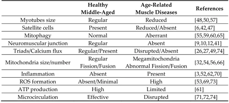
[74]. Main features in adulthood and aging-induced muscle disorders are collected in Table 1.
Table 1. Main features of healthy and age-related skeletal muscle disorders in mammals.
Healthy Middle-Aged
Age-Related
Muscle Diseases References
Myotubes size Regular Reduced [48,50,57]
Satellite cells Present Reduced/Absent [6,42,47]
Mitophagy Normal Aberrant [55,59,60,65]
Neuromuscular junction Regular Absent [9,10,12,41] Triads/Calcium flux Regular/Present Disrupted/Absent [26,27,49,74] Mitochondria size/number Regular
Fission/Fusion
Megamitochondria
Abnormal Fission/Fusion [32,54,56,66]
Inflammation Absent Present [3,52,62,70]
ROS formation Absent/Minimal High [53,69,73]
ATP production High Limited [61]
Microcirculation Effective Disrupted [71,72,74]
*ROS, reactive oxygen species.
2. Melatonin Alleviates Skeletal Muscle Disorders In Vitro and In Vivo
Melatonin (N-acetyl-5-methoxytryptamine) is an evolutionary-conserved molecule originally
isolated from the pineal gland and considered a regulator of circadian rhythms and seasonal breeding
[75,76]. Actually, melatonin has multiple extraordinary functions such as anti-tumor, antioxidant, and
anti-inflammatory indolamine [77–79]. In mammals, melatonin has been identified in all body fluids
and in several extra-pineal sites such as skin, gastrointestinal tract, liver, kidney, immune system, testis,
and skeletal muscles [80]. Remarkably, during the last decade, the indole has been detected in several
edible plants, eggs, and fish, assuming an interesting and promising role as a nutraceutical [81,82]. The
compelling evidence of decreased endogenous melatonin in senescence triggered intense research on
its potential role as a dietary supplement to prevent and treat aging and age-related diseases [83,84].
Intriguingly, in postmenopausal women, the drop of urinary melatonin correlated with
sarcopenia and, in castrated male rats, melatonin supply slowed muscle atrophy, acting as
testosterone [85,86]. Chronic melatonin intake prevented age-related mitochondrial damage in the
heart and the diaphragm muscle of accelerated aged SAMP 8-mice [87]. Muscle strength depends on
constant regular glucose and insulin levels in the blood, but in sarcopenia, insulin-resistance occurred
[88]. Furthermore, inflammation and lower glycolytic potential, assessed as lactate amount, strongly
influenced skeletal muscle metabolism in sarcopenia [89]. In the gastrocnemius muscle of aged mice
altered autophagy, nuclear fragmentation and abnormal lactate production were detected, but all
these adverse changes decreased by oral melatonin intake [90]. Remarkably, melatonin
supplementation in NLRP3 KO mice was particularly beneficial to retard the onset of sarcopenia in
gastrocnemius muscle in aged animals [91]. Interestingly, exogenous melatonin regulated insulin
resistance, ameliorated mitochondrial function in rat muscles, and prevented chemically induced
apoptosis and endoplasmic reticulum stress in different skeletal muscle cells in vitro [92–97].
Coto-Montes and co-workers reviewed the promising utility of safe melatonin dietary intake in
sarcopenia, even if its therapeutic potential in patients is still controversial [98,99]. Recent evidence
in different sarcopenic mice models obtained by single or double KO (DKO) of mitochondrial shaping
proteins (DRP1-KO and Opa1-Drp1 DKO) definitively indicated how, in skeletal muscles
mitohormesis, the mitochondrial size balance was essential [100,101].
Considering the post-mitotic nature of skeletal myofibers, skeletal muscle healing and recovery
after prolonged ischemia are other relevant clinical issues [102]. Several studies demonstrated that
melatonin attenuated ischemic damage and restored microvascular structure and perfusion in rat
cremaster and gracilis muscles during ischemia/reperfusion [103,104].
Moreover, muscular traumas, often during sports performance, are very common evidence
associated with high medical expenses and disabilities. In addition to promising clinical trials with
progenitor cells in humans [105], there are convincing data on the beneficial role of melatonin in
crushed injured muscles in rodents. Chronic melatonin intake reduced apoptosis, increased twitch
force, and accelerated regeneration enhancing satellite cells in muscle injury in mice and in rat
[106,107]. Recently, in an experimental compression model of quadriceps muscle in rats, melatonin
administered two hours after the beginning of the compression and during the following six days
improved redox balance and reduced inflammatory markers and tissue damage [108].
Chronic muscular pain, cognitive dysfunctions, and sleep disorders are all hallmarks of
fibromyalgia linked to reduced urinary secretion of melatonin in women [109]. Considering that an
effective therapy to alleviate pain and muscle damage in this syndrome is still lacking, our group
studied the effects of melatonin in fibromyalgia in rats. Fibromyalgia was induced by reserpine injection
and melatonin administered in tap water, concurrent with reserpine, for one or two months. In rat
gastrocnemius muscle, melatonin reduced oxidative changes and ameliorated mitochondria shape and
cristae, improving voluntary motor activity [110]. More recently, we adopted the same model focusing
on mitochondrial markers in rat gastrocnemius muscle. Interestingly, PGC1-alpha pathway and
mitofusin 2 (MF2), essential indicators of mitochondrial activity and fusion, were affected in reserpine
injected rat but preserved after oral melatonin intake [111]. These data strongly indicated that, in the
muscle, melatonin directly accumulated in the mitochondria where it was able to sustain proper size
and function.
Altered oxidative balance and abnormal mitochondria characterize DMD, a severe genetic
disorder associated with muscle weaning and atrophy [112]. Melatonin, which was successfully
administered as a nutraceutical compound in preclinical mice models and in DMD patients,
ameliorated muscle metabolism and strength [113,114]. Indeed, the indole sustained the antioxidant
muscular potential, increasing total glutathione content and promoting an effective contraction.
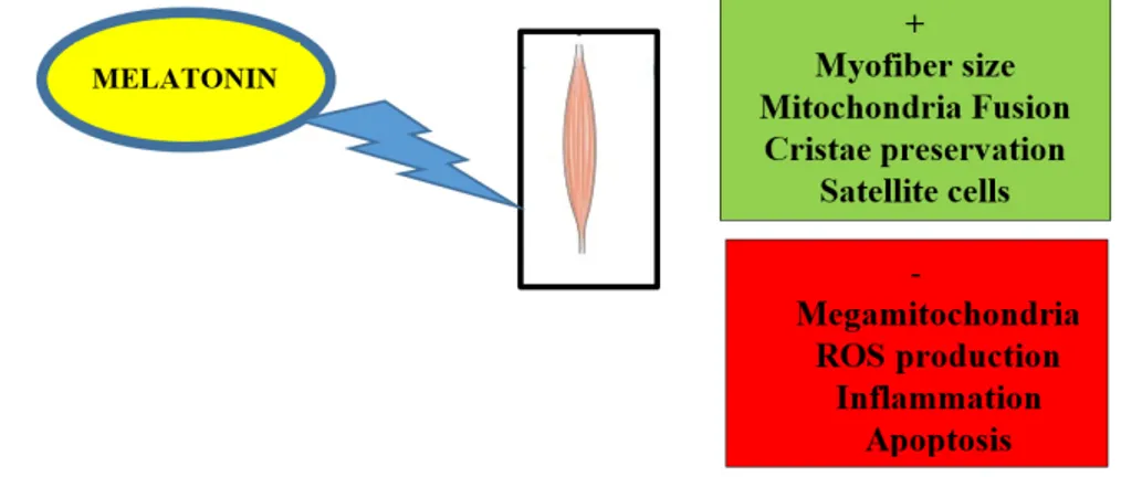
A summary of beneficial or promising actions of melatonin in aged induced skeletal muscle
diseases can be found in Figure 1.
Figure 1. Scheme illustrating improvement (+/ in green) and block (−/ in red) of mitochondrial or muscular events induced by dietary melatonin in aged or damaged skeletal muscle.
3. Exercise—an Anti-Aging Strategy that Preserves Mitochondria in Skeletal Muscle
Physical activity and proper nutrition represent the best lifestyle measures to prevent and to
retard age-related sarcopenia and progressive muscular weakness [115,116]. Remarkably, regular
and controlled exercise with advancing age prolongs lifespan and improves skeletal muscle mass and
performance [117,118].
However, there are different types of physical training with different impact on skeletal muscle
composition and metabolism: acute, chronic, or glycolytic and aerobic exercise [44,119]. Different
types of exercise can be combined in a mixed training. Exercise greatly remodels skeletal muscle
mitochondria size and number and accelerates mitophagy, the peculiar dismantling of damaged
mitochondria [120]. Indeed, the proper balance between new synthesis of mitochondria and
mitochondrial degradation is dramatically altered in aged skeletal muscles [121].
Several studies reported that different muscles aged differently depending on their fiber composition
and metabolism. Generally, mitochondrial respiratory activity is well preserved in slow oxidative muscles
rich in type I, IIA, and 2X fibers, but damaged in aged fast-twitch glycolytic muscles rich in type IIB fibers
[122–124]. Recently, Crupi et al. measured the respiratory activity in isolated mitochondria from fast
glycolytic tibialis anterior muscle versus slow oxidative soleus muscle in aged mice and demonstrated that
oxidative fibers are preserved while the glycolytic ones are damaged [125]. Thus, a fundamental strategy
of physical training in the elderly is to strengthen the oxidative muscles.
At a cellular level, to alleviate sarcopenia is necessary to improve mitochondria number and
turnover of contractile proteins and anti-oxidant enzymes [126]. A proper mitochondrial turnover is
crucial in muscle adaptation to exercise given that abnormal proteostasis leads to loss of contractile
proteins in aged muscles [127,128]. However, despite controlled physical activity, complete muscle
restoration is impossible due to unavoidable oxidative deterioration of fast glycolytic fibers and
enhanced expression of age-related genes insensitive to exercise benefit [129,130].
Remarkably, there is a strict connection between genotype and phenotype in gene
polymorphism for structural proteins and angiotensin-converting enzyme (ACE) [131]. This
connection is particularly evident in differences between endurance or power muscles in athletes
influencing sports performance, degree of vascularization, and mitochondria1 function [132]. A great
deal of evidence indicates that mitophagy in muscle is activated by exercise but is abnormal in aging
if mitophagy flux is excessive and mitochondrial quality reduced [133,134]. Multiple receptors and
signals are involved to drive proper mitochondria turnover in skeletal muscle and to regulate
lysosomal homeostasis. One of the most studied transcription factors activated during exercise and
able to regulate mitophagy is transcription factor EB (TFEB) [135]. This molecule is active when
dephosphorylated by calcium ions released from the sarcoplasmic reticulum during acute exercise
and, after translocation into the nucleus, controls the expression of several genes driving the
autophagy–mitophagy steps [136]. Another master regulator of mitophagy in muscle is PGC1α,
normally activated during acute exercise [137,138]. Recent study in mice indicated that chronic
exercise and stimulated contractile activity ameliorated mitochondria, thus mitophagy was not
necessary [139]. However, the use of colchicine as a microtubule polymerization inhibitor allowed
measuring the enhanced mitophagy flux in muscles of voluntary wheel trained mice [132].
Remarkably, not only mitochondria but also lysosomes are crucial for an efficient mitophagy and the
maintenance of mitohormesis. Recently, Triolo and Hood reviewed the beneficial effect of acute and
chronic exercise in lysosomal biogenesis and reported that exercise might be a peculiar
non-pharmacological “therapy” to clear disrupted cellular components [140]. In particular, the authors
stressed that, after physical training in rodent models of lysosomal diseases such as Pompe disease and
Danon syndrome, muscle mass and strength increased, while mitophagy and lysosomes production
ameliorated. Moreover, the therapeutic potentiality of aerobic and resistance exercise to promote skeletal
muscle performance was reported in patients affected by Pompe disease [141,142].
Emerging evidence indicates that a progressive resistance program better than acute oxidative
exercise is crucial to preserve mobility in aged patients, thus retarding the onset of frailty and related
metabolic and cardiovascular diseases [143]. Another beneficial role of exercise is the secretion of
muscular cytokines, called myokines, which regulate multiple biological functions [144].
Indeed, throughout the blood circulation, myokines influence muscles but also external sites
such as the adipose organ [145]. Among over three thousand myokine, it is necessary here to outline
fibroblast growth factor 21 (FGF21), a hormone-like molecule mainly secreted by the glycolytic fibers,
irisin secreted by the oxidative ones during contraction, and brain-derived neurotrophic factor
(BDNF) activated by aerobic exercise [146–148]. Remarkably, resistance training is able to recover the
secretory activity of aged muscles [149,150].
A recent field of research is the analysis of circulating exosomes, a sort of nanovescicles that
deliver myokines during exercise from muscles to adipose organs or other sites [151]. Interestingly,
new soluble factors delivered by exosomes might represent therapeutic or diagnostic targets of
muscle wasting or walking speed decline in aging [152,153].
4. Impact of Melatonin on Skeletal Muscle Activity and Exercise
Day/night cycles and seasonal rhythms are evolutionary-conserved activities that deeply
influence skeletal muscle mass, performance, and mitochondrial function [154,155]. The circadian
clock conditions whole body homeostasis and mainly the sleep–wake cycles that are essential for
mental and physical fitness [156].
In humans, the master regulator of biological rhythms is the suprachiasmatic nucleus (SCN)
located in the anterior hypothalamus in the brain [157]. This central area is functionally linked to
peripheral sites by factors called Zeitgebers in German, or “synchronizers” in English, such as daylight
exposure, physical activity, sleep, and eating time habit [158,159]. Remarkably, the SCN pathway
linked to retinal ganglion cells produces melatonin, an endogenous Zeitgeber able to control vital
rhythms during the nighttime, to reduce mitochondrial dysfunctions and chronodisruption [160,161].
In particular, melatonin added in vitro to brain slices stimulated SCN phase via melatonin type-2
receptors and protein kinase C activation [162].
Molecular mechanisms regulating circadian rhythms, clock genes, and transcriptional regulators
were firstly characterized in Drosophila and then extended to higher organisms [163]. Compelling
evidence indicates that there is another peripheral clock in skeletal muscle crucial for the maintenance
of mitochondrial balance, muscle metabolism, and energy in sarcopenia [164–167].
A recent study demonstrated that old mice fed an obesogenic diet supplemented with nobiletin,
a polyphenol agonist of muscle circadian regulator retinoid acid receptor-related orphan receptor
(ROR), presented an enhancement of mitochondria activity, energy expenditure, and endurance
exercise in the calf muscles, gastrocnemius and soleus [168]. Moreover, actually single fiber proteomic
allows an unbiased determination of full muscle and mitochondrial proteins that are characteristic of
a specific myofiber to best correlate its status in health, aging, or metabolic diseases [169].
Exercise is another essential nonphotic Zeitgeber that synchronizes the circadian pathway and
sleep depth and controls muscle physiology during all lifespans but greatly in aging [170,171].
Notably, the direct influence of exercise on the endogenous melatonin secretion is still controversial,
probably due to different melatonin estimation in saliva or serum. Indeed, salivary melatonin
evaluated in men in the late evening was inversely related to the time of physical activity because it
was higher in the morning session versus a late afternoon session of steady-state running, thus the
morning fitness may predispose one to regular sleep [172]. Accumulating evidence in rodents
indicates that endogenous melatonin and circadian systems are modulated by repeated vigorous
exercise able to maintain the synchronous phase. Conversely, Escames et al. reported that, in humans,
due to a lack of control on competing Zeitgebers and the difficulty to directly estimate the SCN input,
the effects of exercise on endogenous melatonin are controversial [173]. Moreover, enhanced urinary
melatonin was associated with more grip and quadriceps muscle strength in the elderly population
[174]. However, in previously sedentary men and women aged 40-75 years after one year of moderate
exercise, the urinary melatonin metabolite levels were unchanged [175]. Recently, as a result of a
moderate exercise in a hypoxic status (equivalent to at 4500 m altitude), serum melatonin increased,
probably to protect against oxygen deprivation [176].
In any case, exogenous melatonin is useful as an antioxidant and an anti-inflammatory nutrient
for prolonging muscle strength and adaptation during strenuous exercise in rodents and men in
adulthood and aging [177–180]. Melatonin intake before and during exercise reduces glucose
resistance and ameliorates antioxidant status in various situations, such as during preparatory
training, in a soccer training camp, in resistance, or in high-trained athletes [181–184]. In particular,
during strenuous training and muscular trauma induced in rodents, melatonin supplementation via
different routes improved muscle recovery, inhibiting NF-kB activation and inflammatory cytokines
and downregulating atrophy pathways [185]. However, Beck et al. reported an ergogenic role of
intraperitoneal melatonin in gastrocnemius muscle in rat swimming that mimicked long-duration
aerobic exercise in human but increased inflammation, probably due to excessive physical
performance extension [186,187]. Remarkably, in humans, melatonin was devoid of any side effect
despite administration via several routes [188]. Melatonin induced drowsiness must be considered,
and the best time to assume the indole is during post-exercise recovery or convalescence [189].
On the contrary, the influence of exogenous melatonin on the physical performance is still
debated and controversial. In a recent systematic review, Lopez-Flores et al. indicated that the intake
of melatonin might be effective or ineffective depending on the type of physical activity [190]. Indeed,
melatonin secretion is limited during aerobic exercise but is enhanced during high-intensity exercise,
thus the effects of exogenous melatonin supply are different.
Several studies agreed on the beneficial role of oral melatonin intake to readjust sleep cycles and
jet-lag adverse effects after transcontinental flights, making athletes more prone to optimal
performance [191]. A single “pharmacological“ dose of melatonin (3 mg) should be taken in daytime
to shift the circadian rhythm in the proper direction and to best adapt to the new time zone according
to westward or eastward travel. This suggestion might be very useful, for example, for athletes
participating to the next 2020 Olympic Games in Tokyo, Japan. Moreover, 10 mg melatonin taken
after strenuous exercise in late evening was effective to prolong sleep and to ameliorate short–term
activity in the following morning in teenager athletes [192].
Another interesting property of a single melatonin dose (2.5 mg) is to reduce rectal temperature
during intermittent exercise in a hot environment without any alertness [193]. Independently from
physical training, Liu et al. reported that melatonin (20 mg/kg) intravenously administered for four
weeks enhanced lipolysis in mice vastus lateralis muscle by activating the “browning” effect in the
adipose tissue and thermogenesis [194].
Remarkably, a recent comprehensive review set the point on the urgency to fill the existing gap
on the best therapeutic melatonin dosage in human diseases despite a a great deal of experience in
rodents [195]. Currently, melatonin is not still administered at the best “clinical” dosage, from 40 to
100 mg/day, that is necessary to obtain the best results in metabolic and neurodegenerative diseases.
This “therapeutic range” is defined by the human equivalent dose (HED) considering a 75 kg adult
men and normalization of body surface area [196]
Melatonin schedule treatment for skeletal muscle damage or activity is summarized in Table 2.
Table 2. Melatonin regime in skeletal muscle and exercise in rodents and humans.Subjects/Cells Dose Times of Administration
Reference- Muscle Type or
Exercise
Wistar albino rats 6 mg/kg s.c. 5 weeks [86] Soleus
SAMP8 mice 10 mg/kg oral (water) 10 months [87] Diaphragm C57BL/6J mice 10 mg/kg oral (chow) 2 months [90] Gastrocnemius NLRP3 KO mice 10 mg/kg oral (chow) 2 months [91] Gastrocnemius Pinealectomized Wistar rats 0.5 mg/kg oral (water) 45 days [92] L6 cells 10 nM 24 h [93] C2C12 1–10 nM 20 min [94] C2C12 cells 100 mM 12–24 h [95] C2C12 cells 100 nM 16 h [96]
Primary muscle cells 1–100 µM 24 h [97]
Elderly patients 1 mg/day
oral 4 weeks [99]
Sprague-Dawley rats 10 mg/kg i.p. 30 min prior and immediately after
reperfusion [103] Cremaster
Sprague-Dawley rats 10 mg/kg i.v. 10 min prior and 10 min after reperfusion [104] Gracilis
Wistar rats 10 mg/kg i.p. 4–14 days [106] Soleus
Wistar rats 10 mg/kg i.p. 1–14 days [107] Soleus
Wistar rats 20 mg/kg i.p. 7 days [108]
Sprague-Dawley rats
2.5 mg/kg 5 mg/kg oral (water)
1–2 months [110] Gastrocnemius
Sprague-Dawley rats 5 mg/kg oral
(water) 2 months [111] Gastrocnemius
Wistar rats 1 mg/kg oral
(water) 16 weeks
[177] Treadmill running
Adult men 15 mg oral Before starting exercise [178]
High intensity run Wistar rats 20 mg/kg i.p. Immediately after or 2 h after exercise [179]
Treadmill running Adult subjects 6 mg oral Before starting exercise [180]
30 min graded exercise
Football players 5 mg oral 30 days [181]
Preparatory training Professional
soccer players 6 mg oral 6 days
[182] Intensive training
Adult athletes 100 mg oral 4 weeks [183]
Resistance training
Adult athletes 20 mg oral 2 weeks [184]
High Intensity training
Wistar rat 10 mg/kg i.p. 2 days after exercise [186]
Incremental swimming
Teenage athletes 10 mg oral After exercise [192]
Exhaustive exercise
Adult subjects 2.5 mg oral Before exercise [193]
5. The Emerging Concept of the Gut–Muscle Axis—Role of Exercise and Melatonin in the Gut
Within the past few years, a novel and intriguing concept has been formulated: muscle
composition and metabolism greatly depend on the bacteria population in the gut, called the
microbiome. Thus, factors affecting the inter-individual microbiome such as aging, metabolic
diseases, inflammation, cancer, or malnutrition might condition muscle weakness and induce
sarcopenia [197–199]. Indeed, in age related diseases and obesity, an abnormal intestinal flora, i.e.,
dysbiosis, was detected [200–202]. The analysis of the composition of the intestinal microbiota in the
elderly demonstrated that there were more pathogens such as Enterobacteriaceae and scarce
butyrate-producing healthy bacteria, leading to less tight junctions in the mucosa and altered permeability
[203].
Bindels and Delzenne suggested that the gut bacteria might be a potential therapeutic target to
change muscle mass in cancer cachexia or undernutrition and hypothesized gut–muscle axis [204].
Firstly, Backhed et al. transplanted fecal bacteria from the adult pathogen-free mice to germ-free mice,
in the latter modifying the fat deposition in the liver and the metabolism [205]. This process is defined
as “conventionalization” and indicates the implantation and the colonization of bacteria from the
distal intestine of a lean donor in the gut of a recipient. In another study, the above authors reported
that, in the gastrocnemius muscle of germ free mice lacking the anabolic fasting-induced adipose factor
(Fiaf) and fed a Western diet, there was reduced fatty acid oxidation [206]. On the contrary,
germ-free mice were resistant to a Western diet if compared to control pathogen-germ-free mice with intact gut
bacteria. A crucial study by Yan et al. demonstrated that germ-free mice receiving fecal microbiota
from obese Rongchang pigs presented a similar fatty phenotype with higher intramuscular
triglycerides. In particular, the gastrocnemius muscle of recipient mice presented altered myofibers
composition with more type I slow-contracting versus type II B fast fibers [207]. The study firstly
indicated that there was a direct influence of gut bacteria on skeletal muscle organization and
metabolism in mice. Recently, Lahiri et al. analyzed plasma and muscles in germ-free mice without
any microbiota and fed either a regular diet or a mix of single chain fatty acids (SCFAs) able to
promote beneficial insulin sensitivity and mitochondrial biogenesis [208]. In the first experimental
set, mice presented muscle atrophy and enhanced molecular signaling of atrophy. In the second
experimental set, SCFAs produced by the gut ameliorated muscle strength and composition. Finally,
when germ-free mice were transplanted with a fecal content of pathogen-free mice treated with
antibiotics, they developed muscle atrophy and reduced function due to a reduced healthy
microbiota. These data agreed with Manickam et al.’s study, which demonstrated dysbiosis in
pathogen-free mice treated with the antibiotic metronidazole producing scarce gastrocnemius muscle
mass, abnormal activation of atrophy genes, and insulin resistence [209]. Conversely, fecal
transplantation of old human subjects with high physical performance, called high functioning, in
germ-free mice induced a peculiar microflora rich in Prevotella and Barnesiella species and developed
more grip strength [210].
However, if several studies in animals indicate the possibility of a gut–muscle axis, it is
important to outline that the reverse signaling from the muscle to the gut might also occur. Indeed,
microbes develop a symbiontic reaction with their host and, consequently, reduced physical activity
in sarcopenic subjects may be associated with different microbiota composition and metabolism
[211,212]. This last point might be considered when the physical performance is measured in humans
by gait speed test, because gut bacteria synthesize neurotransmitters such as serotonin,
norepinephrine, GABA, or dopamine are able to influence the neurological control of movement
[213].
Intriguingly, exercise strongly influences microbiota composition and produces beneficial
metabolites in rodents and men. Remarkably, evidence in germ-free mice documents reduced
inflammation and best swim time when colonized with Eubacterium or Clostridium species or highest
treadmill run time if colonized by Veilonella [214–216]. Scheiman et al. reported that, in marathon
runners after exercise, the high microbiota content with Veilonella transplanted in mice produced an
intense treadmill activity [217]. The main metabolite produced by Veilonella in the colon was lactate
then converted into propionate and used to produce short fatty chain acids (SCFAs), such as
n-butyrate, acetate, and propionate, a source of energy for physical performance.
The balance between human microbiota Bacteroidetes or Firmicutes phyla in aerobic exercise is
essential for health, and disrupted equilibrium of gut bacteria colonization may induce inflammation
and metabolic and neurological diseases [218]. On the contrary, aerobic exercise influences Firmicutes
growth, modulating via the microbiome–gut–brain axis the symptoms of irritable bowl syndrome,
axiety, and depression [219,220].
However, if the exercise intensity is too strong, the gut microbiota composition is dysregulated,
and overtrained Kunming mice presented inflammation, inducing Helicobacter pylori and increased
risk of peptic ulcer [221]. Moreover, depletion of gut microbiota in mice treated with antibiotics for
21 days induced low running endurance and less fatigue in the extensorum digitorum longus muscle.
This last result is related to reduced glycogen production by a scarce number of bacteria present in
the gut that directly synthesize glucose [222].
Unfortunately, dysbiosis and low butyrate production in the gut decreased melatonin secretion
and enhanced permeability and inflammation in several organs [223]. Paulose et al. detected in the
human gastrointestinal system that Enterobacter aerogenes is sensitive to melatonin and is
synchronously regulated in the daily swarming [224]. The authors hypothesized that melatonin
might act as a local Zeitgeber in the gut. Moreover, there is evidence that butyrate supplementation
stimulated melatonin secretion in the duodenal tissue and in human colon carcinoma Caco 2 cells
[225].
Firstly, Xu et al. reported that melatonin (50 mg/kg) delivered by gavage was effective as a probiotic
compound ameliorating gut dysbiosis in high fat fed mice [226]. Moreover, in obese mice, melatonin
changed the proportion of Firmicutes to Bacterioides and enhanced Akkermasia species that restored
intestinal barriers in obese mice. Another study reported that melatonin supplementation in drinking
water increased the microbiota variability in obese mice [227]. However, microbiota from obese mice plus
melatonin transplanted in antibiotic-treated high fat fed mice failed to ameliorate lipid metabolism [228].
Probiotic nutrient intake is beneficial for the gut–microbiome–muscle axis, and this is
particularly evident during exercise. Indeed, the analysis of gut microbiome in athletes gives the best
information on the general health status, and proper nutrition may influence its composition to obtain
the best physical performance [229]. Several studies on men after strenuous exercise and athletes
demonstrated that probiotics intake changed microbiota, limited “leaky gut”, reduced inflammatory
cytokines, and potentiated muscle strength [230].
Moreover, a diet rich in protein directly regulated muscle mass, but peculiar protein composition is
crucial to limit sarcopenia in aging [231]. Among the dietary proteins that best sustain muscle mass, whey
protein is effective combined with resistance training to modulate gut microbiota and to prevent
sarcopenia [232]. However, a strong inter-individual response to resistance training exists that is regulated
by myogenic molecular circadian pathways [233]. Remarkably, the gut microbiota influenced anabolic
resistance in the skeletal muscle in the elderly. A recent metabolomic study of fecal content demonstrated
a good correspondence between bacteria in the gut and the visceral fat and obesity grade [234].
Finally, sleep deprived mice showed reduced gut microbiota and limited probiotic species such
as Bacteroides, Akkermasia, and Faecalibacterium [235]. Interestingly, in this animal model, melatonin
reversed abnormal microbiota composition, indicating that sleep deprivation might reduce local
melatonin secretion and melatonin type-1 receptor activity in the gut.
6. Conclusions and Perspectives
The reciprocal influence of aging, diet, exercise, and gut microbiome on skeletal muscle mass
and strength requires urgent innovative research and clinical trials considering the progressive
increase in the age of the population in the world.
Melatonin is a highly evolutionary-conserved ancient molecule that was only recently
rediscovered as a safe dietary supplement in muscle disorders and in exercise. This review attempts
to shed light on potential and promising therapeutic roles of melatonin to limit muscle deterioration,
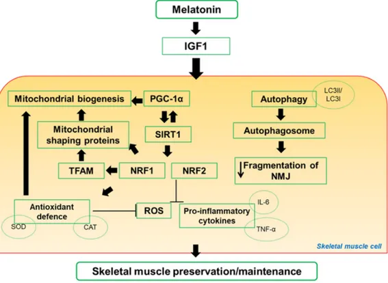
mainly mitochondrial function, and sarcopenia. Main pathways activated by melatonin in skeletal
muscle are drawn in Figure 2.
Figure 2. Scheme representation of proposal pathways regulated by melatonin in skeletal muscle. Notably, melatonin mechanisms of action involved mitochondria signaling. CAT: catalase; IL-6: interleukin-6; IGF1: insulin-like growth factor-1; LC3I/LC3II: microtubule-associated protein 1A/1B-light chain 3 free in the cytosol or conjugated to phosphotidylethanolamine during autophagy; NMJ: neuromuscular junction; NRF1 and NRF2: nuclear respiratory factor 1 and 2; PGC-1α: peroxisome proliferative activated receptor gamma coactivator-1alpha; ROS: reactive oxygen species; SIRT1: sirtuin1; SOD: superoxide dismutase; TFAM: mitochondrial transcription factor; TNFα: tumor necrosis factor alpha.
However, the utility of melatonin in athletes to obtain the best physical performance is strictly
time-dependent, dose-dependent, and exercise-dependent. Finally, the benefit of melatonin on the
gut microbiota is still very limited, and its direct influence on the gut–muscle axis is actually only
speculative. Some limitations must be addressed in this manuscript. Firstly, we did not focus on the
role of melatonin in cancer-induced cachexia due to the high number of existing reviews on cancer.
Second, we did not consider differences in male compared to female or the role of sex hormones on
muscle strength and exercise. Third, most of the studies on the gut–muscle axis were conducted on
germ-free mice, not on humans. However, we are confident that new experimental studies and
comphrensive reviews will be produced on these crucial themes in the future.
Author Contributions: Conceptualization: AS, GF, LFR; Writing, original draft preparation: A.S.; Writing-revised review and editing: AS., GF and LFR. All authors have read and agreed to the published version of the manuscript.
Funding: “This research received no external funding”.
Acknowledgments: Siderurgica Leonessa Srl donation to AS has been greatly appreciated. Conflicts of Interest: “The authors declare no conflict of interest.”
References
1. Janssen, I.; Heymsfield, S.B.; Wang, Z.M.; Ross, R. Skeletal muscle mass and distribution in 468 men and women aged 18-88 yr. J. Appl. Physiol. 1985, 2000, 81–88. DOI: 10.1152/jappl.2000.89.1.81.
2. Frontera, W.R.; Ochala, J. Skeletal muscle: A brief review of structure and function. Calcif. Tissue Int. 2015,
96, 183–195. DOI: 10.1007/s00223-014-9915-y.
3. Shadrin, I.; Khodabukus, A.; Bursac, N. Striated muscle function, regeneration, and repair. Cell. Mol. Life
Sci. 2016, 73, 4175–4202. DOI: 10.1007/s00018-016-2285-z.
4. Giudice, J.; Taylor, J. Muscle as a paracrine and endocrine organ. Curr. Opin. Pharmacol. 2017, 34, 49–55. DOI: 10.1016/j.coph.2017.05.005.
5. Roman, W.; Gomes, E.R. Nuclear positioning in skeletal muscle. Semin. Cell Dev. Biol. 2018, 82, 51–56. DOI: 10.1016/j.semcdb.2017.11.005.
6. Dumont, N.A.; Bentzinger, C.F.; Sincennes, M.C.; Rudnicki, M.A. Satellite cells and skeletal muscle regeneration. Compr. Physiol. 2015, 5, 1027–1059. DOI: 10.1002/cphy.c140068.
7. Gillies, A.R.; Bushong, E.A.; Deerinck, T.J.; Ellisman, M.H.; Lieber, R., L. Three-dimensional reconstruction of skeletal muscle extracellular matrix ultrastructure. Microsc. Microanal. 2014, 20, 1835–1840. DOI: 10.1017/S1431927614013300.
8. Hendrickse, P.; Degens, H. The role of the microcirculation in muscle function and plasticity. J. Muscle Res.
Cell Motil. 2019, 40, 127–140. DOI: 10.1007/s10974-019-09520-2.
9. Lepore, E.; Casola, I.; Dobrowolny, G.; Musaro’, A. Neuromuscolar junction as an entity of nerve-muscle communication. Cells 2019, 8, E906. DOI: 10.3390/cells8080906.
10. Slater, C.R. The structure of human neuromuscular junctions. Some unanswered molecular questions. Int.
J. Mol. Sci. 2017, 18, E 2183. DOI: 10.3390/ijms18102183.
11. Vock, R.; Weibel, E.; Hoppeler, H.; Ordway, G.; Weber, J.; Taylor, C. Design of the oxygen and substrate supply to muscle cells. J. Exp. Biol. 1996, 199, 1675–1688.
12. Boncompagni, S.; Rossi, A.; Micaroni, M.; Beznoussenko, G.; Polishchuk, R., Dirksen, R.; Protasi, F. Mitochondria are linked to calcium stores in striated muscle by developmentally regulated tethering structures. Mol. Biol. Cell. 2009, 20, 1058–1067. DOI: 10.1091/mcb.E08-07-0783.
13. Ferreira, R.; Vitorino, R.; Alves, R.; Appel, H.; Powers, S.; Duarte, J.; Amado, F. Subsarcolemmal and intermyofibrillar mitochondria proteome differences disclose functional specializations in skeletal muscles.
Proteomics 2010, 10, 3142–3154. DOI: 10.1002/pmic.201000173.
14. Dahl, R.; Larsen, S.; Dohlmann, T.; Qvortrup, K.; Helge, J.; Dela, F.; Prats, C. Three-dimensional reconstruction of the human skeletal muscle mitochondrial network as a tool to assess mitochondrial content and structural organization. Acta Physiol. (Oxf.) 2015, 213, 145–155. DOI:10.1111/apha. 12289. 15. Bleck, C.; Kim, Y., Willingham, T.; Glancy, B. Subcellular connectomic analyses of energy networks in
striated muscle. Nat. Commun. 2018, 9, 5111. DOI:10.1038/s41467-018-07676-y.
16. Barbieri, E.; Sestili, P. Reactive oxygen species in skeletal muscle signaling. J. Signal. Transduct. 2012, 2012, 982794. DOI: 10.1155/2012/982794.
17. Glancy, B.; Hartnell, L.; Malide, D.; Yu, Z.; Combs, C.; Connelly, P.; Subramaniam, S.; Balaban, R. Mitochondrial reticulum for cellular energy distribution in muscle. Nature 2015, 523, 617–620. DOI: 10.1038/nature14614.
18. Vincent, A.; White, K.; Davey, T.; Philips, J.; Ogden, T.; Lawless, C.; Warren, C.; Hall, M.; Ng, Y.; Falkous, G.; et al.Quantitative 3D mapping of the human skeletal muscle mitochondrial network. Cell Rep. 2019, 26, 996–1009. DOI: 10.1016/j.celrep.2019.01.010.
19. Liesa, M.; Shirihai, O. Mitochondrial dynamics in the regulation of nutrient utilization and energy expenditure. Cell. Metab. 2013, 17, 491–506. DOI: 10.1016/j.cmet.2013.03.002.
20. Mishra, P.; Varuzhanyan,G.; Pham, A.; Chan, D. Mitochondrial dynamics is a distinguishing feature of skeletal muscle fiber types and regulates organellar compartmentalization. Cell. Metab. 2015, 22, 1033–1044. DOI: 10.1016/j.cmet.2015.09.027.
21. Pette, D.; Staron, R. Myosin isoforms, muscle fiber types, and transitions. Microsc. Res.Tech. 2000, 50, 500– 509. DOI: 10.1002/1097-0029(20000915)50:6<500::AID-JEMT7>3.0.CO;2-7.
22. Schiaffino, S.; Reggiani, C. Fiber types in mammalian skeletal muscles. Physiol. Rev. 2011, 91, 1447–1531. DOI: 10.1152/physrev.00031.2010.
23. Talbot, J.; Maves, L. Skeletal muscle fiber type: Using insights from muscle developmental biology to dissect targets for susceptibility and resistance to muscle disease. WIREs Dev. Biol. 2016, 5, 518–534. DOI: 10.1002/wdev.230.
24. Bourdeau, J.; Sephton, C.; Dutchak, P. Metabolic networks influencing skeletal muscle fiber composition.
Front. Cell Dev. Biol. 2018, 6, 125. DOI: 10.3389/fcell.2018.00125.
25. Szent-Gyorgyi, A. The early history of the biochemistry of muscle contraction. J. Gen. Physiol. 2004, 123, 631–641. DOI: 10.1085/jgp.200409091.
26. Franzini-Armstrong, C.; Boncompagni, S. The evolution of the mitochondria-to-calcium release units relationship in vertebrate skeletal muscles. J. Biomed. Biotechnol. 2011, 2011, 830573. DOI: 10.1155/2011/830573.
27. Rossi, A.; Boncompagni, S.; Dirksen, R. Sarcoplasmic reticulum-mitochondrial symbiosis: Bidirectional signaling in skeletal muscle. Exerc. Sport Sci. Rev. 2009, 37, 29–35. DOI: 10.1097/JES.0b013e3181911fa4. 28. Ogata, T.; Yamasaki, Y. Ultra-high resolution electron microscopy of mitochondria and sarcoplasmic
reticulum arrangement in human red, white, and intermediate muscle fibers. Anat.Rec. 1997, 248, 214–223. 29. Westerblad, H.; Bruton, J.; Katz, A. Skeletal muscle: Energy metabolism, fiber types, fatigue and
adaptability. Exp. Cell Res. 2010, 316, 3093–3099. DOI: 10.1016/j.yexcr.2010.05.019.
30. Zierath, J.; Hawley, J. Skeletal muscle fiber type: Influence on contractile and metabolic properties. Plos
Biology 2004, 2, 1523–1527. DOI: 10.1371/journal.pbio.0020348.
31. Jeon, Y.; Choi, J.; Kim, H.; Lee, H.; Lim, J.; Choi, S. Sex and fiber-type-related contractile properties in human single muscle fiber. J. Exerc. Rehabil. 2019, 15, 537–545. DOI: 10.12965/jer.1938336.168.
32. Hood, D.; Memme, J.; Oliveira, A.; Triolo, M. Maintenance of skeletal muscle mitochondria in health, exercise, and Aging. Ann.Rev.Physiol. 2019, 81, 19–41. DOI: 10.1146/annurev-physiol-020518-114310. 33. Mishra, P.; Chan, D. Metabolic regulation of mitochondrial dynamics. J. Cell. Biol. 2016, 212, 379–387. DOI:
10.1083/jcb.201511036.
34. Chen, H.; Chomyn, A.; Chan, D. Disruption of fusion results in mitochondrial heterogeneity and dysfunction. J. Biol. Chem. 2005, 280, 26185–26192. DOI: 10.1074/jbc.M503062200.
35. Twig, G.; Shirihai, O. The interplay between mitochondrial dynamics and mitophagy. Antioxid. Redox
Signal. 2011, 14, 1939–1951. DOI: 10.1089/ars.2010.3779.
36. Gouspillou, G.; Bourdel-Marchasson, I.; Rouland, R.; Calmettes, G.; Biran, M.; Deschodt-Arsac, V.; Miraux, S.; Thiaudiere, E.; Pasdois, P.; Detaille, D.; et al. Mitochondrial energetics is impaired in vivo aged skeletal muscle. Aging Cell 2014, 13, 39–48. DOI: 10.1111/acel.12147.
37. Picard, M.; Ritchie, D.; Wright, K.; Romestaing, C.; Thomas, M.; Rowan, S.; Taivassalo, T.; Hepple, R.. Mitochondrial functional impairment with aging is exaggerated in isolated mitochondria compared to permeabilized myofibers. Aging Cell 2010, 9, 1032–1046. DOI: 10.1111/j.1474-9726.2010.00628.x.
38. Choi, S.J. Age-related functional changes and susceptibility to eccentric contraction-induced damage in skeletal muscle cell. Integr. Med. Res. 2016, 5, 171–175. DOI: 10.1016/j.imz2016.05.004.
39. Leduc-Gaudet, J.; Picard, M.; St-Jean Pelletier, F.; Sgarioto, N., Auger, M., Vallee, J.; Robitaille, R.; St-Pierre, D.; Gouspillou, G. Mitochondrial morphology is altered in atrophied skeletal muscle of aged mice.
Oncotarget 2015, 6, 17923–17037. DOI: 10.18632/oncotarget.4236.
40. Delbono, O. Expression and regulation of excitation-contraction coupling proteins in aging skeletal muscle.
Curr. Aging Sci. 2011, 4, 248–259. DOI: 10.2174/1874609811104030248.
41. Jang, Y.; Van Remmen, H. Age-associated alterations of the neuromuscular junction. Exp. Gerontol. 2011,
46, 193–198. DOI:. 10.1016/j.exger.2010.08.029.
42. Miljkovic, N.; Lim, J.; Miljkovic, I.; Frontera, W. Aging of skeletal muscle fibers. Ann. Rehabil. Med. 2015, 39, 155–162. DOI: 10.5535/arm.2015.39.2.155.
43. Cruz-Jentoft, A.; Bahat, G.; Bauer, J.; Boirie, J.; Bruyere, O.; Cederholm, T.; Cooper, C.; Landi, F.; Rolland, Y.; Sayer, A.; et al. Sarcopenia: Revised European consensus on definition and diagnosis. Age Ageing 2019,
48, 16–31. DOI: 10.1093/ageing/afy169.
44. Carter, H.; Chen, C.; Hood, D. Mitochondria, muscle health, and exercise with advancing age. Physiology 2015, 30, 208–223. DOI: 10.1152/physiol.00039.2014.
45. Calvani, R.; Joseph, A.; Adhihetty, P.; Miccheli, A.; Bossola, M.; Leeuwenburgh, C.; Bernabei, R.; Marzetti, E. Mitochondrial pathways in sarcopenia of aging and disuse muscle atrophy. Biol. Chem. 2013, 394, 393– 414. DOI: 10.1515/hsz-2012-0247.
46. Fanzani, A.; Conraads, V.; Penna, F.; Martinet, W. Molecular and cellular mechanisms of skeletal muscle atrophy: An update. J. Cachexia Sarcopenia Muscle 2012, 3, 163–179. DOI: 10.1007/s13539-012-0074-6. 47. Hikida, R. Aging changes in satellite cells and their functions. Curr. Aging Sci. 2011, 4, 279–297. DOI:
10.2174/1874609811104030279.
48. Larsson, L.; Degens, H.; Li, M.; Salviati, L.; Lee, Y.; Thompson, W.; Kirkland, J.; Sandri, M. Sarcopenia: Aging-related loss of muscle mass and function. Physiol. Rev. 2019, 99, 427–511. DOI: 10.1152/physrev.00061.2017.
49. Zhao X, Weisleder N, Thornton A, Oppong Y, Campbell, R., Ma J et al. Compromised store-operated Ca2+
entry in aged skeletal muscle. Aging Cell 2008, 7, 561–568. DOI: 10.1111/j.1474-9726.2008.00408.x.
50. Sayed, R.; de Leonardis, E.; Guerrero-Martinez, J.; Rahim, I.; Mokhtar, D.; Saleh, A.; Abdalla, K.; Pozo, M.; Escames, G.; López, L.; et al. Identification of morphological markers of sarcopenia at early stage of aging in skeletal muscle of mice. Exp. Gerontol. 2016, 83, 22–30. DOI: 10.1016/j.exger.2016.07.007.
51. Zhu, S.; Tian, Z.; Torigoe, D.; Zhao, J.; Xie, P.; Sugizaki, T., Sato, M.; Horiguchi, H.; Terada, K.; Kadomatsu, T.; et al. Aging-and obesity-related peri-muscular adipose tissue accelerates muscle atrophy. PlosOne 2019,
14, e0221366. DOI: 10.1371/journal.pone.0221366.
52. Fougere, B.; Boulanger, E.; Nourhashemi, F.; Guyonnet, S.; Cesari, M. Chronic inflammation: Accelerator of biological aging. J. Gerontol. A Biol. Sci. Med. Sci. 2017, 72, 1218–1225. DOI: 10.1093/Gerona/glw240. 53. Marzetti, E.; Picca, A.; Marini, F.; Biancolillo, A.; Coelho Junior, H., Gervasoni, J.; Bossola, M.; Cesari, M.;
Onder, G.; Landi, F.; et al. Inflammatory signatures in older persons with physical frailty and sarcopenia: The frialty “cytokinome” at its core. Exp. Gerontol. 2019, 122, 129–138, 2019. DOI: 10.1016/j.exger.2019.04.019. 54. Szentesi, P.; Csernoch, L., Dux, L.; Keller-Pinter, A. Changes in Redox Signaling in the skeletal muscle with
aging. Oxid. Med. Cell. Long. 2019, 2019, 4617801. DOI: 10.1155/2019/4617801.
55. Del Campo, A. Mitophagy as a new therapeutic target for sarcopenia. Acta Physiol. 2019, 225, e13219. DOI: 10.1111/apha.13219.
56. Sheard, P.; Anderson, R. Age-related loss of muscle fibers is highly variable among mouse skeletal muscles.
Biogerontology 2012, 13, 157–167. DOI: 10.1007/s10522-011-9365-0.
57. Nilwik, R.; Snijders, T.; Leenders, M., Groen, B., van Kranenburg, J., Verdijk, L.; van Loon, L. The decline in skeletal muscle mass with aging is mainly attributed to a reduction in type II muscle fiber size. Exp.
Gerontol. 2013, 48, 492–498. DOI: 10.1016/j.exger.2013.02.012.
58. Del Campo, A.; Contreras-Hernandez, I.; Castro-Sepulveda, M.; Campos, C.; Figueroa, R.; Tevy, M.; Eisner, V.; Casas, M.; Jainovich, E. Muscle function decline and mitochondria changes in middle age precede sarcopenia in mice. Aging 2018, 10, 34–55. DOI: 10.18632/aging.101358.
59. Pernas, L.; Scorrano, L. Mito-Morphosis: Mitochondrial fusion, fission and cristae remodeling as key mediators of cellular function. Ann. Rev. Physiol. 2018, 78, 505–531. DOI: 10.1146/annurev-physiol-021115-105011.
60. Romanello, V.; Sandri, M. Mitochondrial quality control and muscle mass maintenance. Front. Physiol. 2015,
6, 422. DOI: 10.3389/fphys.2015.00422.
61. Le Moal, E.; Pialoux, V.; Juban, G.; Groussard, C.; Zouhal, H.; Chazaud, B.; Mounier, R. Redox control of skeletal muscle regeneration. Antiox. Redox Signal. 2017, 27, 276–310.
62. Zhou, R.; Yazdi, A.; Menu, P.; Tschopp, J. A role for mitochondria in NLRP3 inflammasome activation.
Nature 2011, 469, 221–225. DOI: 10.1038/nature09663.
63. Valentine, J.; Li, M.; Shoelson, S.; Zhang, N.; Reddick, R.; Musi, N. NF-kB regulates muscle development and mitochondrial function. J. Gerontol. A Biol. Sci. Med. Sci. 2018, DOI: 10.1093/Gerona/gly262.
64. Johnson, M.; Robinson, M.; Nair, K. Skeletal muscle aging and the mitochondrion. Trends Endocrinol. Metab. 2013, 24, 247–256. DOI: 10.1016/j.tem.2012.12.003.
65. Yeo, D.; Kang, C.; Gomez-Cabrera, M.; Vina, J.; Ji, L. Intensified mitophagy in skeletal muscle with aging is downregulated by PGC-1 alpha overexpression in vivo. Free Rad. Biol. Med. 2019, 130, 361–368. DOI: 10.1016/j.freeradbiomed.2018.10.456.
66. Huang, D.; Fan, S.; Chen, X.; Yan, X.; Zhang, X.; Ma, B.; Yu, D.; Xiao, W.; Zhuang, C.; Yu, Z. Nrf2 deficiency exacerbates frailty and sarcopenia by impairing skeletal muscle mitochondrial biogenesis and dynamics in an age-dependent manner. Exp. Gerontol. 2019, 119, 61–73. DOI: 10.1016/j.exger.2019.01.022.
67. Ciciliot, S.; Schiaffino, S. Regeneration of mammalian skeletal muscle. Basic mechanisms and clinical implications. Curr. Pharm. Des. 2010, 16, 906–914. DOI: 10.2174/138161210790883453.
68. Abeles, A.; Pillinger, M.; Solitar, B.; Abeles, M. Narrative review: The pathophysiology of fibromyalgia.
Ann. Intern.Med. 2007, 146, 726–734. DOI: 10.7326/0003-4819-146-10-200705150-00006.
69. Chung, C.P.; Titova, D.; Oeser, A.; Randels, M.; Avalos, I.; Milne, G.L.; Morrow, J.D.; Stein, C. Oxidative stress in fibromyalgia and its relationship to symptoms. Clin. Rheumatol. 2009, 28, 435–438.
70. Cordero, M.D.; Alcocer-Gómez, E.; Culic, O.; Carrión, A.M.; de Miguel, M.; Díaz-Parrado, E.; Pérez-Villegas, E.M.; Bullón, P.; Battino, M.; Sánchez-Alcazar, J.A. NLRP3 inflammasome is activated in fibromyalgia: The effect of coenzyme Q10. Antioxid. Redox Signal. 2014, 20, 1169–1180. DOI: 10.1089/ars.2013.5198.
71. Picard, M.; Hepple, R.; Burelle, Y. Mitochondrial functional specialization in glycolytic and oxidative muscle fibers: Tailoring the organelle for optimal function. Am. J. Physiol. Cell Physiol. 2012, 302, C629-641. DOI: 10.1152/ajpcell.00368.2011.
72. Charles, A.; Guilbert, A., Guillot, M.; Talha, S., Lejay, A.; Meyer, A., Kindo, M., Wolff, V.; Bouitbir, J.; Zoll, J.; et al. Muscles susceptibility to ischemia-reperfusion injuries depends on fiber type specific antioxidant level. Front. Physiol. 2017, 8, 52. DOI: 10.3389/fphys.2017.00052.
73. Guiraud, S.; Aartsma-Rus, A.; Vieira, N.; Davies, K.; Van Ommen, G.; Kunkel, L. The pathogenesis and therapy of muscular dystrophies. Ann. Rev. Genom. Human Genet. 2015, 16, 281–308. DOI: 10.1146/annurev-genom-090314-025003.
74. Verhaart, I.; Aartsma-Rus, A. Therapeutic developments for Duchenne muscular dystrophy. Nat. Rev.
Neurol. 2019, 15, 373–386. DOI: 10.1038/s41582-019-0203-3.
75. Tan, D.; Hardeland, R.; Manchester, L.; Paredes, S.; Korkmaz, A.; Sainz, R.; Mayo, J.; Fuentes-Broto, L.; Reiter, R. The changing biological roles of melatonin during evolution: From an antioxidant to signals of darkness, sexual selection and fitness. Biol. Rev. Camb. Philos Soc. 2010, 85, 607–623. DOI: 10.1111/j.1469-185X.2009.00118.x.
76. Reiter, R.; Tan, D.; Rosales-Corral.; Manchester, L. The universal nature, unequal distribution and antioxidant functions of melatonin and its derivatives. Mini-Rev. Med. Chem. 2013, 13, 373–384. DOI: 10.2174/1389557511313030006.
77. Paradies, G.; Paradies, V.; Ruggiero, F.; Petrosillo, G. Protective role of melatonin in mitochondrial dysfunction and related disorders. Arch. Toxicol. 2015, 89, 923–939. DOI: 10.1007/s00204-015-1475-z. 78. Reiter, R.; Tan, D.; Rosales-Corral, S.; Galano, A.; Zhou, X.; Xu, B. Mitochondria: Central organelles for
melatonin’s antioxidant and anti-aging actions. Molecules 2018, 23, E509. DOI: 10.3390/molecules23020509. 79. Reiter, R.; Tan, D.; Galano, A. Melatonin: Exceeding Expectations. Physiology 2014, 29, 325–333. DOI:
10.1152/physiol.00011.2014.
80. Acuña-Castroviejo, D.; Escames, G.; Venegas, C.; Diaz-Casado, M.; Lima-Cabello, E.; Lopez, L., Rosales-Corral, S.; Tan, D.; Reiter, R. Extra-pineal melatonin: Sources, regulation, and potential functions. Cell. Mol.
Life Sci. 2014, 71, 2997–3025. DOI: 10.1007/s00018-014-1579-2.
81. Meng, X.; Li, Y.; Li, S.; Zhou, Y.; Gan, R.; Xu, D.; Li, H. Dietary sources and bioactivities of Melatonin.
Nutrients 2017, 9, 367. DOI: 10.3390/nu9040367.
82. Arnao, M.; Hernandez-Ruiz, J. The potential of Phytomelatonin as a nutraceutical. Molecules 2018, 23, 238. DOI: 10.3390/molecules23010238.
83. Bubenik, G.; Konturek, S. Melatonin and aging: Prospects for human treatment. J. Physiol. Pharmacol. 2011,
62, 13–19.
84. Hardeland, R.; Aging, Melatonin, and the Pro-Inflammatory and Anti-Inflammatory Networks. Int. J. Mol.
Sci. 2019, 20, E1223. DOI: 10.3390/ijms20051223.
85. Lee, J.; Kim, J.; Lee, D. Urine melatonin levels are inversely associated with sarcopenia in postmenopausal women. Menopause 2014, 21, 39–44. DOI: 10.1097/GME.Ob013e318291f6c8.
86. Oner, J.; Oner, H.; Sahin, Z. Melatonin is as effective as testosterone in the prevention of soleus muscle atrophy induced by castration in rats. Anat. Rec. 2008, 29, 448–455. DOI: 10.1002/ar.20659.
87. Rodriguez, M.; Escames, G.; Lopez, L., Garcia, J.; Ortiz, F.; Lopez, A.; Acuña-Castroviejo, D. Melatonin administration prevents cardiac and diaphragmatic mitochondrial oxidative damage in senescence-accelerated mice. J. Endocrinol. 2007, 194, 637–643. DOI: 10.1677/JEO-07-0260.
88. Dardevet, D.; Remond, D.; Peyron, M. Muscle wasting and resistance of muscle anabolism: The “anabolic threshold concept” for adapted nutritional strategies during sarcopenia. Sci. World, J. 2012, 2012, 269531. DOI: 10.1100/2012/269531.
89. McBride, M.; Foley, K., D’Souza, D.; Li, Y.; Lau, T., Hawke, T.; Schertzer, J. The NLRP3 inflammasome contributes to sarcopenia and lower muscle glycolytic potential in old mice. Am. J. Physiol. Endocrinol.
Metab. 2017, 313, E222–232. DOI: 10.1152/ajpendo.00060.2017.
90. Sayed, R.; Fernandez-Ortiz, M., Diaz-Casado, M.; Rusanova, I.; Rahim, I., Escames, G.; Lopez, L., Mokhtar, D.; Acuña-Castroviejo, D. The protective effect of melatonin against age-associated, sarcopenia-dependent tubular aggregate formation, lactate depletion, and mitochondrial changes. J. Gerontol. A Biol. Sci. Med. Sci. 2018, 73, 1330–1338. DOI: 10.1093/gerona/gly059.
91. Sayed, R.; Fernandez-Ortiz, M.; Diaz-Casado, M.; Aranda-Martinez, P.; Fernandez-Martinez, J.; Guerra-Librero, A.; Escames, G.; Lopez, L.; Alsaadawy, R.; Acuña-Castroviejo, D. Lack of NLRP3 inflammasome activation reduces age-dependent sarcopenia and mitochondrial dysfunction, favoring the prophylactict effect of melatonin. J. Gerontol. A Biol. Sci. Med. Sci. 2019, 74, 1699–1708. DOI: 10.1093/gerona/glz079. 92. Teodoro, B.; Baraldi, F.; Sampaio, I.; Bomfim, L.; Queiroz, A.; Passos, M.; Carneiro, E.; Alberici, R.; Amaral,
F.; Cipolla-Neto, J.; et al. Melatonin prevents mitochondria dysfunction and insulin resistance in rat skeletal muscle. J. Pineal Res. 2014, 57, 155–167. DOI: 10.1111/jpi.12157.
93. Favero, G.; Rodella, L.; Nardo, L.; Giugno, L.; Cocchi, M.; Borsani, E.; Reiter, R.; Rezzani, R. A comparison of melatonin and α-lipoic acid in the induction of antioxidant defences in L6 rat skeletal muscle cells. AGE 2015, 37, 83. DOI: 10.1007/s11357-015-9824-7.
94. Ha, E.; Yim, S.; Chung, J.; Yoon, K.; Kang, I.; Cho, Y., Balk, H. Melatonin stimulates glucose transport via insulin receptor substrate-1/phosphatidylinositol 3-kinase pathway in C2C12 murine skeletal muscle cells.
J. Pineal Res. 2006, 41, 67–72. DOI: 10.1111/j.1600-079X.2006.00334.x.
95. Salucci, S.; Battistelli, M.; Baldassarri, V.; Burini, D.; Falcieri, E.; Burattini, S. Melatonin prevents mitochondrial dysfunctions and death in differentiated skeletal muscle cells. Microsc. Res. Tech. 2017, 80, 1174–1181. DOI: 10.1002/jemt.22914.
96. Quan, X.; Wang, J.; Liang, C.; Zheng, H.; Zhang, L. Melatonin inhibits tunicamycin-induced endoplasmic reticulum stress and insulin resistance in skeletal muscle cells. Biochem. Biophys Res. Commun. 2015, 463, 1102–1107. DOI: 10.1016/j.bbrc.2015.06.065.
97. Hibaoui, Y.; Roulet, E.; Ruegg, U. Melatonin prevents oxidative stress-mediated mitochondrial permeability transition and death in skeletal muscle cells. J. Pineal Res. 2009, 47, 238–252. DOI: 10.1111/j.1600-079X.2009.00707.x.
98. Coto-Montes, A.; Boga, J., Tan, D.; Reiter, R. Melatonin as a potential agent in the treatment of sarcopenia.
Int. J. Mol. Sci. 2016, 17, 1771. DOI: 10.3390/ijms17101771.
99. Rondanelli, M.; Peroni, G., Gasparri, C.; Infantino, V.; Nichetti, M.; Cuzzoni, G., Spadaccini, D.; Perna, S. Is a combination of melatonin and amino acids useful to sarcopenic elderly patients? A randomized trial.
Geriatrics 2019, 4, 4. DOI: 10.3390/geriatrics4010004.
100. Romanello, V.; Scalabrin, M., Albiero, M.; Blaauw, B.; Scorrano, L.; Sandri, S. Inhibition of the fission machinery mitigates OPA1 impairment in adult skeletal muscles. Cells 2019, 8, 597. DOI: 10.3390/cells8060597.
101. Favaro, G.; Romanello, V.; Varanita, T.; Desbats, M.; Morbidoni, V.; Tezze, C.-; Albiero, M.; Canato, M.:, Gherardi, G.; De Stefani, D.; et al. DRP1-mediated mitochondrial shape controls calcium homeostasis and muscle mass. Nature Communication 2019, 10, 2576. DOI: 10.1038/s41467-019-10226-9.
102. Messina, A.; Knight, K., Dowsing, B.; Zhang, B.; Phan, L.; Hurley, J.; Morrison, W.; Stewart, A. Localization of inducible nitric oxide synthase to mast cells during ischemia/reperfusion injury of skeletal muscle. Lab.
Invest. 2000, 80, 423–431. DOI: 10.1038/labinvest.3780047.
103. Wang, W., Fang, X.; Stephenson, L., Baynosa, R.; Khiabani, K.; Zamboni, W. Microcirculatory effects of melatonin in rat skeletal muscle after prolonged ischemia. J. Pineal. Res. 2005, 39, 57–65. DOI: 10.1111/j.1600-079X.2005.00215.x.
104. Wang, W.; Fang, X.; Stephenson, L.; Zhang, X.; Khiabani, K.; Zamboni, W. Melatonin attenuates I/R– induced mitochondrial dysfunction in skeletal muscle. J. Surg. Res. 2011, 171, 108–113. DOI: 10.1016/j.jss.2010.01.019.
105. Qazi, T.; Duda, G.; Ort, M.; Perka, C.; Geissler, S.; Winkler, T. Cell therapy to improve regeneration of skeletal muscle injuries. J. Cachexia Sarcopenia Muscle 2019, 10, 501–516. DOI: 10.1002/jcsm.12416.
106. Mehanna, R.; Soliman, G., Hassaan, P.; Sharara, G.; Abdel-Moneim, R. Protective role of melatonin on skeletal muscle injury in rats. Int. J. Clin. Exp. Med. 2017, 10, 1490–1501.