H
UMAN
M
UTATION
MUTATION IN BRIEF
© 2009 WILEY-LISS, INC. DOI: 10.1002/humu.21100
Received 16 May 2009; accepted revised manuscript 14 July 2009.
Mutation within TARDBP Leads to Frontotemporal
Dementia without Motor Neuron Disease
B. Borroni 1, C. Bonvicini 2, A. Alberici 1, E. Buratti 3, C. Agosti 1, S. Archetti 4, A. Papetti 1,
C. Stuani 3, M. Di Luca 5, M. Gennarelli 2, 6*, and A. Padovani 1*
1 Centre for Ageing Brain and Neurodegenerative Disorders, University of Brescia; 2 Genetic Unit, IRCCS Fatebenefratelli,
Brescia; 3 International Centre for Genetic Engineering and Biotechnology, Trieste; 5 Department of Laboratories, Brescia
Hospital; 5 Department of Pharmacology, University of Milan; 6 Department of Biomedical Sciences and Biotechnology,
University of Brescia, Italy.
*These authors equally contributed to this work.
Correspondence to Barbara Borroni. Clinica Neurologica, Università degli Studi di Brescia, Pza Spedali Civili, 1 - 25100 Brescia, Italy; Tel.: +39-0303995632; Fax: +39-0303995027; E-mail: [email protected]
Contract grant sponsor: The work was made possible by grant of “Centro per i disturbi del comportamento e per le malattie neurodegenerative, EULO” to AP. EB and CS are supported by Telethon and Eurasnet.
Communicated by Michel Goossens
ABSTRACT: It has been recently demonstrated that the 43-kDa transactive response (TAR)-DNA-binding protein (TARDBP) is the neuropathological hallmark of Frontotemporal Dementia (FTD) with ubiquitin-positive and tau-negative inclusions. Large series of FTD patients without motor neuron disease have been previously analysed, but no TARDBP mutation was identified. The aim of the present study was to evaluate whether TARDBP gene mutations may be associated with FTD. We report that a pathogenetic TARDBP mutation is causative of behavioural variant FTD
(bvFTD). An aged woman in her seventies initially started to present apathy and depression associated with impairment in executive functions. The diagnosis of bvFTD (apathetic syndrome) was accomplished by three-year follow-up, and structural and functional neuroimaging. By five-years after onset, extensive electrophysiological investigations excluded subclinical motor neuron disease. In this patient, a single base substitution c.800A>G of TARDBP gene was identified. This mutation, already described as causative of ALS, predicted the amino acidic change arginine to serine at position 267 (N267S). In silico analysis demonstrated that this substitution generates a new phosphorylation site, and western blot analysis on lymphoblastoid cells reported a decrease of protein expression in N267S mutation carrier. Our study suggests that TARDBP mutations can be pathogenetic of bvFTD without motor neuron disease. TARDBP screening needs to be considered in FTD cases. © 2009 Wiley-Liss, Inc.
KEY WORDS: TARDBP; TDP-43; behavioral variant Frontotemporal Dementia; Frontotemporal Lobar Degeneration
INTRODUCTION
The 43-kDa transactive response (TAR)-DNA-binding protein (TARDBP; MIM# 605078) was recognized as the major constituent of ubiquitin-positive tau-negative neuronal and glial inclusions in brain autopsy of patients
OFFICIAL JOURNAL
affected by Frontotemporal Dementia (FTD) with ubiquitin inclusions and Amyotrophic Lateral Sclerosis (ALS) [Neumann et al., 2006].
TDP-43 immunoreactive histopathology has also been found in a variable proportion of patients with Alzheimer’s disease, with hippocampal sclerosis, with Pick’s disease and in a subset of patients with Lewy Body related diseases, suggesting the presence of a nosographic entity collectively named as TDP-43 proteinopathies [Cairns et al., 2007; Probst et al., 2007; Cook et al., 2008; Dickson, 2008].
TDP-43 is a ubiquitously expressed and evolutionary conserved ribonucleoprotein, containing two RNA recognition motifs and a C-terminal glycine-rich domain, known to promote protein-protein interactions [Buratti and Baralle, 2008]. TDP-43 can bind to the common microsatellite region (GU/GT)n in RNA and DNA, with a putative role in the regulation of transcriptional activity, of messenger RNA splicing, exon skipping and microRNA biogenesis of several genes. It has been proposed that in neurodegenerative conditions TDP-43 can be redistributed from the nucleus to the cytoplasm, sequestered into inclusions that are mainly composed of abnormally phosphorylated and C-terminally truncated TDP-43 fragments [Buratti and Baralle, 2008; Winton et al., in press].
Additional evidences for the pathogenic role of TDP-43 in neurodegeneration were provided by the identification of mutations within the TARDBP gene as causative of both familial and sporadic ALS [Kabashi et al., 2008; Rutherford et al., 2008; Sreedharan et al., 2008; Van Deerlin et al., 2008]. All mutations with just one exception predominantly affect the conserved C-terminal glycine-rich domain of TDP-43, likely impairing the protein interactions occurring in this domain [Kabashi et al., 2008; Rutherford et al., 2008; Sreedharan et al., 2008; Van Deerlin et al., 2008]. So far, mutations in theTARDBP gene account for 2-3% of ALS patients.
More recently, it has been reported that TARDBP mutations might be causative of FTD cases with motor neuron disease (MND) presenting with a behavioral variant FTD and semantic dementia [Benajiba et al., 2009]. Thus, it can be hypothesized that TARDBP mutations may contribute to the development of TDP-43 proteinopathies,
beyond ALS. Large series of FTD patients without motor neuron disease have been previously analysed, but no TARDBP mutation was identified [Rutherford et al., 2008; Gijselinck et al., 2009].
In the present work, we report the first case of behavioural variant FTD without MND carrying a pathogenetic mutation within TARDBP, namely the N267S (NP_031401.1: p.Asn267Ser).
Our results strongly suggest TARDBP mutations may be causative for FTD, and open a new chapter in the study of FTD pathogenesis, and FTD genetic screening.
MATERIALS AND METHODS
Index case.
We examined the index case performing neurological examination, formal standardized neuropsychological assessment at enrolment and periodically for three years. Brain Single Positron Emission Tomography (SPECT) was undertaken at baseline and analysed by Statistical Parametric Mapping (see Supporting Information) as well as a structural Magnetic Resonance Imaging (MRI). Three years after enrolment, a brain MRI, electroneurography (ENG), electromyography (EMG), motor-evoked potential (MEPs), and somatosensory-evoked potential (SSEPs) were assessed. Genealogic investigation was documented by the Family History Questionnaire.
Genetic Analyses
After obtaining informed consent from the patient and her caregiver, DNA was isolates according to standard procedures. Direct sequencing was performed on genomic DNA for both strands of exons of TARDBP [Rutherford et al., 2008], Progranulin, and MAPT genes by ABI 3130xl DNA analyzer (Applied Biosystems, Foster City, CA, USA), and analyzed using SeqScape Software version 2.6 (Applied Biosystems, Foster City, CA, USA).
The pathogenetic TARDBP mutation was confirmed by three independent analyses, collecting blood from the proband twice.
The following prediction programs have been used: polyphen (http://genetics.bwh.harvard.edu/pph/), SNAP (http://cubic.bioc.columbia.edu/services/SNAP/), PMUT (http://mmb2.pcb.ub.es:8080/PMut/), NetPhos 2.0 (http://www.cbs.dtu.dk/services/NetPhos/), and SIFT (http://sift.jcvi.org).
The reported TARDBP mutation was further excluded in a sample of 98 FTD patients, by direct sequencing of exon 6, and healthy controls.
Biological analysis.
After immortalization of lymphocytes (see Supporting Information), each cell pellet was re-suspended in 1 ml/3 millions of cells of RPMI 1640 complete medium with FCS 10%, and 1 ml cells aliquot per well into a 24 well plate (3 x 10⁶ cells/well). The cells were maintained at 37° C in a CO2 5% incubator. After 1 month in culture,
small to large clumps of cells began to emerge and were visible microscopically; cells were then expanded by transferring into T-25 or T-75 tissue culture flasks. The experiments were done in sterile conditions.
Lymphoblastoid preparations were resuspended in PBS plus Complete Protease Inhibitor Cocktail (Roche Applied Science) and briefly sonicated using a Soniprep 150 (Cellai). Samples were loaded on a 10% SDS-PAGE polyacrylamide gel and blotted on a Hybond-C Extra membrane (Amersham). TDP-43 was detected using a rabbit polyclonal antibody raised against a recombinant form of this protein as previously described [Buratti et al., 2001]. The same blot was stained with a mouse polyclonal antibody obtained at ICGEB against tubulin as a control for total protein loading. Protein bands were detected using ECL (Pierce) according to manufacturer's instructions.
RESULTS Patient description
At age of 74 years, a right-handed woman with 5 years of education started to complain apathy and depression. She had no history for substance abuse and other forms of psychiatric or neurological illnesses. She had a history of diabetes and hypertension under treatment. On the basis of interviews with her relatives, no family history for dementia, MND or other neurodegenerative disorder was reported (see Supp. Figure S1 for pedigree).
Two years later, she was admitted at our clinic because of the worsening in mood disturbances, and personality change along with progressive withdrawn. The neurological examination was unremarkable, but she was severely apathetic and mildly depressed. Executive functions and comprehension of complex syntactic structures were impaired. She had no insight of the deficits (see Table 1, baseline).
Metabolic or infective causes of dementia were excluded by laboratory tests. Apolipoprotein E genotype was ε3/3.
Cognitive deficits at disease presentation were consistent with regional brain dysfunction as shown by SPECT scan (see Figure 1, panel A). Namely, there was a selective hypoperfusion in the dorsolateral frontal cortex, bilaterally (x,y,z=-42,12,8; T=8.57, cluster size=2375; -6,56,2, T=4.53, cluster size=1719; 38,26,-4, T=4.35, cluster size=1685), and in the medial frontal cortex (5,56,20; T=3.95, cluster size=1719) (SPM2, P<0.005). Visual inspection of brain MRI showed bilateral frontotemporal atrophy (Figure 1, panel B).
Therefore, the diagnosisi of behavioural variant FTD was made (apathetic syndrome) [Neary et al., 1998; McKhann et al., 2001].
Neuropsychological assessment at 3 point follow-up is reported in Table 1. By three years after first evaluation, the instrumental activities of daily living (IADL) progressively worsened, and at present she is completely dependent on IADL. Behavioural disturbances have persisted unchanged. Neuropsychological assessment showed progressive decline in language comprehension and in tests tapping executive functions.
Brain MRI performed at 3-year follow-up showed greater frontotemporal atrophy compared to baseline (Figure 1, panel C). At three-year follow-up, no clinical and electrophysiological signs of MND have been developed. The EMG recordings made from the extensor digitorum brevis muscle, brachial biceps muscle, deltoid muscle, anterior tibial muscle, femoral quadriceps muscle, bilaterally, showed no spontaneous activity (fibrillations, fasciculations, and serial discharges), and motor unit potentials were within normal range. ENG and SSEPs studies were unremarkable as well as MEPs (Supp. Table S1).
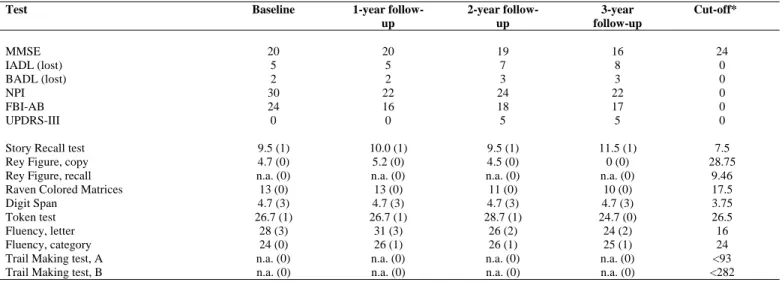
TABLE 1. Neuropsychological assessment of the proband at baseline and at follow-up.
Test Baseline 1-year
follow-up 2-year follow-up 3-year follow-up Cut-off* MMSE 20 20 19 16 24 IADL (lost) 5 5 7 8 0 BADL (lost) 2 2 3 3 0 NPI 30 22 24 22 0 FBI-AB 24 16 18 17 0 UPDRS-III 0 0 5 5 0
Story Recall test 9.5 (1) 10.0 (1) 9.5 (1) 11.5 (1) 7.5
Rey Figure, copy 4.7 (0) 5.2 (0) 4.5 (0) 0 (0) 28.75
Rey Figure, recall n.a. (0) n.a. (0) n.a. (0) n.a. (0) 9.46
Raven Colored Matrices 13 (0) 13 (0) 11 (0) 10 (0) 17.5
Digit Span 4.7 (3) 4.7 (3) 4.7 (3) 4.7 (3) 3.75
Token test 26.7 (1) 26.7 (1) 28.7 (1) 24.7 (0) 26.5
Fluency, letter 28 (3) 31 (3) 26 (2) 24 (2) 16
Fluency, category 24 (0) 26 (1) 26 (1) 25 (1) 24
Trail Making test, A n.a. (0) n.a. (0) n.a. (0) n.a. (0) <93
Trail Making test, B n.a. (0) n.a. (0) n.a. (0) n.a. (0) <282
MMSE: Mini-Mental State Examination; IADL: Instrumental Activities of Daily Living; BADL: Basic Activities of Daily Living; NPI: Neuropsychiatry Inventory; FBI-AB: Frontal Behavioural Inventory, Parts A and B; UPDRS: Unified Parkinson Disease Rating Scale.
Results are expressed as mean values, and equivalent scores between brackets; n.a.: patient was unable to perform the test. * cut-off scores according to Italian normative data.
Genetic analysis
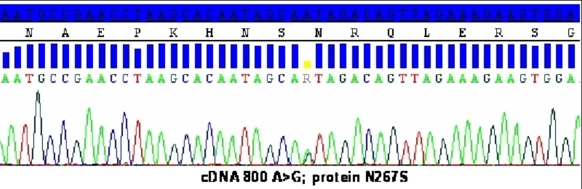
In DNA from the patient, the single base substitution c.800A>G (NM_007375.3) of TARDBP gene predicted the amino acidic change arginine to serine at position 267 (N267S) (see Figure 2).
This mutation was already known to be pathogenic for ALS in an Italian patient [Corrado et al., 2009]. We further analysed 98 FTD (59.2% female, mean age=66.7±7.2; 2 patients with FTD-MND) and 106 healthy controls for this mutation, with negative results.
In silico analysis indicated that the substitution from arginine to serine generates a new phosphorylation site (NetPhos 2.0).
Furthermore, in the index case, no additional mutations were found within the MAPT and Progranulin gene. Progranulin dosage in the serum was within normal range (187 ng/mL), compared to a sample of FTLD patients carrying Progranulin mutations (data not shown).
Figure 1. Brain functional and structural neuroimaging data of the proband at diagnosis, and at 3-year follow-up. Panel A. SPECT-Statistical Parametric Mapping (SPM) analysis performed at diagnosis, superimposed on 3D template. Index case showed a significant reduction of blood flow in the dorsolateral frontal cortex, bilaterally, and in the medial frontal cortex. Panel B. Axial MRI scans assessed at diagnosis. Frontotemporal atrophy was reported. Panel C. Axial MRI scans assessed at 3-year follow-up. Greater frontotemporal atrophy compared to baseline was reported.
Figure 2. Chromatogram indicating proband’s missense mutation. Missense mutation identified within
TARDBP gene in the case index. The observed single base substitution c.800A>G, and predicted amino acid
change arginine to serine at position 267 (N267S) are indicated in the chromatogram. cDNA numbering is according to the largest TARDBP transcript (NM_007375.3), and starting at the translation initiation codon. Protein numbering is relative to the largest TDP-43 isoform (NP_031401.1).
Biological analyses
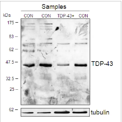
In previous analyses, both Kabashi et al. [2008] and Rutherford et al. [2009] demonstrated the presence of increased TDP-43 degradation fragments with respect to control samples in cell lines derived from ALS patients carrying TDP-43 mutations: G348C, R361S, N390D/S, M337V, N345K, and I383V. A similar result was also obtained by direct analysis of frozen lymphocyte preparations from ALS patients carrying mutations S393L and A382T [Corrado et al., 2009]. Based on these observations, we performed a similar study on lymphoblastoid cells derived from this patient and three appropriate controls, including the healthy son . Compared to the previously published examples we did not detect an increased degradation pattern from the patient cell lines with respect to controls (see Figure 3). Instead, we detected a substantial drop in TDP-43 expression levels in the patient sample. A western blot directed specifically against a C-terminal fragment of TDP-43 did not also detect an increased amount of degraded TDP-43 in the MB patient sample (data not shown). Taken together, these data suggest that the N267S mutation has a direct and aberrant effect on TDP-43 cellular levels in these patient cells with respect to control samples.
Figure 3. Western blot analysis of N267S lymphoblastoid cell line from patient and controls. Western blot analysis of TDP-43 protein from lymphoblastoid lysates of controls (CON) and the FTD patient carrying N267S mutation (lane TDP-43+). Samples were standardized using tubulin as an internal control. Similar results were obtained in two independent western blot experiments (data not shown).
DISCUSSION
The search for TARDP mutations in affected FTD with ubiquitin inclusions and ALS patients has been quite intensive in order to establish a clear molecular connection with disease, and thus rule out the possibility that TDP-43 might be just a pathological curiosity [Rothstein, 2007].
Initial studies in FTD cohorts failed to identify a genetic association for the disease when analyzing common TARDBP variations, especially with regards to single-nucleotide-polymorphisms [Rollinson et al., 2007; Schumacher et al., 2009]. Nonetheless, despite the evidence of causative role of TARDBP mutations in approximately 2-3% of sporadic and familial forms of ALS[Banks et al., 2008; Corrado et al., 2009], monogenic forms of FTD has not been described yet. Up to now, two cases with FTD but with MND have been described [Benajiba et al., 2009].
In the present study, we report the first robust clinical evidence of a pathogenic mutation, namely N267S, in the TARDBP gene as causative of behavioural variant of FTD (apathetic syndrome) without MND. The same mutation was previously found as causative of a spinal form of ALS, and excluded in 771 neurologically healthy controls (1.542 chromosomes) [Corrado et al., 2009]. In silico analyses using a variety of prediction programs indicated that the substitution from arginine into serine may generate a new phosphorylation site; the novel creation of this site may alter the properties of TDP-43 or the interaction with other molecular partners. From a functional point of view, analysis of the patient lymphoblasts highlighted a substantial reduction on TDP-43 expression levels in patient’s cells with respect to controls, suggesting that only the wild-type TDP-43 protein produced by the non-mutated allele can be observed in our analysis. Further work is currently in progress to better characterize the effects of the N267S mutation on impairing the cellular levels of TDP-43 inside the cells.
Up to now, the described TARDBP pathogenetic mutations cause MND with or without dementing illness [Kabashi et al., 2008; Rutherford et al., 2008; Sreedharan et al., 2008; Van Deerlin et al., 2008; Benajiba et al., 2009].
In our case, the pathogenic heterozygous N267S variation was found in an aged woman, who has displayed since her late seventies progressive behavioural disturbances along with cognitive deficits in executive functions. Functional brain imaging study was consistent with dorsolateral and frontal medial hypoperfusion, in accordance with previous work on apathetic presentation of bvFTD [Franceschi et al., 2005].
Extensive clinical investigations, three-year follow-up assessment, and electrophysiological studies excluded an evolution towards a form associated with MND. We cannot definitively exclude that the patient will develop MND over disease progression, but it is however true that she has had no preclinical signs or symptoms of MND by five years of estimated disease onset. The two patients reported by Benajiba and colleagues both presented with isolated FTD at onset, but they developed MND two years after [Benajiba et al., 2009].
Autopsy analyses will be needed to definitely clarify the neuropathology of our patient, but these data strikingly highlight the effect of a new genetic determinant in FTD.
There is evidence that neuropathologically TDP-43 disorders include ALS, FTD, and ALS with FTD
[McCluskey et al., 2009]; accordingly, our study argues for a further extension of the pathogenetic role of
TARDBP gene mutations in these neurodegerative processes. This suggests that TARDBP mutation screening should be considered even in FTD cases without signs or symptoms of MND.
ACKNOWLEDGMENTS
The authors wish to thank the patient and her family for kindly participating to this study. The authors are indebted with Dr Maura Cosseddu for clinical assistance, to Ms Maria Ferrari for technical support, and to Dr Enrico Premi for figure editing.
REFERENCES
Banks GT, Kuta A, Isaacs AM, Fisher EM. 2008. TDP-43 is a culprit in human neurodegeneration, and not just an innocent bystander. Mamm Genome 19(5):299-305.
Benajiba L, Le Ber I, Camuzat A, Lacoste M, Thomas-Anterion C, Couratier P, Legallic S, Salachas F, Hannequin D, Decousus M, Lacomblez L, Guedj E, Golfier V, Camu W, Dubois B, Campion D, Meininger V, Brice A; French Clinical and Genetic Research Network on Frontotemporal Lobar Degeneration/Frontotemporal Lobar Degeneration with Motoneuron Disease. 2009. TARDBP mutations in motoneuron disease with frontotemporal lobar degeneration. Ann Neurol 65(4):470-3. Buratti E, Dörk T, Zuccato E, Pagani F, Romano M, Baralle FE. 2001. Nuclear factor TDP-43 and SR proteins promote in vitro
and in vivo CFTR exon 9 skipping. EMBO J 20(7):1774-84.
Buratti E, Baralle FE. 2008. Multiple roles of TDP-43 in gene expression, splicing regulation, and human disease. Front Biosci 13:867–878.
Cairns NJ, Bigio EH, Mackenzie IR, Neumann M, Lee VM, Hatanpaa KJ, White CL 3rd, Schneider JA, Grinberg LT, Halliday G, Duyckaerts C, Lowe JS, Holm IE, Tolnay M, Okamoto K, Yokoo H, Murayama S, Woulfe J, Munoz DG, Dickson DW, Ince PG, Trojanowski JQ, Mann DM; Consortium for Frontotemporal Lobar Degeneration. 2007. Neuropathologic diagnostic and nosologic criteria for frontotemporal lobar degeneration: consensus of the Consortium for Frontotemporal Lobar Degeneration. Acta Neuropathol. 114(1):5-22.
Cook C, Zhang YJ, Xu YF, Dickson DW, Petrucelli L. 2008. TDP-43 in neurodegenerative disorders. Expert Opin Biol Ther 8(7):969-78.
Corrado L, Ratti A, Gellera C, Buratti E, Castellotti B, Carlomagno Y, Ticozzi N, Mazzini L, Testa L, Taroni F, Baralle FE, Silani V, D'Alfonso S. 2009. High frequency of TARDBP gene mutations in Italian patients with Amyotrophic Lateral Sclerosis. Hum Mutat 30(4):688-94.
Dickson DW. 2008. TDP-43 immunoreactivity in neurodegenerative disorders: disease versus mechanism specificity. Acta Neuropathol 115(1):147-9.
Franceschi M, Anchisi D, Pelati O, Zuffi M, Matarrese M, Moresco RM, Fazio F, Perani D. 2005. Glucose metabolism and serotonin receptors in the frontotemporal lobe degeneration. Ann Neurol 57(2):216-25.
Gijselinck I, Sleegers K, Engelborghs S, Robberecht W, Martin JJ, Vandenberghe R, Sciot R, Dermaut B, Goossens D, van der Zee J, De Pooter T, Del-Favero J, Santens P, De Jonghe P, De Deyn PP, Van Broeckhoven C, Cruts M. 2009. Neuronal inclusion protein TDP-43 has no primary genetic role in FTD and ALS. Neurobiol Aging 30(8):1329-31.
Kabashi E, Valdmanis PN, Dion P, Spiegelman D, McConkey BJ, Vande Velde C, Bouchard JP, Lacomblez L, Pochigaeva K, Salachas F, Pradat PF, Camu W, Meininger V, Dupre N, Rouleau GA. 2008. TARDBP mutations in individuals with sporadic and familial amyotrophic lateral sclerosis. Nat Genet 40(5):572-574.
McCluskey LF, Elman LB, Martinez-Lage M, Van Deerlin V, Yuan W, Clay D, Siderowf A, Trojanowski JQ. 2009.
Amyotrophic Lateral Sclerosis–Plus syndrome with TAR DNA-Binding Protein-43 pathology. Arch Neurol 66:121-124.
McKhann GM, Albert MS, Grossman M, Miller B, Dickson D, Trojanowski JQ; Work Group on Frontotemporal Dementia and Pick's Disease. 2001. Clinical and pathological diagnosis of frontotemporal dementia: report of the Work Group on Frontotemporal Dementia and Pick's Disease. Arch Neurol 58(11):1803-1809.
Neary D, Snowden JS, Gustafson L, Passant U, Stuss D, Black S, Freedman M, Kertesz A, Robert PH, Albert M, Boone K, Miller BL, Cummings J, Benson DF. 1998. Frontotemporal lobar degeneration: a consensus on clinical diagnostic criteria. Neurology 51(6):1546-1554.
Neumann M, Sampathu DM, Kwong, LK, Truax AC, Micsenyi MC, Chou TT, Bruce J, Schuck T, Grossman M, Clark CM, McCluskey LF, Miller BL, Masliah E, Mackenzie IR, Feldman H, Feiden W, Kretzschmar HA, Trojanowski JQ, Lee VM. 2006. Ubiquitinated TDP-43 in frontotemporal lobar degeneration and amyotrophic lateral sclerosis. Science 314:130–133. Probst A, Taylor KI, Tolnay M. 2007. Hippocampal sclerosis dementia: a reappraisal. Acta Neuropathol 114(4):335-45. Rollinson S, Snowden JS, Neary D, Morrison KE, Mann DM, Pickering-Brown SM. TDP-43 gene analysis in frontotemporal
lobar degeneration. 2007. Neurosci Lett 419(1):1-4.
Rothstein JD. 2007. TDP-43 in amyotrophic lateral sclerosis: pathophysiology or patho-babel? Ann Neurol 61(5):382-384.
Rutherford NJ, Zhang YJ, Baker M, Gass JM, Finch NA, Xu YF, Stewart H, Kelley BJ, Kuntz K, Crook RJ, Sreedharan J, Vance C, Sorenson E, Lippa C, Bigio EH, Geschwind DH, Knopman DS, Mitsumoto H, Petersen RC, Cashman NR, Hutton M, Shaw CE, Boylan KB, Boeve B, Graff-Radford NR, Wszolek ZK, Caselli RJ, Dickson DW, Mackenzie IR, Petrucelli L,
Rademakers R. 2008. Novel mutations in TARDBP (TDP-43) in patients with familial amyotrophic lateral sclerosis. PLoS Genet 4(9):e1000193.
Schumacher A, Friedrich P, Diehl-Schmid J, Ibach B, Perneczky R, Eisele T, Vukovich R, Foerstl H, Riemenschneider M. 2009. No association of TDP-43 with sporadic frontotemporal dementia. Neurobiol Aging 30(1):157-159.
Sreedharan J, Blair IP, Tripathi VB, Hu X, Vance C, Rogelj B, Ackerley S, Durnall JC, Williams KL, Buratti E, Baralle F, de Belleroche J, Mitchell JD, Leigh PN, Al-Chalabi A, Miller CC, Nicholson G, Shaw CE. 2008. TDP-43 mutations in familial and sporadic amyotrophic lateral sclerosis. Science 319:1668–1672.
Van Deerlin VM, Leverenz JB, Bekris LM, Bird TD, Yuan W, Elman LB, Clay D, Wood EM, Chen-Plotkin AS, Martinez-Lage M, Steinbart E, McCluskey L, Grossman M, Neumann M, Wu IL, Yang WS, Kalb R, Galasko DR, Montine TJ, Trojanowski JQ, Lee VM, Schellenberg GD, Yu CE. 2008. TARDBP mutations in amyotrophic lateral sclerosis with TDP-43 neuropathology: a genetic and histopathological analysis. Lancet Neurol 7(5):409-416.
Winton MJ, Igaz LM, Wong MM, Kwong LK, Trojanowski JQ, Lee VM. Disturbance of nuclear and cytoplasmic Tar DNA binding protein (TDP-43) induces disease-like redistribution, sequestration and aggregate formation. J Biol Chem in press.
SUPPORTING INFORMATION
Methods
Regional Cerebral Blood Flow (rCBF) measurement.
SPECT imaging of rCBF for the index case and for 14 control subjects (mean age±SD=66.3±15.4) was performed using a maximum dose of 1110 MBq 99mTc-ethyl cysteinate dimer (ECD), injected intravenously, as previously published. Statistical Parametric Mapping (SPM2, Welcome Department of Cognitive Neurology, University College, London), and Matlab 6.1 (Mathworks Inc., Sherborn, MA) were used for image pre-processing. Images were spatially normalized to a reference stereotactic template (Montreal Neurological Institute, MNI), and smoothed by a Gaussian kernel of 8x8x8mm FWHM.
Immortalization of lymphocytes.
The patients carrying TARDBP N267S mutation and three controls were considered. Blood drawing occurred while fasting at 9.00-10.00 am. A blood sample (10 ml) was taken, and collected into EDTA and NaCl. Blood underlayered with 5 ml Histopaque 1077 was centrifuged at 2000 rpm for 30 min at room temperature. The opaque lymphocyte layer along with the Histopaque, was collected. The cells were washed twice by repeated centrifugations in NaCl. The cells were re-suspended in 1 ml/3 millions of cells of EBV supernatant. After an incubation of 3 hours at 37° C with CO2 5%, a centrifugation at 1200 rpm for 10 min at room temperature was
Supp. Figure S1. Pedigree of the case index carrying TARDBP N267S mutation. Solid symbol indicated the proband (II:4). To protect confidentiality, sex of individuals is disguised. The proband’s parents (I:1 and I:2) died in their seventies for unknown causes; the three brothers died for cancer at 76 (II:1), 70 (II:2), and 53 (II:3) year-old. The proband’s son (III:1) is in his fifties, and he does not complain of cognitive and behavioural disturbances.
Supp. Table S1. Motor evoked potential at 3 year follow-up (5 years from estimated onset of symptoms) showed values within normal range
Limb Stimulation Latency
(msec) Amplitude (mV) CMCT (msec) Cut-off values (msec)
Upper, right Cervical 13.5
Cortical 21.65 4.60 8.15 < 10
Upper, left Cervical 13.50
Cortical 21.60 2.70 8.10 <10
Lower, right Cervical 14.80
Cortical 28.80 3.80 14.00 <17
Lower, left Cervical 14.50
Cortical 28.70 3.20 14.20 <17