UNIVERSITA' DEGLI STUDI DI
CATANIA
Dipartimento di Scienze Biologiche, Geologiche e Ambientali
D
OTTORATO DIR
ICERCA INB
IOLOGIAE
VOLUZIONISTICA E DELL’A
MBIENTECoordinatore: Prof. Salvatore Saccone
G
IUSEPPE
D
IEGO
P
UGLIA
Meccanismi fisiologico-molecolari di
germinazione in specie spontanee
_____________________________
Tesi di dottorato
_____________________________Tutor: Prof. Pietro Pavone
Co-Tutor: Dr. Peter Toorop
XXVI ciclo
AA.AA. 2010÷2013
CATANIA
Dipartimento di Scienze Biologiche, Geologiche e Ambientali
D
OTTORATO DIR
ICERCA INB
IOLOGIAE
VOLUZIONISTICA E DELL’A
MBIENTECoordinatore: Prof. Salvatore Saccone
G
IUSEPPE
D
IEGO
P
UGLIA
Physiological and molecular
mechanisms of seed germination in
weed species
_____________________________
Ph.D. thesis
_____________________________
Tutor: Prof. Pietro Pavone
Co-Tutor: Dr. Peter Toorop
XXVI cycle
AA.AA. 2010÷2013
Seed dormancy provides a mechanism for plants to delay germination until conditions are optimal for survival of the next generation. Knowledge of dormancy release and dormancy induction requirements is important to increase our understanding about the entire process. Furthermore, assess the association between gene expression patterns and physiological phase in dormancy is a major goal to allow a positive definition of this condition.
In Chrysanthemum coronarium var. concolor and var. discolor, anatomical and germination analyses showed that non-deep physiological dormancy (PD) is the more plausible type of dormancy for both of them. The genetic composition of Chrysanthemum coronarium varieties was characterized using Inter Simple Sequence Repeat (ISSR) PCR together with a DNA-Barcoding approach.
Seed priming treatments were used in a non-dormant species, Leucanthemum vulgare, to induct secondary dormancy at specific temperature range and a long-priming treatment was used for its release. Fluridone and giberellic acid were tested to investigate the metabolic pathway involved in the onset of secondary dormancy and in its maintenance. Five probable housekeeping genes, useful as reference genes for quantitative PCR (qPCR) analysis in seed tissue, were identified in Chrysanthemum coronarium and in Leucanthemum vulgare, isolated and confirmed with comparative analysis with homologous genes in model species. One radicle-protrusion associated gene, EXP (Expansin), was identified in the two species, isolated and confirmed. Three dormancy-associated genes, DOG1 (Delay Of Germination 1), FLC (Flowering Locus C), HUB2 (Histone Mono-Ubiquitination 2) were identified in Chrysanthemum coronarium and in Leucanthemum vulgare.
Table of contents
Introduction... 7
Seed germination... 7
Radicle growth... 8
Seed dormancy... 8
Physical dormancy (PY)... 9
Physiological dormancy (PD)... 9
PD deep ... 10
PD intermediate... 10
PD non-deep... 10
Endosperm-weakening and embryo growth potential...11
Molecular aspects of seed dormancy... 12
Dormancy release... 13
Seed priming... 14
Hydro-priming ... 14
Osmo-priming ... 14
Chrysanthemum coronarium ... 16
Species description and distribution ... 16
Heterocarpy and dormancy in Asteraceae... 17
Seed germination... 18
Leucanthemum vulgare... 19
Species description and distribution... 19
Seed germination... 21
Varietal genotyping... 23
Aim of the thesis... 25
Materials and Methods... 26
Study sampling... 26
Germination experiments... 27
Seed excision... 28
Water uptake... 28
Anatomical analyses of pericarp tissue... 28
Germination in dark... 28
Cold stratification... 29
Dry storage... 29
Dry after ripening... 29
Wet warm stratification... 29
Seed osmo-priming ... 29
Seed hydro-priming... 30
ABA synthesis... 30
Incubation in agitated water... 30
Giberellic acid... 30
Statistical analyses... 31
Molecular analyses... 32
Chrysanthemum coronarium varietal genotyping... 32
DNA extraction... 32
ISSR profiling... 32
rbc-L gene amplification and sequencing... 33
mat-K gene amplification and sequencing... 34
Data analysis... 34
Identification and isolation of reference and dormancy associated genes in C. coronarium and L. vulgare... 35
Reference genes... 35
Dormancy associated genes ... 35
DNA extraction... 36
Germination behaviour ... 39
Seed excision... 40
Comparison of the germination behaviour of the two varieties... 41
Water uptake ... 42
Anatomical analyses of pericarp tissue... 42
Germination in dark... 45
Cold stratification... 46
Dry storage... 48
Dry after ripening... 49
Wet warm stratification... 51
ABA synthesis... 56
Genotyping of Chrysanthemum coronarium concolor and discolor varieties...57
DNA extraction ... 57
ISSR analyses... 59
Sequencing of rbcL and mat-k regions... 60
Physiology of germination in Leucanthemum vulgare... 63
Germination behaviour ... 63
Seed osmo-priming ... 63
Hydro-priming... 65
ABA synthesis... 66
Isolations of dormancy genetic markers in Leucanthemum vulgare and Chrysanthemum coronarium... 69
Genes selection... 69
DNA extraction... 71
RNA extraction ... 71
PCR products cloning with TA-Vector... 72
Sequences analyses... 77
Phosphoglycerate kinase (PGK) gene... 77
18s gene:... 78 UBC 21 gene... 79 UBC 8-28 gene... 80 UBC 9-10 gene... 81 UBC sequences:... 82 Expansin gene:... 83 Discussion... 84
Chrysanthemum coronarium germination physiology... 84
Chrysanthemum coronarium varietal genotyping... 85
Leucanthemum vulgare germination physiology... 86
Cloning of reference and dormancy associated genes:... 87
Conclusions... 88
_____________________________________________________________________Introduction
Introduction
Seed traits are recognized as decisive adaptations of spermatophytes that are subjected to natural selection and optimize plant fitness (Fenner 2005), (Donohue 2010). Among these traits, seed dormancy is considered one of the main factors determining the adaptive value of germination (W. E.-M. Finch-Savage 2006) (Venable 2007) (Donohue 2010). Upon dispersal, seeds may show total (i.e. no germination under any conditions) or conditional dormancy (i.e. germination under a restricted set of conditions) (J. M. Baskin 2004). In response to certain environmental signals (the so-called dormancy breaking factors), seeds gradually lose dormancy and their range of germination conditions broadens until eventually they become non-dormant. This stimulus can be related to the season of major risk for the seedling in many species (Vleeshouwers 1995) (Mattana 2012) (Wagmann 2012).
Seed germination
Seed germination process starts with water imbibition of a dry seed and ends with radicle protrusion (W. E.-M. Finch-Savage 2006) (Bassel 2011). The elongation of embryonic axis protruding through the endosperm tissue is clear evidence of the completion of germination. From the physiological point of view, germination consists of reactivation of metabolism, followed by protein synthesis and radicle cells extension (J. D. Bewley 1997). Metabolism resumption is triggered by water entrance in seed tissues, which activates a sequence of enzymatic mechanisms.
Water imbibition is a tri-phasic process. The phase I is characterized by a rapid water uptake, with a consequent immediate leakage of solutes (Crowe 1992), followed by mitochondria repairing and a resumption of principal metabolic pathways (Botha 1992) (J. D. Bewley 1994). Within 3 – 6 hours after imbibition the mRNAs transcribed during seed development are degraded and a distinct germination transcriptional program commences (K. O. Nakabayashi 2005). Conversely, at the same time, the amount of new transcripts increases gradually to support cell metabolism for seed germination (K. O. Nakabayashi 2005) (Carrera 2007). In the phase II water absorption is prevented, DNA is repaired to correct the reduction in DNA integrity resulting from storage in the dry state (Osborne 1994), and DNA replicative synthesis takes place (Zlatanova 1987).
In the phase III the increase in osmotic concentration promoted by the hydrolysis of storage solutes lead to a further water uptake. At the end of this phase radicle extension, through the structures surrounding the embryo, occurs and this is the event that terminates germination and marks the commencement of seedling growth. This extension may or may not be accompanied by cell division, but it always requires the yielding of hypocotyl cell walls (Cosgrove 1997) (J. D. Bewley 1997).
Radicle growth
Radicle growth may be exerted by a decrease in water potential of the seed (ψπ),
cell wall-loosening of tissues surrounding the radicle tip or by a partial wall degradation allowing elongation of the embryonic axis driven by turgor ψp (J. D. Bewley 1997)
(Koornneef 2002). Previous authors (Pritchard 1993) (Wu 1994) observed an increase of activity of xyloglucan endotransglycosylase activity (XET) (which is decreased in presence of Fluridone and enhanced by ABA) specifically in the elongation zone with a low ψw.
Moreover, a class of proteins named expansins was shown (Cosgrove 1997) (McQueen-Mason S. 1993) to induce cell wall extension in concert with a variety of enzymes that cut and restructure the wall in cucumber (McQueen-Mason 1995) (Cosgrove 1997) and in rice cultivars (Magneschi 2009). In tobacco, endosperm ß-Glucanases induction was observed to be positively correlated with endosperm and testa rupture (G. F.-L. Leubner-Metzger 1995). This hypothesis was supported with the application of hormones that inhibit (ABA) or promotes (GA) seed germination (G. F.-L. Leubner-Metzger 1995): ABA presence prevents radicle cell wall extension, whilst it does not interfere with ψs, solute potential and
with water uptake.
Seed coats and surrounding structures may have a further influence on the ability of a seed to germinate interfering with water uptake, gas exchange, diffusion of endogenous inhibitors, or by mechanical restriction of embryo growth (Clark 1989) (Durán 1989) (C. C. Baskin 1998). Conversely, even the endosperm tissue may restrict embryo growth: in pepper mechanical resistance of the endosperm appeared to affect germination (Watkins 1983).
Seed dormancy
Seed dormancy, could be defined as a block to the completion of germination of an intact viable seed under favourable conditions (W. E.-M. Finch-Savage 2006). It forms a
_____________________________________________________________________Introduction protection mechanism for species survival, avoiding undesirable germination during unfavourable environmental conditions for seedling growth (A. G.-M. Linkies 2010). Recent seed physiology literature (J. M. Baskin 1998) (J. M. Baskin 2004) discriminates five classes of dormancy depending on which factor causes seed dormancy: physical (PY), physiological (PD), morphological (MD), morphophysiological (MPD) and combinational (PY+PD) dormancy. In this manuscript, only physical and physiological dormancy will be introduced because of their pertinence with the species presented in this study.
Physical dormancy (PY)
In physical dormancy seed/fruit covering layers impose dormancy preventing embryo/seed imbibition (J. M. Baskin 2000). Water-impermeable seed (or fruit) is obtained during maturation drying of the seeds (Van Staden 1989) (C. C. Baskin 1998) by a layer of palisade cells acting as a physical barrier against water entrance. Heavy lignified cells, waxes or suberin-cutin usually make up this cell barrier.
In PY once the seed or fruit becomes permeable to water by a loss of barrier integrity, the seed can imbibe and germinate over a wide range of temperatures (C. C. Baskin 1998) (J. M. Baskin 2000). As a consequence, once the physical barrier has been disaggregated, it generally cannot revert to impermeability (Hamley 1932). Release of physical dormancy is usually exerted by external factors that could affect impermeable cell layer integrity. In nature, these factors include high temperatures, widely fluctuating temperatures, fire, drying, freezing/thawing and passage through the digestive tracts of animals (C. C. Baskin 1998).
Physiological dormancy (PD)
In physiological dormancy (PD) a block of germination occurs, and it is removed only with the presence of specific environmental cues (W. E.-M. Finch-Savage, Seed dormancy and the control of germination 2006). Within A. thaliana physiological dormant seed the ABA- and GA-biosynthesis and catabolism were modelled (C. T.-S. Cadman, Gene expression profiles of Arabidopsis Cvi seeds during dormancy cycling indicate a common underlying dormancy control mechanism 2006) (W. E. Finch-Savage 2007), where hormones catabolism and metabolism are influenced by prevailing environmental conditions. This balance can make the difference between germination and the seed remain dormant.
Furthermore, presence of inhibitors in seed coat layers or endosperm-weakening can inhibit embryo growth.
Depending on the intensity of physiological dormancy in seeds, three different types of PD were identified.
PD deep
PD-deep seeds excised embryos do not grow or will produce abnormal seedlings. They usually need long pre-chilling treatments and chemicals do not exert any remarkable effect on seed germination. Pyrus seeds re-enter in dormancy during post-stratification drying after a pre-chilling treatment (Shen 2001).
PD intermediate
Embryos excised from PD-intermediate seeds produce normal seedlings. Usually a long pre-chilling, up to 6 months, is necessary. GA, kinetin or thiourea could reduce cold stratification period.
PD non-deep
Non-deep physiological dormancy is the most common physiological dormancy class (J. M. Baskin 2004). Excised embryos produce normal seedling, and dormancy is usually released after a short cold stratification time period. Moreover, these seeds, depending on their distribution area, they need a cold or warm stratification to release dormancy. This, could be broken by chemicals (potassium nitrate, thiourea, kinetin, ethylene, and gibberellins. Non-deep physiological dormant seeds, usually, need light to germinate. Seeds with a PD non-deep gradually lose dormancy and enter in a conditional dormancy state (Naylor 2008). At the beginning of this stage, an increasing amount of seeds is able to germinate over a narrow range of temperatures. As the conditional dormancy is gradually lost an increasing proportion of seeds germinate over a more ample range of environmental conditions (J. M. Baskin 2004).
There is a general agreement on the identification of five types of non-deep PD proposed by Baskin & Baskin (J. M. Baskin 2004), which is based on the patterns of change in physiological responses to temperature during dormancy break (see Figure 1). In the first type temperature range at which seeds can germinate gradually increases from low to high temperatures along with dormancy release. Arabidopsis thaliana is a species with non-deep PD type 1 (Meinke 1998).
_____________________________________________________________________Introduction In the second type, during dormancy release temperature range required for germination decreases as a continuum from high to low, example: Helianthus annuus (Corbineau 1990) (M. T. Le Page-Degivry 1990).
In type 3 temperature range for germination increases during dormancy release as a continuum from medium to high and low, example: Aster ptarmacoides (M. B. McDonald 2005).
In non-deep PD type 4 and 5 temperature and dormancy pattern change does not result in continuum trend, but non-dormant seeds germinate only at high temperature (type 4), example: Callicarpa americana (M. B. McDonald 2005) or low temperature (type 5), example: Gentianella quinquefolia (J. M. Baskin 2004).
Figure 1 Five types of non-deep physiological seed dormancy (PD) according to Baskin and Baskin (2004).
Endosperm-weakening and embryo growth potential
Differently from physical dormancy imposed by a physical barrier to water entrance, a mechanical constraint exerted by endosperm combined with low embryo growth potential can result in seed dormancy. This condition falls in non-deep physiological dormancy and it can be released by light and GA, which increase embryo osmotic potential and/or reduce endosperm weakening (Casal 1998), (G. M. Leubner-Metzger 2001) (Sanchez 2004) (Kucera 2005). The force required to break seed coats depends on the different seed surrounding layers features and it can range from 9.9MPa in Pancratium marithimum to 133.2MPa in Iris lorteti (Blumenthal 1986). Appropriate germination or dormancy break conditions as presence of light (Scheibe 1965) or darkness (S. Chen 1968), giberellic acid (J. M. Baskin 1971), incubation temperatures (Juntilla 1973) or cold stratification (Carpita 1983) could promote embryo growth potential enough for radicle protrusion through the seed coat (C. C. Baskin 1998). Moreover, seed coat scarification may increase germination percentage removing the mechanical constraint as in cucumber and in sunflower seeds, where, after scarification, a 50% increase of
germination was observed in light at an increased osmotic potential (McDonough 1967). In tomato the endosperm cap weakening is a biphasic process correlated with endo-β-mannase activity for the first phase and controlled by ABA in the latter one (Toorop 2000).
Molecular aspects of seed dormancy
Notwithstanding the function of dormancy in different plant species to spread germination across time avoiding unfavourable environmental condition for seedling growth (C. C. Baskin 1998) there is a common underlying molecular mechanism that manage seed dormancy in plants (C. S.-S. Cadman 2006) (A. G.-M. Linkies 2010). However, the regulation of this process at the molecular level is far to be unveiled (Footitt 2011). Recent studies on non-deep physiological dormancy in A. thaliana have provided new insights (C. T.-S. Cadman, Gene expression profiles of Arabidopsis Cvi seeds during dormancy cycling indicate a common underlying dormancy control mechanism 2006) (W. E. Finch-Savage 2007) (S. D.-S.-S. Footitt 2011) showing that ABA and GA hormones balance is finely tuned at molecular level. As an example, it has been shown that the expression of genes involved in the ABA/GA level regulation is sensitive to seed maturation temperature (S. L. Kendall 2011). Genetic approaches have led to the identification of specific loci correlated with altered dormancy, and -omics approaches in many species have identified numerous transcripts and proteins correlated with dormant versus non-dormant seeds (W. E.-M. Finch-Savage, Seed dormancy and the control of germination 2006) (L. J. Bentsink 2006) (G. C. Chiang 2009) (Bassel 2011). DELAY OF GERMINATION 1 (DOG1) gene is a major quantitative trait gene more specifically involved in seed dormancy and germination timing (L. J. Bentsink 2006), it is so far only known within the Brassicaceae and its expression appeared to be the dominant factor influencing ABA signalling (S. H.-S. Footitt 2013) (Graeber 2013). Other studies (G. C. Chiang 2011) (S. C.-S. Footitt 2014) have shown
DOG1 expression to be negatively correlated to soil temperature which protein
accumulation drives changes in germination potential acting as a timer for seed dormancy release (K. B. Nakabayashi 2012). Furthermore, the expression of GA3ox1 gene (GA biosynthesis) was observed to follow seed water potential cycle decreasing upon burying, with a subsequent continuum increase until dormancy release (S. H.-S. Footitt 2013). Other researches shown the gene FLC (Flowering Locus C) to have a role in germination timing with a similar expression pattern as GA3ox1 gene (G. C. Chiang 2009) (Chiang G. C. 2013).
_____________________________________________________________________Introduction Analysis on Arabidopsis thaliana mutants with reduced dormancy (rdo) (Peeters 2002) and successive investigations (Liu 2007) showed that HISTONE MONOUBIQUITINATION (HUB1) protein influences gene expression of dormancy-related genes. Recently in
Castanea sativa a clear association of genes encoding histone mono-ubiquitinase HUB2
with dormancy behaviour was shown (Santamaría 2011).
Dormancy release
Recent speculations on seed dormancy release mechanisms have led to consider dormancy as a continuum process, where the expression of dormancy-related genes is highly sensitive to the soil temperature history, nitrate, light and GA (C. S.-S. Cadman 2006) (W. E.-M. Savage, Seed dormancy and the control of germination 2006) (W. E. Finch-Savage 2007) (S. D.-S.-S. Footitt 2011) (W. E. Finch-Finch-Savage 2012). This continuum is made up by layers of dormancy being progressively removed by environmental cues until only light is required, in the absence of which seeds remain dormant and enter into another dormancy cycle as the seasons change (W. E. Finch-Savage 2012) (S. C.-S. Footitt 2014). Therefore, any cue that enlarges the environmental required conditions allowing germination has to be intended as a release factor (W. E. Finch-Savage 2007).
Dormancy-release factors are usually related to the specific environment where the species is spread. So that species adapted to regions with a dry season where lack of water represents a serious obstacle for seedling growth, the dry after-ripening, i.e. a period of dry storage for usually several months (J. D. Bewley 1997) (Kucera 2005) (Probert 2000), is often the major dormancy release factor.
During dry storage physiological changes takes place within the seed, which result in a decrease in the level of dormancy and, as consequence, germination requirements become less specific (Probert 2000). In particular, in tobacco dormant seeds, dry after-ripening treatment led to a widening of the temperature range for germination, a decrease in ABA level within the seed and an increase in GA sensitivity, a loss of the requirement for nitrate, and an increase of germination promptness (G. Leubner-Metzger 2005).
Furthermore, even cold and warm incubation of pre-imbibed seeds can play a role in both release and induce dormancy (Probert 2000). In winter annuals high winter temperatures release dormancy and low summer temperatures induce dormancy (J. M. Baskin 1978) (J. M. Baskin 1978). On the contrary, in summer annuals low winter temperatures release dormancy, whereas high summer temperatures induce dormancy (J. M. Baskin 1977) (J.
Seed priming
Among treatments that could alter seed physiology, which, in turn, could promote dormancy release, seed priming is widely used in crops as a standard technique (S. G. Kaur 2005) (Guan 2009). It was shown to widen the temperature range suitable for germination (McDonald 2000) (Jisha 2013), and provide faster and synchronous seedling emergence (Taylor 1990) accomplish uniform emergence, and give better crop stand in many horticultural and field crops (Ashraf 2005).
It consists in the acquisition of the potential to rapidly imbibe and revive the seed metabolism thus enhancing the germination rate (McDonald 2000).
Seed priming could be implemented with different priming factors: water (hydropriming), osmoticum (osmopriming), chemicals (chemical priming), etc. In this manuscript only hydropriming and osmopriming will be introduced because of their pertinence with the presented experimental procedure.
Hydro-priming
Hydro-primed seeds are immersed in water and kept at appropriate temperature for a determined length of time (Kaya 2006). Afterwards seeds are dried to their original weight (Bennett 1987) (Thomas 2000). Hydro-priming promotes metabolism reactivation even if germination is prevented (S. M. Basra 2003). Protoplasm of hydro-primed seeds is found to have a lower viscosity and exhibit higher permeability to water and nutrients and also hold water against dehydrating forces (Thomas 2000). In onion germination hydro-priming was found to be the most effective method for improving seed germination (Caseiro 2004). In Zea mays L. priming widened germination temperature range and shortened germination time without decreasing final percentage emergence (W. E. Finch-Savage 2004).
Osmo-priming
Osmo-primed seeds are exposed to low-water-potential solutions to restrict the rate and extent of imbibition. This process prolongs the early imbibition phase promoting a gradual progression of various pre-germinative metabolic activities (Bradford 1986).
Polyethylene glycol (PEG) is the most common agent used to create low-water potential solution because of its nontoxic nature and large molecular size, which lowers water potential without penetrating into the seeds during soaking (Thomas 2000). In comparison with hydropriming, osmopriming can preserve plasma membrane structures resulting in
_____________________________________________________________________Introduction better germination frequency because of controlled long hydration in seeds (Jett 1996). In rice, osmopriming treatment was observed to enhance starch hydrolysis triggering a series of events leading to more vigorous seedling production, improved allometric, kernel yield and quality attributes (Farooq 2006).
In contrast to the copious literature on positive effects of seed priming, to date just few studies report a negatively interference of this treatment with germination in non-dormant seeds. In Amitiphale et al. (D. S. Amritphale 2000) cucumber seeds imbibition within an osmoticum allowed examining the dormancy breaking in isolation from the germination process. In fact the osmotic treatment caused germination inhibition, whereas a seed exposure to far red light induced secondary dormancy. Researchers hypothesized that change in plasma membranes fluidity led to the transition from dormant to germinable state.
Chrysanthemum coronarium
Species description and distribution
Chrysanthemum coronarium L. (family Asteraceae; tribe Anthemideae; genus
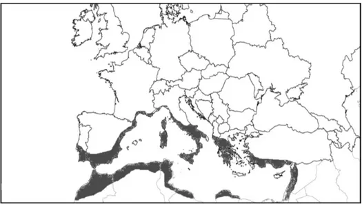
Figure 2 Chrysanthemum coronarium distribution according to H. Meusel. Figure re-drawn from (Meusel 1965).
Dumont d’Urville described two varieties under Chrysanthemum coronarium: var.
concolor d’Urv., from the island of Malta, with yellow ray florets (“radio discoque luteis sola reperitur inter segetes insulae Melitae”), and var. discolor d’Urv., from the Aegean
islands, with white ray florets (“radio albo discoque luteo copiosè occurrit in insulis
Archipelagi”) (d’Urville 1822).
The two entities appear to be widespread in the Mediterranean region and show no obvious correlation with geographic distribution (Turland 2004). Recently, investigation on the
_____________________________________________________________________Introduction different population occurrence of the two varieties shown that the discolor variety occurs in thermo- and meso-Mediterranean areas of Greece, Italy, Spain, Portugal and the north of Morocco, whereas no occurrence of the concolor var. was reported in the meso-Mediterranean environment (E. C.-O. Cano 2012) (Cano-Ortiz 2007). In the thermo-Mediterranean belt the populations of both variants tend to appear separately with only occasional concurrences (E. C. Cano 2013).
Furthermore, a kariomorphological study (El-Twab 2008) on Chrysanthemum coronarium varieties shown that, although the two varieties are both 2n = 18, as previously reported for
Chrysanthemum coronarium (Bhattacharyya 1977) (Pavone 1981) (Razaq Z.A. 1988)
(Nirmala A. 1986) (Vogt R. 1993) (Kaul M.L. 1995) (Strother 1997) (Carr 1999), they differ in the arrangement of two chromosomes (submedian-centromeric in discolor and median-centromeric in concolor).
In other studies, basing on the disposition of the intercostal glands, the colour of the ligules, and the size and wings of the cypselas of the flosculous flowers and their distribution, some researchers (E. C. Cano 2013) proposed to elevate the discolor var. to the rank of species as Glebionis discolor (d’Urville 1822).
Therefore, a different approach based on molecular data is needed to further investigate the genetic grounds underneath the differentiation of the two varieties.
Figure 3 Co-occurence of Chrysanthemum coronarium concolor and discolor varieties in an abandoned field (for geographic reference see Table 1 sampling ID #5)
Heterocarpy and dormancy in Asteraceae
Sixty-three and three-tenths percent of the species and 52.5% of the genera belong to the Asteraceae and 8.3 and 10%, respectively, to the Chenopodiaceae. In Asteraceae, fruit heteromorphism, i.e. the difference between central and peripheral achenes (McDonough 1975), may be associated with a difference in germination behaviour, e.g. in Bidens, Senecio, Picris and Crepis (McEvoy 1984), (Rocha 1996), (E. E. Imbert 1999), (E. Imbert 2002), (Brändel 2004), (Brändel 2007). Central achenes are often less dormant and germinate over a wider range of temperatures, to higher percentages and at faster rates than peripheral ones (J. B. Baskin 1976), (Forsyth C. 1982), (Corkid 1991) (Rocha 1996) (Brändel 2007).
In seed heteromorphism the different fruit or seed morphs usually have differential ecological behaviour, mainly germination requirements and dispersal abilities. Peripheral achenes are frequently heavier, show more limited dispersal and have stricter requirements for germination (p.e. (E. E. Imbert 1999) (Clavijo 2001). In most of the heterocarpic Asteraceae central achenes germinate immediately when favourable conditions occur while peripheral achenes show delayed germination. Differential dispersal and germination allow spreading offspring in space and time, thus reducing density dependent effects such as sib competition, predation or pathogen attack (D. H. Janzen 1971) (Augspurger 1983) (Levin S. 1984) (Harms 2000). In addition, this different ecological behaviour leads to a risk-reducing or bet-hedging strategy that is advantageous in variable and unpredictable habitats such as Mediterranean biomes and highly disturbed, man-made habitats.
Discerning the differential ecology of achene morphs in heterocarpic weedy Asteraceae is of interest in weed population dynamics and basic weed biology studies, which, in turn, can be useful in designing appropriate weed management practices (Bhowmik 1997).
Seed germination
Chrysanthemum coronarium emergence occurs in autumn and seedlings develop into
overwinter rosettes that bolt in January and February. Flowering and fruiting start in late February or March and continue to early summer (F. M. Bastida 2004). Flowers consist of central hermaphrodite disc florets and marginal female ray florets with inferior ovaries (Cockshull 1984). These florets develop two morphologically distinct lignified fruit called cypselae (hereafter named more merely “achenes”), which are prominently composed by sclerotic cells (Mukherjee 1992). The two morphotypes are distinguishable for their average size, peripheral much bigger than central, and for their shape: one-winged central, and three-winged peripheral achenes (Figure 4).
_____________________________________________________________________Introduction
Figure 4 a) One-winged central achene; b) Three-winged peripheral achene.
Both central and peripheral achenes show dormancy in a high proportion, especially peripheral achenes are more dormant than central ones (F. M. Bastida 2004).
Even thought some studies (M. P. Chiang 1994) (Vicente 2002) (Hwang 2008) (Banon 2009) (F. J. Bastida 2010) have been investigating the germination behaviour in Chrysanthemum coronarium, there is still a bit of uncertainty in defining precisely the type of dormancy of this species.
In a previous study (F. M. Bastida 2004) germination was tested against different conditions: temperature regimes, dark, with a pre-chilling treatment, in presence of GA3, of
nitrate solution, and finally excising seeds from achenes. From the highest germination frequency shown in response to seed excision in presence of the pericarp tissue authors concluded that dormancy in C. coronarium is physically imposed by pericarp.
Conversely, in another study (Banon 2009) authors tested C. coronarium germinability in presence of several commercial gibberellins and excising seeds from achenes. Interestingly, when excised seeds were irrigated with pericarp leachate this produced an inhibition of germinability (from 92% to 43%), so they concluded that dormancy lies on a combination of physical and chemical dormancy.
Therefore, even though this plant is very common in Mediterranean basin, invasive outside its native environment and produces a large number of seeds, the germination behaviour remains unclear and worthy of study.
Leucanthemum vulgare
Species description and distribution
Leucanthemum vulgare (Vaill.) Lam. (family Asteaceae; tribe Anthemideae; genus
Leucanthemum) is a monocarpic perennial herb 20 - 40 (100) cm high, with a lignified base, hairless or hairy, simple or branched with a wiggling root-stock where new sprouts originate; flower heads are made up of 15 – 30 white rays that circle a yellow button,
depressed center; florets are clustered in composite heads 2-5 cm wide, with white ray florets radiating out from a yellow centre of disk florets (Pignatti 1982) (Douglas 1998) (Jauzein 2011). His chorological type is Eurasiatic, Euri-Mediterranean and Euro-Siberian (Pignatti 1982) (Lauber 2001) (Meusel 1965) (Figure 5). In northern Europe flowers bloom from June to August.
Figure 5 Leucanthemum vulgare distribution according to H. Meusel. Figure re-drawn from (Meusel 1965) with the permission of AG Chorology & Macroecology of Vascular Plants, University of Halle. Red dots represents neophytic occurrences, green dots original distribution area.
Leucanthemum vulgare is highly adaptable to a variety of sites. It can grow in course to medium textured soils and can be found in moist to moderately dry sites; however, it does prefer abundant sunlight. Once planted as an ornamental, it escaped cultivation and is now common in native meadows, pastures, fields in open and thick woodlands, along waterways and roadsides and it was reported as invasive plants Figure 7 (Khuroo 2010). It is also found in disturbed areas, hay fields, gardens and lawns, and irrigation ditches.
_____________________________________________________________________Introduction
Figure 6 Leucanthemum vulgare
Figure 7 A Conifer forest-opening invaded by Leucanthemum vulgare
Seed germination
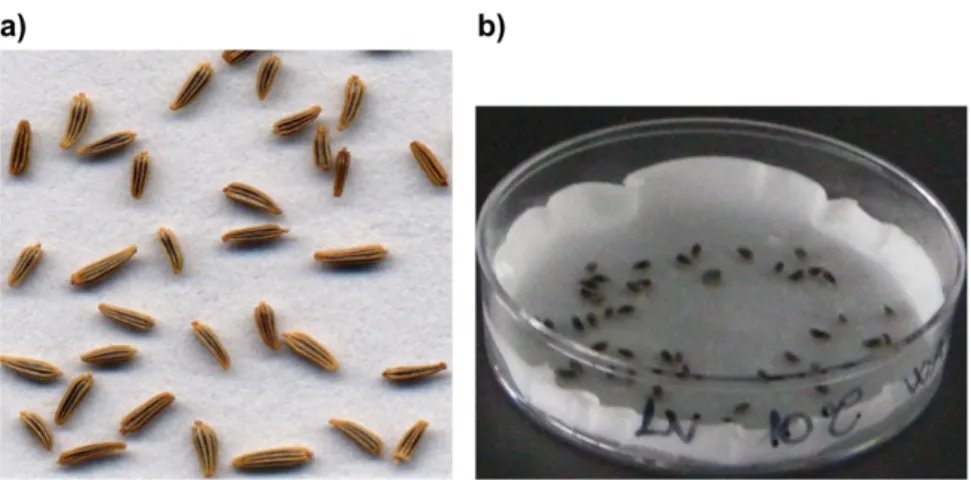
The fruit of Leucanthemum vulgare is a small flat seed (Figure 8), dark gray in color with no pappus. One plant can produce over 500 seeds and seeds can remain viable in the seedbank for up to three years.
This plant does not show any dormant behaviour and has a germination temperature between 15 and 20° C and it may germinate either in spring or in early autumn (Keller 1999). Seed germination is inhibited by continuous darkness but otherwise not affected by variation in light (K. R. Thompson 1989).
Figure 8 a) Leucanthemum vulgare seeds; b) Leucanthemum vulgare seeds imbibed in a Petri dish used for germination tests
_____________________________________________________________________Introduction
Varietal genotyping
The assessing of genotypic diversity of closely related taxonomic entities is a challenging goal and its difficulty mainly relies on the reproductive system and species distribution.
Chrysanthemum sensu lato consisted of 27 genera which has been taxonomically placed in
the subtribe Chrysantheminae O. Hoffm., the tribe Anthemideae Cass (Shih 1983) while it has been placed in different subtribes in the tribe Anthemideae (Bremer 1993). The main centres of distribution of Chrysanthemum sensu lato are two areas: one in the Mediterranean basin and the other in China and Japan, which are considered as ancestors of the Mediterranean species (Dowrick 1952).
The varietal genotyping in Chrysanthemum with traditional techniques markers such as morphological and cytological markers was observed to be not effective (Wolff 1994). Therefore, other techniques for identifying molecular markers such as random amplified polymorphic DNA (RAPD) have been developed and applied (Williams 1990). The RAPD technique is more straightforward compared to RFLP analysis and requires only nanograms of genomic DNA. It has been employed in different Brassica species for varietal identification and genetic diversity studies (Jain 1994). However, this technique is sensitive to subtle changes in reaction conditions and hence difficult to reproduce.
Inter simple sequence repeat (ISSR) markers with low cost and low labour requirement but with high reliability have been developed since 1994 (Zietkiewicz E. 1994). Like any other PCR-based marker, it is rapid and require only small amount of the template DNA. ISSR amplification does not require genome sequence information but produce highly polymorphic patterns, and it seem to have the reproducibility of SSR’s and the usefulness of RAPD’s (Bornet 2001), and thus combine the advantages of SSR and the utility of RAPD. ISSR markers have been used to determine the genetic diversity (Qian 2001). It was also applied to the study of genetic relationships and phylogenetic analysis of Anthemideae tribe (El-Twab A. M. H. 2010).
On the other hand, DNA barcoding is a diagnostic technique for species identification, using a short, standardized DNA region, i.e., the ‘‘DNA barcode’’ (www.barcoding.si.edu) (Lahaye 2008). A DNA barcoding to properly work the sequence variation must be high enough between species so that they can be discriminated from one another; however, it must be low enough within species that a clear threshold between
barcoding for identification and taxonomy has been controversial (Ebach 2005) (Will 2005) a growing scientific community has embraced DNA barcoding as a practical tool for biodiversity studies, for example to facilitate inventories of very diverse but taxonomically poorly known regions (Blaxter 2004) (D. H. Janzen 2005).
DNA barcoding in plants is now better established (Hollingsworth 2009) then in the past (Rubinoff 2006) (Pennisi 2007) after the creation of the stable Plant Working Group (http://www.barcoding.si.edu/plant_working_group.html). This consortium approved the usage of matK and rbcL as the BARCODE regions for Land Plants. So that nowadays, plant barcode resources are catalogued in the Barcode Of Life Database (BOLD) (http://www.boldsystems.org/) web portal and available for search for species identification.
__________________________________________________________________Aim of the thesis
Aim of the thesis
The aim of this work was to perform a comparative study on two common Asteraceae weeds, a dormant species, Chrysanthemum coronarium var. concolor and discolor, and a non-dormant one, Leucanthemum vulgare, where a secondary dormancy could be inducted. In particular, the focus of this research was to determine their germination physiology, and investigating their dormancy release and dormancy induction requirements. Beyond the characterization of germination and dormancy patterns, this study aims to analyse these processes with a molecular approach through isolation of candidate genes associated with dormancy onset and its regulation.
Materials and Methods
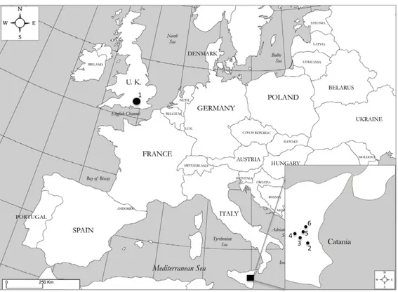
Study samplingLeucanthemum vulgare accession was collected in summer 2010 by Millennium Seed Bank collector team (Figure 9 and Table 1). Seeds were then cleaned and kept at +5° C with controlled RH conditions.
For Chrysanthemum coronarium two different seed lots were used in this study: a seed lot harvested in early summer of 2012 and another one harvested in the subsequent year in the same period, see Table 1 and Figure 9. In the older collection only Chrysanthemum coronarium var. concolor was sampled, while in 2013 field sampling concolor and discolor varieties were collected.
In 2012 sampling achenes were collected from around 20 healthy plants in a pure ample concolor meadow. On the other hand, in the 2013 sampling since the paucity of discolor plants, the harvesting was limited to 16 individuals which was the maximum number of discolor occurences. Discolor var. was always found together with concolor variety. During the early spring 2013 thirty-two flowering plants, sixteen individuals for each variety, were identified and labelled. From each individual an adequate amount of fresh leaves was taken and kept on ice until stored at -80° C for subsequent genotyping analyses. Plants labelling was carried out for subsequent discrimination during seed dispersal season when is not possible to distinguish the two C. coronarium varieties from florets colour. Achenes of the two varieties were collected from marked plants and kept at 15% of Relative Humidity and 15° C before used in germination tests.
Collection ID Year of collection Taxon Height above sea level City - Country Latitude Longitude
1 2010 Leucanthemum vulgare 240 m Ardingly -England 51° 3'58.40"N 0° 5'41.24"O 2 2012 Chrysanthemum coronarium var. concolor 110 m Catania – Italy 37°29'25.75"N 14°59'23.63"E 3 2013 Chrysanthemum coronarium var. concolor 529 m Catania – Italy 37°35'11.79"N 14°59'11.68"E 4 2013 Chrysanthemum coronarium var. concolor 297 m Catania – Italy 37°33'18.86"N 14°55'52.88"E 5 2013 Chrysanthemum coronarium var. concolor 252 m Catania – Italy 37°32'49.65"N 14°56'45.60"E 6 2013 Chrysanthemum coronarium var. concolor 320 m Catania – Italy 37°33'41.28"N 14°58'21.60"E 3 2013 Chrysanthemum coronarium var. discolor 529m Catania – Italy 37°35'11.79"N 14°59'11.68"E 4 2013 Chrysanthemum coronarium var. discolor 297 m Catania – Italy 37°33'18.86"N 14°55'52.88"E 5 2013 Chrysanthemum coronarium var. discolor 252 m Catania – Italy 37°32'49.65"N 14°56'45.60"E 6 2013 Chrysanthemum coronarium var. discolor 320 m Catania – Italy 37°33'41.28"N 14°58'21.60"E
_____________________________________________________________Materials and Methods
Figure 9 Map of collection of Leucanthemum vulgare and Chrysanthemum coronarium
Germination experiments
Germination experiments consisted of three replica samples of 50 seeds each. Basing on the different samples size seeds (L. vulgare) and achenes (C. coronarium) were sowed in two layers of moist filter paper (Whatmann) in plastic Petri dishes, respectively, with 5 cm diameter and with 9 cm diameter. For Chrysanthemum coronarium the two morphotypes were always tested separately. Filter paper imbibition was carried out always with double-distilled water when not differently indicated. Since the vast mould proliferation during germination tests, achenes of Chrysanthemum coronarium were always washed with 1% of commercial chlorine and then rinsed 3 times in distilled water before sowing them in Petri dishes.
Experiments were conducted in temperature (±1 ° C) and light (40 μmol m-2 s-1) controlled
conditions using a 12 h or 8/16 h daily thermo- (for alternating temperatures) and photo-period (= light hereafter). In the incubators actual temperature were checked repeatedly. Germination was defined as radicle emergence from the testa by at least 1 mm often with a small radicle flexing. All emerging seedlings were counted and removed from the dish. At the end of the experiments (upon full germination, or after 2 weeks without any further
germination) cut tests determined the number of un-germinated but viable seeds; defective seeds (i.e. empty, damaged and infected) were excluded from the all calculations.
In Chrysanthemum coronarium seed germination was observed to occur always after the fruit coat rupture, as a preparatory event that precedes germination, hence, FCR was recorded and used for subsequent data elaborations.
For climatic data of the collecting sites and to choose adequately the germination temperatures to use World Climate (http://www.climate-charts.com) was used as climatic reference database.
Seed excision
To observe the germination behaviour in C. coronarium without pericarp tissue, seeds were excised, using a razor blade, from both peripheral and central achenes and sowed at 5, 10, 15, 20, and 25° C with 12h light. For this test both seeds lots were used (2012 and 2013) to compare seeds viability. Once deduced the optimum temperature range, germination of excised seed was tested in alongside pericarp tissue to observe any possible chemical inhibitory effect.
Water uptake
In order to test the presence of physical dormancy in C. coronarium water uptake of intact achenes was monitored. Achenes were, hence, imbibed at 10° C and weighed with a 4-place precision balance at regular intervals of time until reaching a stable plateau. Isolated seeds were also included in this test for comparison.
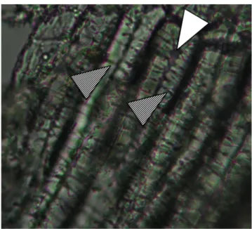
Anatomical analyses of pericarp tissue
To observe the anatomical structures in the Chrysanthemum coronarium pericarp a tissue dissection was carried out in both achene morphotypes by using a razor blade and visualizing tissues with a microscope (Primo Star - Carl Zeiss Microscopy) using different magnifications (10X, 40X and 100X).
Germination in dark
In order to test whether Chrysanthemum coronarium germination is dependent on the presence of light or not, achenes from 2013 collection were tested in continuous darkness at 5, 10, 15, 20 and 25° C for 12 weeks in the dark and checked once at the end of the test. Darkness was achieved by wrapping Petri dishes in 2 aluminium foil layers and kept inside
_____________________________________________________________Materials and Methods a thick-textured black bag for the duration of the experiment. In the meantime a control experiment was carried out in light presence.
Cold stratification
A chilling stratification to mimic natural conditions during winter was carried out in Chrysanthemum coronarium in order to observe its impact in dormancy release. Cold stratification was carried out sowing achenes for 0, 4, 8 and 12 weeks at 5° C in darkness (see germination in darkness for methodology), and transferring them at 20, 25, 30, and 35° C after stratification.
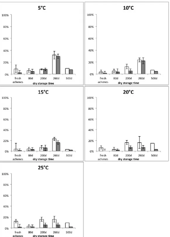
Dry storage
Chrysanthemum coronarium central achenes from the 2012 collection were used for a long-term dry storage test that was carried out keeping achenes at 10-22° C with a Relative Humidity between 20 and 30% up to 500 days. Achenes germination were tested after 80, 200, 260 and 500d and compared with seed germination of fresh achenes.
Dry after ripening
Achenes from the 2013 collection of Chrysanthemum coronarium were used to test the effect of dry after-ripening treatment on seed dormancy. Hot and dry native environment summer climatic conditions were simulate keeping achenes for 0, 4, and 12 weeks at alternating temperature of 35°/20° C (night/day) with a 8/16 photoperiod with a Relative Humidity of, respectively, 35% and 58%. Afterwards achenes were imbibed at 5, 10, 15 and 20° C.
Wet warm stratification
Wet warm stratification was tested in achenes from the 2013 collection of Chrysanthemum coronarium to analyse the different germination response in comparison with cold stratification. Thus, achenes were imbibed at 35/20° C with 8/16 hours of photoperiod. This treatment was performed for 3, 5 or 8 weeks and after which achenes were transferred to germination temperatures 5, 10, 15 and 20° C.
Seed osmo-priming
To determine the effect on germination behaviour of seed osmo-priming treatment in Leucanthemum vulgare seeds were primed imbibing them with Polyethylene-glycol (PEG)
(concentration and to have an osmotic potential of -0.072 MPa) and incubated at 5° C for 1, 5, and 9 weeks. Osmo-priming at 10° C were carried out with an osmotic potential of -0.069 MPa for 1, 5, and 9 weeks. Light role in affecting germination response during priming was also tested imbibing seeds in continuous darkness for 1 week. Both for light and darkness experiments, after priming treatment seeds were washed with tap water and imbibed in double distilled water at 10, 15, 20, 25, and 30° C.
Seed hydro-priming
Leucanthemum vulgare seeds were hydro-primed by soaking seeds in water or with different osmotic potentials: -0.02, -0.072, -0.292 MPa. Afterwards seeds were washed with tap water and imbibed in double distilled water at 10, 15, 20, 25, and 30° C.
ABA synthesis
In both Chrysanthemum coronarium and Leucanthemum vulgare the ABA biosynthesis was investigated to observe its role in dormancy maintenance. To do that seeds (L. vulgare) or achenes (C. coronarium) were imbibed with different concentration 0.03, 0.1, 0.3, 1, 3, 10, 30, 50, and 100µM of fluridone (1-methyl-3-phenyl-5-[3-(trifluoromethyl)phenyl]-4(1H)-pyridinone) which inhibits the carotenoid pathway depleting the ABA biosynthesis (Lem 1981).
Incubation in agitated water
In Chrysanthemum coronarium achenes were washed in 200 ml water for 24 h on a rotating shaker to remove endogenous ABA as published in (M. T. Le Page-Degivry 1990). Afterwards, soaked achenes were used to run simultaneously two different tests: i) achenes were imbibed in double distillied water and incubated at 20° C; ii) achenes were imbibed with a 100µM solution of fluridone and incubated at 20° C. In the first test, water washing was done to remove the ABA already present in the achene, while in the second test, in addition to removing the pre-existing ABA, fluridone was added to prevent any further ABA biosynthesis during the achene imbibition.
Giberellic acid
After the osmo-priming treatment, Leucanthemum vulgare seeds appeared to be inducted in a non-deep physiological dormancy status. Thus, in order to investigate the degree of physiological dormancy, osmo-primed seeds were imbibed with giberellic acid at different
_____________________________________________________________Materials and Methods concentrations 0.01, 0.03, 0.1, 0.3, 1, 3, and 10 µM. control experiment was carried out by imbibing seeds in double distilled water.
Statistical analyses
Germination results statistical support was carried out testing normality and homogeneity of data, and subsequently the relative analysis of variance (Anova). All the elaborations were carried out by using R program (R Development Core Team, 2008).
Molecular analyses
Chrysanthemum coronarium varietal genotyping
To investigate the genetic relationship between the two varieties of Chrysanthemum coronarium, two different approaches were carried out: an intra-specific analysis using markers which produce highly polymorphic patterns to evaluate their genetic diversity; an inter-specific approach to investigate whether the genetic diversity degree arises from differences at the species rank or not. In the first system the profiles of Inter Simple Sequence Repeats (ISSRs) were used, and in the second one ribulose-bisphosphate carboxylase gene (rbcL) and maturase K (mat-K) gene were sequenced on both strands.
DNA extraction
For each of the 32 individuals (16 concolor + 16 discolor var.) around 90 mg of fresh leaf tissue was used to extract DNA following the CTAB/Chloroform-Isoamyl alcohol protocol published by (J. J. Doyle 1987) (J. J. Doyle 1987) (Cullings 1992).
Even though the presence of 4% Polyvinylpyrrolidone (PVP) in the extraction buffer the polyphenolic compounds exerted an inhibitory effect on the Taq polymerase activity during the amplification DNA. Thus, a final concentration of 2% of Polyvinylpyrrolidone was added to the PCR mix (Koonjul 1999) to have reproducible and stable DNA amplifications. From DNA extraction 25 ng of DNA was used as template for subsequent PCR analyses.
ISSR profiling
Internal Sequence Simple Repeats (ISSR) PCR were carried out for all the samples in a total volume of 25 µL containing 25 ng of DNA template, 2.5 μL of 10 × PCR buffer (100 mM Tris–HCl, 15 mM MgCl2, 500 mM KCl), 2% of PVP, 0.5 μL of dNTPs (10 mM each),
0.5 μL of primer (10 mM), 1.5 unit of Solis HotStart Taq DNA polymerase (Solis BioDyne, Tartu, Estonia).
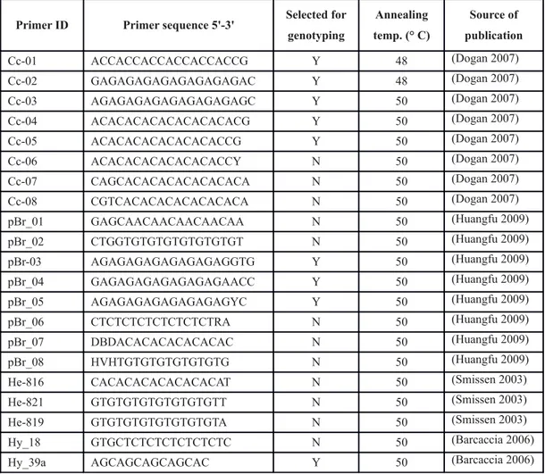
Twenty-one ISSR markers, 3’ or 5’ anchored primers (synthesized by Eurofins MWG Operon, Ebersberg, Germany), were tested and 9 of them were selected basing on their band pattern and reproducibility (see Table 2).
_____________________________________________________________Materials and Methods
Primer ID Primer sequence 5'-3' Selected for genotyping Annealing temp. (° C) Source of publication Cc-01 ACCACCACCACCACCACCG Y 48 (Dogan 2007) Cc-02 GAGAGAGAGAGAGAGAGAC Y 48 (Dogan 2007) Cc-03 AGAGAGAGAGAGAGAGAGC Y 50 (Dogan 2007) Cc-04 ACACACACACACACACACG Y 50 (Dogan 2007) Cc-05 ACACACACACACACACCG Y 50 (Dogan 2007) Cc-06 ACACACACACACACACCY N 50 (Dogan 2007) Cc-07 CAGCACACACACACACACA N 50 (Dogan 2007) Cc-08 CGTCACACACACACACACA N 50 (Dogan 2007) pBr_01 GAGCAACAACAACAACAA N 50 (Huangfu 2009) pBr_02 CTGGTGTGTGTGTGTGTGT N 50 (Huangfu 2009) pBr-03 AGAGAGAGAGAGAGAGGTG Y 50 (Huangfu 2009) pBr_04 GAGAGAGAGAGAGAGAACC Y 50 (Huangfu 2009) pBr_05 AGAGAGAGAGAGAGAGYC Y 50 (Huangfu 2009) pBr_06 CTCTCTCTCTCTCTCTRA N 50 (Huangfu 2009) pBr_07 DBDACACACACACACAC N 50 (Huangfu 2009) pBr_08 HVHTGTGTGTGTGTGTG N 50 (Huangfu 2009) He-816 CACACACACACACACAT N 50 (Smissen 2003) He-821 GTGTGTGTGTGTGTGTT N 50 (Smissen 2003) He-819 GTGTGTGTGTGTGTGTA N 50 (Smissen 2003) Hy_18 GTGCTCTCTCTCTCTCTC N 50 (Barcaccia 2006) Hy_39a AGCAGCAGCAGCAC Y 50 (Barcaccia 2006)
Table 2 Primers sequences used for varietal genotyping in Chrysanthemum coronarium
The amplification reactions were carried out using a Quanta biotech SI-96 Thermal Cycler (Quanta Biotech Ltd, Surrey, UK) with the following PCR profile: 95° C for 15 min; 35 cycles at 94° for 30 sec, specific annealing temperature for each primer (Table 2) for 1 min, 72° C for 1 min; and final extension at 72° C for 10 min.
Ten ul of PCR products were electrophoresed on 2.0% agarose gels during 2.0 h at 100 V in 1Å~ TAE buffer (40 mM Tris, 20 mM acetic acid, 1 mM EDTA, pH 8.4) and a 100 base-pair ladder (100 bp DNA Ladder, Solis BioDyne, Tartu, Estonia) was used as molecular size marker. Gel was stained for 30 min with 1X Gel Red (© Biotium, USA) and then visualized and photographed using a gel documentation system (BioDoc-It® Imaging
System, CA).
rbc-L gene amplification and sequencing
The entire rbcL gene was amplified using the “1f” and “1369 r” primers as described in (Olmstead 1992) in a total volume of 70 µL containing 25 ng of DNA template, 7 μL of 10 × PCR buffer (100 mM Tris–HCl, 15 mM MgCl2, 500 mM KCl), 8.4 μL of MgCl2 (25
mM), 2% of PVP, 1.4 μL of dNTPs (20 mM each), 1.75 μL of primer (10 mM), 2 unit of Solis HotStart Taq DNA polymerase (Solis BioDyne, Tartu, Estonia).
Amplifications were carried out in the thermal cycler aforementioned with the following PCR profile: 95° C for 15 min; 35 cycles at 94° for 30 sec, 1 min at 48°, 72° C for 1 min; and final extension at 72° C for 10 min. The obtained PCR products were purified and sequenced on both strands by Macrogen Inc. (Netherlands) using PCR primers. Sequencing was conducted under BigDyeTM terminator cycling conditions and products
purified using ethanol precipitation and run using automatic sequencer ABI-3730XL.
mat-K gene amplification and sequencing
The central region of mat-K gene was amplified using the “390f” and “1326 r” primers as described in (Schaefer 2011) in a total volume of 70 µL containing 25 ng of DNA template, 7 μL of 10 × PCR buffer (100 mM Tris–HCl, 15 mM MgCl2, 500 mM KCl), 8.4
μL of MgCl2 (25 mM), 2% of PVP, 1.4 μL of dNTPs (20 mM each), 1.75 μL of primer (10
mM), 2 unit of Solis HotStart Taq DNA polymerase (Solis BioDyne, Tartu, Estonia).
Amplifications were carried out in the thermal cycler aforementioned with the following PCR profile: 95° C for 15 min; 35 cycles at 94° for 30 sec, 1 min at 48°, 72° C for 1 min and 30 seconds; and final extension at 72° C for 10 min. The obtained PCR products were purified and sequenced on both strands by Macrogen Inc. (Netherlands) using PCR primers. Sequencing was conducted under BigDyeTM terminator cycling conditions and
products purified using ethanol precipitation and run using automatic sequencer ABI-3730XL.
Data analysis
ISSR Band analysis was carried out using Total Lab 100 (TotalLab Ltd, UK) and only non-overlapping and highly reproducible bands with a molecular size comprised from 350 to 1700bp were considered for fragments detection.
Genetic diversity was measured by the percentage of polymorphic bands (P), which was calculated by dividing the number of polymorphic bands at population and species levels by the total number of bands surveyed. Shannon indices of diversity, namely both the total diversity (Hsp) and the intra-population diversity (Hpop), were also calculated using the computer program POPGENE software package Version 1.32. Principal Coordinates Analysis was carried out with GeneAlex program (Peakall, 2006)
Sequences of rbc-L and mat-k genes were blasted against BOLD databases (http://www.boldsystems.org/) (Ratnasingham 2007) for confirmation. Sequence alignment
_____________________________________________________________Materials and Methods was carried out using CLUSTAL W algorithm (Larkin 2007), and phyologenetic elaboration was carried out with PAUP program (Swofford, 2003).
Identification and isolation of reference and dormancy associated genes in C.
coronarium and L. vulgare
Reference genes
Quantifying gene expression levels through reverse transcription–quantitative real-time PCR (RT–qPCR) is the preferred method for targeted gene expression measurements. However, normalization, is necessary to correct for sample input and reverse transcriptase efficiency, is a crucial step to obtain reliable RT–qPCR results. Stably expressed genes (i.e. genes whose expression is not affected by the treatment or developmental stage under study) are indispensable for accurate normalization of RT–qPCR experiments. Lack of accurate normalization could affect the results and may lead to false conclusions.
Some of the house keeping genes involved in basic cellular activities such as 18s rRNA, 25s rRNA, glyceraldehyde-3-phosphate dehydrogenase (GAPDH) and ubiquitin (UBQ) are some of the commonly used internal control genes as they are likely to be expressed at constant levels regardless of experimental conditions (Schmittgen 2000) (Czechowski 2005).
Therefore, selected genes from literature (Dekkers 2011) were used to find their homologous gene family for Asteraceae consulting “Nucleotide” dataset of “NCBI” database. Afterwards, each matched sequences was searched at the Arabidopsis electronic fluorescent pictographic (eFP) browser (http://bar.utoronto.ca/efp/cgi-bin/efpWeb.cgi) (Schmid 2005) (Kilian 2007) (Winter 2007) to check how much stable is the expression of the selected gene compared with the treatment as reported in the browser. Most stable gene sequences, hence, were aligned for cloning primers designing to use in downstream C. coronarium and L. vulgare PCR amplifications.
Dormancy associated genes
On the basis of the specific literature on molecular regulation of seed dormancy (Liu 2007) (G. C. Chiang 2009) (L. H.-L.-B. Bentsink 2010) (S. D.-S.-S. Footitt 2011) DOG1 (DELAY OF GERMINATION 1), FLC (FLOWERING LOCUS C), HUB2 (HISTONE MONOUBIQUITINATION 2) genes were included. In addition the EXP Expansin gene was selected for his role in mediating cell expansion during radicle elongation (F. D. Chen 2001).
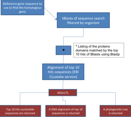
The overall workflow used for marker selection is illustrated in Figure 10.
Figure 10 Workflow for primer designing of dormancy associated genes in C. coronarium and L.
vulgare
DNA extraction
Fifty seedlings of Leucanthemum vulgare about 1 cm long each along with their seed coat were grounded in a mortar pestle using liquid nitrogen for DNA extraction. A CTAB/Chloroform-Isoamyl Alcohol procedure was used for DNA extraction as in (Kocik 1996). Samples were diluited 1000 times, shared through vigorous vortexing to make DNA more accessible for downstream amplification purposes.
Both L. vulgare and C. coronarium (herein for this chapter just concolor variety, DNA extracted following method described at the beginning of this chapter) extracted DNAs were used as templates for amplification of reference genes and dormancy-associated genes.
RNA extraction
Total RNA was isolate from different stage of dormancy of L. vulgare seeds: dry seeds (as reference control), 1 week primed seeds at 5° C, 1 week primed seeds at 10° C, 5 weeks primed seeds at 5° C, 5 weeks primed seeds at 10° C, and after imbibition upon germination (endosperm rupture and when upon radicle protrusion not longer then 1 mm).
_____________________________________________________________Materials and Methods Each sample was made up by three replicas of 50 seeds each. RNA was extracted using the hot borate protocol as in (Wan 1994) (Toorop 2005). Pellets were dried over-night using a freeze-dryer (Maxi Dry Lyo, Gemini BV) and, afterwards, resuspended in 50 μl of RNA-secure™ (Ambion®, Life Technologies) to inactivate RNases. RNA was quantified with a
spectrophotometer WPA Lightwave (Biochrom, Cambridge, UK). Half μg per each sample was loaded in a electrophoresis gel well, run at 100 V in 1Å~ TAE buffer (40 mM Tris, 20 mM acetic acid, 1 mM EDTA, pH 8.4) for 1 hour and visualized through a Gene Genius (Syngene, Synoptics Ltd) bio imaging system.
cDNA first strand synthesis
One μg of total RNA was used as a template for first cDNA strand with Transcriptor First Strand cDNA Synthesis kit (Roche, UK), using anchored-oligo(dT)18 primers. First strand
polymerization was carried out in PCR tubes using a thermal cycler DNA Engine Dyad, Peltier Thermal Cycler. Synthesized cDNA was used as template for amplification of reference genes and dormancy-associated genes.
PCR products cloning
Three μL of PCR products were used for ligation with a TA cloning vector, pGEM®-T easy
Vector kit (Promega), following the manufacturer instructions with some modifications: 1 μL of ligation reaction was used for transformation and LB medium (for 1 L: 10 g BactoTryptone, 5 g of BactoYeast, and 5 g NaCl) was used. Bacteria were grown in a 1.5 mL polypropylene tube for 1.5 h and 100 μL were plated in a LB/ampicillin plate and incubated at 37° C over-night. Single colonies were picked up with wooden sticks and rinsed with double distilled sterile water in an Eppendorff tube. Ten μL were used as template for PCR colony using plasmid specific primers to amplify the inserted DNA fragment. Electrophoretic gel runs were carried out for evaluating the size of the incorporated fragment and comparing it with the supposed gene length as observed during homologous gene alignment.
When positive colonies were found bacteria were picked up from the twin colony and grown over night at 37° C in 3 mL of LB-medium containing the ampicillin antibiotic. Isolation of plasmid DNA was carried out using the High Pure Plasmid Isolation kit (Roche) starting from a 3 mL of cell culture (transferring two times 1.5 ml to an Eppendorf tube and centrifuging at 14000 rpm for 30 sec). Isolated plasmid DNA samples were quantified using a spectrophotometer (see above for instruments specifications), checked
through inserted fragment amplification, and sequenced by University College London sequencing service.
Sequences analysis
Plasmid sequences were trimmed against vector database to remove any contaminations and then blasted using BLASTn algorithm (NCBI) against Asteraceae dataset sequence or if no results were returned enlarging the dataset including more plant groups. The most similar sequences resulted in homologous genes and the most similar sequences were aligned using CLUSTAL W algorithm (Larkin 2007). This procedure gave an evidence of the origin of isolated sequences (Figure 11).
_______________________________________________________________________Results
Results
Physiology of germination of Chrysanthemum coronarium Germination behaviour
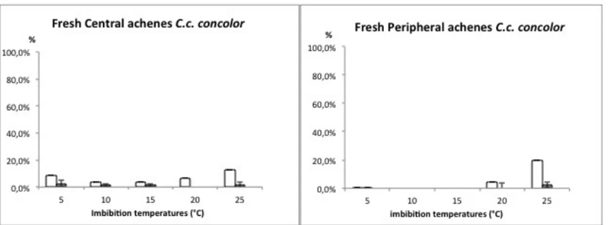
Chrysanthemum coronarium germination of fresh achenes in the two varieties (see Figure 12 and Figure 13) showed a very low response (under 5 %). Radicle emergence always occurred in achenes with an evident rupture in the fruit coat, but not all the ruptured achenes germinated. Higher temperatures produced a significantly increase in FCR events then lower temperatures.
Figure 12 Chrysanthemum coronarium var. concolor germination at constant temperatures. Fruit coat rupture: open histograms; Seed germination: solid histograms.
Figure 13 Chrysanthemum coronarium var. discolor germination at constant temperatures. Fruit coat rupture: open histograms; Seed germination: solid histograms.
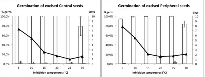
Seed excision
Excising seeds from pericarp resulted in very high germination frequency (100%) for both peripheral and central morphotypes (Figure 14). Values on germination speed (t-50) indicated a slower response at low temperatures (Figure 14).
On the other hand, intact achenes germinated fewer than 5% in all the tested temperatures.
Figure 14 Germination of central and peripheral excised seeds of Chrysanthemum coronarium var
concolor. Histograms follow principal axis (final germination %), while curve follows the secondary
axis (number of days until 50% of final germination). Open histograms: seed germination of excised seeds. Solid histograms: seed germination of intact achenes. Solid line: excised seed germination t-50.
Furthermore, sowing excised seeds in close contact with pericarp tissue did not showed any significant inhibitory effect (Figure 15).
Figure 15 Germination at 15° C of central and peripheral excised seeds of Chrysanthemum coronarium var concolor in close contact with pericarp tissue; Open circle: intact achenes (control experiment); Solid line: excised seeds with pericarp tissue; Open triangles: excised seeds without pericarp tissue.
_______________________________________________________________________Results
Comparison of the germination behaviour of the two varieties
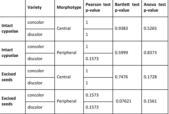
The germination frequency of the two varieties of Chrysanthemum coronarium was compared (Figure 16) and statistical support was tested. Normality and homogeneity were respected and Anova analysis indicated no significant differences within the germination behaviour of the two varieties (see Table 3).
Figure 16 Final germination of intact cypselae (represented with dark bars) and excised seeds (light bars)
Variety Morphotype Pearson test p-value Bartlet test p-value Anova test p-value Intact cypselae concolor Central 1 0.9383 0.5265 discolor 1 Intact cypselae concolor Peripheral 1 0.5999 0.8373 discolor 0.1573 Excised seeds concolor Central 1 0.7476 0.1728 discolor 1 Excised seeds concolor Peripheral 0.1573 0.07621 0.1561 discolor 0.1573
Table 3 Homogeneity (Pearson’s test), normality (Bartlett’s test), and analysis of variance (ANOVA); P-value >0.05 null hypothesis is accepted = not significant difference; P-value <0.05 null hypothesis is rejected = significant difference.