Antibiotic resistance genes in the viral DNA
fraction of environmental samples
Gens de resistència a antibiòtics en el DNA de la fracció vírica
de mostres ambientals
Marta Colomer Lluch
,
Aquesta tesi doctoral està subjecta a la llicència Reconeixement 3.0. Espanya de Creative Commons.
Esta tesis doctoral está sujeta a la licencia Reconocimiento 3.0. España de Creative
Commons.
ANTIBIOTIC RESISTANCE GENES IN THE VIRAL DNA FRACTION
OF ENVIRONMENTAL SAMPLES
GENS DE RESISTÈNCIA A ANTIBIÒTICS EN EL DNA DE LA
FRACCIÓ VÍRICA DE MOSTRES AMBIENTALS
Marta Colomer Lluch
ANTIBIOTIC RESISTANCE GENES IN THE VIRAL DNA FRACTION OF
ENVIRONMENTAL SAMPLES
GENS DE RESISTÈNCIA A ANTIBIÒTICS EN EL DNA DE LA FRACCIÓ VÍRICA DE
MOSTRES AMBIENTALS
Thesis presented by Marta Colomer Lluch to obtain the degree of Doctor by the University of Barcelona
Memòria presentada per Marta Colomer Lluch per optar al grau de Doctora per la Universitat de Barcelona
EHEA Doctorate programme: Environmental Microbiology and Biotechnology Programa de doctorat EEES: Microbiologia Ambiental i Biotecnologia
Thesis developed under the supervision of Dr. Joan Jofre Torroella and Dr. Mª Teresa Muniesa Pérez in the Department of Microbiology, Faculty of Biology, University of Barcelona
Tesi realitzada sota la direcció del Dr. Joan Jofre Torroella i la Dra. Mª Teresa Muniesa Pérez al Departament de Microbiologia, Facultat de Biologia, Universitat de Barcelona
Supervisor, Supervisor, Author,
El director, La directora, Autora,
Joan Jofre Torroella Mª Teresa Muniesa Pérez Marta Colomer Lluch Barcelona, April 2014
Departament de Microbiologia Facultat de Biologia Universitat de Barcelona
Professors Joan Jofre Torroella and Mª Teresa Muniesa Pérez from the University of Barcelona
DECLARE THAT,
Marta Colomer Lluch has performed the work entitled Antibiotic resistance genes in the viral DNA fraction of environmental samples / Gens de resistència a antibiòtics en el DNA de la fracció vírica de mostres ambientals under our supervision in order to obtain the degree of Doctor by the University of Barcelona and that this thesis fulfils the requirements to obtain the International Doctor Mention, and that this work is ready to be presented from the present day.
Signature,
Joan Jofre Torroella Mª Teresa Muniesa Pérez
“Learn from yesterday, live for today, hope for tomorrow. The important thing is to not stop questioning.” Albert Einstein.
"Le rôle de l'infiniment petit dans la nature est infiniment grand". Louis Pasteur.
Aknowledgements/Agraïments
i
AKNOWLEDGEMENTS/AGRAÏMENTS
En aquest punt ja només em queda donar les gràcies a tots els que heu fet possible aquesta tesi i que m’heu acompanyat, d’una o altra manera al llarg d’aquesta experiència. Sense tots vosaltres no hauria estat possible!!!
En primer lloc, vull donar les gràcies a la Maite i al Joan, per donar-me l’oportunitat d’entrar al grup, al principi com a estudiant de col·laboració, i per oferir-me després la possibilitat de fer una tesi doctoral. Moltíssimes gràcies per la confiança rebuda al llarg de tots aquests anys i per contagiar-me el vostre esperit per la feina ben feta.
Maite, moltíssimes gràcies per tot, i per tot vull dir per la teva confiança des del principi, per
donar-me suport i ajudar-me sempre que ho he necessitat, i per orientar-me en tot moment. Per la teva visió a l’hora d’encarar les dificultats o problemes que han sorgit i per veure sempre la part positiva de les coses. Amb tu he après moltíssim en tot aquest temps. Tant a mi com a la resta sempre has aconseguit motivar-nos i gran part del mèrit que tot el lab siguem una pinya és teu. Ets una jefa única! També m’alegro molt d’haver pogut compartir amb tu anècdotes, històries, dinars, cafès (amb terrasseta, clar!), congressos (amb piscina i sense ;)), sortides de grup, algun que altre “cotilleo” (que sempre van bé per animar el dia) i tants i tants moments que recordo amb molt i molt de carinyo.
Joan, moltíssimes gràcies, la teva experiència i els teus consells m’han ajudat a entendre
moltes coses al llarg d’aquests anys. Els teus comentaris i reflexions m’han permès millorar i despertar el meu sentit crític. Les teves entrades sorpresa al laboratori, moltes vegades enganxant-nos de ple en allò que no toca i tot i així riure amb nosaltres, no tenen preu!
També voldria donar les gràcies a la resta de professors del grup. A l’Anicet, per ser sempre tan amable amb mi, i al Francisco por tus reflexiones y comentarios que no nos dejan indiferentes. També a la Rosina, a la Rosa, a la Susana i al Joan T. per la vostra amabilitat i per ajudar-me sempre que ho he necessitat.
Vull agrair també a tots els membres de secretària del departament, a la Macu, a la Susana, a la Bea i al Manolo, per estar sempre disposats a donar-me un cop de mà, sobretot amb els
Aknowledgements/Agraïments
ii
temes de “papeleo” (que no han estat pocs...), i per organitzar cosetes tan divertides com el carnestoltes, la rifa de nadal,...seguiu sempre així! Molt especialment, gràcies a la Rosario, sempre amb bona cara i a punt per ajudar-me, i per fer que gràcies a ella preparar les pràctiques de cada any sigui molt més senzill.
A totes les persones de Fase I, pels increïbles sopars de departament, per les festes de tesi, per les reunions de responsables, per les trobades a la cuina,...moltes gràcies a tots!
Ja a Fase II, voldria agrair als membres del lab 7, tant els que ja no hi són però van coincidir amb mi, al Markus i a la Ivania, com als que encara hi sou, a la Raquel, a la Maite, i a la resta del seu lab. Sobretot, merci per deixar-me electroporar sempre que ho he necessitat i patir amb mi aquell segon de tancar els ulls i creuar els dits perquè no peti res.
Arribant al lab 8, vull agrair-vos a tots els bons moments que hem compartit tant a dins com a fora del departament. Als rosinots i als araujos! Merci per ser tots tan macos!!! Sílvies,
Cervero i Bofill, Tarik, Laura, Marta, Sandra, Ayalke (i els Chus, Byron, Anna que van marxar
no fa tant). I no em podia deixar el Persi! Ets un sol!!! Les teves visites dia sí dia també al nostre lab són la prova que Vircont s’està estenent! Sempre em fas riure amb una sortida de les teves (maldita peluquera...!) i quan em pensava que ja no podia tenir més noms, arribes tu amb lo de pigeon...!
Moltíssimes gràcies també als meus veïns del lab 9!!! Sou tots genials! A través de la porta que ens connecta sempre acabem sentint els vostres riures, les vostres converses (a vegades una miqueta surrealistes), el catxonedeo general....i no podem evitar treure el nas i xafardejar com bons veïns que som! Sou un grup maquíssim! Gràcies a la Raquel per compartir amb mi la teva passió per la música i per alegrar-me el dia amb els teus “m’agraden les teves sabates!” (sobretot les Ted Mosby, oi? Jaja!), a la Míriam per estar sempre disposada a resoldre’m qualsevol dubte i per fer-me riure de veritat quan et poses a explicar alguna de les teves històries i, sobretot, merci a tu i a l’Arnau per tenir tant integrat l’APM (m’encantaaaaa!). Del lab 9 no em voldria deixar de donar les gràcies a Sílvia M,
Camilo, Mateu, Sergi, Ángela, i com no, el relleu del lab 9, Laura i Julia, totes dues trepitjant
Aknowledgements/Agraïments
iii A la Cris, i a l’Eli, merci pel teu “Hola!” tan eufòric cada vegada que entres al lab i pel teu entusiasme en tot allò que fas.
Aquest llarg camí ha estat millor gràcies a tots els companys del lab 10, que heu fet que aquests anys amb vosaltres hagin estat increïbles. Gràcies per liar-la de tant en tant i fer que un dia qualsevol pugui ser genial, per col·laborar a les frases cèlebres de la nostra llibreta (que no passi res el dia que surtin a la llum...). Merci per compartir dies de feina i més feina, frustracions i alegries, sopars, sortides, viatges, congressos, rialles i més rialles,....és que amb vosaltres és un non-stop i realment és veritat que amb els anys hem esdevingut una petita gran família.
Gràcies a tots, a les que em vau iniciar al lab 10, a la Fanny i a la Ruth, i als que han passat pel lab, a la Laura (grazie mille!), a la Clara, a l’Ana M, a la Míriam 2 (o hauria de dir lab 9?, tu sempre m’has fet costat en la meva desesperació els divendres Disney...).
Moltíssimes gràcies als meus tècnics preferits!!! El tàndem Andreu&Jordi,...Jordi&Andreu! Vau marcar una època inoblidable! Sempre m’heu cuidat moltíssim i us heu portat genial amb mi! Enyoro els nostres partidets!
A l’Aiora, moltíssimes gràcies per estar disposada a ajudar-me en tot moment i tenir paciència (infinita moltes vegades...) amb mi i amb les meves preguntes (ja saps que el word i l’excel em tenen una mania especial). Ets una crack! Merci pels teus viatges amb moto quan encara mitja ciutat està dormint (és que anem molt d’hora!) i per les nostres converses a primera hora del matí quan el departament encara no s’ha despertat. En tu he trobat una aliada per fer front a l’amenaça Disney, ja m’entens..., jo crec que ho aconseguirem!
A l’Anna i a la Marta, o més ben dit, a l’Annis i a la Gómez, estic convençuda que juntes fem un trio espectacular!!! Per on començar...vam fer la carrera juntes, vam entrar col·laborant al departament alhora, vam fer el màster plegades i finalment vam començar l’aventura del doctorat!!! És que ara que hi penso...portem quasi 10 anys juntes! Que fooooort!!! Ha estat un plaer tenir-vos al costat de la meva poiata durant tots aquests anys. Amb vosaltres he passat moments increïbles, magnífics, he après un munt amb i de vosaltres, sou genials!!! Congressos, dinars, sopars, festes, calçotades, excursions, sortides inoblidables, les festes de
Aknowledgements/Agraïments
iv
Cadaqués, viatges per la llunyana Califòrnia amb parada obligada a Las Vegas,...quedaran gravats per sempre a la nostra memòria i els conservo amb molt i molt de carinyo. Durant tots aquests anys heu aportat el toc freak al lab i això us fa encara més autèntiques! Heu hagut de suportar les meves queixes de les sessions Disney (sempre amb carinyo, ja ho sabeu), els meus moments de stress màxim al lab,...però sempre heu estat allà per tot el que he necessitat. No tinc prou paraules per agrair-vos tots els anys que hem compartit juntes. Sense vosaltres dues res d’això no hauria estat el mateix.
A l’Àlex, el nostre valencià (¿a ver cuándo te sueltas i parles en català, no?), sin ti nada de esto tampoco habría sido igual. Muchas gracias por arrancarme una sonrisa siempre que lo he necesitado (aunque a veces haya sido a costa de tus bromas...lo sé, soy tu víctima número uno), por compartir momentos geniales e imposibles de olvidar y demostrarme que puedo contar contigo para lo que sea. Hemos pasado días largos de trabajo en el lab pero tus historias surrealistas, anécdotas, vídeos...siempre lo han hecho mucho más llevadero e infinitamente más divertido. Me has aportado muchísimo en todo este tiempo y en ti he descubierto a una gran persona, y quiero que sepas que eres una pieza clave en nuestro lab. Pero… ¿ahora tampoco te lo vayas a creer mucho, vale? ;)
A Pablo, nuestro gran fichaje asturiano!!! Gracias por desprender siempre tan buen rollo y buen humor!!! Siempre dispuesto a ayudar en lo que haga falta. Me has tenido que sufrir enseñándote durante tu practium y siento haberte iniciado en el gran mundo de las “muestras de origen humano”, ¿no me lo tengas en cuenta, vale?
A Willliam, mejor dicho Sir William, siempre tan educado y con gran voluntad, también me has tenido que sufrir un poquito...muchos ánimos que seguro que todo te irá muy bien.
A las noves incorporacions: Carmen, Ferran, Maryuri... ¡No sabéis donde os metéis! Jajaja!
Thank you very much Dr. Sommer from Systems Biology Department at DTU, in Denmark to allow me to work in your research group for my short stay. You and all people from sommerlab were very kind to me and it was really a pleasure to spend four lovely months there.
Aknowledgements/Agraïments
v De manera molt especial vull donar les gràcies a dues persones espectaculars que admiro moltíssim:
A la Lejla, Lejlissss! Para mí siempre serás un poquito mi teacher, tú me enseñaste muchísimas de las cosas que he aprendido y por eso siempre te estaré muy agradecida (bueno...menos por los litros y litros de chromocult que hicimos ;)). En el lab hemos pasado de todo juntas, reído un montón y nuestra mezcla de idiomas lo ha hecho todo aún más divertido (está todo en la libretaaaaa!) Con el tiempo nos hicimos buenas amigas y siempre he podido contar contigo. Siempre te has preocupado mucho por mí y juntas hemos compartido mucho: el frío de Dinamarca (y el sol de Estocolmo...), vinitos y sesiones chillout, nuestras ganas de viajar por el mundo…y una laaaaarga lista de momentos geniales. ¡Muchas gracias por todo!
A la Maru, mongaaaa! Mil gracias por ser como eres, ya sabes que esto no habría sido igual sin ti. Nuestros cafés, charlas y más charlas, sueños en voz alta, cenitas, fiestas, Cadaqués, viajecitos, y taaaaantos momentos especiales e irrepetibles los guardo con mucho cariño. Quiero que sepas que te has convertido en una más en mi familia (y no lo digo porque nos debes un roscón...;)) y que aunque ahora estemos separadas puedes contar conmigo para lo que quieras! Merci mongaaa!
Fora del departament també vull donar les gràcies a tots els amics que han estat amb mi durant tot aquest temps.
A “Las Piltrafillas”! Noies sou genials!!! Vull donar-vos les gràcies per totes les nits de divendres amb partits inoblidables, victòries de “subidón”, jugant amb pluja, fred, vent,... feu que cada setmana esperi amb més ganes l’arribada del divendres!
I com no, gràcies també a “Los Piltrafillas”! Nois, sou uns cracks! Molts de vosaltres ja heu passat pel doctorat i us heu posat a la meva pell moltes vegades. Els vostres consells i ànims m’han ajudat moltíssim al llarg d’aquests anys. Vull donar les gràcies, especialment al Manu, por conocernos des de hace tantos años, mejor dicho, por conocerme tan bien (da un poquito de rabia y todo...jeje!) y por estar ahí siempre. També al Xavi R., Quique, Bruno,
Aknowledgements/Agraïments
vi
De manera molt especial vull donar les gràcies a la Sara, la Maria i la Luisa. Noies, no sé què faria sense vosaltres!!!! “Quedamos???” És sinònim dels nostres cafès, sopars, curses, excursions,...amb vosaltres he viscut moments molt i molt divertits, hem compartit rialles, preocupacions, projectes, ambicions,... Les tres heu estat amb mi des del principi (en les alegries i també en les penes...jaja!) i m’heu recolzat en tot sempre que ho he necessitat. Per tot això i més, merci a les tres. Sou úniques!
A la Blanca, por tener siempre tanto interés en cómo van “mis bichitos” y animarme con tu alegría.
A la Carmen, per l’entusiasme que sempre has demostrat per la meva feina, per les nostres sessions de teràpia (i n’hem fet unes quantes entre cafè i cafè!) i pels teus consells, i sobretot pels moments únics que hem compartit al llarg de tots aquests anys d’amistat.
A l’Ares, tot i que últimament des de la distància, sempre que ens retrobem en un dels nostres cafès, brunchs, acabem parlant de tot i és com si no haguessis marxat de Barcelona. Et desitjo el millor per a la teva tesi! Segur que t’anirà genial!
I per acabar, vull donar les gràcies a la meva família.
A l’Anna Maria i la Consol, la vostra experiència i consells m’han ajudat a tenir una perspectiva diferent de les coses.
A l’Ana, pel teu entusiasme des del primer moment en voler col·laborar en la portada de la tesi i plasmar les teves idees d’una manera tant espectacular. Al Jordi i la Lucy, pel vostre interès, per les nostres converses i inquietuds compartides, i per fer-me descobrir que fa més de cinquanta anys la nostra família ja havia començat a investigar en bacteriòfags...i ja ho veieu! La tradició continua!
A les meves àvies, àvia Lluïsa i àvia Elena, i a la tieta Teresa, a qui sé que els fa molta il·lusió això de tenir una néta científica tot i no acabar d’entendre ben bé què és el què faig. M’heu demostrat sempre el vostre carinyo i per això us estic enormement agraïda.
Aknowledgements/Agraïments
vii A la meva germana, Miri!!! Sister!!! Què t’haig de dir...merci per ser com ets i per confiar tant en la teva germana. Amb tu he compartit moltes alegries però també moments de “bajón” i frustracions, però sempre t’he tingut amb mi i he pogut comptar amb tu. Ets la millor germana que podia tenir!
I sobretot als meus pares. Mama, papa, sempre heu cregut en mi i m’heu recolzat i animat des que vaig començar la tesi. Tot i que moltes vegades sé que és complicat entendre el què faig i comprendre que de sobte alguns caps de setmana haig d’anar al lab, la vostra curiositat i el vostre interès ho diuen tot. Vosaltres m’heu ensenyat que amb esforç, dedicació i fent la feina ben feta es pot aconseguir tot allò que una es proposi. Per oferir-me sempre el vostre suport i estimar-me com ho feu, moltes gràcies.
A tots vosaltres, perquè en tots aquests anys m’heu enriquit com a persona i em feu sentir molt afortunada, gràcies.
Presentation of the thesis
ix
PRESENTATION OF THE THESIS
The PhD thesis presented here has as a main objective the study of antibiotic resistance genes clinically relevant in the DNA fraction of bacteriophage particles isolated from environmental samples of different origin in order to determine the importance of bacteriophages as vehicles for the mobilization of antibiotic resistance genes between bacteria.
Specifically, a broad range of antibiotic resistance genes were studied as representative of the main groups recently described in our geographical area belonging to three β-lactamases (blaTEM, blaCTXM-1 and blaCTX-M-9), the mecA gene conferring resistance to methicillin in
staphylococci, and the quinolones resistance genes qnrA and qnrS.
To achieve these goals samples of urban wastewater, river water and animal faecal wastes were analysed quantifying the antibiotic resistance genes of interest in bacteriophages DNA. During the development of this Thesis, it was attempted to optimize the available methodology for bacteriophage DNA extraction, as well as the necessary controls to guarantee the amplification of encapsidated DNA and to remove any free DNA in the samples and any possible vesicles containing DNA.
In addition, the ability of phage-encoded genes to confer antibiotic resistance in bacterial strains was assessed by performing transfection experiments.
It was also studied the influence of various compounds involved in the induction of the lytic cycle of temperate bacteriophages, on the abundance of antibiotic resistance genes in DNA from the phage fraction in wastewater samples.
Finally, due to the importance of horizontal gene transfer as a mechanism for antibiotic resistance dissemination in clinical and environmental settings, transduction experiments were attempted, although unsuccessfully, to reproduce in vitro the process that would take place in nature.
Presentation of the thesis
x
The research developed in this Thesis is divided into 5 studies included in 4 chapters: (1) Antibiotic resistance genes in the bacteriophage DNA fraction of water samples (urban wastewater, river water and wastewater with animal faecal wastes); (2) Quinolone resistance genes (qnrA and qnrS) in bacteriophage particles from wastewater samples and the effect of inducing agents on packaged antibiotic resistance genes; (3) Evaluation of ARGs in the DNA of bacterial and bacteriophage fraction in wastewater samples from Tunisia and comparison with results obtained in Barcelona area; (4) Detection of quinolone-resistant Escherichia coli isolates belonging to clonal groups O25b:H4-B2-ST131 and O25b:H4-D-ST69 in water samples from Barcelona area.
Each of the studies has given rise to a scientific article already published or submitted for scientific publication.
In accordance with the requirements for the international mention, English and Catalan were the chosen languages for the development of this memory. This thesis is based on the published and submitted articles that have been included in the publications section. All articles were written in English and are accompanied by a summary in Catalan at the beginning of each publication. It has also been included an introduction, the main objectives and the conclusions in both languages. The discussion has been written entirely in English.
Presentació de la tesi
xi
PRESENTACIÓ DE LA TESI
La tesi doctoral que es presenta a continuació té com a objectiu principal l’estudi de gens de resistència a antibiòtics de rellevància clínica en la fracció de DNA de partícules de bacteriòfags aïllades de diferents tipus de mostres ambientals per tal de determinar la importància dels bacteriòfags com a vehicles de mobilització de gens de resistència a antibiòtics entre bacteris.
Concretament, s’ha estudiat un ampli espectre de gens de resistència a antibiòtics com a representants dels grups principals descrits actualment en la nostra àrea geogràfica corresponent a tres β-lactamases (blaTEM, blaCTXM-1 i blaCTX-M-9), el gen mecA de resistència a
meticil·lina en estafilococs, i els gens de resistència a quinolones qnrA i qnrS.
Per això s’han analitzat diversos tipus de mostres procedents d’aigua residual municipal, d’aigua de riu i d’aigua residual amb contaminació fecal animal per tal de quantificar els gens de resistència a antibiòtics d’interès en DNA aïllat de bacteriòfags.
Durant els diferents estudis s’ha intentat optimitzar la metodologia d’extracció de DNA de bacteriòfags així com els controls corresponents per garantir l’amplificació de DNA encapsidat i l’eliminació de qualsevol DNA lliure present a les mostres i de qualsevol possible vesícula amb DNA al seu interior.
Per altra banda, també s’ha determinat la capacitat funcional dels gens de resistència detectats en DNA de fags i per això s’han realitzat experiments de transfecció a partir de soques bacterianes sensibles a un determinat antibiòtic amb l’objectiu d’incorporar la resistència i per tant, esdevenir resistents a l’antibiòtic en qüestió.
També, s’ha estudiat la influència de determinats compostos implicats en la inducció del cicle lític de bacteriòfags temperats, en l’augment en el nombre de còpies de gens de resistència a antibiòtics en DNA present en la fracció de fags de l’aigua residual.
Presentació de la tesi
xii
Finalment, degut a la importància de la transferència horitzontal de gens com a mecanisme de dispersió de la resistència a antibiòtics en el medi ambient i en clínica s’han dut a terme experiments de transducció, malauradament sense èxit, per tal d’intentar reproduir in vitro el procés que tindria lloc de manera natural.
El treball desenvolupat al llarg d’aquesta tesi s’ha dividit en 5 estudis agrupats en 4 capítols: (1) Gens de resistència a antibiòtics en la fracció d’DNA de bacteriòfags en mostres d’aigua (aigua residual municipal, aigua de riu i aigua residual amb contaminació fecal animal); (2) Gens de resistència a quinolones (qnrA i qnrS) en partícules de bacteriòfags en mostres d’aigua residual i avaluació de l’efecte d’agents inductors en els gens de resistència a antibiòtics encapsidats; (3) Avaluació de gens de resistència a antibiòtics en DNA de la fracció bacteriana i de bacteriòfags en mostres d’aigua residual amb contaminació fecal humana i animal de Tunísia i comparació amb els resultats obtinguts a l’àrea de Barcelona; (4) Detecció d’aïllaments d’Escherichia coli resistents a quinolones dels grups clonals O25b:H4-B2-ST131 i O25b:H4-D-ST69 en mostres d’aigua de l’àrea de Barcelona.
Cadascun dels capítols ha donat lloc a un o més articles científics publicats o sotmesos a revistes científiques.
D’acord amb els requisits exigits per a l’obtenció de la menció internacional, les llengües escollides per a realitzar aquesta memòria han estat el català i l’anglès. La base d’aquesta tesi són els articles publicats i sotmesos que s’inclouen a l’apartat de publicacions de la tesi. Tots els articles han estat escrits en anglès i s’acompanyen del corresponent resum en català a l’inici de cada publicació. També s’ha incorporat una introducció, uns objectius i conclusions finals en ambdues llengües. La discussió s’ha realitzat íntegrament en anglès.
List of abbreviations/Llistat d’abreviatures
xiii
LIST OF ABBREVIATIONS/LLISTAT D’ABREVIATURES
ºC Degree Celsius
ARGs Antibiotic resistance genes
bp Base pairs
CDC Centers for Disease Control and Prevention CFU Colony forming units
DNA Deoxyribonucleic acid
dNTP Deoxyribonucleotide
OD Optical density
ECDC European Center for Disease Control and Prevention
EDTA Ethylenediaminetetraacetic acid
ESBLs Extended-spectrum ß-lactamases
g Centrifugal force
GC Gene copy
HGT Horizontal gene transfer
kb Kilobases
kDa KiloDaltons
MDR Multidrug-resistant bacteria
MGEs Mobile genetic elements
MRSA Methicillin-resistant Staphylococcus aureus
NCBI National Center for Biotechnology Information
n.d. Not determinated
P Probability, variable of statistical significance
PCR Polymerase chain reaction
PFU Plaque forming units
qPCR Quantitative PCR
RNA Ribonucleic acid
rRNA Ribosomal ribonucleic acid
List of abbreviations/Llistat d’abreviatures
xiv
SSCmec Staphylococcal cassette chromosome mec
SOMCPH Somatic coliphages
spp Specie
TBE Tris/Borate/EDTA
TSA Tryptone soya agar
UV Ultraviolet light
V Volts
WHO World Health Organization
Content of the thesis/Contingut de la tesi
xv
CONTENT OF THE THESIS/CONTINGUT DE LA TESI
Acknowledgments/Agraïments ... i Presentation of the thesis... ix Presentació de la tesi ... xi Abbreviations/Llistat d’abreviatures ... xiii 1. General introduction ... 1-50
1.1.Bacteriophages ... 3 1.1.1. Bacteriophages life cycles ... 4 1.1.2. Inducers of lytic cycle in temperate bacteriophages ... 7 1.1.3. Bacteriophage transduction ... 8 1.1.4. Ubiquity and abundance of bacteriophages ... 11 1.1.5. Persistence of bacteriophages in the environment ... 13 1.2.Antibiotics ... 16 1.2.1. Antibiotics: definition and history ... 16 1.2.2. Use of antibiotics ... 18 1.2.3. Antibiotics classification... 19 1.3.Antibiotic resistance ... 22 1.3.1. Causes of antibiotic resistance... 25 1.3.2. Consequences of antibiotic resistance ... 28 1.3.3. Mechanisms of antibiotic resistance ... 28 1.4.Antibiotic resistance genes ... 34 1.4.1. β-lactams and β-lactam resistance genes ... 34 1.4.2. Quinolones and quinolone resistance genes ... 43 1.4.3. Antibiotic resistance determinants in viral communities ... 45 1.4.4. Antibiotic resistance dissemination and the role of the environment ... 49
1. Introducció general ... 51-88 1.1.Bacteriòfags ... 51
1.1.1. Cicles de vida dels bacteriòfags ... 52 1.1.2. Inductors del cicle lític en bacteriòfags temperats ... 55 1.1.3. Transducció ... 56 1.1.4. Ubiqüitat i abundància dels bacteriòfags ... 60 1.1.5. Persistència dels bacteriòfags en el medi ambient ... 61
Content of the thesis/Contingut de la tesi
xvi
1.2.Antibiòtics ... 64 1.2.1. Antibiòtics: definició i història ... 64 1.2.2. Ús dels antibiòtics ... 65 1.2.3. Classificació dels antibiòtics ... 66 1.3.Resistència a antibiòtics ... 68 1.3.1. Causes de la resistència a antibiòtics ... 70 1.3.2. Conseqüències de la resistència a antibiòtics ... 72 1.3.3. Mecanismes de resistència a antibiòtics ... 72 1.4. Gens de resistència a antibiòtics ... 76 1.4.1. β-lactàmics i gens de resistència a β-lactàmics ... 76 1.4.2. Quinolones i gens de resistència a quinolones ... 84 1.4.3. Determinants de resistència a antibiòtics en poblacions víriques ... 86 1.4.4. Disseminació de la resistència a antibiòtics i el paper del medi ambient ... 88
2. Objectives ... 89-94 2. Objectius ... 95-98 3. Publications ... 99-192 3.1.Chapter 1/Capítol 1 ... 101-128
Resum article 1 ... 101 Informe del factor d’impacte i de participació de l’article 1 ... 106 Article 1: Antibiotic resistance genes in the bacteriophage DNA fraction of environmental samples ... 107 Resum article 2 ... 119 Informe del factor d’impacte i de participació de l’article 2 ... 123 Article 2: Bacteriophages carrying antibiotic resistance genes in fecal waste from cattle, pigs and poultry ... 125 3.2.Chapter 2/Capítol 2 ... 129-144 Resum article 3 ... 129 Informe del factor d’impacte i de participació de l’article 3 ... 134 Article 3: Quinolone resistance genes (qnrA and qnrS) in bacteriophage particles from wastewater samples and the effect of inducing agents on packaged antibiotic resistance genes ... 135
Content of the thesis/Contingut de la tesi
xvii
3.3.Chapter 3/Capítol 3 ... 145-178 Resum article 4 ... 145 Informe del factor d’impacte i de participació de l’article 4 ... 151 Article 4: Antibiotic resistance genes in Tunisian and Spanish wastewaters and their use as markers of antibiotic resistance patterns within a population ... 153 3.4.Chapter 4/Capítol 4 ... 177-190 Resum article 5 ... 177 Informe del factor d’impacte i de participació de l’article 5 ... 181 Article 5: Detection of quinolone-resistant Escherichia coli strains belonging to clonal groups O25b:H4-B2-ST131 and O25b:H4-D-ST69 with high virulence gene content in raw sewage and river water in Barcelona, Spain ... 183
4. General discussion ... 191-210 5. Conclusions ... 211-214 5. Conclusions ... 215-218 6. References ... 219-246 7. Appendices ... 247-269 7.1.Appendix 1 ... 249 7.2.Appendix 2 ... 255 7.3.Appendix 3 ... 261 7.4.Appendix 4 ... 267 7.5.Appendix 5 ... 273
1. General introduction
3
1. GENERAL INTRODUCTION 1.1. Bacteriophages
Mobile genetic elements (MGEs) are typically identified as fragments of DNA that encode a
variety of virulence and resistance determinants as well as the enzymes that mediate their own transfer and integration in the new host (Frost et al., 2005). MGEs may consist of insertion sequences (IS), transposons, intragenic chromosomal elements (ICEs), plasmids, pathogenicity islands, chromosome cassettes and some bacteriophages (Lupo et al., 2012). In this thesis, we will focus on bacteriophages and especially in their potential role as reservoris and vehicles in the dissemination of antibiotic resistance genes (ARGs).
Bacteriophages (or phages) are viruses that infect bacteria and were discovered
independently in 1915 by Twort and in 1917 by d’Herelle (Adams, 1959; Duckworth, 1976). Bacteriophages are ubiquitous in nature and are probably the most abundant entities on Earth with an estimated total population of 1030-1032 (Suttle, 1994; Brüssow and Hendrix,
2002; Ashelford et al., 2003; Jofre, 2003; Chibani-Chennoufi et al., 2004).
Bacteriophages are extremely diverse showing different morphologies, lifestyles and genomic composition, with dsDNA tailed phages accounting for 95% of all the phages reported in the literature (Mc Grath and van Sinderen, 2007). Phages also vary in structure, ranging from the most simple to the most elaborated and complex, with different sizes and shapes but essentially each phage particle (virion) contains its nucleic acid genome (DNA or RNA) enclosed in a protein or lipoprotein coat, or capsid; the combined nucleic acid and capsid form the nucleocapsid. Many phages also contain additional structures such as tails or spikes (Kutter and Sulakvekidze, 2005).
Although phages carry all the information to direct their own reproduction in an appropriate host, they lack of machinery for generating energy and have no ribosomes for making proteins. Thus, bacteriophages can only replicate in a susceptible host bacterial cell by using the enzymatic cell system in order to duplicate their structures, mainly proteins and nucleic acids (Adams, 1959; Waldor and Friedman, 2005).
1. General introduction
4
The spectrum of bacteria that can be infected by a given phage, termed host range, depends on the presence of bacterial receptors recognizable by the phage. The target host for each phage is a specific group of bacteria, often a subset of one species, but several related species can sometimes be infected by the same phage (Kutter and Sulakvekidze, 2005). Many phages have a narrow host range infecting a limited number of strains of a given bacteria species. However, others known as polyvalent bacteriophages have been reported to have a wide host range that crosses the boundaries of different bacterial species in a genus, as in Enterococcus (Mazaheri et al., 2011); of different genera in a family, for example in Enterobacteriaceae (Souza et al., 1972; Evans et al., 2010); or different taxa, for example between Gammaproteobacteria and Betaproteobacteria (Jensen et al., 1998); and between Gram-positive and Gram-negative bacteria (Khan et al., 2002). As an example, similar prophages have been detected in bacteria of different species of Clostridium and Bacillus (Shan et al., 2012).
1.1.1. Bacteriophages life cycles
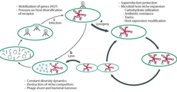
Based on the outcome of phage infection of the host cell, phages can follow two different life cycles (Adams, 1959) (Figure 1):
• Lytic cycle: Following adsorption of the phage to the specific receptor of the
susceptible bacterial host cell, the phage genome is injected into the bacterial cytoplasm. In the lytic pathway, the genome is replicated independently of the bacterial chromosome and several copies of the phage genome are produced. Meanwhile, the phage late genes are transcribed and translated to yield the protein components of the heads and/or tails, which assemble independently in the bacterial cytoplasm. The phage capsid protein assembles and the genome is then spooled into the new phage heads. The DNA-containing heads are then joined to the performed tails (if tailed phages) leading to the production of new phages. Further phage-encoded proteins (very late proteins) burst the host cell and release the progeny of phages into the medium finally causing bacterial cell death.
1. General introduction
5 The phages that can only follow the lytic pathway are called virulent phages. T4 phage and ФX174 are examples of virulent phages.
• Lysogenic cycle: Certain phages, once they infect the host cell, can either enter lytic
or lysogenic cycle and are known as temperate phages.
In the lysogenic pathway, the genome of the temperate phage becomes part of the bacterial genome, replicating along with the host, either integrated in the host chromosome (this is the case for most prophages), or by making the phage genome circular and remaining as an independent replicon (e.g. P1 phage).
More in detail, after phage attachment to the bacterial receptor, the phage genome is injected within the host cell and usually circularizes. By means of the product of the phage gene encoding for an integration enzyme (integrase), the phage genome becomes integrated in the bacterial chromosome usually in a specific locus yielding the quiescent phage, called prophage. By means of the integrase, there is a recognition of a specific site in the bacterial chromosome, called attB (attachment site of the bacteria). Phage and chromosomal DNAs become joined by a sequential series of DNA breaking and joining reactions. As a result of integrative recombination, the phage chromosome becomes attached to the bacterial chromosome, afterward resembling a normal cellular gene. It remains in this condition indefinitely, being replicated as its host cell replicates to make a clone of cells all containing prophages. Cells containing prophages are said to be lysogenic (or
lysogens). Therefore, the bacterial cell incorporates phage genes and is able to
express them; the process is called lysogenic conversion.
Temperate prophages may occasionally come out of its quiescent condition and switch to the lytic cycle, in a process called induction. Once in the lytic cycle, phages propagate causing at the end the lysis of the host cell and being released as new phage particles (virions).
1. General introduction
6
Figure 1. Lytic and lysogenic cycles of bacteriophages (Reyes et al., 2012).
It is important to highlight that usually these virions cannot infect other cells from the lysogenic culture since all of them are carrying the same prophage in their genome giving immunity to the attack of a virion of the same phage, although cells still vulnerable to the infection by other phages. Because of their immunity condition, temperate phages only make plaques when there are plated in a non-lysogenic strain. Some exceptions have been however found, as temperate Shiga toxin encoding bacteriophages (Serra-Moreno et al., 2008).
Summarizing, temperate phages have then a choice of reproductive modes when they infect a new host cell. Sometimes the infecting phage initiates a lytic cycle, resulting in the lysis of the cell and the release of new phage particles as previously mentioned. Alternatively, the infecting phage may initiate a quiescent state called a prophage, often integrated into the host genome, but sometimes maintained as a plasmid. It remains in this condition indefinitely, being replicated as its host cell reproduces to make a clone of cells all containing prophages (lysogenic cells). Occasionally, one of these prophages comes out of its quiescent condition and enters the lytic cycle. The factors affecting the choice to lysogenize or to reenter into a lytic cycle (induction) are called inductors or inducing agents and are described in the next section.
1. General introduction
7 The plaques of lysis generated by temperate phages could be slightly different compared to the ones formed by virulent phages. Instead of being clear plaques as virulent phages do, they are usually slightly turbid. Plaques are visible for the lysis of the cell following the lytic cycle and the turbidity observed is caused because many of the phages are conducting lysogenic cycles instead of strictly lytic cycle and the lysogenic cells are growing within the plaque.
Lambda (λ), P1, P22, and Mu bacteriophages infecting Enterobacteriaceae and various phages infecting Lactobacillaceae are among the best-known temperate phages.
Temperate phages can help protect their hosts from infection by other similar phages (superinfection) and by means of lysogenic conversion can lead to significant changes in the properties of their hosts. Some authors have emphasized the importance of temperate phages as “replicons endowed with horizontal transfer capabilities” (Briani et al., 2001) and they have probably been major factors in bacterial evolution by moving segments of genomes into different bacteria.
1.1.2. Inducers of lytic cycle in temperate bacteriophages
Prophage induction can take place either spontaneously or stimulated by inductors. Some
compounds, either natural or introduced by human activity in the environment, may act as inducing agents leading to phage replication and increase in the number of phage particles that will be released from the host cell. The most relevant inducers are described below:
• Mutagenic agents: Mutagenic agents or any agent that can cause DNA damage, such
as radiations as UV, can induce the phage lytic cycle in many lysogens.
• Different classes of antibiotics: Certain antibiotics, used in human therapy or animal husbandry as growth promoters, are able to induce phages from their lysogens. Among others, those antibiotics affecting DNA or those activating the bacterial SOS response will cause phage induction. Quinolones, such as ciprofloxacin or norfloxacin have been widely used for induction of phages (Goerke et al., 2006; Rolain et al., 2009; Looft, 2012; Meessen-Pinard et al., 2012) and Qnr-prophage induction in the
1. General introduction
8
presence of quinolones has been demonstrated with intestinal populations (Modi et al., 2013).
Other antibiotics like trimethoprim, furazolidone and ciprofloxacine are potent SOS inducers and they are reported to induce stx gene expression in EHEC O157:H7 (Kimmitt et al., 2000). Ampicillin has also been shown to induce prophages (Maiques et al., 2006).
• Mitomycin C, which damages DNA by cross-linking complementary strands, is considered an antitumoral agent. This compound is commonly used in laboratory practice as an agent for temperate phage induction through activation of the SOS response (Fuchs et al., 1999; Livny and Friedman, 2004; Muniesa et al., 2004).
• Chelating agents: EDTA and sodium citrate are chelating agents. EDTA was reported
to increase the number of copies of Shiga toxin gene (stx) in temperate Stx phages when a culture of a lysogenic strain for a Stx phage is treated with 20mM EDTA, even in the absence of RecA (Imamovic and Muniesa, 2012).
• Oxidizing agents: Reactive oxygen species (ROS), such as hidrogen peroxide, are strong oxidizing agents that can decompose into free radicals and cause DNA damage to bacteria, which leads to prophage induction (Kutter and Sulakvekidze, 2005).
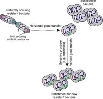
Such factors may play important roles in enhancing the frequency of gene exchange in environments such as farms, hospitals, and sewage systems, which provide ideal conditions for ARGs acquisition.
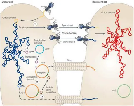
1.1.3. Bacteriophage transduction
Bacteriophages mediate horizontal gene transfer (HGT) through transduction. Transduction refers to the process by which a DNA fragment is transferred from one bacterial cell to another using a bacteriophage particle as a vector. The size of DNA fragments that can be
1. General introduction
9 packaged into a phage particle is limited by the size of the phage capsid, but can reach upwards of 100 kb.
Transduction by bacteriophages includes any sort of bacterial DNA, from linear chromosome fragments to all sorts of MGEs such as plasmids, genomic islands, transposons and insertion elements (Mann and Slauch, 1997).
Transduction was traditionally considered to occur at very low frequencies around once every 107-109 phage infections, but recent studies show that transduction takes place in the environment at a remarkably high rate with frequencies greater than previously thought (Chiura, 1997; Evans et al., 2010; Kenzaka et al., 2010). For example, transduction of genes for the global marine phage population between marine bacteria has been calculated to take place in the oceans at the rate of about 20 million billion times per second, although the real numbers will probably be lower due to smaller transduction efficiency and more rapid phage decay in the ocean than in the laboratory. (Bushman, 2002).
There are two types of transduction, generalized transduction and specialized transduction: • Generalized transduction: Generalized transduction is so named because essentially
any fragment of bacterial DNA from any location in its genome can be packaged into a phage head, instead of phage DNA, and then be transferred between cells by this mechanism. Phage particles that have encapsidated bacterial DNA are called transducing particles. Generalized transducing particles can be produced during lytic growth of either virulent or temperate phages (Thierauf et al., 2009). Phages that package DNA by a headful mechanism can occasionally misfire and package a headful of host cell DNA instead. Upon infection of a new host cell, the DNA can sometimes become incorporated into the genome of the new host.
Generalized transduction is a prominent means of gene transfer between bacteria. The generalized transducing particles filled with host DNA rather than phage DNA cannot propagate, but may infect a new cell, hence still contribute substantially to bacterial exchange in nature.
1. General introduction
10
Examples of phages known to be capable of generalized transduction are P1, Mu, P22, T1, T4, KB1 and ES18 phages.
Generalized transduction process can be exemplified by bacteriophage P1, a temperate phage that infects a variety of Gram-negative bacteria, which was isolated from a lysogenic strain of Escherichia coli. Upon translocation into the host cytoplasm, the linear double-stranded P1 DNA circularizes by recombination. During lytic growth, the circular genome of P1 initiates several rounds of bidirectional replication before switching to rolling circle replication that produces long concatamers of double-stranded P1 DNA. P1 encodes a phage endonuclease that recognizes a specific pac sequence on the phage DNA and cuts the DNA at this site to initiate headful packaging into an empty phage head. When packaging has been completed, the excess DNA is cleaved by a sequence independent mechanism. Subsequent rounds of packaging are then initiated from the cleaved DNA and these packaging reactions continue processively until several heads are filled with DNA. When a cell lyses, it releases the new phage particles. (Sternberg and Coulby, 1990; Thierauf et al., 2009). The generalized transduction occurs when the endonuclease cuts by mistake sequences on the host bacterial chromosome that are homologous to the P1 pac site. When P1 infects a cell, occasionally the P1 endonuclease cuts one or more of these chromosomal sites DNA and package it into P1 phage heads. These phage heads contain only bacterial DNA and no phage DNA. The P1 particles carrying bacterial DNA (transducing particles) can inject this DNA into a new hostand the DNA once inside the new cell can then recombine into the chromosome by homologous recombination. These transducing particles will not be able, however, to conduct lysis or lysogeny in the new host.
About 30% of the phage particles in a P1 lysate contain host DNA rather than phage DNA. Given the relative sizes of the E. coli and P1 genomes, approximately 1 in 1,500 phage particles in a lysate will carry a given gene from the donor. All genes of the donor (including plasmid genes) present the same probabilities of being mobilized by this mechanism.
1. General introduction
11 • Specialized transduction: In contrast to generalized transduction, it results from the
aberrant excision of a prophage from a specific integration site in the bacterial chromosome, packaging both phage DNA and a fragment of adjacent DNA from the bacterial genome into a single phage particle. By means of specialized transduction the phage can package only its DNA and the specific bacterial DNA flanking the attachment site of an integrated prophage (Miller et al., 2004; Thierauf et al., 2009). Because the types of sequences that can be transferred are so restricted, it seems likely that specialized transduction is not a major contributor to gene transfer in the environment compared to generalized transduction. Nevertheless, the frequency of transduction of this specific DNA fragment is high since all the phage particles produced after induction of the prophage carry this particular fragment.
A few examples of phages capable of specialized transduction have been reported being lambda phage the most well-known.
Usually, temperate phages that cause specialized transduction immediately circularise their DNA after infection of the bacterial host, by using the cos (cohesive end) sites. When these phages package their DNA after induction of lytic cycle they use the cos sites to cleave their DNA concatemers and these DNA segments cut at the cos sites are packaged in the phage heads. Therefore, theoretically, they are not able to produce generalized transduction, which needs to package DNA by a pac-mechanism. Only few examples of cos-packaging phages causing generalized transduction are found in the literature (Sternberg, 1986; Campoy et al., 2006).
1.1.4. Ubiquity and abundance of bacteriophages
Bacteriophages exist in large quantities and are widely distributed in different natural environments, wherever their bacterial hosts live, such as gastrointestinal tracts of humans and animals where gut bacteria are associated with their specific phages communities (Breitbart et al., 2003, 2008; Minot et al., 2011), sewage water (Cantalupo et al., 2011), human and animal faeces (Letarov and Kulikov, 2009; Victoria et al., 2009; Reyes et al.,
1. General introduction
12
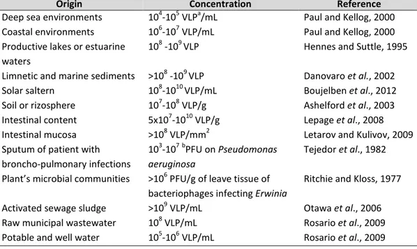
2010), soil (Weinbauer, 2004), plants (Gill and Abedon, 2003), marine systems (Angly et al., 2006), lakes (Ogunseitan et al., 1990), river water, etc. and even in extreme environments (Le Romancer et al., 2007), with variable numbers that seem to depend on bacterial abundance and activity (Table 1).
Origin Concentration Reference
Deep sea environments 104-105 VLPa/mL Paul and Kellog, 2000 Coastal environments 106-107 VLP/mL Paul and Kellog, 2000 Productive lakes or estuarine
waters
108 -109 VLP Hennes and Suttle, 1995 Limnetic and marine sediments >108 -109 VLP Danovaro et al., 2002
Solar saltern 108-1010 VLP/mL Boujelben et al., 2012
Soil or rizosphere 107-108 VLP/g Ashelford et al., 2003
Intestinal content 5x10⁷-1010 VLP/g Lepage et al., 2008 Intestinal mucosa >108 VLP/mm2 Letarov and Kulivov, 2009 Sputum of patient with
broncho-pulmonary infections
103-107bPFU on Pseudomonas aeruginosa
Tejedor et al., 1982 Plant’s microbial communities >106 PFU/g of leave tissue of
bacteriophages infecting Erwinia
Ritchie and Kloss, 1977 Activated sewage sludge >109 VLP/mL Otawa et al., 2006 Raw municipal wastewater 108 VLP/mL Rosario et al., 2009 Potable and well water 105-106 VLP/mL Rosario et al., 2009
Table 1. Ubiquity and abundance of bacteriophages (Muniesa et al., 2013). a
VLP: Virus-like particles ; bPFU: Plaque forming units.
Transmission electron microscopy and epifluorescence microscopy, together with the development of molecular techniques, such as genome sequencing and metagenomic analysis, have allowed the detection of bacteriophages in environments in a way that was not possible previously, independently of their infectivity, revealing that phages are much more abundant than previously thought (Breitbart et al., 2004; Weinbauer, 2004; Srinivasiah et al., 2008), and indicating that natural phage communities are reservoirs of the greatest uncharacterized diversity on Earth, with an enormous variety of environmental niches and survival strategies (Weinbauer, 2004). Many of these studies are based in analysis of the viral fraction of a given sample but since in most environments studied phages are the main part of the viral fraction (Dinsdale et al., 2008), it can be assumed that the viral DNA evaluated in these studies will belong mostly to bacteriophages.
1. General introduction
13 The idea that bacteriophages play an important role in microbial ecology is widely accepted nowadays. On the one hand, by infecting and lysing infected bacteria, they contribute remarkably to bacteria mortality, thus they regulate the numbers of certain bacterial populations in a given environament; and by releasing organic compounds through cell lysis they have an important impact on the cycling of organic matter in the biosphere (Suttle, 1994). On the other hand, they control microbial diversity by selecting for some types of bacteria that are resistant to their attacker (Scanlan and Buckling, 2012), thus changing the proportions of bacterial species or strains in a community, and also influencing the evolution of bacterial genomes through HGT by transduction.
Considering the wide occurrence of phages in the environment, and the high concentration of phages in some water bodies (Weinbauer, 2004; Srinivasiah et al., 2008), and since they can transduce genes among their bacterial hosts, they are expected to play a crucial role of genetic transfer in water habitats mediated by phages (Ripp and Miller, 1995; Brabban et al., 2005; Parsley et al., 2010b). Transduction probably constitutes one of the main gene transfer mechanisms and of genome evolution for bacteria in water habitats.
1.1.5. Persistence of bacteriophages in the environment
Phages, either virulent or temperate, show a higher resistance to natural and anthropogenic stress factors than bacteria usually persisting better in aquatic environments than their bacterial hosts do (IAWPRC, 1991; Muniesa et al., 1999; Durán et al., 2002; Jofre, 2007). This higher survival and the abundance of phages carrying ARGs in animal and human wastewater (Muniesa et al., 2004; Minot et al., 2011; Looft et al., 2012) makes them suitable candidates as vehicles for the mobilization of the environmental pool of ARGs between bacteria in different biomes that contribute to the maintenance and emergence of new resistances (Sano et al., 2004). In addition, due to the structural characteristics of phages, with phage-encapsidated DNA protected from degradation, their persistence in the environment is higher than free DNA (either linear fragments or plasmids), which is more sensitive to nucleases, temperature, predation and radiation (Lorenz and Wackernagel, 1994; Dupray et al., 1997; Zhu, 2006).
1. General introduction
14
Phages may survive in special environments without the loss of their infectious capabilities and are able to transfer genes by transduction. This observation supports the notion that the contribution of phages to gene transfer in natural extra-intestinal environments and in human-generated environments could be greater than that of plasmids or transposons, with a lower persistence in the environment (Lorenz and Wackernagel, 1994; Dupray et al., 1997; Zhu, 2006). In clinical settings, however, plasmids and transposons are probably most relevant MGEs for horizontal antibiotic resistance transfer because their lower environmental persistence is not a limitation in a human body.
Indeed, their high level of specificity, long-term survivability, and ability to reproduce rapidly in appropriate hosts displayed by phages contribute to the maintenance of a dynamic balance among the wide variety of bacterial species in any natural ecosystem. When no appropriate hosts are present, many phages can maintain their ability to infect for a long period of time unless damaged by external agents.
Although phages vary greatly in their sensitivity to various chemical and physical agents, certain general principles have been described. For example, DNA phages are very susceptible to UV light in the range of 260 nm as well as in the far UV. Other factors potentially affecting phages include:
• pH: Phages generally are stable at pH 5 to 8, and many are stable down to pH 3 or 4, but it depends on each phage.
• Temperature: Temperature is a crucial factor for bacteriophage replication and survavility (Hurst et al., 1980; Yates et al., 1985; Nasser and Oman, 1999; Olson et al., 2004). It plays a fundamental role in attachment, penetration, multiplication, and the length of the latent period (in the case of lysogenic phages). At lower than optimal temperatures, fewer phage genetic material penetrate into bacterial host cells; therefore, fewer of them can be involved in the multiplication phase. Higher temperatures can prolong the length of the latent stage. Moreover, temperature determines the occurrence, viability, and storage of bacteriophages.
1. General introduction
15 • Urea and urethane: Phages are often quite sensitive to protein-denaturing agents
such as urea and urethane, but the level of inactivation depends on both concentration and temperature and differs for different phages.
• Detergents: Not surprisingly, detergents generally have far less effect on phages than they do on bacteria, although the few phages that are enveloped in membranes are quite susceptible.
• Chelating agents: Chelating agents, in contrast, have strong effects on some phages depending mainly on cations requirements for adsorption or capsid assembly. • Chloroform: Chloroform has little or no effect on nonenveloped phages.
• Mutagenic agents: such as mustard gas, nitric oxide, and UV light can inactivate phages and, as mentioned before, can also induce the lytic cycle in many lysogens.
1. General introduction
16
1.2. Antibiotics
1.2.1. Antibiotics: definition and history
The term antibiotic (Greek. anti, "against"; bios, "life") was originally referred to a natural compound produced mainly by moulds or other microorganisms that kills bacteria which cause disease in humans or animals. Nowadays, we refer to an antibiotic as a chemical substance produced by a microorganism or a synthetic derivative that inhibits the growth of (bacteriostatic) or kills (bactericidal) other sensitive microorganisms.
Before the early 20th century, treatments for infections were based on traditional medicine. Many ancient cultures, including the ancient Egyptians, the ancient Greeks, the Chinese, and Indians of central America used specially selected mould and plant materials and extracts to treat infections, but without understanding the connection of their antibacterial properties and the treatment of diseases.
In the 19th century, prior to the introduction of antibiotics and antiseptic treatments, more
than half of all surgical patients developed infections so scientists began to devote time to searching for drugs that would kill these disease-causing bacteria. In Germany, Paul Ehrlich discovered the synthetic antibacterial Salvarsan (arsphenamine). But it was not until in 1928 that Alexander Fleming observed that colonies of the bacterium Staphylococcus aureus could be destroyed by a product secreted by the fungus of the genus Penicillium, demonstrating antibacterial properties, although he was not able to purify the molecule (penicillin).
In 1932, at the Bayer Laboratories in Germany, a research team developed the first sulfonamide and first commercially available antibacterial, Prontosil, which had a relatively broad effect against Gram-positive cocci, but not against enterobacteria.
A key breakthrough came in 1945 with the development, mass production and distribution of penicillin. Purified penicillin displayed potent antibacterial activity against a wide range of bacteria and had low toxicity in humans. Furthermore, its activity was not inhibited by biological constituents unlike the synthetic sulfonamides. The discovery of such a powerful antibiotic was unprecedented and the development of penicillin led to renewed interest in
1. General introduction
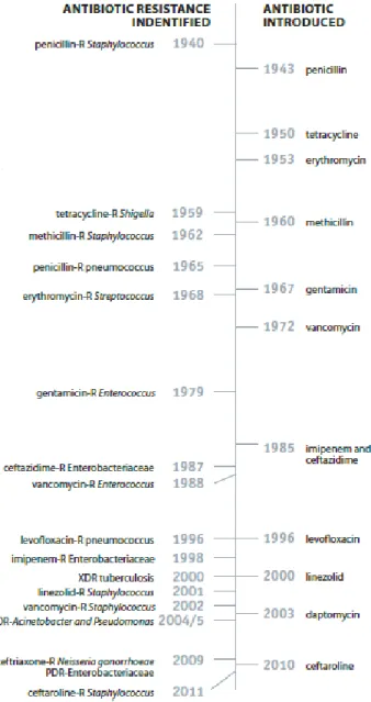
17 the search for antibiotic compounds with similar efficacy and safety. These findings, together with the discovery of streptomycin by Selman Waksman, opened the era of antibiotics. Initially, after being introduced into clinical practice in the 1940s, antibiotics were extremely efficient and transformed medical care by dramatically reducing illnesses and deaths from bacterial infectious diseases that were leading causes of human morbidity and mortality. Importantly, it was recognized that certain types of bacteria, particularly actinomycetes and streptomycetes, often produced compounds with antibiotic properties leading to systematic efforts to isolate environmental antibiotic producing bacteria.
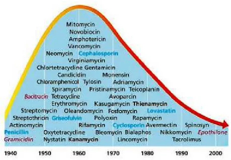
The world discovered and produced more than 20 novel classes of antibiotics between 1930 and 1962 with a peak during the 1960s (Coates et al., 2002; Powers, 2004) (Figure 2). Since then, new antibiotic development has dramatically fallen off and only five new classes of antibiotics have been marketed (Butler and Buss, 2006; Hair and Kean, 2007) mainly due to the problem of bacteria becoming resistant very fast every time a new antibiotic is introduced. The mainstrean approach to combat emerging and re-emerging resistent pathogens has been the modification of existint antibiotics.
1. General introduction
18
1.2.2. Use of antibiotics
As mentioned before, the discovery and industrial production of antibiotics in the middle of the previous century has been one of medicine’s greatest achievements. The use of antibiotics has revolutionised the treatment of bacterial infectious diseases both in humans and in animals by reducing their morbidity and mortality and has also contributed substantially to human’s increased life expectancy. They are also an essential tool for modern medicine and common procedures such as surgeries or to to prevent infections in chemotherapy for cancer, which could not be performed without the availability of potent antibiotics.
Since then, ever-increasing amounts of antibiotics have been extensively produced and used in different applications, some of them mentioned below:
• Therapeutic/prophylactic use in humans
• Therapeutic/prophylactic use in animals (e.g. livestock, poultry, pigs) including aquaculture
• Production purposes: to enhance growth food-producing animals (growth promoters) and to improve feed efficiency in animals
• Therapeutic/prophylactic use in agriculture • Therapeutic/prophylactic use in household pets
• Use as biocides in toiletries and in hand care and household cleaning products • Culture sterility, cloning, and selection in research and industry
It should be noted that therapeutic use in humans accounts for less than half of all applications of antibiotics produced commercially. Actually, antibiotics are used in greater quantities in healthy food-producing animals than in the treatment of disease in human patients. (Davies and Davies, 2010). Although the use of antibiotics for promoting growth is not necessary and was abandoned in the EU in 2006, there are still many other countries using them for this purpose.
1. General introduction
19
1.2.3. Antibiotics classification
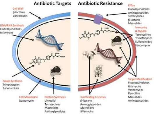
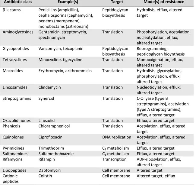
Nowadays different classes of antibiotics are known and they are commonly classified based on their mechanisms of action, chemical structure, or spectrum of activity (Neu, 1992). Antibiotics can for instance inhibit protein synthesis, like aminoglycosides, chloramphenicol, macrolides, and tetracyclines or interact with the synthesis of DNA and RNA, such as quinolones and rifamycins. Other groups inhibit the synthesis of, or damage the bacterial cell wall as β-lactams and glycopeptides do, or modify the energy metabolism of the bacterial cell like sulfonamides and trimethoprim. In Table 2 are presented the main classes of antibiotics currently used and their mechanism of action.
Antibiotic
class Antibiotic Species range Primary target Pathways affected
Fluoroquinolones DNA
synthesis inhibitor
Nalidixic acid, ciprofloxacin, norfloxacin, levofloxacin and gemifloxacin
Aerobic Gram-positive and
Gram-negative species, some anaerobic Gram-negative species (C. perfringes) and M. tuberculosis
Topoisomerase II (DNA gyrase), topoisomerase IV
DNA replication, SOS response, cell division, ATP generation, TCA cycle, Fe–S cluster synthesis, ROS formation, and envelope and redox-responsive two-component systems Trimethoprim–sulfamethoxazole DNA synthesis inhibitor Co-trimoxazole (a combination of trimethoprim and sulfamethoxazole in a 1:5 ratio)
Aerobic Gram-positive and Gram-negative species
Tetrahydrofolic acid synthesis inhibitors
Nucleotide biosynthesis and DNA replication
Rifamycins RNA synthesis inhibitor
Rifamycins, rifampin and rifapentine
Gram-positive and Gram-negative species, and M. tuberculosis
DNA-dependent RNA polymerase
RNA transcription, DNA replication and SOS response β-lactams Cell wall synthesis inhibitors Penicillins (penicillin, ampicillin, oxacillin), cephalosporins (cefazolin, cefoxitin ceftriaxone, cefepime) and carbapenems (imipenem)
Aerobic and anaerobic Gram-positive and Gram-negative species
Penicillin-binding proteins
Cell wall synthesis, cell division, autolysin activity (regulated by LytSR–VncRS two-component system), SOS response, TCA cycle, Fe–S cluster synthesis, ROS formation, and envelope and
redox-responsive two-component systems Glycopeptides and glycolipopeptides
Cell wall synthesis inhibitor
Vancomycin; teicoplanin Gram-positive species Peptidoglycan units (terminal d-Ala-d-Ala dipeptide)
Cell wall synthesis, transglycosylation, transpeptidation and autolysin activation (VncRS two-component system)
Lipopeptides Cell wall synthesis inhibitors
Daptomycin and polymixin B Gram-positive species (daptomycin), Gram-negative species (polymixins)
Cell membrane Cell wall synthesis and envelope two-component systems