Structural Determinants of the Specificity for Synaptic
Vesicle-associated Membrane Protein/Synaptobrevin of Tetanus
and Botulinum Type B and G Neurotoxins*
(Received for publication, February 29, 1996, and in revised form, May 21, 1996) Rossella Pellizzari‡, Ornella Rossetto‡, Luisa Lozzi§, Silvia Giovedi’‡, Eric Johnson¶,
Clifford C. Shonei, and Cesare Montecucco‡**
From the ‡Centro Consiglio Nazionale delle Ricerche di Biomembrane and Dipartimento di Scienze Biomediche, Universita` di Padova, Via Trieste 75, Padova, Italy, the §Dipartimento di Biologia Molecolare, Le Scotte, Universita` di Siena, Italy, the¶Food Research Institute, University of Wisconsin, Madison, Wisconsin 52706, andiCenter for Applied Microbiology and Research, Porton Down, Salisbury, Wiltshire SP4 0JG, United Kingdom
Tetanus and botulinum neurotoxins type B and G are zinc-endopeptidases of remarkable specificity. They recognize and cleave a synaptic vesicle-associated mem-brane protein (VAMP)/synaptobrevin, an essential pro-tein component of the vesicle docking and fusion appa-ratus. VAMP contains two copies of a nine-residue motif, also present in SNAP-25 (synaptosomal-associated pro-tein of 25 kDa) and syntaxin, the two other substrates of clostridial neurotoxins. This motif was suggested to be a determinant of the target specificity of neurotoxins. An-tibodies raised against this motif cross-react among VAMP, SNAP-25, and syntaxin and inhibit the proteo-lytic activity of the neurotoxins. Moreover, the various neurotoxins cross-inhibit each other’s proteolytic ac-tion. The role of the three negatively charged residues of the motif in neurotoxin recognition was probed by site-directed mutagenesis. Substitution of acidic residues in both copies of the VAMP motif indicate that the first one is involved in tetanus neurotoxin recognition, whereas the second one is implicated in binding botulinum B and G neurotoxins. These results suggest that the two copies of the motif have a tandem association in the VAMP molecule.
Tetanus neurotoxin (TeNT)1 and botulinum neurotoxins
(BoNTs, seven types from A to G) are three-domain protein toxins that bind selectively to the neuronal presynaptic mem-brane. They are internalized inside intracellular compartments from which the amino-terminal 50-kDa domain (termed L chain) enters into the cytosol (1– 4). The L chains of TeNT and BoNTs are zinc-endopeptidases that cleave specifically three proteins of the neuroexocytosis apparatus, thereby blocking neurotransmitter release (4 –7). TeNT and BoNT/B, BoNT/D, BoNT/F, and BoNT/G recognize and cleave specifically a syn-aptic vesicle-associated membrane protein (VAMP, also re-ferred to as synaptobrevin) at different single peptide bonds (4, 8 –12). BoNT/A and BoNT/E specifically recognize and cut
SNAP-25 (synaptosomal-associated protein of 25 kDa) at two different peptide bonds near the COOH terminus (10, 13, 14), whereas BoNT/C cleaves syntaxin (15, 16) and SNAP-25 (17– 19). VAMP, SNAP-25, and syntaxin are collectively termed SNARE proteins, because they act as receptors of soluble N-ethylmaleimide-sensitive factor accessory proteins, involved in vesicle-membrane fusion (5–7).
Sequence comparison of the L chains of the eight clostridial neurotoxins show strong similarities (20), which are even more extensive at the level of predicted secondary structure (21). These similarities suggest that they derive from a common ancestral metalloproteinase. On this basis, to account for their different substrate specificity, we considered the possibility that the three SNAREs contain a common neurotoxin recogni-tion site in addirecogni-tion to the cleavage sites specific for each neurotoxin type. We identified a nine-residue-long motif (SNARE motif) present in eukaryotes only in the three proteins known to be proteolytic substrates of the neurotoxins (22). The SNARE motif is included within regions predicted to adopt an a-helical conformation in the three SNAREs (23). This motif is characterized by the presence of three negatively charged res-idues and three hydrophobic resres-idues spaced in such a way that the Edmundson wheel plot shows a negatively charged surface contiguous to a hydrophobic face. Preliminary experiments showed that peptides corresponding to the motif sequence of the three SNARES inhibit neurotoxin activity both in vitro and
in vivo in injected Aplysia neurons (22). We also suggested that
the specificity of the clostridial neurotoxin’s action is based on a double recognition of their substrates via the SNARE motif and via the segment containing the cleavage site (4, 22, 24).
Here we report on studies focusing on the interaction among TeNT, BoNT/B and BoNT/G, and VAMP. Results obtained with different experimental approaches provide strong evidence for the involvement of the SNARE motif in this specific interac-tion. Moreover, they show that negatively charged residues of the motif play a major role in this interaction.
MATERIALS AND METHODS
Proteins, Peptides, and Chemicals—TeNT and BoNT/A, BoNT/B, BoNT/C, BoNT/E, and BoNT/G were prepared as detailed before (25– 27). Immobilized metal ion affinity chromatography was used to remove traces of contaminant proteases (28). Synaptosomes were isolated from rat brain cortex as detailed before (10). Peptides V2 (ELDDRADALQ), S3 (MLDEQGEQLER), and X2 (LEDMLESGN) were prepared with an SMP 350 automatic synthesizer (Zynsser Analytic, Frankfurt), employ-ing an Fmoc (N-(9-fluorenyl)methoxycarbonyl) chemistry, and were purified by reverse phase chromatography on a C8 Ultra-Sphere pre-parative column (Beckman). The composition of soybean mixed lipids (asolectin) was as described previously (29). Liposomes were obtained by mixing chloroform/methanol (2:1) stock solutions of lipids. After * This work was supported by Telethon-Italia Grant 763. The costs of
publication of this article were defrayed in part by the payment of page charges. This article must therefore be hereby marked “advertisement” in accordance with 18 U.S.C. Section 1734 solely to indicate this fact.
‡ To whom correspondence should be addressed: Dipartimento di Scienze Biomediche, Via Trieste 75, 35100 Padova, Italy. Tel.: 39-49-8286058; Fax: 39-49-8276049.
1The abbreviations used are: TeNT, tetanus neurotoxin; GST,
gluta-thione S-methyl transferase; BoNT, botulinum neurotoxin; VAMP, ves-icle-associated membrane protein; VAMP-2, VAMP isoform 2; SNAP-25, synaptosomal-associated protein of 25 kDa; SNARE, soluble N-ethylmaleimide-sensitive factor accessory protein receptor.
THEJOURNAL OFBIOLOGICALCHEMISTRY Vol. 271, No. 34, Issue of August 23, pp. 20353–20358, 1996
© 1996 by The American Society for Biochemistry and Molecular Biology, Inc. Printed in U.S.A.
20353
by guest on April 24, 2019
http://www.jbc.org/
drying under N2flux, lipids were resuspended in diethyl ether, dried,
and sonicated until optical clarity was achieved.
Antibodies and Immunoblotting—Rabbit polyclonal antisera against peptides V2, S3, and X2 were prepared as described by Kreis (30). The antibodies were purified by affinity chromatography using activated CH-Sepharose 4B (Pharmacia Biotech Inc.) conjugated with each pep-tide. Antibody concentration was determined by the Bradford method (31). Synaptosomes were transferred onto nitrocellulose as described elsewhere (10) and treated with anti-V2, anti-S3, and anti-X2 antisera (1:200). Primary antibodies were detected by immunostaining with an anti-rabbit antibody (1:10,000 dilution, Boehringer Mannheim) conju-gated with alkaline phosphatase (1:1,000; Sigma).
Bacterial Strains, Plasmid Construction, and VAMP Mutagenesis— The VAMP-2 gene was generated by inserting a PCR-derived DNA fragment from the rat VAMP-2 cDNA clone characterized previously (32). This DNA fragment covers the complete sequence of the VAMP-2 gene and has BamHI and EcoRI restriction sites at the 59 and 39 ends, respectively. It was inserted into the pEMBL8- plasmid (33). Uracil containing single-stranded DNA was produced within BW313, an Esch-erichia coli dut- ung- strain, for generation of VAMP-2 mutants by site-directed mutagenesis. The in vitro reactions were performed as described by Kunkel et al. (34). The following oligonucleotides were used to generate the corresponding mutants: 59-TGCGCGATTAT-TCAGTTCC-39 for VAMP-2 D64N,D65N; 59-GGAGGGCATTTGCGC-GAT-39 for VAMP-2 D68N; the combination of the two previous oligo-nucleotides for VAMP-2 D64N,D65N,D68N; 5 9-CTGCGCGACTAC-TCAGTTCC-39 for VAMP-2 D64S,D65S, and 59-TCCACCACCTGAT-TCACCTGG-39 for VAMP-2 D40N,E41Q. The sequence of the mutated genes was checked by dideoxy sequencing using the fmole DNA se-quencing System (Promega). Wild type VAMP-2 and mutants were subcloned into BamHI and EcoRI sites of pGEX-KG vector (35) and transformed into the AB1899 strain of E. coli.
VAMP Protein Expression and Purification—Rat VAMP-2 and rat VAMP-2 mutants were expressed as GST fusion proteins and were purified by affinity chromatography on GSH-agarose matrix (Pharma-cia) as before (12).
Assay of Proteolytic Activity—GST-fusion VAMPs (50 mg/ml final concentration) were incubated with a sonicated clear liposome suspen-sion (final concentration, 1 mg of soya bean mixed phospholipids/ml) at 37 °C for 1 h. After treatment with dithiothreitol (10 mMfor 30 min at
37 °C), TeNT (200 nM), BoNT/B, or BoNT/G (40 nM) was added to the
reaction mixture, containing one of the different GST fusion proteins. Proteolysis was carried out in 150 mMNaCl, 10 mMNa2HPO4, pH 7.4,
at 37 °C for variable periods of time. In some experiments 1mg of GST-VAMP-2 was preincubated for 30 min with affinity-purified anti-bodies (1mM), specific for the different peptides, before neurotoxin addition and proteolysis, as described above. Samples were analyzed in a 15% polyacrylamide SDS gels and, after silver staining of proteins, scanned with a dual wavelength Shimadzu CS-630 densitometer.
For neurotoxin competition experiments, GST-VAMP-2 was preincu-bated for 1 h at 37 °C with each one of the three neurotoxins, BoNT/A, BoNT/E, and BoNT/C, which do not cleave VAMP-2 (1mM) in 150 mM
NaCl, 10 mMNa2HPO4, 0.3 mMCaCl2, 2 mMMgCl2, pH 7.4. Proteolytic
cleavage was carried out for 2 h at 37 °C with TeNT 200 nMor BoNT/B 40 nM. All neurotoxins were preincubated with dithiothreitol 10 mMin the same cleavage buffer for 30 min at 37 °C.
RESULTS
Anti-SNARE Motif Antibodies Specifically Recognize VAMP, SNAP-25, and Syntaxin—Available models for neuroexocytosis
assign a central role to VAMP, SNAP-25, and syntaxin, the three targets of clostridial neurotoxins (5–7). These proteins form an SDS-resistant heterotrimeric complex able to bind SNAPs and N-ethylmaleimide-sensitive factor (23, 36, 37). Fig. 1 depicts the three SNAREs and includes membrane-spanning segments and sites of cleavage of clostridial neurotoxins. There are two copies of the motif in VAMP (V1 and V2), four copies in SNAP-25 (S1, S2, S3, and S4), and two copies in syntaxin (X1 and X2). Little structural information is available for isolated SNARE proteins. If the motif is to serve as neurotoxin recog-nition site, it has to be exposed at the protein surface, where it may act as an epitope and bind antipeptide specific antibodies. To test this possibility, rabbits were immunized with keyhole limpet hemocyanin coupled with the V2, S3, or X2 peptide, and IgGs were purified after booster immunization with the same antigens. Fig. 2 shows that the rabbit IgGs specific for V2 do recognize VAMP, but they also stain syntaxin and SNAP-25, FIG. 1. Schematic structure of VAMP, SNAP-25, and syntaxin with cleavage sites of neurotoxins and positions of SNARE motif (a). The SNARE motif is composed of nine residues and contains three conserved carboxylate residues (2) at positions 2, 3, and 6; in an Edmundson wheel plot of the motif Glu or Asp residues clusters on one-third of the helix. This motif is present in two copies in VAMP-2 (V1 and V2), two copies in syntaxin (X1 and X2), and four in SNAP-25 (S1, S2, S3, and S4). Capital letters correspond to the proteolytic cleavage site of tetanus toxin (T) and of the seven botulinum neurotoxins from A to G. Dark dots identify the transmembrane region of VAMP and syntaxin. The lower part of the figure reports the sequences of the various copies of the SNARE motif (b).
by guest on April 24, 2019
http://www.jbc.org/
although with lower intensity. In parallel, anti-S3 rabbit IgGs show binding to syntaxin and to VAMP in addition to the expected recognition of SNAP-25, and a similar pattern of staining is observed with X2-specific antibodies. The immuno-genicity of the SNARE motif, at the basis of the SNARE cross-reactivity, is also supported by the fact that rabbit polyclonal antisera prepared against recombinant VAMP or SNAP-25 or
syntaxin stain all three SNARE proteins (Fig. 2). This result puts a word of caution on the interpretation of immunofluores-cence studies performed with antibodies raised against an en-tire SNARE protein.
Anti-SNARE Motif Antibodies Interfere with VAMP Proteol-ysis by Tetanus and Botulinum Neurotoxins—The involvement
of the SNARE motif in neurotoxin binding leads to the predic-tion that antimotif-specific antibodies should inhibit the pro-teolytic activity of neurotoxins. Fig. 3a shows that antibodies prepared against V2, S3, or X2 have an inhibitory effect on TeNT and BoNT/B proteolysis of VAMP. The extent of protec-tion is variable and incomplete. This may be accounted for by variable affinities of the different antibodies and by the fact that the antibody competes with the neurotoxin for only one of the two sites of interaction between VAMP and the neurotoxin.
SNAP-25 and Syntaxin-specific Neurotoxins Inhibit Tetanus and Botulinum Neurotoxin Proteolysis of VAMP—Another
pre-diction of the model, outlined in the Introduction, is that each clostridial neurotoxin should be able to bind any SNARE pro-tein, although only one of them (two in the case of BoNT/C) would be cleaved, depending on the fitting of a particular sequence into each neurotoxin active site (4). As a consequence, binding of any neurotoxin type to a SNARE should inhibit its proteolysis by the specific neurotoxin. Fig. 3b shows the results of neurotoxin cross-inhibition experiments performed with FIG. 2. Antibody cross-reactions among VAMP, SNAP-25, and
syntaxin. Rat brain synaptosomes were electrophoresed and blotted
onto nitrocellulose membranes. Samples were incubated with rabbit anti-SNARE motif-specific polyclonal antibodies anti-V2, anti-S3, and anti-X2 affinity-purified as detailed under “Materials and Methods.” The last three samples were incubated with rabbit VAMP-2, anti-SNAP-25, or antisyntaxin antisera and were stained with the appro-priate alkaline phosphatase-conjugated anti-IgG antibodies.
FIG. 3. Inhibition of VAMP cleavage
by tetanus and botulinum B neuro-toxins with anti-SNARE motif-spe-cific antibodies and non-VAMP-spe-cific neurotoxins. a, the amount of
cleavage of GST-VAMP2 fusion protein incubated with tetanus neurotoxin (TeNT) or botulinum neurotoxin B (BoNT/B) alone or in the presence of af-finity-purified antibodies against pep-tides V2, S3, and X2 after 2 h at 37 °C. b, GST-VAMP-2 was preincubated with BoNT/A, BoNT/E, or BoNT/C, previously reduced with dithiothreitol, and then re-duced TeNT and BoNT/B were added and incubated for 2 h at 37 °C. Data are the average of three independent experi-ments, and bars represent S.D. values.
Neurotoxin VAMP Binding
20355
by guest on April 24, 2019
http://www.jbc.org/
VAMP as a substrate: TeNT and BoNT/B proteolysis of VAMP is inhibited by BoNT/A, BoNT/C, and BoNT/E. The inhibitory effect is not complete and differs for BoNT/A, BoNT/C, and BoNT/E. This is to be attributed to the fact that BoNT/A, BoNT/C, and BoNT/E bind VAMP via a single interaction with the SNARE motif, whereas TeNT and BoNT/B interact with VAMP at two sites: the SNARE motif and the segment around the cleavage site. The result of Fig. 3b indicates that BoNT/A interacts with the SNARE motif more strongly than BoNT/C and BoNT/E. Similar inhibitory effects were found when TeNT or BoNT/B were present in assays of proteolysis of SNAP-25 by BoNT/A and BoNT/E (not shown).
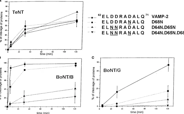
Mutation of the Negatively Charged Residues of V2 Affects VAMP Cleavage by Botulinum B and G—As shown in Fig. 1,
the SNARE motif is characterized by the presence of three negatively charged residues, which cluster on a face if the motif
is arranged in ana-helix. As a first approach to the study of the
role of the different residues of the motif in neurotoxin speci-ficity, each of the three aspartic residues of the V2 segment of VAMP was replaced with asparagine. This mutation was cho-sen because it is the most conservative substitution compatible with the removal of the negative charge of the carboxylate residue. Such mutant VAMPs were purified as GST fusion proteins from E. coli extracts, and the proteolytic activity of VAMP-specific clostridial neurotoxins was tested. Previous studies with VAMP segments of different lengths (11, 23, 38 – 40) implicated V2 in BoNT/B and BoNT/G binding of VAMP (4, 24).
Fig. 4 shows that progressive replacement of the three Asp residues of the V2 segment of VAMP with Asn residues causes a progressive loss of proteolytic activity of BoNT/B and BoNT/G. The effect of the D68N replacement appears to be
larger for BoNT/G than for BoNT/B, although the lower enzy-mic activity of BoNT/G may be partially responsible for this effect. On the contrary, TeNT cleaves the three VAMP mutants at similar rates. On the basis of the close structural similarity between clostridial neurotoxins, this result was unexpected. However, it is possible that TeNT binds V2 via hydrogen bond-ing interactions, which can be formed both with Asp and with Asn lateral chains. Therefore, the two Asp residues 64 and 65 of V2 were converted into Ser residues, which are hydrophilic but have a much smaller lateral chain. The VAMP-2 D64S, D65S mutant is, however, cleaved by TeNT at a rate very similar to that of wild type VAMP-2 (Fig. 5).
Mutation of the Negatively Charged Residues of V1 Affects VAMP Cleavage by Tetanus Neurotoxin, but Not by Botulinum B and G Neurotoxins—Another possibility that may account for
the results of Fig. 5a is that TeNT recognizes VAMP via an interaction with V1, rather than V2. To test this possibility,
Asp40and Glu41of VAMP-2 were replaced by the corresponding
Asn and Gln residues. Fig. 6 shows that this VAMP-2 mutant is not cleaved by TeNT (panel a), while the same protein is proteolyzed by BoNT/B as efficiently as the wild type VAMP-2 (panel b). These results indicate that TeNT and BoNT/B, the only two clostridial neurotoxins that recognize and cleave the same target protein at the same peptide bond (8), actually differ from each other. They are identical with respect to the consequence of their proteolysis because the same two VAMP fragments are generated. But they differ in their mode of sub-strate recognition, because this recognition is mediated by two different segments of VAMP.
VAMPs mutated in V1 and V2 were also tested as proteolytic substrates of TeNT and BoNT/B in the presence of BoNT/A. The ability of this latter toxin to inhibit TeNT and BoNT/B FIG. 4. Neurotoxin proteolysis of VAMPs containing mutations that progressively remove the negative charges of the V2 SNARE
motif. The rate of proteolysis of GST-VAMP-2 and the GST-VAMP-2 mutants are indicated (top right) with TeNT (panel a), BoNT/B (panel b), and
BoNT/G (panel c), determined as detailed under “Materials and Methods.” Samples were removed at 5, 30, and 120 min, electrophoresed, and silver-stained, and the amount of protein was determined by densitometric scanning. Data are the average of four different experiments, and bars represent S.D. values.
by guest on April 24, 2019
http://www.jbc.org/
proteolysis of VAMP, documented in Fig. 3b, was reduced when
VAMP-2 D64S,D65S mutant and the VAMP-2 Asp40and Glu41
were assayed (not shown). However, results did not allow us to draw a clear conclusion of a possible preference of BoNT/A between V1 and V2.
DISCUSSION
The light chain of the eight clostridial neurotoxins have a unique specificity for VAMP, SNAP-25, and syntaxin, termed SNAREs (5–7). Their three-dimensional structure is not known. Available spectroscopic and biochemical data, as well as comparison of their primary and predicted secondary struc-ture, indicate that they are closely similar. Nonetheless, they show unique proteolytic activity exerted on different peptide bonds of three different protein substrates. Short peptides
en-compassing the cleavage site of VAMP-2 cannot be cleaved by TeNT and BoNT/B. For proteolysis to occur, long VAMP pep-tides, including V2 and/or V1, are required (4, 38 – 41). Hence, the neurotoxin’s specificity cannot be explained by the sole recognition of the cleavage site (4, 39). A negatively charged motif was identified in the three SNAREs, and it was suggested that it is implicated in their selective recognition by clostridial neurotoxins (22, 24). Here, we report strong evidence in favor of the involvement of the SNARE motif in the interaction between VAMP and TeNT and BoNT/B and BoNT/G. 1) Antibodies, raised against motif peptides or recombinant proteins, recog-nize and cross-react among the three SNAREs, despite subtle sequence differences in the specific SNARE motifs of VAMP, SNAP-25, and syntaxin. This result indicates that this is a FIG. 5. Tetanus and botulinum B neurotoxin proteolysis of VAMP mutants containing Asp 3 Ser substitutions in the V2 motif. GST-VAMP-2 and mutants with Ser residues replacing Asp residues at positions 64 and 65 (D64S.D65S) were incubated with TeNT (panel a) or BoNT/B (panel b) as described in the legend to Fig. 4. Data are the average of three independent experiments, and bars are S.D. values.
FIG. 6. Substitution of the carboxylate residues in positions 40 and 41 of VAMP abolishes proteolysis with tetanus neurotoxin but
not with botulinum B neurotoxin. GST-VAMP-2 and GST-VAMP-2 mutants in which two carboxylated residues of the V1 motif were replaced
with the corresponding amide groups (D40N.E41Q) were incubated with TeNT (panel a) or BoNT/B (panel b) under the same conditions of Fig. 4. Samples were removed at 5, 30, 60, and 120 min, electrophoresed, silver-stained, and quantitated by densitometric scanning. Data are averages of three independent experiments, and bars are S.D. values.
Neurotoxin VAMP Binding
20357
by guest on April 24, 2019
http://www.jbc.org/
common structural element of SNAREs. 2) Occupation of the motif with an antibody or with a non-VAMP-specific neurotoxin inhibits VAMP proteolysis by TeNT and BoNT/B. The partial and variable inhibitory effects observed can be explained by the fact that antibodies and neurotoxins can compete for only one of the two sites involved in the specific binding of the appro-priate neurotoxin type. 3) Mutation of the acidic residues of the motif leads to resistance of VAMP to the neurotoxin proteoly-sis. Based on the clostridial neurotoxin similarity mentioned above, we had assumed that TeNT and BoNT/B, which cleave VAMP at the same peptide bond, were also similar in their recognition. Hence, we were expecting that mutation in V2, the SNARE motif copy next to the cleavage site, would affect to a similar extent TeNT and BoNT/B proteolysis of VAMP. The results of the mutagenesis experiments reported here clearly show that TeNT, BoNT/B, and BoNT/G are all different from each other, not only from the serological point of view, but also with respect to their interaction with VAMP. The two botuli-num neurotoxins interact with the same V2 segment but cleave VAMP at two different peptide bonds (8, 12). Conversely, TeNT and BoNT/B cleave VAMP at the same peptide bond (8) but recognize VAMP via two additional different segments: V1 and V2, respectively. This result explains the recent finding of Foran et al. (40) that BoNT/B, but not TeNT, cleaves a peptide corresponding to the VAMP-2-(55–94) segment and that TeNT requires an amino-terminal extension of 12 residues to cleave efficiently. The present results are potentially relevant also with respect to VAMP structure. All available data suggest that the L chains of TeNT and BoNT/B fold very similarly. If this is the case, a corresponding structural similarity must be present in the VAMP structure. In other words, the present results are best explained by assuming that V1 and V2 occupy
spatially equivalent positions with respect to the Gln76–Phe77
peptide bond cleaved by TeNT and BoNT/B. We suggest that V1 and V2 are arranged as a tandem association in the VAMP structure and that this particular three-dimensional organiza-tion plays a role in the biological funcorganiza-tion of VAMP. Recently, Kelly and colleagues (42, 43) have analyzed exoendocytosis in cells transfected with VAMP mutants deleted in various por-tions of the molecule. It was found that deletion of V1 leads to a deficient targeting of VAMP-2 to the synaptic vesicles. Hence, it appears that tetanus neurotoxin interacts with a crucial part of the VAMP molecule, a part that cannot be altered without negative consequences for a fundamental physiological process. Our findings are very relevant to the possible engineering of TeNT- or BoNT-cleavable domain in a protein whose function is to be abolished at any given time in a cell by toxin exposure. This is suggested by a recent study in which tetanus toxin light chain was expressed in Drosophila and it was found that only one of the two VAMP isoforms was cleaved by TeNT. The two
Drosophila VAMPs have an identical sequence at the cleavage
site, but in the TeNT-resistant VAMP isoform a Gly residues replaces the third Asp residue within the V1 motif (44).
Acknowledgments—We thank W. Tepp for the kind gift of samples of BoNT/A and BoNT/B, S. Censini and J. Telford for the synthesis of
oligonucleotides, and G. Schiavo and P. Washbourne for critically read-ing the manuscript.
REFERENCES
1. Simpson, L. L. (ed) (1989) Botulinum Neurotoxins and Tetanus Toxin, Aca-demic Press, Inc., New York
2. Halpern, J. & Neale, E. A. (1995) Curr. Top. Microbiol. Immunol. 195, 221–241 3. Montecucco, C., Papini, E. & Schiavo, G. (1994) FEBS Lett. 346, 92–98 4. Montecucco, C. & Schiavo, G. (1995) Q. Rev. Biophys. 28, 423– 472 5. Rothman, J. E. (1994) Nature 372, 55– 63
6. Ferro-Novick, S. & Jahn, R. (1994) Nature 370, 191–193 7. Su¨ dhof, T. C. (1995) Nature 375, 645– 653
8. Schiavo, G., Benfenati, F., Poulain, B., Rossetto, O., Polverino de Laureto, P., DasGupta, B. R. & Montecucco, C. (1992) Nature 359, 832– 835 9. Schiavo, G., Shone, C. C., Rossetto, O., Alexander, F. C. G. & Montecucco, C.
(1993) J. Biol. Chem. 268, 11516 –11519
10. Schiavo, G., Rossetto, O., Catsicas, S., Polverino de Laureto, P., DasGupta, B. R., Benfenati, F. & Montecucco, C. (1993) J. Biol. Chem. 268, 23784 –23787 11. Yamasaki, S., Baumeister, A., Binz, T., Blasi, J., Link, E., Cornille, F., Roques, B., Fykse, E. M., Su¨ dhof, T. C., Jahn, R. & Niemann, H. (1994) J. Biol.
Chem. 269, 12764 –12772
12. Schiavo, G., Malizio, C., Trimble, W. S., Polverino de Laureto, P., Milan, G., Sugiyama, H., Johnson, E. A. & Montecucco, C. (1994) J. Biol. Chem. 269, 20213–20216
13. Schiavo, G., Santucci, A., DasGupta, B. R., Mehta, P. P., Jontes, J., Benfenati, F., Wilson, M. C. & Montecucco, C. (1993) FEBS Lett. 335, 99 –103 14. Binz, T., Blasi, J., Yamasaki, S., Baumeister, A., Link, E., Su¨ dhof, T., Jahn, R.
& Niemann, H. (1994) J. Biol. Chem. 269, 1617–1620
15. Blasi, J., Chapman, E. R., Yamasaki, S., Binz, T., Niemann, H., Jahn, R. (1993)
EMBO J. 12, 4821– 4828
16. Schiavo, G., Shone, C. C., Bennett, M. K., Scheller, R. H. & Montecucco, C. (1995) J. Biol. Chem. 270, 10566 –10570
17. Williamson, L. C., Halpern, J. L., Montecucco, C., Brown, E. & Neale, E. A. (1996) J. Biol. Chem. 271, 7694 –7699
18. Osen-Sand, A., Staple, J. K., Naldi, E., Schiavo, G., Rossetto, O., Petitpierre, S., Malgaroli, A., Montecucco, C. & Catsicas, S. (1996) J. Comp. Neurol. 367, 222–234
19. Foran, P., Lawrence, G. W., Shone, C. C., Foster, K. A. & Dolly, J. O. (1996)
Biochemistry 35, 2630 –2636
20. Minton, N. (1995) Curr. Top. Microbiol. Immunol. 195, 161–194
21. Lebeda, F. J. & Olson, M. A. (1994) Proteins Struct. Funct. Genet. 20, 293–300 22. Rossetto, O., Schiavo, G., Montecucco, C., Poulain, B., Deloye, F., Lozzi, L. &
Shone, C. C. (1994) Nature 372, 415– 416
23. Chapman, E. R., Hanson, P. I., An, S. & Jahn, R. (1995) J. Biol. Chem. 270, 23667–23671
24. Rossetto, O., Deloye, F., Poulain, B., Pellizzari, R., Schiavo, G. & Montecucco, C. (1995) J. Physiol. 89, 43–50
25. Das Gupta, B. R. (1994) in Therapy with Botulinum Toxin (Jankovic, J. &
Hallett, M., eds) pp. 15–39, Marcel Dekker, New York
26. Shone, C. C. & Tranter, H. S. (1995) Curr. Top. Microbiol. Immunol. 195, 143–160
27. Schiavo, G. & Montecucco, C. (1995) Methods Enzymol. 248, 643– 652 28. Rossetto, O., Schiavo, G., Polverino de Laureto, P., Fabbiani, S. & Montecucco,
C. (1992) Biochem. J. 285, 9 –12
29. Schiavo, G., Demel, R. & Montecucco, C. (1991) Eur. J. Biochem. 199, 705–711 30. Kreis, T. (1986) EMBO J. 5, 931–941
31. Bradford, M. M. (1976) Anal. Biochem. 72, 248 –254
32. Elferink, L. A., Trimble, W. S. & Scheller, R. H. (1989) J. Biol. Chem. 264, 11061–11064
33. Dente, L. & Cortese, R. (1987) Methods Enzymol. 155, 111–119
34. Kunkel, T. A., Roberts J. D. & Zakour R. A., (1987) Methods Enzymol. 154, 367–389
35. Guan, K. L. & Dixon, J. E. (1991) Anal. Biochem. 192, 262–267
36. Hayashi, T., McMahon, H., Yamasaki, S., Binz, T.,Hata, Y., Su¨dhof, T. C. & Niemann, H., (1994) EMBO J. 21, 5051–5061
37. Pellegrini, L. L., O’Connor, V., Lottspeich, F. & Betz, H. (1995) EMBO J. 14, 4705– 4713
38. Shone, C. C., Quinn, C. P., Wait, R., Hallis, B., Fooks, S. G. & Hambleton, P. (1993) Eur. J. Biochem. 217, 965–971
39. Shone, C. C. & Roberts, A. K. (1994) Eur. J. Biochem. 225, 263–270 40. Foran, P., Shone, C. C. & Dolly, J. O. (1994) Biochemistry 31, 15365–15374 41. Cornille, F., Goudreau, N., Ficheux, D., Niemann, H. & Roques, B. P. (1994)
Eur. J. Biochem. 222, 173–181
42. Grote, E., Hao, J. C., Bennet, M. K. & Kelly, R. B. (1995) Cell 81, 581–589 43. Desnos, C., Clift-O’Grady, L. & Kelly, R. B. (1995) J. Cell Biol. 130, 1041–1049 44. Sweeney, S. T., Broadie, K., Keane, K., Niemann, H. & O’Kane, J. O. (1995)
Neuron 14, 341–351
by guest on April 24, 2019
http://www.jbc.org/
Shone and Cesare Montecucco
Rossella Pellizzari, Ornella Rossetto, Luisa Lozzi, Silvia Giovedi', Eric Johnson, Clifford C.
Protein/Synaptobrevin of Tetanus and Botulinum Type B and G Neurotoxins
Structural Determinants of the Specificity for Synaptic Vesicle-associated Membrane
doi: 10.1074/jbc.271.34.20353
1996, 271:20353-20358.
J. Biol. Chem.
http://www.jbc.org/content/271/34/20353
Access the most updated version of this article at
Alerts:
When a correction for this article is posted
•
When this article is cited
•
to choose from all of JBC's e-mail alerts
Click here
http://www.jbc.org/content/271/34/20353.full.html#ref-list-1
This article cites 42 references, 11 of which can be accessed free at
by guest on April 24, 2019
http://www.jbc.org/