RESEARCH ARTICLE
Testing the vagal withdrawal hypothesis during light exercise under
autonomic blockade: a heart rate variability study
Timothée Fontolliet,1Vincent Pichot,2Aurélien Bringard,1Nazzareno Fagoni,3XAlessandra Adami,4 XEnrico Tam,5Raffaello Furlan,6Jean-Claude Barthélémy,2and XGuido Ferretti1,3
1Départements d’Anesthésiologie, de Pharmacologie et des Soins Intensifs/des Neurosciences Fondamentales, Université de Genève, Geneva, Switzerland;2Système Nerveux Autonome-Epidémiologie, Physiologie, Ingénierie, Santé, EA 4607, Université Jean-Monnet, Saint-Étienne, France;3Dipartimento di Medicina Molecolare e Traslazionale, Universita` di Brescia, Brescia, Italy;4Department of Kinesiology, College of Health Sciences, University of Rhode Island, Kingston, Rhode Island;5Dipartimento di Scienze Neurologiche e del Movimento, Universita` di Verona, Verona, Italy; and6Division of Internal Medicine, Humanitas Clinical and Research Center, Rozzano Humanitas University, Rozzano, Italy
Submitted 9 July 2018; accepted in final form 10 October 2018
Fontolliet T, Pichot V, Bringard A, Fagoni N, Adami A, Tam E, Furlan R, Barthélémy JC, Ferretti G. Testing the vagal withdrawal hypothesis during light exercise under autonomic blockade: a heart rate variability study. J Appl Physiol 125: 1804 –1811, 2018. First published October 11, 2018; doi:10.1152/japplphysiol.00619.2018.—We per-formed the first analysis of heart rate variability (HRV) at rest and during exercise under full autonomic blockade on the same subjects, to test the conjecture that vagal tone withdrawal occurs at exercise onset. We hypothesized that between rest and exercise there would be 1) no differences in total power (PTOT) under parasympathetic blockade, 2) a PTOTfall under1-sympathetic blockade, and 3) no differences in PTOT under blockade of both autonomic nervous system branches. Seven men [24 (3) yr, mean (SD)] performed 5-min cycling (80 W) supine, preceded by 5-min rest during control and with administration of atropine, meto-prolol, and atropine ⫹ metoprolol (double blockade). Heart rate and arterial blood pressure were continuously recorded. HRV and blood pressure variability were determined by power spectral analysis, and baroreflex sensitivity was determined by the sequence method. At rest, PTOTand the powers of low- and high-frequency components of HRV (LF and HF, respectively) were dramatically decreased with atropine and double blockade compared with control and metoprolol, with no effects on LF-to-HF ratio and on the normalized LF (LFnu) and HF (HFnu). During exercise, patterns were the same as at rest. Comparing exercise with rest, PTOT varied as hypothesized. For systolic and diastolic blood pressure, resting PTOTwas the same in all conditions. During exercise, in all conditions, PTOTwas lower than in control. Baroreflex sensitivity decreased under atropine and double blockade at rest and under control and metoprolol during exercise. The results support the hypothesis that vagal suppression determined disappearance of HRV during exercise.
NEW & NOTEWORTHY This study provides the first demonstra-tion, by systematic analysis of heart rate variability at rest and during exercise under full autonomic blockade on the same subjects, that suppression of vagal activity is responsible for the disappearance of spontaneous heart rate variability during exercise. This finding sup-ports previous hypotheses on the role of vagal withdrawal in the control of the rapid cardiovascular response at exercise onset.
arterial blood pressure; atropine; baroreflexes; cardiovascular regula-tion; metoprolol
INTRODUCTION
At exercise start, the characteristics of the heart rate (HR) kinetics under vagal blockade (12) suggest that sudden with-drawal of vagal tone may occur. This hypothesis may explain the concomitant sudden increase in cardiac output (13, 25). Recently, vagal withdrawal was called upon also to explain the early changes in baroreflex sensitivity upon exercise start (4). If this is so, we would expect that the stronger the vagal modulation of heart activity at rest, the greater the amplitude of the rapid HR and cardiac output responses.
The experimental evidence, however, is not conclusive in this respect, and several data seem to contradict the vagal withdrawal hypothesis. For instance, although we know that resting vagal activation is greater in supine than in upright position (35, 47, 49), the amplitude of the rapid cardiac output response at exercise onset was found to be smaller in supine than in upright posture (27, 55). On the other hand, vagal activity is reduced and sympathetic activation is increased in acute hypoxia compared with normoxia (5, 18, 23, 57, 58): despite this, even in hypoxia, HR determined a large fraction of a significant cardiac output response (26). These data represent a serious challenge to the vagal withdrawal hypothesis at exercise onset.
The vagal withdrawal hypothesis at exercise onset may also be tested by investigating the neural modulation of the heart-beat under pharmacological blockade of either the vagal or the sympathetic or both branches of the autonomic nervous system (ANS; 2, 6, 15, 17, 21, 24, 29, 32, 33, 35, 43, 53). The analysis of spontaneous HRV demonstrated that vagal blockade re-duced the total power (PTOT) of HRV, acting on the reduction
of both its high- and low-frequency components (HF and LF, respectively). Nevertheless, little attention has been given so far to the analysis of HRV during exercise combined with pharmacological blockade. Warren et al. (53) reported that the powers of both the LF and the HF peaks were by far lower during exercise than at rest under placebo, but they did not find differences under vagal blockade with glycopyrrolate;
more-Address for reprint requests and other correspondence: T. Fontolliet, Dept. d’Anesthésiologie, de Pharmacologie et des Soins Intensifs/des Neurosciences Fondamentales, Univ. de Genève, 1 Rue Michel Servet, CH-1211 Geneva 4, Switzerland (e-mail: [email protected]).
over, esmolol administration provided results similar to those of placebo. The interpretation of their results was undermined by the type of drug used, and their study was limited by the fact that they did not analyze blood pressure variability, another important indirect feature of sympathetic modulation of the cardiovascular system. Polanczyk et al. (42) showed that atro-pine and propranolol administration did not vary the spectrum components of HRV, contrary to their expectations.
If the vagal withdrawal hypothesis is correct, we would predict that when comparing rest and exercise, 1) no differ-ences in PTOT, LF, and HF under full vagal blockade would be
found; 2) a drastic fall in PTOT, LF, and HF under selective
1-sympathetic blockade would occur; and 3) no differences in
PTOT, LF, and HF under simultaneous blockade of the two
branches of the ANS would appear. Moreover, we expect that arterial blood pressure variability would not follow the same pattern of response as HRV, because the former reflects the peripheral sympathetic vascular modulation more than the central cardiac modulation.
These predictions were tested in the present study, the aim of which was to investigate the effects of vagal blockade, of selective1-sympathetic blockade, and of simultaneous
block-ade of both branches of the ANS, at rest and during exercise, on HRV and blood pressure variability.
METHODS
Participants. Seven healthy nonsmoking young participants volun-teered for the experiments. They were 24.3 (2.6) yr old, 181.2 (3.1) cm tall, and weighed 78.9 (6.1) kg [means (SD)]. Exclusion criteria were presence of history of cardiopulmonary disease and regular use of drugs at the time of the study. Participants were instructed to avoid caffeine consumption 24 h before the visit and to refrain from performing strenuous exercise the day before testing.
All participants were preliminarily informed of the design and risks associated with the experiments, and they signed a written informed consent form. The study was conducted in accordance with the Declaration of Helsinki, and the protocol was approved by the local institutional ethical committee.
Protocol and measurements. The experiments were performed in the Clinical Physiology Laboratory of the University of Geneva, Geneva, Switzerland. The volunteers reported to the laboratory on 4 different days, with at least a 48-h recovery between visits. Experi-ments were performed in supine posture, to reduce potential mechan-ical effects related to the remarkable, sudden increase in venous return at exercise start upright. After instrumentation, a 20-gauge catheter was placed in the antecubital vein of the right arm to administer drugs. A unique 5-min monitoring at rest preceded a series of three 5-min constant-load leg exercises, on cycle ergometer, at 80 W, to avoid lactate threshold. Between repetitions a 5-min recovery was admin-istered.
For the entire duration of the protocol, we obtained continuous recordings of electrocardiogram (ETM 2000; ELMED, Heiligenhaus, Germany) and arterial pulse pressure profiles, obtained at a fingertip of the left arm by means of a noninvasive cuff pressure recorder (Portapres; Finapres Medical Systems, Amsterdam, The Netherlands). The R-R interval (RR) and its reciprocal, HR, were computed beat by beat. Systolic and diastolic blood pressure (SAP and DAP, respec-tively) values were obtained from each pulse pressure profile, using the BeatScope software package (Finapres Medical Systems). Beat-by-beat mean arterial pressure (MAP) was computed as the integral mean of each pressure profile, using the same software package. Breathing frequency was also calculated from the electrocardiogram plot.
All the signals were digitalized in parallel by a 16-channel analog-to-digital converter (MP150; Biopac Systems, Goleta, CA) and stored on a computer. The acquisition rate was 400 Hz.
The protocol was performed under four experimental conditions, administered in random order: 1) control, i.e., with placebo infusion; 2) parasympathetic blockade with atropine administration; 3) selective 1-adrenergic blockade with metoprolol administration; and 4) double blockade of both branches of the ANS with simultaneous atropine and metoprolol administration.
Drug administration. Parasympathetic blockade was achieved by administering atropine in a single 0.04 mg/kg dose [mean 3.06 (0.23) mg, range 2.7–3.4 mg], which was used in previous studies to attain full vagal blockade (14, 17, 31, 59). The half-life of a single atropine dose is 180 min (52), so that blockade was maintained during the entire duration of each experiment.
The 1-adrenergic blockade was attained by using metoprolol tartrate (Loprésor; Novartis, Basel, Switzerland). After an initial bolus of 15 mg, metoprolol tartrate was continuously infused in an antecu-bital vein at a rate of 45 mg/h, by means of an infusion pump. The efficacy of adrenergic blockade with respect to time was evaluated in a separate session, by analyzing the HR response following isopren-aline injection, as previously described (14). The correct metoprolol maintenance dose was identified as the dose ensuring an 80% reduc-tion of the HR response to isoprenaline for the entire protocol duration.
For the experiments with double, simultaneous sympathetic and parasympathetic blockade, the same atropine and metoprolol dose and administration procedure described above were applied.
Data treatment. After construction of the time series of RR, SAP, and DAP from the continuous recordings of electrocardiogram and pulse pressure profiles, fast Fourier transform was used to evaluate spontaneous variability of RR, SAP, and DAP (35). The data length used was 5 min at rest and 3 min during exercise. In the latter case, one repetition, that with the most stable and cleanest trace, was analyzed. The PTOT(0.0 – 0.5 Hz) of RR, SAP, and DAP variabilities, corresponding to variance, was initially computed. Subsequently, the powers and frequencies of low-frequency (0.03– 0.14 Hz) and high-frequency (0.15– 0.5 Hz) components of the power spectrum (LF and HF, respectively) were computed and expressed in absolute units (ms2). The very low frequency component (VLF) was neglected. The ratio of LF to HF (LF/HF) was also calculated. Normalized LF and HF (LFnu and HFnu, respectively) were computed as
LF⫻ 100
PTOT⫺ VLF (1)
(shown for LFnu only) and expressed in normalized units (28). The spontaneous baroreflex sensitivity (BRS, expressed in ms/ mmHg) was estimated from SAP and RR by means of the sequence method (3). Sequences of at least three heartbeats, corresponding to an increase or decrease in SAP and identifying a change that agreed in RR, were selected. Linear regression analysis was applied on these sequences, and the calculated slope was retained. BRS was then calculated as the mean of the slopes of all sequences per each participant in each condition. Only sequences showing a coefficient of determination of at least 0.85 were analyzed.
Spectral analysis and BRS were performed in the MATLAB environment as previously described (41). Breathing frequency was calculated with the electrocardiogram-derived respiration method used by Moody et al. (30).
Statistics. Data are reported as group means (SD). The effects of medication and exercise type on the main outcomes were analyzed by two-way ANOVA for repeated measurements. When applicable, a Tukey post hoc test was used to locate significant differences. Dif-ferences were considered significant if P ⬍ 0.05. All data were analyzed with Statistica 12 (StatSoft, Tulsa, OK).
RESULTS
All participants successfully completed the study maintain-ing a normal sinus beat durmaintain-ing the four experimental conditions (no arrhythmic beats were observed). The mean values of measured and calculated variables at rest and during exercise for all conditions are reported in Table 1. At rest, in the control condition, HR was 62.7 (8.5) min⫺1. Under sympathetic block-ade, no significant differences with respect to control were observed. Under atropine, it was significantly higher than in control and under metoprolol. Under double blockade, it was higher than in control and under metoprolol but lower than under atropine. During exercise, in the control condition, HR was 105.0 (12.4) min⫺1and was higher under atropine than in control. With respect to the corresponding values at rest, HR during exercise increased in all conditions except double blockade.
At rest, in the control condition, SAP was 112.0 (9.5) mmHg, and DAP was 55.0 (9.6) mmHg. With respect to control, no differences were observed for either SAP or DAP with any investigated pharmacological treatment, although with double blockade, DAP tended to be higher than in control and was significantly higher than under metoprolol. MAP was 74.0 (8.6) mmHg in control and did not differ in the three investigated pharmacological conditions, except that it was higher under double blockade than with metoprolol. Breathing frequency was 0.23 (0.06) Hz in control and did not change in the three conditions. During exercise, in the control condition, SAP was 138.5 (17.5), and DAP was 60.9 (7.5) mmHg. With respect to control, SAP was significantly lower under the three pharmacological conditions. No differences were observed for DAP. MAP was 86.8 (9.9) mmHg in control and did not vary significantly among conditions. With respect to the corre-sponding values at rest, MAP during exercise was higher only
in control. Breathing frequency was 0.42 (0.07) Hz in control and did not change in the three other conditions.
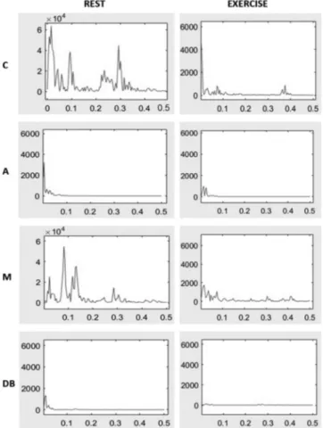
HRV data are shown in Table 2. Examples of HRV spectra are shown in Fig. 1. At rest, with respect to control, PTOTwas
not affected by metoprolol administration, but it was largely and significantly decreased under atropine and double block-ade, because of drastically lower values of both LF and HF. No differences between atropine and double blockade were found. The same was the case during exercise, although the differ-ences were much smaller than at rest, because when moving from rest to exercise, PTOTwas drastically reduced in control
and under metoprolol. No differences for LF and HF between sympathetic blockade and control, or between atropine and double blockade, were observed.
At rest, LF/HF was unaffected by drug treatment, the only significant difference being between atropine and double blockade. The same was the case for LFnu. No differences were observed concerning HFnu. At exercise, LF/HF did not differ under metoprolol or atropine with respect to control, but it was lower under double blockade than in control and in the other pharmacological conditions. The same was the case for LFnu. Coherently, HFnu was higher in double blockade than in any other condition.
All data concerning spontaneous SAP and DAP variability are shown in Table 3. At rest, concerning SAP, no differences among conditions were observed for PTOT. Concerning LF, no
differences between sympathetic blockade and control were found, but it was lower under atropine and double blockade than in control and sympathetic blockade. HF in atropine and double blockade was lower than in control and under meto-prolol, although for the latter the level of significance was not attained. During exercise, PTOTwas lower in all three
investi-gated pharmacological conditions than in control, but no
dif-Table 1. Mean steady-state values for the cardiovascular variables monitored during rest and exercise in the four
experimental conditions: control, atropine, metoprolol, and double blockade
Measured Variables Control Metoprolol Atropine Double Blockade
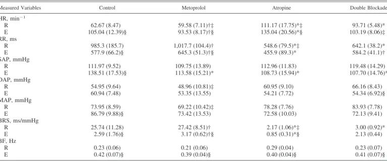
HR, min⫺1 R 62.67 (8.47) 59.58 (7.11)†‡ 111.17 (17.75)*‡ 93.71 (5.48)* E 105.04 (12.39)§ 93.53 (8.17)†§ 135.04 (20.56)*§ 103.19 (8.06)‡ RR, ms R 985.3 (185.7) 1,017.7 (104.4)† 548.6 (79.5)*‡ 642.1 (38.2)* E 577.9 (66.2)§ 645.3 (51.3)†§ 455.9 (89.3)* 584.2 (41.1)† SAP, mmHg R 111.97 (9.52) 109.75 (13.89) 112.96 (11.83) 119.48 (14.29) E 138.51 (17.53)§ 113.58 (15.21)* 108.73 (15.94)* 107.70 (14.76)* DAP, mmHg R 54.95 (9.64) 48.96 (10.81)‡ 60.95 (9.10) 66.16 (8.43) E 60.94 (7.48) 53.35 (13.55) 54.21 (7.72) 54.34 (6.92)§ MAP, mmHg R 73.95 (8.59) 69.22 (10.42)‡ 78.28 (7.76) 83.93 (7.78) E 86.79 (9.88)§ 73.42 (13.53) 72.58 (10.03) 72.13 (9.41) BRS, ms/mmHg R 25.74 (11.28) 27.42 (8.51)† 2.17 (1.06)*‡ 3.00 (0.92)* E 2.59 (1.76)§ 3.17 (0.62)†§ 0.85 (0.31)*§ 2.13 (0.44) BF, Hz R 0.23 (0.06) 0.21 (0.06) 0.29 (0.04) 0.23 (0.07) E 0.42 (0.07)§ 0.39 (0.04)§ 0.40 (0.04)§ 0.41 (0.07)§
Values are means (SD); no. of participants n⫽ 7. BF, breathing frequency; BRS, spontaneous baroreflex sensitivity; DAP, diastolic arterial pressure; E, exercise; HR, heart rate; MAP, mean arterial pressure; R, rest; RR, R-R interval; SAP, systolic arterial pressure. Two-way ANOVA for repeated measurements;
P⬍ 0.05. *Significantly different from control; †significantly different from atropine; ‡significantly different from double blockade; §exercise significantly
ferences among conditions were observed for either LF or HF. In control and under atropine, LF was higher during exercise than at rest. LF/HF was unchanged in all conditions.
At rest, concerning DAP, no changes in PTOTwere found in
any pharmacological condition with respect to control. HF did not vary among conditions, whereas LF was lower in atropine and double blockade than in control. LF/HF was lower in all conditions than in control. During exercise, there were no significant differences among conditions or with respect to the same condition at rest.
The BRS values at rest and during exercise are shown in Fig. 2. At rest, BRS was significantly lower under atropine and under double blockade than in control and under sympathetic blockade, which in turn did not differ between them. During exercise, BRS under atropine and double blockade was lower than in control and under sympathetic blockade. BRS was lower during exercise than at rest in all conditions except double blockade.
DISCUSSION
The analysis of spontaneous HRV at rest showed that 1) atropine administration drastically reduced PTOT, because of
the fall of both LF and HF, with respect to control; 2) simultaneous double blockade with atropine and metoprolol provided the same results as atropine administration only; and
3) metoprolol administration had no effects on HRV.
When moving from rest to exercise, our results showed that
1) no differences in PTOT, LF, and HF appeared under atropine
or under simultaneous atropine and metoprolol administration with respect to rest and 2) PTOT, LF, and HF were decreased to
the same extent under metoprolol as in control. However, during exercise, PTOT, LF, and HF were lower under atropine
and double blockade than in control or with metoprolol. These results are in line with the predictions made and thus do not allow refutation of the vagal withdrawal hypothesis, but
rather reinforce it. Although, taken separately, similar consis-tent results can be found in the previous literature (2, 6, 8, 10, 11, 15, 17, 21, 24, 29, 32, 33, 35, 43, 44), this is the first time that a complete picture of the role of the ANS in determining HRV at rest and during exercise was obtained.
Heart rate variability. The significant increase in HR after
atropine administration is in line with previous studies (9, 21, 22, 48, 50) and was opposed by the observation that after metoprolol administration, despite a slight decrease, HR did not change significantly compared with control. These results were similar in size to those obtained in a previous study with the same drug (48). However, they are at variance with those of other studies, performed in upright posture, showing a significant HR reduction at rest with beta-blockade (11, 14, 15, 19). In supine posture, the predominance of vagal modulation of HR (20, 35) may explain the lack of HR changes with metoprolol.
Concerning HRV, metoprolol failed in changing PTOT, LF,
and HF at rest, indicating that a selective blockade of cardiac -adrenergic receptors has no effects on spontaneous HR oscillations. This suggests that the sympathetic outflow to the heart may not be the main determinant of HRV, although the PTOTvalues under double blockade appear to be lower (just a
tendency) than under atropine. These results for PTOT, although
in agreement with those of some previous studies (15, 53), are in contrast with those given by Cogliati et al. (11), who showed an increase in PTOTunder atenolol, supporting the idea that the
pattern was mostly due to an increase in the HF peak. This finding suggested stronger cardiorespiratory coupling under atenolol than in control. Comparable results were obtained by others (40) using propranolol.
Spontaneous HR oscillations were almost suppressed after atropine administration, as previously found (8, 15, 21, 29, 32, 33, 53), supporting the notion that parasympathetic outflow to the heart is the major determinant of HRV in resting humans. Table 2. Means (SD) of all parameters calculated by means of heart rate variability in the four investigated conditions:
control, atropine, metoprolol, and double blockade
Heart Rate Variability Control Metoprolol Atropine Double Blockade
Absolute values PTOT, ms2/Hz R 6,351.4 (4,476.4) 7,883.2 (5,965.9) 22.5 (13.8)*† 12.9 (4.9)*† E 185.4 (77.1)§ 93.6 (30.9)* 10.1 (3.3)*† 14.8 (4.7)*† LF, ms2/Hz R 1,717.5 (1,290.6) 2,711.9 (2,061.8) 1.5 (1.2)*† 1.1 (0.5)*† E 40.6 (29.3)§ 41.3 (29.3)§ 1.7 (1.4)*† 1.6 (1.5)*† HF, ms2/Hz R 1,441.0 (1,296.1) 2,552.3 (2,245.0) 0.9 (0.5)*† 2.6 (0.8)*† E 10.8 (7.8)§ 11.2 (9.2)§ 0.3 (0.13)* 3.1 (1.6)*† Relative values LF/HF R 1.4 (1.0) 1.4 (0.8) 1.8 (0.9) 0.5 (0.3)‡ E 4.1 (2.0)§ 4.0 (2.0)§ 4.0 (2.6) 0.3 (0.1)*†‡ LFnu, % R 46.8 (19.3) 46.1 (14.7) 57.7 (28.2) 25.9 (13.8)‡ E 69.6 (16.5) 65.3 (21.2) 61.6 (22.7) 12.7 (8.0)*†‡ HFnu, % R 51.1 (18.3) 51.1 (15.4) 38.3 (26.9) 62.6 (15.6) E 15.5 (8.7)§ 17.4 (5.0)§ 22.6 (14.4) 45.5 (23.5)*†‡
Values are means (SD); no. of participants n⫽ 7. E, exercise; HF, high-frequency power; HFnu, relative high-frequency power; LF, low-frequency power; LF/HF, ratio of low-frequency power to high-frequency power; LFnu, relative low-frequency power; PTOT, total power; R, rest. Two-way ANOVA for repeated
measurements; P ⬍ 0.05. *Significantly different from control; †significantly different from metoprolol; ‡significantly different from atropine; §exercise significantly different from rest.
This was so also under simultaneous sympathetic and vagal blockade, indicating that suppression of the parasympathetic modulation of the heartbeat was the most important determi-nant of the present results. Breathing frequency did not change in the three conditions, being obviously higher during exercise than at rest. This implies that changes in HF were not due to any change in breathing frequency.
Coherently, in the present study, passing from rest to exer-cise implied a large fall in LF and HF in control and under metoprolol. Conversely, under atropine and double blockade, in which a suppression of the vagal modulation of HR was obtained already at rest, no changes were found during exercise with respect to rest. These results demonstrate that the well-known fall of HRV that is usually observed during exercise (37) is essentially a consequence of the withdrawal of the vagal outflow to the heart occurring at exercise onset (12, 25), as hypothesized. As such, our results suggest that vagal with-drawal is incomplete at the investigated powers, because LF and HF during exercise were still higher in control than with atropine or double blockade, the two conditions in which a full blockade of muscarinic receptors was attained. On the other hand, the fact that passing from rest to exercise generated comparable results with metoprolol as in control is coherent with the reported decrease of the LF peak in humans (37, 39).
These data are in contrast with the generally accepted notion that during exercise, the degree of sympathetic activation increases (46, 54), and the modulation of the heartbeat by the sympathetic efferents becomes predominant (38, 45). This may mean that HRV in exercise does not reflect the degree of ongoing sympathetic activation.
When we look at the normalized variables at rest, none of the investigated drugs could change the LF/HF significantly with respect to control: this reflects the finding that the effects of drug administration on LF and HF at rest were of the same size. In contrast, during exercise, there was a tendency toward a lower HF than LF. Yet this tendency, though not significant, was such as to provide, during exercise compared with at rest, significantly lower HFnu values in control and under sympa-thetic blockade (only a tendency in atropine and in double blockade). Consequently, LF/HF results were higher during exercise than at rest, at least in these two cases.
In the context of the present hypothesis, this would suggest that the withdrawal of vagal tone at exercise onset might have had greater effects on the HF than on the LF component of HRV. Alternatively, the relative increase of the LF component of RR variability may suggest an increase of the cardiac sympathetic modulation. Nevertheless, LFnu in double block-ade was significantly lower, and HFnu significantly higher, than in control despite the lack of differences in LF/HF. This may be due to the nonautonomic effect of an increase in ventilation that is reflected in HRV through changes in venous return during exercise. A similar condition can be observed in a neurodegenerative disease such as pure autonomic failure. This condition is characterized by both a cardiac sympathetic and parasympathetic denervation leading to PTOTvalues
mim-icking high-dosage atropine administration (16), in which a HF component of HRV, nonautonomic in origin, is present (39). These apparently contradictory results prevent us from arriving at clear-cut conclusions concerning the mechanisms at the basis of relative powers in this study.
Blood pressure variability. Arterial blood pressure at rest
was unaffected by drug administration. The fact that atropine did not act on systemic blood pressure, in agreement with previous studies (15, 21), is coherent with the notion that there is no cholinergic innervation in most regional circulations. On the other hand, metoprolol is a selective blocker of1
-adren-ergic receptors that are expressed specifically in the heart, not in arterioles, so that it is not expected to induce changes in blood pressure.
Coherently, SAP variability was much less affected by atropine and double blockade than HRV. According to Zhang et al. (61), who investigated spontaneous blood pressure vari-ability under ganglionic blockade with trimethaphan, the HF peak of blood pressure variability is mediated by mechanical effects due to the breathing cycle and cardiac filling: if this is so, one would not expect effects of any of the drugs used in this study on the HF for blood pressure. In fact, the changes in HF due to drug administration in the present study were much smaller than for HRV, although significant under atropine and double blockade. Zhang et al. (61) also attributed the LF of blood pressure variability to either sympathetic activity or intrinsic vascular rhythmicity: if this is so, no changes in LF would be found with atropine, metoprolol, or double blockade. In fact, we found much smaller differences in LF due to drug administration for blood pressure variability than for HRV. Yet
Fig. 1. Heart rate variability (HRV) spectra resulting from the experiments, in which the HRV segments shown are as follows: rest (panels at left), exercise (panels at right), control (C, first row), atropine (A, second row), metoprolol (M, third row), and double blockade (DB, fourth row). Here, n⫽ 7; x-axis, frequency (Hz); y-axis, R-R interval power (ms2/Hz). Note differences in y-scales.
these changes were in agreement with those of HF, being significant under atropine and double blockade. These effects might have been an indirect consequence of the role that the ANS may play in modulating the dynamic relationship be-tween HRV and blood pressure variability (7, 61), with an involvement of its parasympathetic branch.
Most remarkable are the differences observed when passing from rest to exercise: the LF for SAP increased in control, as expected (37, 39), and with atropine, but not with metoprolol and in double blockade. This indicates that the increase in LF for SAP may be a consequence of increased sympathetic modulation during exercise. No effects were observed under
any drug on HF: this means that the HF of SAP is independent of the activity of the two branches of the ANS. The lack of exercise effects on HF under drug stimulation explains why the PTOTdid not differ significantly during exercise with respect to
rest under atropine.
DAP variability was unaffected by drug administration: this suggests that the exercise effect on the LF of SAP, related to a selective blockade of1-adrenergic receptors, is mediated by a
central (cardiac) rather than a peripheral (arteriolar muscle vasodilation) action of the sympathetic branch of the ANS.
Baroreflex sensitivity. At rest, BRS was drastically lower
under atropine and double blockade than in control. This observation was consistent with what we observed for the LF peak of blood pressure variability: reduced under atropine and double blockade and unchanged under metoprolol with respect to control. Coherently, when comparing rest with exercise in a given condition, BRS decreased in control and under meto-prolol but did not change under atropine or double blockade. These results for BRS appear to be in agreement with previous observations (1, 11, 56). Bringard et al. (4) postulated that BRS is mainly modulated by the parasympathetic efferent branch of the ANS. These data support this hypothesis. Muscarinic re-ceptors do not modulate smooth muscle tone in most arterioles, including those of skeletal muscles. Thus, the parasympathetic effect on arterial blood pressure variability indexes must be indirect. Based on the present results, we speculate that baro-reflexes may participate in the modulation of the LF of arterial blood pressure. The reduction of BRS observed during exercise (51) supports the idea of ␣-index changes as previously re-ported (36). In the present study, the BRS reduction during Table 3. Parameters resulting from the analysis of spontaneous variability of systolic and diastolic arterial pressures in the
four investigated conditions: control, atropine, metoprolol, and double blockade
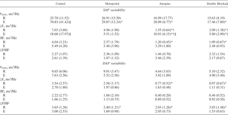
Control Metoprolol Atropine Double Blockade
SAP variability PTOT, ms2/Hz R 25.70 (11.52) 26.91 (15.50) 16.99 (17.77) 15.63 (8.19) E 70.83 (41.42)§ 29.07 (12.24)* 28.09 (6.77)* 17.46 (7.00)* LF, ms2/Hz R 7.03 (3.60) 4.96 (1.90) 1.55 (0.64)*† 2.09 (1.38)*† E 18.68 (17.97)§ 5.51 (1.52) 10.93 (6.15)*†§ 5.80 (2.90)*‡ HF, ms2/Hz R 4.04 (3.21) 2.57 (1.79) 1.20 (0.45)* 1.09 (0.67)* E 5.49 (4.20) 5.48 (3.96) 3.29 (1.80) 2.48 (0.93) LF/HF R 2.27 (1.07) 2.36 (1.09) 1.46 (0.70) 2.32 (1.54) E 2.61 (1.39) 1.87 (1.32) 3.46 (2.39) 2.17 (0.67) DAP variability PTOT, ms2/Hz R 9.65 (6.06) 9.01 (3.47) 4.64 (3.03) 5.10 (2.32) E 7.63 (2.56) 5.52 (2.56) 3.92 (1.00) 4.90 (3.40) LF, ms2/Hz R 3.54 (2.57) 2.56 (1.17) 0.77 (0.52)* 0.97 (0.67)* E 2.70 (1.80) 1.97 (0.86) 1.63 (0.48) 1.11 (0.31) HF, ms2/Hz R 2.22 (2.77) 1.88 (2.10) 0.40 (0.20) 0.46 (0.52) E 1.66 (1.25) 1.13 (0.75) 0.89 (0.52) 0.92 (0.50) LF/HF R 3.65 (1.26) 2.40 (1.21)* 2.03 (1.26)* 3.03 (1.48)* E 3.00 (2.53) 1.69 (0.98) 2.05 (0.73) 1.53 (0.63)
Values are means (SD); no. of participants n⫽ 7. E, exercise; HF, high-frequency power; LF, low-frequency power; LF/HF, ratio of low-frequency power to high-frequency power; PTOT, total power; R, rest. Two-way ANOVA for repeated measurements; P⬍ 0.05. *Significantly different from control; †significantly
different from metoprolol; ‡significantly different from atropine; §exercise significantly different from rest.
Fig. 2. Means (SD) of spontaneous baroreflex sensitivity in each investigated condition (control, atropine, metoprolol, and double blockade) at rest and during exercise. Here, n⫽ 7; two-way ANOVA for repeated measurements;
P⬍ 0.05. *Significantly different from control. #Significantly different from
double blockade. §Significantly different from atropine. $Significantly differ-ent from the same condition at rest.
exercise was observed only in control and with metoprolol, but not with atropine and double blockade. This finding reinforces the notion that withdrawal of vagal tone is responsible for the fall of BRS at exercise onset (4, 34). Coherently, no differences in BRS among the four investigated conditions were observed during exercise.
Study limitations. A limitation of this study may be
sug-gested by the lack of differences between control and meto-prolol, as this may also suggest that the1-adrenergic blockade
might have been incomplete. It is of note, however, that we used the same dose and followed the same procedure for metoprolol administration as in a previous study (14) in upright posture, which showed a significant resting HR decrease both in normoxia and in acute hypoxia at rest as during exercise. Moreover, we observe that the isoprenaline test provided unambiguous evidence of quasi-complete1-adrenergic
block-ade.
Another possible limiting factor is related to the fact that HR differed remarkably among conditions. This may affect the HRV indexes in the time domain per se (60), thus possibly undermining the relation to the action of the ANS.
Conclusion. The results of this study support the tested
hypothesis that vagal suppression is responsible for the disap-pearance of spontaneous HRV during exercise. The observed effects on arterial blood pressure variability are indirectly related to the action of the administered drugs, supporting the notion that blood pressure and HRV are only partially associ-ated phenomena, possibly controlled by different physiological mechanisms.
ACKNOWLEDGMENTS
We thank the participants for the time and dedication given to complete the study.
GRANTS
This study was supported by Swiss National Science Foundation Grants 3200B0_102181 and 32003B_143427 to G. Ferretti.
DISCLOSURES
No conflicts of interest, financial or otherwise, are declared by the authors. AUTHOR CONTRIBUTIONS
T.F. and G.F. conceived and designed research; A.B., N.F., A.A., and E.T. performed experiments; T.F. and V.P. analyzed data; T.F. interpreted results of experiments; T.F. prepared figures; T.F. drafted manuscript; T.F., V.P., A.B., N.F., A.A., R.F., J.-C.B., and G.F. edited and revised manuscript; R.F. and G.F. approved final version of manuscript.
REFERENCES
1. Airaksinen KE, Niemelä MJ, Huikuri HV. Effect of beta-blockade on baroreflex sensitivity and cardiovascular autonomic function tests in pa-tients with coronary artery disease. Eur Heart J 15: 1482–1485, 1994. doi:10.1093/oxfordjournals.eurheartj.a060418.
2. Beckers F, Verheyden B, Ramaekers D, Swynghedauw B, Aubert AE. Effects of autonomic blockade on non-linear cardiovascular variability indices in rats. Clin Exp Pharmacol Physiol 33: 431–439, 2006. doi:10. 1111/j.1440-1681.2006.04384.x.
3. Bertinieri G, Di Rienzo M, Cavallazzi A, Ferrari AU, Pedotti A, Mancia G. Evaluation of baroreceptor reflex by blood pressure monitor-ing in unanesthetized cats. Am J Physiol Heart Circ Physiol 254: H377– H383, 1988. doi:10.1152/ajpheart.1988.254.2.H377.
4. Bringard A, Adami A, Fagoni N, Fontolliet T, Lador F, Moia C, Tam E, Ferretti G. Dynamics of the RR-interval versus blood pressure rela-tionship at exercise onset in humans. Eur J Appl Physiol 117: 619 –630, 2017. doi:10.1007/s00421-017-3564-6.
5. Buchheit M, Richard R, Doutreleau S, Lonsdorfer-Wolf E, Branden-berger G, Simon C. Effect of acute hypoxia on heart rate variability at rest and during exercise. Int J Sports Med 25: 264 –269, 2004. doi:10. 1055/s-2004-819938.
6. Castiglioni P, Parati G, Di Rienzo M, Carabalona R, Cividjian A, Quintin L. Scale exponents of blood pressure and heart rate during autonomic blockade as assessed by detrended fluctuation analysis. J
Physiol 589: 355–369, 2011. doi:10.1113/jphysiol.2010.196428. 7. Cevese A, Gulli G, Polati E, Gottin L, Grasso R. Baroreflex and
oscillation of heart period at 0.1 Hz studied by␣-blockade and cross-spectral analysis in healthy humans. J Physiol 531: 235–244, 2001. doi:10.1111/j.1469-7793.2001.0235j.x.
8. Challapalli S, Kadish AH, Horvath G, Goldberger JJ. Differential effects of parasympathetic blockade and parasympathetic withdrawal on heart rate variability. J Cardiovasc Electrophysiol 10: 1192–1199, 1999. doi:10.1111/j.1540-8167.1999.tb00295.x.
9. Chamberlain DA, Turner P, Sneddon JM. Effects of atropine on heart-rate in healthy man. Lancet 290: 12–15, 1967. doi: 10.1016/S0140-6736(67)90057-8.
10. Chen X, Mukkamala R. Selective quantification of the cardiac sympa-thetic and parasympasympa-thetic nervous systems by multisignal analysis of cardiorespiratory variability. Am J Physiol Heart Circ Physiol 294: H362– H371, 2008. doi:10.1152/ajpheart.01061.2007.
11. Cogliati C, Colombo S, Ruscone TG, Gruosso D, Porta A, Montano N, Malliani A, Furlan R. Acute beta-blockade increases muscle sympathetic activity and modifies its frequency distribution. Circulation 110: 2786 – 2791, 2004. doi:10.1161/01.CIR.0000146335.69413.F9.
12. Fagraeus L, Linnarsson D. Autonomic origin of heart rate fluctuations at the onset of muscular exercise. J Appl Physiol 40: 679 –682, 1976. doi:10.1152/jappl.1976.40.5.679.
13. Faisal A, Beavers KR, Robertson AD, Hughson RL. Prior moderate and heavy exercise accelerate oxygen uptake and cardiac output kinetics in endurance athletes. J Appl Physiol (1985) 106: 1553–1563, 2009. doi:10. 1152/japplphysiol.91550.2008.
14. Ferretti G, Licker MJ, Anchisi S, Moia C, Susta D, Morel DR. The effects of 1-adrenergic blockade on cardiovascular oxygen flow in
normoxic and hypoxic humans at exercise. Eur J Appl Physiol 95: 250 –259, 2005. doi:10.1007/s00421-005-1393-5.
15. Formes KJ, Wray DW, O-Yurvati AH, Weiss MS, Shi X. Sympathetic cardiac influence and arterial blood pressure instability. Auton Neurosci 118: 116 –124, 2005. doi:10.1016/j.autneu.2005.01.002.
16. Furlan R, Piazza S, Bevilacqua M, Turiel M, Norbiato G, Lombardi F, Malliani A. Pure autonomic failure: complex abnormalities in the neural mechanisms regulating the cardiovascular system. J Auton Nerv
Syst 51: 223–235, 1995. doi:10.1016/0165-1838(94)00135-7.
17. Goldberger JJ, Challapalli S, Tung R, Parker MA, Kadish AH. Relationship of heart rate variability to parasympathetic effect. Circulation 103: 1977–1983, 2001. doi:10.1161/01.CIR.103.15.1977.
18. Halliwill JR, Minson CT. Effect of hypoxia on arterial baroreflex control of heart rate and muscle sympathetic nerve activity in humans. J Appl
Physiol (1985) 93: 857–864, 2002. doi:10.1152/japplphysiol.01103.2001. 19. Hawkins MN, Barnes Q, Purkayastha S, Eubank W, Ogoh S, Raven PB. The effects of aerobic fitness and1-adrenergic receptor blockade on
cardiac work during dynamic exercise. J Appl Physiol (1985) 106: 486 – 493, 2009. doi:10.1152/japplphysiol.90795.2008.
20. Hughson RL, Yamamoto Y, McCullough RE, Sutton JR, Reeves JT. Sympathetic and parasympathetic indicators of heart rate control at alti-tude studied by spectral analysis. J Appl Physiol (1985) 77: 2537–2542, 1994. doi:10.1152/jappl.1994.77.6.2537.
21. Kamimori GH, Bellar D, Fein HG, Smallridge RC. Hormonal and cardiovascular response to low-intensity exercise with atropine adminis-tration. Mil Med 174: 253–258, 2009. doi:10.7205/MILMED-D-00-4608. 22. Kelbaek H, Marving J, Hvid-Jacobsen K, Nielsen SL. Effects of atropine on left ventricular volumes and ejection and filling rates at rest and during exercise. Br J Clin Pharmacol 32: 585–589, 1991. doi:10.1111/ j.1365-2125.1991.tb03956.x.
23. Koller EA, Drechsel S, Hess T, Macherel P, Boutellier U. Effects of atropine and propranolol on the respiratory, circulatory, and ECG re-sponses to high altitude in man. Eur J Appl Physiol Occup Physiol 57: 163–172, 1988. doi:10.1007/BF00640657.
24. Krediet RT, Dunning AJ. Baroreflex sensitivity in hypertension during beta-adrenergic blockade. Br Heart J 41: 106 –110, 1979. doi:10.1136/hrt. 41.1.106.
25. Lador F, Azabji Kenfack M, Moia C, Cautero M, Morel DR, Capelli C, Ferretti G. Simultaneous determination of the kinetics of cardiac output, systemic O2delivery, and lung O2uptake at exercise onset in men. Am J Physiol Regul Integr Comp Physiol 290: R1071–R1079, 2006.
doi:10.1152/ajpregu.00366.2005.
26. Lador F, Tam E, Azabji Kenfack M, Cautero M, Moia C, Morel DR, Capelli C, Ferretti G. Phase I dynamics of cardiac output, systemic O2
delivery, and lung O2uptake at exercise onset in men in acute normobaric
hypoxia. Am J Physiol Regul Integr Comp Physiol 295: R624 –R632, 2008. doi:10.1152/ajpregu.00797.2007.
27. Leyk D, Essfeld D, Hoffmann U, Wunderlich HG, Baum K, Stege-mann J. Postural effect on cardiac output, oxygen uptake and lactate during cycle exercise of varying intensity. Eur J Appl Physiol Occup
Physiol 68: 30 –35, 1994. doi:10.1007/BF00599238.
28. Malliani A, Pagani M, Lombardi F, Cerutti S. Cardiovascular neural regulation explored in the frequency domain. Circulation 84: 482–492, 1991. doi:10.1161/01.CIR.84.2.482.
29. Montano N, Cogliati C, Porta A, Pagani M, Malliani A, Narkiewicz K, Abboud FM, Birkett C, Somers VK. Central vagotonic effects of atropine modulate spectral oscillations of sympathetic nerve activity.
Circulation 98: 1394 –1399, 1998. doi:10.1161/01.CIR.98.14.1394. 30. Moody GB, Mark RG, Zoccola A, Mantero S. Derivation of respiratory
signals from multi-lead ECGs. Comput Cardiol 12: 113–116, 1985. 31. Morikami Y, Yasue H, Okumura K, Horio Y, Fujii H, Matsuyama K.
Effects of phentolamine and atropine on angina pectoris induced by handgrip test in patients with variant angina. Am J Cardiol 61: 71–76, 1988. doi:10.1016/0002-9149(88)91307-0.
32. Mullen TJ, Appel ML, Mukkamala R, Mathias JM, Cohen RJ. System identification of closed-loop cardiovascular control: effects of posture and autonomic blockade. Am J Physiol Heart Circ Physiol 272: H448 –H461, 1997. doi:10.1152/ajpheart.1997.272.1.H448.
33. Ng J, Sundaram S, Kadish AH, Goldberger JJ. Autonomic effects on the spectral analysis of heart rate variability after exercise. Am J Physiol
Heart Circ Physiol 297: H1421–H1428, 2009. doi:10.1152/ajpheart. 00217.2009.
34. Ogoh S, Fisher JP, Dawson EA, White MJ, Secher NH, Raven PB. Autonomic nervous system influence on arterial baroreflex control of heart rate during exercise in humans. J Physiol 566: 599 –611, 2005. doi:10. 1113/jphysiol.2005.084541.
35. Pagani M, Lombardi F, Guzzetti S, Rimoldi O, Furlan R, Pizzinelli P, Sandrone G, Malfatto G, Dell’Orto S, Piccaluga E. Power spectral analysis of heart rate and arterial pressure variabilities as a marker of sympatho-vagal interaction in man and conscious dog. Circ Res 59: 178 –193, 1986. doi:10.1161/01.RES.59.2.178.
36. Pagani M, Somers V, Furlan R, Dell’Orto S, Conway J, Baselli G, Cerutti S, Sleight P, Malliani A. Changes in autonomic regulation induced by physical training in mild hypertension. Hypertension 12: 600 –610, 1988. doi:10.1161/01.HYP.12.6.600.
37. Perini R, Veicsteinas A. Heart rate variability and autonomic activity at rest and during exercise in various physiological conditions. Eur J Appl
Physiol 90: 317–325, 2003. doi:10.1007/s00421-003-0953-9.
38. Persson PB. Modulation of cardiovascular control mechanisms and their interaction. Physiol Rev 76: 193–244, 1996. doi:10.1152/physrev.1996.76. 1.193.
39. Piazza S, Furlan R, Dell’Orto S, Porta A, Lombardi F, Pagani M, Malliani A. Mechanical effects of respiration and stepping on systolic arterial pressure variability during treadmill exercise. J Hypertens 13: 1643–1647, 1995. doi:10.1097/00004872-199512010-00024.
40. Pichot V, Gaspoz JM, Molliex S, Antoniadis A, Busso T, Roche F, Costes F, Quintin L, Lacour JR, Barthélémy JC. Wavelet transform to quantify heart rate variability and to assess its instantaneous changes. J
Appl Physiol (1985) 86: 1081–1091, 1999. doi:10.1152/jappl.1999.86.3. 1081.
41. Pichot V, Roche F, Celle S, Barthélémy JC, Chouchou F. HRVanalysis: a free software for analyzing cardiac autonomic activity. Front Physiol 7: 557, 2016. doi:10.3389/fphys.2016.00557.
42. Polanczyk CA, Rohde LE, Moraes RS, Ferlin EL, Leite C, Ribeiro JP. Sympathetic nervous system representation in time and frequency domain indices of heart rate variability. Eur J Appl Physiol Occup Physiol 79: 69 –73, 1998. doi:10.1007/s004210050475.
43. Pomeranz B, Macaulay RJ, Caudill MA, Kutz I, Adam D, Gordon D, Kilborn KM, Barger AC, Shannon DC, Cohen RJ, Benson H. Assess-ment of autonomic function in humans by heart rate spectral analysis. Am
J Physiol Heart Circ Physiol 248: H151–H153, 1985. doi:10.1152/ ajpheart.1985.248.1.H151.
44. Porta A, Castiglioni P, Di Rienzo M, Bari V, Bassani T, Marchi A, Takahashi AC, Tobaldini E, Montano N, Catai AM, Barbic F, Furlan R, Cividjian A, Quintin L. Short-term complexity indexes of heart period and systolic arterial pressure variabilities provide complementary infor-mation. J Appl Physiol (1985) 113: 1810 –1820, 2012. doi:10.1152/ japplphysiol.00755.2012.
45. Rimoldi O, Furlan R, Pagani MR, Piazza S, Guazzi M, Pagani M, Malliani A. Analysis of neural mechanisms accompanying different intensities of dynamic exercise. Chest 101, Suppl: 226S–230S, 1992. doi:10.1378/chest.101.5_Supplement.226S.
46. Robinson BF, Epstein SE, Beiser GD, Braunwald E. Control of heart rate by the autonomic nervous system. Studies in man on the interrelation between baroreceptor mechanisms and exercise. Circ Res 19: 400 –411, 1966. doi:10.1161/01.RES.19.2.400.
47. Sanderson JE, Yeung LY, Yeung DT, Kay RL, Tomlinson B, Critch-ley JA, Woo KS, Bernardi L. Impact of changes in respiratory frequency and posture on power spectral analysis of heart rate and systolic blood pressure variability in normal subjects and patients with heart failure. Clin
Sci (Lond) 91: 35–43, 1996. doi:10.1042/cs0910035.
48. Smith ML, Hudson DL, Graitzer HM, Raven PB. Blood pressure regulation during cardiac autonomic blockade: effect of fitness. J Appl
Physiol (1985) 65: 1789 –1795, 1988. doi:10.1152/jappl.1988.65.4.1789. 49. Sundblad P, Haruna Y, Tedner B, Linnarsson D. Short-term cardio-vascular responses to rapid whole-body tilting during exercise. Eur J Appl
Physiol 81: 259 –270, 2000. doi:10.1007/s004210050041.
50. Toska K, Eriksen M. Respiration-synchronous fluctuations in stroke volume, heart rate and arterial pressure in humans. J Physiol 472: 501– 512, 1993. doi:10.1113/jphysiol.1993.sp019958.
51. Vallais F, Baselli G, Lucini D, Pagani M, Porta A. Spontaneous baroreflex sensitivity estimates during graded bicycle exercise: a compar-ative study. Physiol Meas 30: 201–213, 2009. doi:10.1088/0967-3334/30/ 2/007.
52. Virtanen R, Kanto J, Iisalo E, Iisalo EU, Salo M, Sjövall S. Pharmacokinetic studies on atropine with special reference to age. Acta
Anaesthesiol Scand 26: 297–300, 1982. doi:10.1111/j.1399-6576.1982. tb01770.x.
53. Warren JH, Jaffe RS, Wraa CE, Stebbins CL. Effect of autonomic blockade on power spectrum of heart rate variability during exercise. Am
J Physiol Regul Integr Comp Physiol 273: R495–R502, 1997. doi:10. 1152/ajpregu.1997.273.2.R495.
54. White DW, Raven PB. Autonomic neural control of heart rate during dynamic exercise: revisited. J Physiol 592: 2491–2500, 2014. doi:10. 1113/jphysiol.2014.271858.
55. Wieling W, Harms MP, ten Harkel AD, van Lieshout JJ, Sprangers RL. Circulatory response evoked by a 3 s bout of dynamic leg exercise in humans. J Physiol 494: 601–611, 1996. doi:10.1113/jphysiol.1996. sp021518.
56. Wray DW, Formes KJ, Weiss MS, O-Yurvati AH, Raven PB, Zhang R, Shi X. Vagal cardiac function and arterial blood pressure stability. Am J Physiol Heart Circ Physiol 281: H1870 –H1880, 2001. doi:10.1152/ ajpheart.2001.281.5.H1870.
57. Xie A, Skatrud JB, Puleo DS, Morgan BJ. Exposure to hypoxia produces long-lasting sympathetic activation in humans. J Appl Physiol
(1985) 91: 1555–1562, 2001. doi:10.1152/jappl.2001.91.4.1555. 58. Yamamoto Y, Hoshikawa Y, Miyashita M. Effects of acute exposure to
simulated altitude on heart rate variability during exercise. J Appl Physiol
(1985) 81: 1223–1229, 1996. doi:10.1152/jappl.1996.81.3.1223. 59. Yasue H, Horio Y, Nakamura N, Fujii H, Imoto N, Sonoda R,
Kugiyama K, Obata K, Morikami Y, Kimura T. Induction of coronary artery spasm by acetylcholine in patients with variant angina: possible role of the parasympathetic nervous system in the pathogenesis of coronary artery spasm. Circulation 74: 955–963, 1986. doi:10.1161/01.CIR.74.5. 955.
60. Zaza A, Lombardi F. Autonomic indexes based on the analysis of heart rate variability: a view from the sinus node. Cardiovasc Res 50: 434 –442, 2001. doi:10.1016/S0008-6363(01)00240-1.
61. Zhang R, Iwasaki K, Zuckerman JH, Behbehani K, Crandall CG, Levine BD. Mechanism of blood pressure and R-R variability: insights from ganglion blockade in humans. J Physiol 543: 337–348, 2002. doi:10.1113/jphysiol.2001.013398.