DOCTORAL SCHOOL IN BIOLOGY
Biology Applied to Human Health
XXVII DOCTORAL PROGRAM
Identification and characterization of
regulatory networks controlling
the expression and activity of
the alternative sigma factor PvdS in
Pseudomonas aeruginosa
Daniela VisaggioSupervisors:
Prof. Paolo Visca
, Roma Tre University, RomeAbstract
Identification and characterization of regulatory networks controlling the expression and activity of the alternative sigma factor PvdS in Pseudomonas
aeruginosa
Pseudomonas aeruginosa is one of the most dreaded opportunistic pathogens in the hospital setting and represents the first cause of morbidity and mortality in cystic fibrosis patients. P. aeruginosa is able to produce several virulence factors; among them, the siderophore pyoverdine plays a critical role in P. aeruginosa pathogenicity. This siderophore is not only the main iron uptake system of P. aeruginosa but also a signaling molecule which promotes the expression of other virulence factors (i.e. exotoxin A, extracellular protease PrpL). The key role of pyoverdine in P. aeruginosa infections makes this siderophore a promising target for the development of anti-virulence drugs.
The antimicrobial agents currently used in clinical practice induce a strong selective pressure which causes the development and spreading of resistant bacteria. An alternative antibacterial strategy, which should reduce the emergence of resistance, is the development of anti-virulence drugs able to disarm pathogens without inhibiting their growth.
In the first part of this PhD thesis the use of a drug repurposing approach to search for anti-pyoverdine compounds is described. This approach is based on the search for side activities in old drugs already used in humans. A library of 1,120 marketed drugs was screened with a purpose-generated reporter strain, leading to the identification of a promising hit compound, the antimycotic drug flucytosine, which strongly reduced pyoverdine production in vitro without affecting bacterial growth. The anti-virulence activity of flucytosine was also confirmed in vivo in a mouse model of lung infection. This work provided the first evidence that pyoverdine inhibition is a suitable strategy for anti-virulence therapy against P. aeruginosa, and that drug repurposing is a cheap and rapid approach to search for novel anti-virulence compounds.
The prominent role of pyoverdine in P. aeruginosa virulence emphasized the importance of pyoverdine regulation for full understand P. aeruginosa pathogenicity. Previous transcriptomic studies suggested that the Gac system, which regulates the switch between the planktonic and biofilm lifestyles, could also influence pyoverdine gene expression in P. aeruginosa, although different studies obtained opposite results.
of Gac system in the regulation of pyoverdine production has been investigated. It has been demonstrated that the Gac system and high intracellular levels of the intracellular signaling molecule bis-(3’-5’)-cyclic dimeric guanosine monophosphate (c-di-GMP) coordinately promote pyoverdine production. In more detail, these two systems indirectly control pyoverdine genes by modulating the expression of the operons encoding the enzymes involved in the synthesis of the two aggregative exopolysaccharides Pel and Psl. The deletion of both the pel and psl operons caused a strong reduction in the production of pyoverdine and pyoverdine-dependent virulence factors, irrespective of the Gac activation state or the intracellular c-di-GMP levels, indicating that these exopolysaccharides plays an important role in the regulation of this siderophore. We also found that the effect of Pel and Psl on pyoverdine production depends on their ability to support the formation of planktonic aggregates, rather than on the exopolysaccharides per se. Indeed, we observed that the simulation of aggregation in a Pel- and Psl-independent manner is able to restore pyoverdine production in the exopolysaccharide-null mutant. These results indicate that pyoverdine, and consequently, pyoverdine-dependent virulence factors are also controlled by a new regulatory system activated by the cellular aggregation.
In conclusion, this thesis shows that cell aggregation is not only important in the first stages of biofilm formation, but also in the regulation of virulence in P. aeruginosa. The formation of cellular aggregates depends on the production of the Pel and Psl exopolysaccharides, which are regulated by the Gac system. Therefore, given the role of Gac also in the activation of pyoverdine-dependent virulence, this system could represent a potential target to inhibit simultaneously biofilm formation and the production of several virulence factors. The drug repurposing approach was thus used to search for Gac inhibitors. Although the initial screening led to identify three promising anti-Gac compounds, subsequent experiments revealed that these molecules promote, rather than inhibit, biofilm formation in P. aeruginosa, in a Gac-independent manner. Consequently, they were not further investigated as potential anti-virulence drugs.
Riassunto
Identificazione e caratterizzazione dei sistemi di regolazione deputati al controllo del fattore sigma alternativo PvdS in Pseudomonas aeruginosa
Pseudomonas aeruginosa è un batterio gram-negativo che rappresenta una delle principali cause di infezione in ambito ospedaliero e soprattutto nei pazienti affetti da fibrosi cistica. La patogenicità di P. aeruginosa dipende dalla sua capacità di resistere a molteplici classi di farmaci e di produrre numerosi fattori di virulenza. Tra i fattori di virulenza prodotti da P.aeruginosa, il sideroforo pioverdina è uno dei più importanti, in quanto non solo rappresenta il principale sistema di acquisizione del ferro del batterio, ma agisce anche da molecola segnale che attiva l’espressione dei geni coinvolti nella sintesi della pioverdina stessa e di altri fattori di virulenza. Il ruolo chiave svolto dalla pioverdina nelle infezioni causate da P. aeruginosa rende la via biosintetica e regolativa di questa molecola bersagli promettenti per lo sviluppo di farmaci anti-virulenza. I farmaci anti-virulenza, che mirano a disarmare il batterio senza inibirne la crescita, rappresentano una potenziale alternativa ai comuni farmaci antibatterici i quali, avendo un’azione generalmente battericida o batteriostatica, esercitano una forte pressione selettiva che favorisce l’insorgenza e la diffusione di resistenze. Nella prima parte della presente tesi di dottorato è stata descritta la ricerca di farmaci anti-pioverdina attraverso l’utilizzo di un approccio noto come “drug repurposing”. Tale approccio consiste nella ricerca, in farmaci comunemente utilizzati in clinica, di attività diverse rispetto a quelle per cui ciascun farmaco viene utilizzato. A tale scopo è stato generato un ceppo ricombinante di P. aeruginosa che permettesse di monitorare in maniera rapida l’espressione dei geni della pioverdina, la produzione di pioverdina stessa e la crescita batterica. L’effetto anti-pioverdina è stato saggiato in una collezione di 1,120 composti ed è stato selezionato un farmaco antimicotico, la 5-fluorocitosina, in grado di causare una forte riduzione della produzione di pioverdina e dei fattori di virulenza pioverdina-dipendenti senza però avere effetti rilevanti sulla crescita batterica. La riduzione della virulenza di P. aeruginosa, causata dall’inibizione della sintesi della pioverdina mediata dall’azione della 5-fluorocitosina, è stato confermata anche in vivo in un modello murino di infezione polmonare. Questo studio ha dimostrato che la pioverdina è un bersaglio eccellente per un’eventuale terapia finalizzata a ridurre la virulenza di P. aeruginosa e che il drug repurposing rappresenta una strategia rapida ed efficace per la ricerca di farmaci anti-virulenza.
Dato il ruolo chiave della pioverdina nella virulenza di P. aeruginosa, è stato essenziale approfondire i meccanismi che regolano la produzione di tale sideroforo, al fine di poter comprendere a pieno la patogenicità di P. aeruginosa. A tale scopo sono stati condotti degli studi, descritti nella seconda parte della presente tesi di dottorato, mirati a chiarire il ruolo del sistema Gac nella regolazione della pioverdina. Il sistema Gac è un sistema a due componenti coinvolto principalmente nella regolazione dell’espressione dei geni responsabili della sintesi dei polisaccaridi Pel e Psl, che rappresentano una componente essenziale della matrice del biofilm. Tali esopolisaccaridi sono regolati a livello trascrizionale anche dal bis-(3’-5’)-diguanosina monofosfato ciclico (c-di-GMP), un secondo messaggero intracellulare che svolge un ruolo chiave nella formazione del biofilm. Nel presente lavoro di tesi è stato dimostrato che lo stato di attivazione del sistema Gac ed elevati livelli intracellulari di c-di-GMP promuovono la produzione di pioverdina. Questi due sistemi di regolazione controllano la produzione di pioverdina indirettamente, attraverso la modulazione dell’espressione dei geni per gli esopolisaccaridi Pel e Psl. La generazione di un doppio mutante negli operoni pel e psl ha consentito di scoprire che questi operoni svolgono un ruolo essenziale nella regolazione della pioverdina, in quanto la mancata produzione di entrambi gli esopolisaccaridi determina una drastica riduzione della sintesi del sideroforo e dei fattori di virulenza da esso regolati. I risultati ottenuti in questo studio suggeriscono fortemente che l’effetto degli esopolisaccaridi sulla produzione di pioverdina sia dovuto alla loro capacità di indurre l’aggregazione cellulare in P. aeruginosa. Simulando l’aggregazione in maniera indipendente dagli esopolisaccaridi è stato infatti possibile ripristinare la produzione di pioverdina nel doppio mutante pel psl a livelli paragonabili a quelli del ceppo parentale. I risultati ottenuti indicano quindi la presenza di un nuovo sistema di regolazione, mediato dall’aggregazione cellulare, che influenza la produzione di pioverdina e dei fattori di virulenza pioverdina-dipendenti.
Nel presente lavoro di tesi è stato quindi dimostrato che l’aggregazione cellulare non solo svolge un ruolo chiave nelle prime fasi della formazione del biofilm, ma anche nella regolazione della virulenza. L’aggregazione è mediata principalmente dagli esopolisaccaridi, i quali sono regolati dal sistema Gac. Pertanto, questo sistema di regolazione rappresenta un potenziale bersaglio per inibire al contempo la produzione di alcuni fattori di virulenza e la formazione del biofilm. E’ stato quindi utilizzato l’approccio del drug repurposing per identificare composti anti-Gac. Sebbene siano
stati individuati tre derivati flavinici in grado di ridurre significativamente l’attivazione del sistema Gac, tali farmaci hanno mostrato un forte effetto di promozione della formazione del biofilm in P. aeruginosa, del tutto indipendente dal sistema Gac, e pertanto le loro potenzialità come farmaci anti-virulenza sono compromesse.
Table of contents
Chapter1
Introduction and aims
1 Pseudomonas aeruginosa 2
2 P. aeruginosa pathogenicity 2
2.1 Acute infection 4
2.2 Chronic infection 5
3 P. aeruginosa iron uptake: different strategies
in different type of infections 7
3.1 Siderophores: Pyoverdine and Pyochelin 8
3.2 Other iron uptake systems 12
4 The regulatory switch from acute to chronic infection 13
4.1 Gac regulatory network 13
4.2 Bis-(3’-5’)-cyclic dimeric guanosine monophosphate (c-di-GMP) 16
5 Aims 20
Chapter 2
Repurposing the antimycotic drug flucytosine for suppression of
Pseudomonas aeruginosa pathogenicity 29
Chapter 3
The Gac/Rsm and cyclic-di-GMP signaling networks coordinately
regulate iron uptake in Pseudomonas aeruginosa 43
Chapter 4
Exopolysaccharide-mediated cell aggregation promotes pyoverdine
dependent iron uptake and virulence in Pseudomonas aeruginosa 63
Chapter 5
Search for biofilm inhibitors through inhibition of the
Gac regulatory system 114
Chapter 6
Concluding remarks 126
Chapter 1
Chapter 1
Introduction and aims
1
Pseudomonas aeruginosa
Pseudomonas aeruginosa is a non-spore forming, Gram-negative, rod-shaped γ-Proteobacterium measuring 0.5 to 0.8 µm by 1.5 to 3.0 µm. This bacterium grows aerobically but is capable of using nitrate or arginine as a final electron acceptor in the absence of oxygen, allowing anaerobic growth (Vander Wauven et al., 1984; Haas et al., 1992). Because of its remarkable adaptability, P. aeruginosa can be found in diverse environments such as soil, water, plants and animals (Rahme et al., 1995; Mahajan-Miklos et al., 2000). P. aeruginosa synthesizes 2-aminoacetophenone (2-AA), producing a fruity, grape-like smell. This feature, along with the fact that this bacterium produces the blue colored soluble pigment pyocyanin, greatly helps in identifying an unknown colony as P. aeruginosa. The complete genome of the widely used laboratory strain P. aeruginosa PAO1, a wound isolate (Holloway, 1955), was sequenced and published in 2000 (Stover et al., 2000). The genome has a high G+C content (66.6 %) and consists of 6.3 million base pairs. The presence of 5,570 predicted open reading frames reflects the genetic complexity of the P. aeruginosa genome underlying the capability of this bacterium to grow in different ecological niches (Stover et al., 2000). Consistent with its larger genome size and environmental adaptability, P. aeruginosa was argued to contain the highest proportion of regulatory genes observed for a bacterial genome (Stover et al., 2000).
2
P. aeruginosa
pathogenicity
P. aeruginosa is typical opportunistic pathogen which can cause a wide range of infections in different tissues, including lung, eyes, ears, urinary tract and burns (Lyczak et al., 2000). Airway infections, caused by this pathogen, could be classified in two different types: acute and chronic infections (Arancibia et al., 2002). The best example of an acute respiratory nosocomial infection is the ventilator associated pneumonia (VAP). VAP is generally a consequence of the damage of the airway from mechanical ventilation which is followed by the acute pneumonia. The high mortality rate of VAP (34-48%) appears to be related to dysregulated host-pathogen interaction with an excessive host response to pneumonia (William et al., 2010). If the acute infection is not fully eradicated, P. aeruginosa can adapt to the lung environment and
fibrosis (CF) and chronic obstructive pulmonary disease (COPD) (Doring et al., 2011). CF is a congenital disease affecting 1:2,500 newborns in the Caucasian population; it is due to loss-of-function mutations in the cystic fibrosis membrane regulator (CFTR), which result in a dehydrated and thickened airway surface liquid that impairs the normal clearance of the airway. Chronic P. aeruginosa infections represent the main cause of morbidity and mortality in CF patients (www.cftr2.org). Chronic P. aeruginosa lung infections are also associated with people who have COPD. This pathology is caused by chronic inflammation of lung tissues leading to the restriction of airway passage. The incidence of P. aeruginosa infection in patients with COPD ranges from 4 to 15% (William et al., 2010) and the mortality rate due to exacerbation is high (22-49%).
Therapeutic options to treat acute and chronic P. aeruginosa infections are limited due to the ability of P. aeruginosa to resist to many antimicrobial agents. P. aeruginosa has developed many different mechanisms of resistance, that can be classified as intrinsic or acquired (Moore and Flaws, 2011). The intrinsic mechanisms by which P. aeruginosa exerts its ability to resist to many antimicrobial agents are mainly three. The first is the low permeability of the outer membrane, which reduces the entrance of the antimicrobial agents into the cell. Second, P. aeruginosa expresses many efflux pumps, which are proteins able to eject a wide range of antibiotics out of the cell (Benz and Hancock, 1981; Schweizer, 2003). The third intrinsic mechanism is the production of the beta lactmase AmpC which is localized in the periplasm; it is expressed at low levels but can be induced by sub-inhibitory concentrations of certain beta-lactams (Juan et al., 2005). It has been demonstrated that P. aeruginosa could also acquire antibiotic resistance by taking up resistance genes from other pathogens and/or by mutation in the antibiotic targets or in the regulatory systems which reinforce the intrinsic resistance (Breidestein et al., 2011). At present, few new drugs are available to fight P. aeruginosa infections and there has been a return of old drugs. In particular, colistin (a polymixin family drug), which was originally fallen out due to its toxic side effects, is now routinely administered via inhalation in CF patients suffering recurrent infections whit multi drug resistant strains of P. aeruginosa (Falagas and Kasiakou, 2006).
2.1 Acute infection
Acute P. aeruginosa infections are characterized by the massive production of virulence factors and by a strong immune response. P. aeruginosa has the genetic
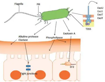
potential to produce an arsenal of virulence factors; some of them are present on the cell surface, such as type IV pili, flagella and type III secretion system (T3SS), while others are secreted, such as extracellular proteases, toxins, phospholipases (Fig 1), (Sadikot et al., 2005). The type IV pili and flagella are mainly involved in P. aeruginosa motility, but they also play a role in the pathogenesis by eliciting an inflammatory response (Kipnis et al., 2006). The type IV pili are the most important adhesins and the retractile properties of these structures make them responsible for the twitching motility, which enables bacterial cells to move on solid surfaces. They also facilitate the swarming motility (Mattick, 2002). Swarming is a special form of motility; it depends on flagella and type IV pili and allows bacteria to move across semi-solid surface. This type of motility is thought to be relevant to the movement of P. aeruginosa through mucus layers because the conditions that trigger swarming (intermediate viscosity and amino acids as a poor nitrogen source) exist in the lung (Overhage et al., 2008). P. aeruginosa is also able to swim in aqueous environments by using the flagellum which is a complex protein structure localized at the pole of the bacterium (Feldman et al., 1998). These motile surface appendances are responsible for bacterial motility and progression towards epithelial contact. The adhesion to epithelial cells mediated by pili and the flagellum represents the first step of colonization (Feldman et al., 1998). The contact with the host cells activates T3SS; it is a major determinant of virulence which allows to directly inject toxins into the host cell, through a syringe-like apparatus (Hauser et al., 1998). The toxins secreted by the P. aeruginosa T3SS are ExoU, ExoS, ExoT, ExoY. These toxins are variably expressed in different strains and all participate in the cytotoxicity. ExoT and ExoS have ADP-ribosyltrasferase activity which causes the destruction of the cytoskeleton of the host cell. ExoU has a phospholipase activity which destroys the eukaryotic cell membranes, while ExoY is an adenylate cyclase which causes the increase of the cAMP in the cytosol leading to increased epithelial barrier permeability in the lung (Hauser et al., 2009). Other virulence factors involved in the invasion and dissemination of P. aeruginosa are secreted through the type II secretion system (T2SS). They are the phospholipase C (PlpC), the exotoxin A (ToxA) and the proteases LasB, LasA, PrpL. The elastases LasA and LasB are not only involved in the damage of the respiratory epithelium through the destruction of the cellular junctions (tight junction) but they also induces an inflammatory response by the host (Azghani, 1996; Kon et al., 1999). The PlpC, also called the hemolytic phospholipase, targets the eukaryotic membrane phospholipids and participates to lung injury and inflammation (Konig et al., 1993). A
major role in P. aeruginosa virulence is played by the ToxA. This toxin inhibits eukaryotic protein synthesis by blocking the elongation factor 2 (EF-2), and induces cell death (Ochsner et al., 1996). ToxA and the protease PrpL are mainly expressed under iron-depleted conditions (Paragraph 2.1). The extracellular protease PrpL (or Protease IV) is an endoprotease which cleaves casein, lactoferrin, transferrin, elastin, and decorin, and was found to contribute to the ability of P. aeruginosa to persist in a rat chronic pulmonary infection model (Wilderman et al., 2001). Another important protease produced by P. aeruginosa during the infections is the alkaline protease AprA. This protease is a zinc metallo-protease released by the type I secretion system. It has been demonstrated that AprA degrades several components of the host immune system such as the cytokines INF-γ TNF-α and the protein C1q e C3 inhibiting the activation of the complement (Hong et al., 1992; Parmely et al., 1990). AprA also cleaves the iron-binding protein transferrin (Laarman et al., 2012). The proteolysis of transferrin causes the release of iron which could be taken up by the bacterium favoring its spreading (Kim et al., 2006). The damage caused by the above-mentioned virulence factors and the resulting strong inflammatory response make the acute infections by P. aeruginosa severe and often lethal (Gellantly et al., 2013)
Fig 1. Virulence factors produced by P. aeruginosa during acute infections. Flagella, pili and
T3SS are present on the cell surface. Pili and flagella represent the mains adhesions involved in the contact with the host epithelial cells. The contact activates the T3SS and consequently the injection of the cytotoxins (ExoU, ExoS, ExoT and ExoY) directly into the host cells . During the lung colonization P. aeruginosa also secretes several virulence factors such as the proteases, phospholipase C and exotoxin A (Modified from Gellantly and Hancock, 2013).
2.2 Chronic infection
CF, COPD or immune compromised patients are generally unable to completely eradicate the acute infections and it could evolves in chronic infection in which P. aeruginosa is completely adapt to the lung environment (Deretic et al., 1995). During the chronic infection P. aeruginosa grows and persists as a biofilm, which is a surface attached community of bacteria encased in a self-produced polymeric matrix (Bjarnsholt et al., 2009). The polymeric matrix consists of polysaccharides, nucleid acids, proteins and lipids and represents 50-90% of the biofilm volume. Biofilm formation is a multistep process. The first contact of bacterial cells with the surface is followed by a strong (irreversible) attachment. After the attachment bacteria grow as microcolonies and begin to produce the extracellular polymeric matrix. The last step relies on the maturation of the biofilm structure, characterized by the formation of mushroom shaped structures and of the fluid filled channels for the exchange of nutrients and waste products (Klausen et al., 2003). The microbial biofilm is a relevant problem in clinical setting, since it allows bacteria to persist in the human body and on medical devices such as catheters and endotracheal tubes. Bacteria growing as biofilm show enhanced tolerance to the host immune response and to the activity of antimicrobial agents. In particular, P. aeruginosa growing as a biofilm has a minimal inhibitory concentration (MIC) and a minimal bacterial concentration (MBC) for many antibiotics 100-1,000 fold higher than planktonic cells (Moskowitz et al., 2004). The increased tolerance of bacteria growing as biofilm likely depends on the limited penetration of the antimicrobial agents through the biofilm matrix and on the reduced metabolic activity of biofilm-forming bacterial cells (Walters et al., 2003; Chiang et al., 2013; Pamp et al., 2008). Many antibiotics target DNA replication, protein synthesis or cell wall biogenesis and are highly active on actively replicating planktonic bacteria; however, since the metabolic activity of bacteria growing as biofilm is much higher in the upper part of biofilm compared to the inner part, biofilm-forming bacterial cells present in the inner part of the biofilm are more tolerant to these classes of antimicrobial agents (Werner et al., 2004; Pump et al., 2008). The ability of P. aeruginosa growing as biofilm to persist on biotic and abiotic surfaces and to resist to antimicrobial agents makes chronic infections difficult to eradicate.
3
P. aeruginosa
iron uptake: evidence of different strategies in
different types of infections
Iron is an essential element in almost all living organisms. Although iron is the one of the most abundant elements on the earth, the concentration of biologically useful iron is generally extremely low. In aerobic inorganic environment, iron is mainly present in the oxidized ferric form Fe(III) which aggregates into insoluble oxy-hydroxide polymers and, consequently, it is not easily available to microorganisms. Once in the human host, pathogens such as P. aeruginosa are forced with the problem of iron acquisition. In human fluids iron is sequestered by proteins with high affinity for Fe(III), such as transferrin and lactoferrin, while in the cells iron is fastened in heme, iron-sulfur clusters and ferritins. The limited availability of iron is addressed by bacteria through the development of different systems able to actively acquire iron under depleted conditions (Ratledge and Dover 2000). P. aeruginosa has evolved different strategies to acquired iron, which are: (i) the production of low-molecular weight compounds which scavenge iron from various sources (siderophores); (ii) the uptake of siderophore and iron chelators which are not synthesized by P. aeruginosa itself, (iii) the uptake of heme molecule from host hemoproteins and (iv) the reduction and/or uptake of Fe(II) (Poole and McKay, 2003).
Except for the last one, these uptake systems require the presence of specific receptors on the outer membrane. These receptors need a complex of three proteins localized in the cytoplasmic membrane (TonB, ExbB and ExbD) called the “TonB complex”. The TonB complex converts the transmembrane proton gradient in energy to internalize the iron carrier (siderophore or hemophore or hemoprotein) into the periplasm. Once in the periplasm the transport across the inner membrane to the cytoplasm is carried out by ABC permeases (Wandersman and Stojiljkovic, 2000). The cytoplasmic concentration of iron is strictly regulated, and an overload of iron is toxic for bacterial cells because the reduced form Fe(II) activates the Fenton reaction generating reactive oxygen species which damage many biological macromolecules. In order to prevent an excessive iron uptake, in P. aeruginosa, as well as in many gram-negative bacteria, the systems involved in iron uptake are gram-negatively regulated by the ferric uptake regulator Fur. Fur is a 15 KDa protein which has been proposed to be essential in P. aeruginosa, since deletion mutants in the fur gene were not obtained in this bacterium (Vasil and Ochsner, 1996). Fur acts as an iron intracellular sensor which represses directly or indirectly the expression of iron uptake genes under iron replete conditions. When Fur is loaded with Fe(II) it forms homodimers that bind the
regulatory elements of its target genes polymerase and hence transcription (Och
that P. aeruginosa could vary the iron uptake s
infection in order to best fulfill its needs without spending too much energy and Dingemans , 2013)
3.1 Siderophores: Pyoverdine a
Pyoverdine is a green fluorescent siderophore which represents the primary iron uptake system of P. aeruginosa
three parts: (i) a fluorescent dihydroxyquinoline chromophore, (ii) an acyl si
bound to amino group of the chromophore and (iii) a variable peptide chain linked by an amino group to the C1 carboxyl
Fig 2. Structure of A) type I pyoverdine and B) pyochelin (Modified from Crosa and Walsh;
2002)
Each P. aeruginosa strain can produce II or III; Bodilis et al., 2009
(Kf≈1024M-1) and a 1:1 stoichiometric ratio (Visca
pyoverdine for Fe(III) makes it capable to scavenge iron from host protein transferrin and lactoferrin,
(Ankenbauer et al., 1985; Sriyosachati and Cox, 1986). The ability to acquire iron is not the only
pyoverdine also acts as a signaling molecule involved in the regulation of virulence genes expression (Fig 3) (Lamont
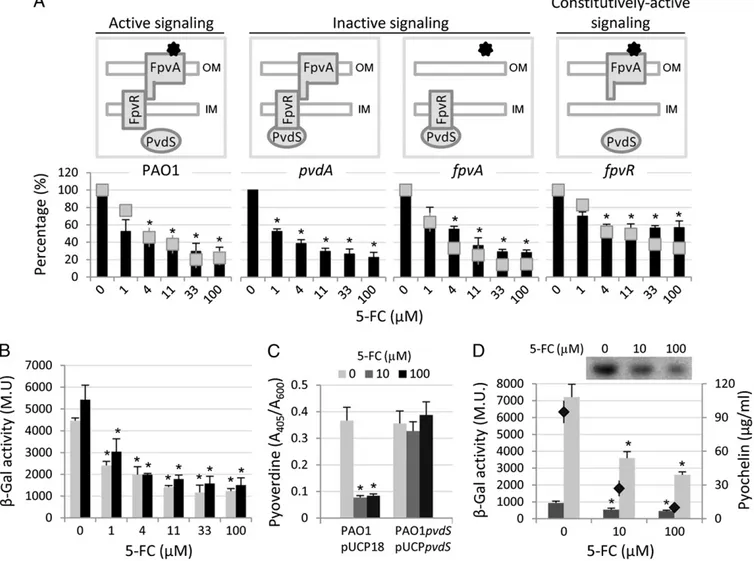
siderophore is called pyoverdine si FpvA, the antisigma factor FpvR single transmembrane helix,
regulatory elements of its target genes and prevent the bind
transcription (Ochsner and Vasil, 1996). It has been
could vary the iron uptake strategies according to the type of in order to best fulfill its needs without spending too much energy
Siderophores: Pyoverdine and Pyochelin
Pyoverdine is a green fluorescent siderophore which represents the primary iron P. aeruginosa (Meyer et al., 2000). This siderophore is composed by three parts: (i) a fluorescent dihydroxyquinoline chromophore, (ii) an acyl si
bound to amino group of the chromophore and (iii) a variable peptide chain linked by an amino group to the C1 carboxyl group of the chromophore (Fig 2
Structure of A) type I pyoverdine and B) pyochelin (Modified from Crosa and Walsh;
can produce one of three different pyoverdine types ., 2009). Pyoverdine is able to chelate Fe(III
) and a 1:1 stoichiometric ratio (Visca et al., 2007). The h makes it capable to scavenge iron from host protein
, allowing bacterial growth also in human serum
., 1985; Sriyosachati and Cox, 1986).
on is not the only property of this siderophore;
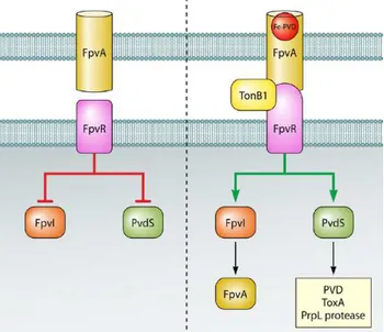
a signaling molecule involved in the regulation of virulence (Lamont et al., 2002). The signaling mediated by this siderophore is called pyoverdine signaling and involves the TonB dependent receptor FpvA, the antisigma factor FpvR, which spans the cytoplasmic membrane through a single transmembrane helix, and the alternative sigma factors PvdS and FpvI. FpvA is prevent the binding of the RNA
It has been proposed according to the type of in order to best fulfill its needs without spending too much energy (Cornelis
Pyoverdine is a green fluorescent siderophore which represents the primary iron ., 2000). This siderophore is composed by three parts: (i) a fluorescent dihydroxyquinoline chromophore, (ii) an acyl side chain bound to amino group of the chromophore and (iii) a variable peptide chain linked by
2).
Structure of A) type I pyoverdine and B) pyochelin (Modified from Crosa and Walsh;
three different pyoverdine types (type I, III) with high affinity 2007). The high affinity of makes it capable to scavenge iron from host proteins such as
allowing bacterial growth also in human serum
of this siderophore; indeed, a signaling molecule involved in the regulation of virulence ., 2002). The signaling mediated by this gnaling and involves the TonB dependent receptor , which spans the cytoplasmic membrane through a and the alternative sigma factors PvdS and FpvI. FpvA is
the pyoverdine outer membrane receptor; it has a beta barrel structure with a globular domain (plug domain) which occludes the interior of the barrel. Once pyoverdine is loaded with iron, it binds FpvA and induces a conformational change of the plug domain that enables the entry of the ferri-siderophore in the periplasm. The binding of ferri-pyoverdine to FpvA also promotes the interaction between FpvA and the periplasmic domain of the antisigma factor FpvR, and this interaction results in the transmission of a signal to the cytoplasmic domain of FpvR. This signal leads to the release of FpvI and PvdS from the FpvR antisigma, ultimately determining the activation of these two sigma factors. The alternative sigma factor FpvI drives the expression of the gene encoding the receptor FpvA, while PvdS controls the expression of almost 30 genes (Ochsner et al., 2002).
Fig 3. Schematic representation of the pyoverdine signaling cascade. In the absence of
pyoverdine, FpvR represses the activity of the alternative sigma factors FpvI and PvdS. The binding of pyoverdine loaded with iron to its outer membrane receptor FpvA induces a signal through FpvR which leads to the release of the sigma factors and consequent activation of FpvI- and PvdS-dependent genes (Jimenez et al., 2012).
Among the genes regulated by PvdS there are genes encoding virulence factors which play a critical role in P. aeruginosa pathogenicity, such as pyoverdine, the protease PrpL and the exotoxin A (Lamont et al., 2002). The prominent role of pyoverdine in pathogenicity has been demonstrated in different mouse model of infections (Meyer et al., 1996; Takase et al., 2000). In particular, in the burned mouse model of infection used by Meyer and colleagues, mice were infected post-burned with 102 CFU (colony forming unit) of wild type strain or mutants unable to produce pyoverdine. The mortality of the mice infected with the wild type strain was almost 100% while the pyoverdine defective mutants appeared fully avirulent since no mice died. The
injection of pyoverdine into the infected mice restored the virulence of the pyoverdine defective mutants and increased the mortality of burned mice (Meyer et al., 1996). The other experimental model in which was tested the virulence of pyoverdine defective mutant is the immunosuppressed mice. The wild type strain or the pyoverdine defective mutant (approximately 106) were inoculated intranasally in
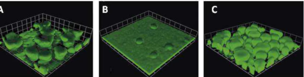
immunosuppressed mice. The virulence of pyoverdine defective mutant appeared to be attenuated because the mutant took somewhat longer to kill the mice then wild type strain (12 hours versus 48 hours post inoculum), (Takase et al., 2000). Given that pyoverdine is involved in the regulation of virulence factors important for the colonization and invasion of human host, it seems to be essential to cause acute infection (Cornelis and Dingemans, 2013). Interestingly, pyoverdine production is also important for biofilm formation which is a typical trait of chronic infection. It has been demonstrated that mutants which do not produce pyoverdine are unable to generate mature biofilms under iron limited conditions, but the addition of exogenous pyoverdine to the growth media restores the biofilm formation ability of pyoverdine defective mutants (Banin et al., 2005; Patriquin et al., 2008) (Fig 4). Although pyoverdine is involved in different biological processes, ranging from host invasion to biofilm formation, its production is extremely energy consuming for P. aeruginosa, given that its biogenesis occurs through non ribosomal peptide synthesis and requires the activity of many different enzymes (Visca et al. 2007).
Fig 4. Biofilm development in flow cell of A) P. aeruginosa PAO1 (wild type), B) an isogenic
mutant in the biosynthetic gene pvdA which is unable to produce pyoverdine, and C) the pvdA mutant in the presence of exogenous pyoverdine The image represent a 6 day biofilm on a glass surface in a flow of 1% of TSB (tryptic soy broth), (modified by Banin et al., 2005) This could plausibly explain why PvdS activity and pyoverdine production are finely regulated by the pyoverdine signaling cascade (see above). Indeed, this regulatory network allows maximum siderophore production only when pyoverdine is effective in catching iron from the environment or from host proteins, given that PvdS activity is fully activated only when iron-loaded pyoverdine binds to the FpvA receptor.
In addition, pvdS expression is directly repressed at the transcriptional level by Fur-Fe(II) and, consequently, the production of pyoverdine is shut-off under iron replete conditions (Ochsner et al., 1996). More recently, other proteins have been described to positively influence pvdS expression, such as the LipA lipase and the transcriptional regulator CysB (Funken et al., 2011; Imperi et al., 2010): LipA is an extracellular lipolytic enzyme, and its absence causes a strong reduction in pvdS expression and pyoverdine production, although the mechanism is still unknown (Funken et al., 2011). CysB is a LysR-type transcriptional regulator which plays a central role in sulfur metabolism. It has been demonstrated that CysB binds to the promoter region of pvdS and positively controls its expression and, consequently, the production of PvdS-dependent virulence factors (Imperi et al., 2010).
The second siderophore produced by P. aeruginosa is pyochelin (Fig 2). Pyochelin is a condensation product of salicylate and two molecules of cysteine; it chelates iron with a 2:1 (pyochelin:iron) stoichiometry (Tseng et al., 2006). The affinity for iron of pyochelin (kf≈ 105M-2) and the number of genes involved in its biosynthesis are lower
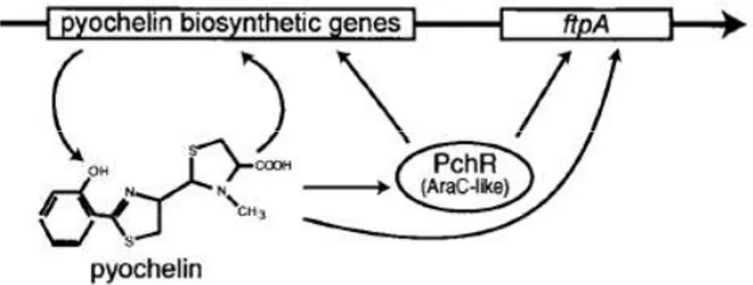
than pyoverdine (Youard et al., 2011). The production of this siderophore is regulated by the cytoplasmic regulator PchR, which is a protein of the AraC/XylS family. These kind of regulators can act as transcriptional repressor or activator depending on the presence of specific effector molecules. In particular, the pyochelin biosynthetic genes are repressed by PchR in the absence of pyochelin, while they are induced when PchR is activated by pyochelin binding (Fig 5) (Michel et al., 2007). Thus, as in the case of pyoverdine, also pyochelin is only produced when it is effective in feeding the cell with iron. Moreover, pyochelin production is also subjected to the repression by Fur, which controls the expression of the genes involved in the synthesis (pchDCBA, pchEFGHI), regulation (pchR) and internalization (fptA) of this siderophore. Recently, it has been demonstrated that P. aeruginosa firstly produces pyochelin and then switches to pyoverdine when iron concentration becomes extremely low (Dumas et al., 2013). Given that the cost associated with the synthesis of pyochelin is much lower compared to that of pyoverdine, it has been proposed that this regulatory strategy could allow P. aeruginosa cells to save energy until the pyoverdine synthesis is absolutely required for growth (Dumas et al., 2013).
Fig 5. Schematic representation of the pyochelin genes regulation. In the presence of
pyochelin, PchR triggers the transcription of the pyochelin biosynthetic genes and of the gene
ftpA, encoding the outer membrane receptor for ferri-pyochelin (Modified from Crosa and
Walsh, 2002)
3.2. Other iron uptake systems
Although endogenous siderophores, in particular pyoverdine, represent the main iron carriers of P. aeruginosa, this bacterium has evolved other strategies to acquire iron in different environments. During the infection, a possible source of iron is represented by the heme and hemoglobin (Wandersman and Stojiljkovic, 2000). Two different systems for heme uptake have been identified and characterized in P. aeruginosa, namely the Phu and Has systems. The Phu system consists of a TonB-dependent outer membrane receptor for heme (PhuR) which binds heme and transport it into the periplasm. Once in the periplasm, the periplasmic protein PhuS and the ATP-dependent permease system PhuTUV direct the transfer of heme into the cytoplasm (Ochsner et al., 2000). The other heme uptake system (Has) reminds the siderophore-dependent iron uptake, since this system is based on the activity of the haemophore HasA, an extracellular protein secreted by the bacterial cell that catches heme with high affinity and delivers it to its TonB-dependent receptor HasR (Letoffé et al., 1998; Takase et al., 2000). Once heme is in the cytoplasm, the heme oxygenase HemO degrades heme to form biliverdin, CO and Fe(II) (Barker et al., 2012).
During the polymicrobial infection, P. aeruginosa could be benefited from the ability to recognize and use siderophores produced by other species (xenosiderophores) or other exogenous iron chelators. For this reason, P. aeruginosa has the genetic potential to encode several TonB-dependent receptors for exogenous iron carriers, such as PfeA and PirA for the E. coli siderophore enterobactin (Dean and Poole, 1993), FoxB and FiuA for the uptake of ferioxamine and ferrichrome, respectively (Llamas et al., 2006), FecA for the Fe-citrate (Marshall et al., 2009), FemA for the utilization of
aerobactin (Cuiv et al., 2006) and FvbA for the uptake of vibriobactin (Elias et al., 2011).
The last iron uptake system used by P. aeruginosa is the Feo system, which internalizes Fe(II). Fe(II) is soluble and is mainly present under anaerobic conditions and/or in the presence of low pH (Andrews et al., 2003). The ferrous iron diffuses through the outer membrane through porins and is transported in the cytoplasm by FeoB. FeoB is a GTP-dependent transporter and it is associated with other two proteins of the inner membrane, FeoA and FeoC (Cartron et al., 2006). It has recently been proposed that the Feo system could play a role in iron uptake during lung chronic infections in CF patients, due to the ability of phenazines, which are secondary metabolites produced by P. aeruginosa, to reduce Fe(III) to Fe(II). Indeed, it has been demonstrated that phenazines and Fe(II) accumulate in the lung of CF patients during chronic P. aeruginosa infection (Hunter et al., 2012; Hunter et al., 2013)
4
The regulatory switch from acute to chronic infection
During chronic and acute infections P. aeruginosa expresses different sets of genes involved respectively in biofilm formation or in virulence factor production. The ability to modulate gene expression on the basis of the type of infection is due to the presence of intricate regulatory networks which can control, by repressing or activating, different sets of genes (Stover et al., 2000). Among the systems involved in the regulation of the switch between the planktonic and biofilm lifestyles, the most studied are the Gac system and the bis-(3’-5’)-cyclic dimeric guanosine monophosphate (c-di-GMP) intracellular signal.
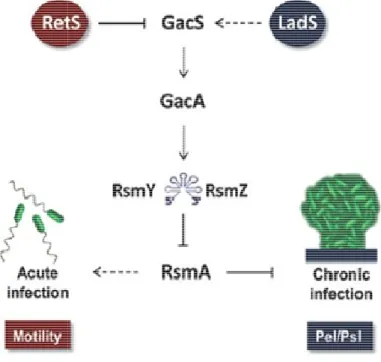
4.1 Gac regulatory network
The Gac regulatory network (global activator of antibiotic and cyanide synthesis) is a two component system typically present in gamma proteobacteria (Fig 6). This system consists of the inner membrane sensor kinase GacS, the response regulator GacA, two small RNAs and the translational repressor RsmA (regulatory of secondary metabolism). In response to a still unknown signal, GacS phosphorylates and activates GacA, which in turn recognizes and binds the conserved Gac sequence (TGTAAGN6CTTACA) present in the rsmZ and rsmY promoter regions, thereby
promoting their transcription (Brencic et al., 2009). These small RNAs are the exclusive targets of the GacA regulator, and are part of an uncharacterized feedback
mechanism by which they inhibit their prediction of these small RNAs
motif in the single strand region of binding of these sRNA to RsmA. translational repressor activity of
small translational regulatory proteins; it binds GGA untranslated region of target mRNA
consequently inhibiting translation (Lapouge been described to modulate the
RetS and LadS, which exert transcription, respectively. While heterodimerizes with GacS, GacA activation, LadS (lost
through a still unknown molecular mechanism effect of RetS on GacA activity is
phenotype very similar to that
Fig 6. Schematic representation of
The effect of rsmA deletion
characterized by several research groups analyses. In particular, rsmA
mechanism by which they inhibit their own transcription (Kay et al., 2006). small RNAs suggests the presence of multiple copies of motif in the single strand region of a steam loop; this motif is responsible
RsmA. RsmZ and/or RsmY binding to RsmA
translational repressor activity of this protein. RsmA is a member of the CsrA family of small translational regulatory proteins; it binds GGA motifs present within the
of target mRNAs, occluding the ribosome binding site and translation (Lapouge et al., 2008). Additional regulators have been described to modulate the activity of the Gac system, namely
which exert a negative and positive effect on
While RetS (regulator of exopolysaccharides an , preventing its autophosphorylation and
ost adherence sensor) promotes the activation of GacA molecular mechanism (Ventre et al., 2006). The inhibitory activity is confirmed by the fact that retS
to that of rsmA mutants (Goodman et al., 2004).
Schematic representation of the Gac system (modified from Moscoso
deletion on P. aeruginosa physiology has been
characterized by several research groups through transcriptomic and phenotypic rsmA mutants show strongly increased
et al., 2006). Structural esence of multiple copies of a GGA is responsible for the to RsmA inhibits the member of the CsrA family of present within the 5’ occluding the ribosome binding site and onal regulators have , namely the hybrid sensors on rsmY and rsmZ xopolysaccharides and T3SS) preventing its autophosphorylation and, consequently, promotes the activation of GacA ., 2006). The inhibitory retS mutants have a ., 2004).
Gac system (modified from Moscoso et al., 2011).
has been extensively through transcriptomic and phenotypic biofilm formation,
reduced T3SS expression and cytotoxicity, and diminished twitching motility. This characteristic phenotype depends on the ability of RsmA to directly inhibit the translation of genes responsible in the biosynthesis of the exopolysaccharide Psl, and to indirectly promote the transcription of the genes involved in the production of the T3SS, type IV pili and flagella (Irie et al., 2010; Brencic and Lory 2009, Burrowes et al., 2006). Moreover transcriptomic analyses of rsmA mutants indicated that RsmA negatively influence the expression of the genes encoding the type VI secretion system and of the pel operon which encodes the enzymes involved in the synthesis of the exopolysaccaride Pel (Brencic and Lory 2009). Transcriptomic analyses also suggested that the Gac system influences the expression of several iron uptake genes, although different studies obtained opposite results (Brencic and Lory 2009; Burrowes et al., 2006). In one study, the deletion of rsmA in the P. aeruginosa reference strain PAO1 caused an increase in the transcription of some iron uptake genes (Burrowes et al., 2006), while in another study, performed on the P. aeruginosa strain PAK, the rsmA deletion resulted in a decrease in iron uptake gene expression (Brencic and Lory 2009). Additional studies are clearly required to clarify the role of the Gac system in the regulation of iron uptake in P. aeruginosa. Irrespective of its effect on iron uptake, the ability of the Gac system to coordinately control the expression of many virulence genes involved in the acute infection and those related to chronic infection makes the Gac system an important factor in P. aeruginosa pathogenicity, as it has been experimentally demonstrated in different models of infection. Indeed, deletion mutants in gacA showed a strongly reduced ability to cause infection in mice, plants, insects and nematodes (Rahme et al., 1995; Tan et al., 1999; Jander et al., 2000, Coleman et al., 2003). Accordingly it has been demonstrated that the deletion of rsmA, hence constitutive activation of Gac system, causes an increase in the ability of P. aeruginosa to persist in the mouse lung (Mulcahy et al., 2007). The enhanced persistence of the P. aeruginosa rsmA mutant could depend on the fact that in this mutant the production of the exopolysaccharides Pel and Psl is not repressed by RsmA, ultimately resulting in increased exopolysaccharide synthesis and biofilm formation compared to the wild type strain. Indeed, in vitro experiments have demonstrated that the deletion of rsmA causes a strong increase in the ability to form biofilms, while the deletion of the two small RNA genes rsmY and rsmZ or of gacA almost completely abolishes biofilm formation (Brencic et al., 2009).
Very recently, it has been proposed that the Gac system could also indirectly influence the transcription of the exopolysaccharide operons pel and psl and, therefore, biofilm
formation in P. aeruginosa by modulating the intracellular levels of the signaling molecule c-di-GMP. In particular it has been demonstrated that deletion of retS, which is the negative regulator of the activation state of Gac system, causes an increase of the intracellular level of c-di-GMP (Moscoso et al., 2011).
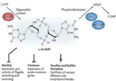
4.2 Bis-(3’-5’)-cyclic dimeric guanosine monophosphate (c-di-GMP)
The signaling molecule c-di-GMP has been identified in different bacteria and seems to take part in the control of the switch between the planktonic and biofilm lifestyles (Jenal et al., 2004). The intracellular levels of this molecule depend on the activity of two classes of enzymes which are the diguanilate cyclases (DGCs) and phospodiesterase (PDEs) (Fig 7).
Fig 7. Structure and functions of c-di-GMP. The intracellular levels of c-di-GMP depend on the
activity of diguanylate cyclases and phosphodiesterases. High levels of c-di-GMP promote biofilm lifestyle while inhibiting virulence gene expression and motility (Modified from Hengge, 2009)
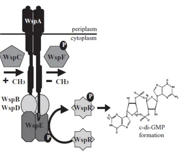
The active DGC is a dimer of two subunits with a GGDEF domain, which is essential for the production of c-di-GMP from two molecules of GTP. The catalytic site is located at the interface between the two subunits. Each subunit binds one molecule of GTP, and point mutations in the GGDEF motif impair the catalytic activity (Malone et al., 2007). The PDE activity is associated with two different motifs: EAL and HD-GYP. The EAL-type PDE is a monomeric enzyme that linearizes c-di-GMP to 5’-pGpG which is then degraded by other cellular enzymes, while the HD-GYP proteins are able to directly degrade c-di-GMP to two molecules of GMP (Christen et al., 2005). Notably, some enzymes have both the domains EAL and GGDEF, although the function of these proteins remains unclear (Kulasakara et al., 2006). In P. aeruginosa, about 41 genes
have been predicted to encode proteins with DGC and/or PDE domains (Kulasakara et al., 2006). The high numbers of genes involved in the metabolism of c-di-GMP makes the signaling network mediated by this molecule difficult to characterize. One of the best characterized protein involved in the synthesis of c-di-GMP is the DGC WspR, belonging to the Wsp chemosensory system. The Wsp system is a multi component chemosensory system with homology to the well characterized chemotaxis pathway. This system consists of a putative methyl-accepting chemotaxis protein (WspA), an histidine kinsase (WspE), two adaptor proteins (WspB and WspD) predicted to link WspA with WspE, a methytransferase (WspC),a methyesterase (WspF) and a response regulator WspR (Fig 8). Likely in response to surface contact, WspA activates the histidine kinase WspE resulting in phosphorylation of WspR receiver domain and DGC activation (Guvener and Harwood, 2007). The methylesterase WspF is a negative regulator of Wsp system. The inactivation of WspF causes an increase in the intracellular levels of c-di-GMP which in turn promote cell aggregation and biofilm formation (Hickman et al., 2005). A typical feature of strains with high intracellular levels of c-di-GMP, as in the case of wspF mutant, is the small colony variant (SCV) phenotype (Hickman et al., 2005). These colonies appear wrinkly and it is due to the very high production of exopolysaccharides, which causes an increase in surface adherence and aggregation. The link between SCVs and intracellular levels of c-di-GMP has been demonstrated by different research groups through the analysis of the effect of mutations in the negative regulators of two different DGCs, WspR and YfiN (Hickman et al., 2005; Malone et al., 2010). The deletion of these inhibitors caused, in both cases, an increase in the intracellular level of c-di-GMP and the concomitant appearance of the SCV phenotype. The SCVs are characterized by enhanced levels of persistence and resistance to many different antimicrobial agents and represent a quite common morphotype of P. aeruginosa isolated from the sputum of chronically infected CF patient (von Götz et al., 2004, Drenkard and Ausubel, 2002; Starkey et al., 2009), suggesting that evolution towards increased c-di-GMP levels occurs during P. aeruginosa colonization of the CF lung.
As the Gac/Rsm system, c-di-GMP regulates biofilm formation and motility in opposite ways. High levels of c-di-GMP cause an increase in exopolysaccharide production and biofilm formation, but, at the same time, they result in the decreased expression of genes encoding the flagellum and the type IV pilus, involved in the swimming, swarming and twitching motilities (Choy et al., 2004; Hickman et al., 2005; Huang et al., 2003).
Fig 8. Schematic representation of Wsp system (modified from Guvener and Harwood, 2007) In the last few years, an increasing number of systems involved in the regulation of intracellular levels of c-di-GMP has been discovered. However, the molecular mechanism by which c-di-GMP controls diverse cellular processes remains largely unknown. This is the case of the psl operon, encoding the enzymes for the synthesis one of the two aggregative exopolysaccharides produced during the biofilm formation; although transcriptomic analysis indicates that c-di-GMP positively controls Psl production, the underlying molecular mechanisms remain to be elucidated (Hickman et al., 2005). Recently, the mechanism by which c-di-GMP exerts its regulatory activity on the production of the Pel exopolysaccharide and flagella has been partly elucidated. The Pel exopolysaccharide is responsible for the pellicle formation and it is the only exopolysaccharide produced by P. aeruginosa strain PA14. It has been demonstrated that c-di-GMP controls the production of Pel at transcriptional and post transcriptional level (Baraquet et al., 2012). The expression of the operon encoding the enzymes involved in the synthesis of Pel is regulated by the transcriptional regulator FleQ. FleQ could act as a repressor or activator of pel operon expression depending on the intracellular levels of c-di-GMP. In the absence of c-di-GMP, two molecules of FleQ bind two different sites into the promoter region of pel operon and prevent RNA polymerase binding. The binding of c-di-GMP to FleQ induces a conformational change in the protein and in the DNA promoter region which promotes the binding of RNA polymerase and consequently transcription of the pel operon. In addition to regulating pel genes for biofilm formation, FleQ has a second, better-known role as master regulator of flagella gene expression. In particular FleQ
activates the expression of genes involved in the regulation and assembly of the flagella export apparatus and basal body (Hickman and Harwood, 2008). FleQ contains an N-terminal domain, a central ATPase domain, and a C-terminal helix-turn-helix DNA-binding domain. In general, ATP hydrolysis, by the ATPase domain, provides energy for loading of the template strand of DNA into the active site of the RNA polymerase (Bose et al., 2008). Baraquet and colleagues have demonstrated that the binding of c-di-GMP inhibits FleQ ATPase activity and consequently causes a reduction in the FleQ ability to activate flagella gene expression (Baraquet and Harwood, 2013).
Thus, high intracellular levels of c-di-GMP promote the expression of pel operon and at the same time repress the expression of flagella genes. This signaling molecule could also be involved in the regulation of biofilm formation at post transcriptional level. Indeed, the production of Pel is also regulated by c-di-GMP during the generation of exopolysaccharide chain (Lee et al., 2007). It has been demonstrated that PelD, encoded by one of the genes within the pel operon, specifically binds c-di-GMP. PelD is predicted to be an inner membrane protein with four transmembrane helices and a large cytosolic region (Whitney et al., 2012). The expression of PelD is required for Pel polysaccharide production and mutations in the c-di-GMP binding site of PelD impair the ability of the P. aeruginosa strain PA14 to produce biofilm (Lee et al., 2007). Although the role of PelD in the synthesis of Pel is still unknown it is thought that it is involved in the transport of the saccharide polymer across the inner membrane (Franklin et al., 2011). The different types of regulation described above are emblematic examples of the versatility of c-di-GMP regulation; this molecule could be considered a transcriptional or post-transcriptional regulator depending on the role of its effector proteins (Hengge et al., 2009).
Recently another regulator has been proposed to play an important role in the modulation of the switch from the planktonic to the biofilm lifestyle: AmpR. This protein belongs to the LysR family of transcriptional regulators and was originally discovered as a regulator of the ampC gene (paragraph 1). AmpR seems to affect both the intracellular levels of c-di-GMP and the activation state of Gac system. In particular, AmpR negatively regulates RsmA activity by upregulating the expression of
ladS, therefore promoting the planktonic lifestyle and acute infection
(Balasubramanian et al., 2014). A proteomic study has demonstrated that AmpR also positively regulates the transcription of three different phosphodiesteres (PA4367,
PA4969 and PA4781) and, as a consequence, it causes a decrease in the intracellular c-di-GMP levels, again promoting the acute infection (Kumari et al., 2014).
5
Aims of the thesis
Pyoverdine plays a key role in P. aeruginosa pathogenicity. It is important for iron uptake and as a signaling molecule which promotes the expression of others virulence factors. The main aims of this PhD thesis are (1) to search for inhibitors of the synthesis or regulation of this siderophore and (2) to clarify the role of the Gac system in pyoverdine. Given the increasing emergence of P. aeruginosa resistant strains, mainly due to the strong selective pressure caused by the use of antibiotics, new therapeutic options are needed. A possible strategy is to disarm bacteria by inhibiting the regulatory networks involved in the regulation of virulence gene expression. To this aim, a drug repurposing approach has been used to search for inhibitors of pyoverdine production. Drug repurposing relies on the search for side activities in old drugs already approved for use in humans, and could represent a fast and cheap strategy to identify new uses for old drugs. The compounds identified with this strategy will then be tested in vitro and in vivo for their anti-virulence activity. Given the crucial role of pyoverdine in the P. aeruginosa infection process, the characterization of the mechanisms which control the synthesis of this siderophore is essential to fully understand P. aeruginosa pathogenicity. Recently, two independent transcriptomic studies on the influence of the Gac system on pyoverdine production provided opposite results. To clarify the actual role of this regulatory network on pyoverdine gene regulation, deletion mutants in specific components of the Gac system has been generated and assessed for pyoverdine production and expression of pyoverdine genes. After having defined the role of the Gac system in pyoverdine regulation, the mechanism(s) by which this system controls pyoverdine production has been investigated, by verifying whether it acts through any of the already known regulators of pyoverdine production or through a still unexplored regulatory mechanism(s).
References
Andrews SC, Robinson AK, Rodríguez-Quiñones F (2003) Bacterial iron homeostasis.
FEMS Microbiol Rev. 27:215-37.
Ankenbauer R, Sriyosachati S, Cox CD (1985) Effects of siderophores on the growth of
Pseudomonas aeruginosa in human serum and transferrin. Infect Immun. 49:132-40.
Arancibia F, Bauer TT, Ewig S, Mensa J, Gonzalez J, Niederman MS, Torres A (2002)
Community-acquired pneumonia due to gram-negative bacteria and Pseudomonas aeruginosa: incidence, risk, and prognosis. Arch Intern Med. 162:1849-58.
Azghani AO (1996) Pseudomonas aeruginosa and epithelial permeability: role of virulence
factors elastase and exotoxin A Am J Respir Cell Mol Biol. 15:132-40.
Balasubramanian D, Kumari H, Jaric M, Fernandez M, Turner KH, Dove SL, Narasimhan G, Lory S, Mathee K (2014) Deep sequencing analyses expands the
Pseudomonas aeruginosa AmpR regulon to include small RNA-mediated regulation of iron
acquisition, heat shock and oxidative stress response. Nucleic Acids Res. 42:979-98.
Banin E, Vasil ML, Greenberg EP (2005) Iron and Pseudomonas aeruginosa biofilm
formation. Proc Natl Acad Sci U S A. 102:11076-81.
Baraquet C, Murakami K, Parsek MR, Harwood CS (2012) The FleQ protein from
Pseudomonas aeruginosa functions as both a repressor and an activator to control gene
expression from the pel operon promoter in response to c-di-GMP. NucleicAcids Res. 40:7207-18.
Baraquet C, Harwood CS (2013) Cyclic diguanosine monophosphate represses bacterial
flagella synthesis by interacting with the Walker A motif of the enhancer-binding protein FleQ. Proc Natl Acad Sci U S A. 110:18478-83.
Barker KD, Barkovits K, Wilks A (2012) Metabolic flux of extracellular heme uptake in
Pseudomonas aeruginosa is driven by the iron-regulated heme oxygenase (HemO). J Biol
Chem. 287:18342-50
Benz R, Hancock RE (1981) Properties of the large ion-permeable pores formed from protein
F of Pseudomonas aeruginosa in lipid bilayer membranes. Biochim Biophys Acta. 646:298-308.
Bjarnsholt T, Jensen PØ, Fiandaca MJ, Pedersen J, Hansen CR, Andersen CB, Pressler T, Givskov M, Høiby N (2009) Pseudomonas aeruginosa biofilms in the respiratory tract of
cystic fibrosis patients. Pediatr Pulmonol. 44:547-58.
Bodilis J, Ghysels B, Osayande J, Matthijs S, Pirnay JP, Denayer S, De Vos D, Cornelis P (2009) Distribution and evolution of ferripyoverdine receptors in Pseudomonas aeruginosa.
Environ Microbiol. 11:2123-35.
Bose D, Joly N, Pape T, Rappas M, Schumacher J, Buck M, Zhang X (2008) Dissecting
the ATP hydrolysis pathway of bacterial enhancer-binding proteins. Biochem Soc Trans. 36:83-8.
Breidenstein EB, de la Fuente-Núñez C, Hancock RE (2011) Pseudomonas aeruginosa:
allroads lead to resistance. Trends Microbiol. 19:419-26.
Brencic A, McFarland KA, McManus HR, Castang S, Mogno I, Dove SL, Lory S (2009)
The GacS/GacA signal transduction system of Pseudomonas aeruginosa acts exclusively through its control over the transcription of the RsmY and RsmZ regulatory small RNAs. Mol
Brencic A, Lory S (2009) Determination of the regulon and identification of novel mRNA
targets of Pseudomonas aeruginosa RsmA. Mol Microbiol.72:612-32.
Burrowes E, Baysse C, Adams C, O'Gara F (2006) Influence of the regulatory protein RsmA
on cellular functions in Pseudomonas aeruginosa PAO1, as revealed by transcriptome analysis. Microbiology. 152:405-18.
Cartron ML, Maddocks S, Gillingham P, Craven CJ, Andrews SC (2006) Feo--transport of
ferrous iron into bacteria. Biometals. 19:143-57.
Chiang WC, Nilsson M, Jensen PØ, Høiby N, Nielsen TE, Givskov M,Tolker-Nielsen T
(2013) Extracellular DNA shields against aminoglycosides in Pseudomonas aeruginosa biofilms. Antimicrob Agents Chemother. 57:2352-61.
Choy WK, Zhou L, Syn CK, Zhang LH, Swarup S (2004) MorA defines a new class of
regulators affecting flagellar development and biofilm formation in diverse Pseudomonas species. J Bacteriol. 186:7221-8.
Christen M, Christen B, Folcher M, Schauerte A, Jenal U (2005) Identification and
characterization of a cyclic di-GMP-specific phosphodiesterase and its allostericcontrol by GTP. J Biol Chem. 280:30829-37.
Coleman FT, Mueschenborn S, Meluleni G, Ray C, Carey VJ, Vargas SO, Cannon CL,Ausubel FM, Pier GB (2003) Hypersusceptibility of cystic fibrosis mice to chronic
Pseudomonas aeruginosa oropharyngeal colonization and lung infection. ProcNatlAcadSci U S
A. 100:1949-54.
Cornelis P, Dingemans J (2013) Pseudomonas aeruginosa adapts its iron uptake strategies
in function of the type of infections. Front Cell Infect Microbiol. 3:75.
Crosa JH, Walsh CT (2002) Genetics and assembly line enzymology of siderophore
biosynthesis in bacteria. Microbiol Mol Biol Rev. 66:223-49.
Cuív PO, Clarke P, O'Connell M (2006) Identification and characterization of an
iron-regulated gene, chtA, required for the utilization of the xenosiderophores aerobactin, rhizobactin 1021 and schizokinen by Pseudomonas aeruginosa. Microbiology. 152:945-54
Dean CR, Poole K (1993) Cloning and characterization of the ferric enterobactin receptor
gene (pfeA) of Pseudomonas aeruginosa. J Bacteriol. 175:317-24.
Deretic V, Schurr MJ, Yu H (1995) Pseudomonas aeruginosa, mucoidy and the chronic
infection phenotype in cystic fibrosis. Trends Microbiol. 3:351-6.
Döring G, Parameswaran IG, Murphy TF (2011) Differential adaptation of
microbialpathogens to airways of patients with cystic fibrosis and chronic obstructive pulmonary disease. FEMS Microbiol Rev. 35:124-46.
Drenkard E, Ausubel FM (2002) Pseudomonas biofilm formation and antibiotic resistance are
linked to phenotypic variation. Nature. 416:740-3.
Dumas Z, Ross-Gillespie A, Kümmerli R (2013) Switching between apparently redundant
iron-uptake mechanisms benefits bacteria in changeable environments. Proc BiolSci.
280:20131055
Elias S, Degtyar E, Banin E (2011) FvbA is required for vibriobactin utilization in