45
Journal of the Medical Society of the Republic of Srpska
Časopis Društva doktora medicine Republike Srpske
Vol. 45 • No 2 • October 2014. Medical Society of the Republic of Srpska
Godina: 45. • Broj 2 • oktobar 2014. Časopis Društva doktora medicine Republike Srpske
ORIGINAL ARTICLE
Comparison Of Abdominal Puncture And Diuretics During Ascites Treatment
Z. MAVIJA, M. MAVIJAORIGINAL ARTICLE
Sentinel Lymph Node Biopsy In Breast Cancer: Validation Study And
Comparison Of Lymphatic Mapping Techniques
A. GUZIJAN, B. BABIĆ, Z. GOJKOVIĆ, R. GAJANIN, J. ĆULUM, D. GRAHOVAC ORIGINAL ARTICLE
Life Quality Of Patients Treated With Fixed And Mobile Dentures
S. GNJATOPROFESSIONAL PAPER
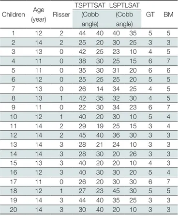
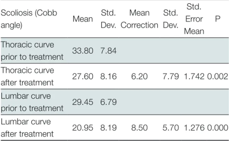
Cheneau Brace In The Treatment Of Idiopathic Scoliosis
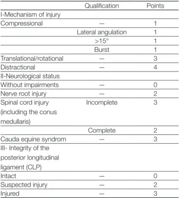
D. DRAGIĆ, Đ. STEVANOVIĆ-PAPIĆ, G. TALIĆ, N. TOMIĆ, V. ŠOLAJA-KOŠČICA PROFESSIONAL PAPERIs The Thoracolumbar Injury Severity Score (TLISS) Still A Good Base For The
Education Of Residents In Orthopaedics And Traumatology
L. MECCARIELLO, S. CARTA, M. FORTINA, M. MARAN, M. MUGNAINI, F. VITALIANO MUZIÌ, P. FERRATA CASE REPORT
Chronic Recurrent Biliary Ascites: An Unusual Scenario
G. KUTTY, S. GUPTA, M. SEKOSANCASE REPORT
Obturator Hernia With Meckel’s Diverticulum In Hernial Sac
I. NOVIĆ, J. MIŠIĆ, P. LAZIĆCASE REPORT
Melkersson-Rosenthal Syndrome
D. TADIĆ, V. ĐAJIĆ, S. MILJKOVIĆ, M. NAZALEVIĆ, LJ. POPOVIĆ CASE REPORT
Ultrasonographic Assessment Of Collateral Cerebral Circulation In Patient With
Internal Carotid Artery Occlusion
Z. VUJKOVIĆ, S. MILJKOVIĆ, V. ĐAJIĆ, D. RAČIĆ, S. MAVIJA, S. DRAGIĆ
EDITORIAL BOARD Editor–in- chief Predrag Grubor Senior Editors Milka Mavija Snježana Milićević Slavica Jandrić
Tamara Kovačević Preradović Siniša Ristić
Members
Vanda Marković Peković Šefik Hasukić Siniša Maksimović Mirza Biščević PUBLISHING COUNCIL Co-Presidents Siniša Miljković, PhD Milan Skrobić, PhD Members Zoran Vujković, PhD Goran Spasojević, PhD Radoslav Gajanin, PhD Nikola Gavrić, PhD Duško Vasić, PhD Milenko Krneta, M.D
International Advisory Board
Milorad Mitković, Niš, Srbija
Marko Bumbaširević, Beograd,Srbija Dragan Micić, Beograd, Srbija Đorđe Radak, Beograd, Srbija Dušan Stefanović, Beograd, Srbija Branislav Antić, Beograd, Srbija Vinka Vukotić, Beograd, Srbija Nebojša Bojanić, Beograd, Srbija Miodrag Ostojić, Beograd, Srbija Bosiljka Vujisić Tešić, Beograd,Srbija Milorad Žikić, Novi Sad, Srbija Branislav Bobić, Novi Sad, Srbija Beatrice Amann Vesti, Cirih, Švicarska Franz Wolfgang Amann, Cirih, Švicarska Luigi Meccariello, Siena, Italija
Web site Editor: Čedomir Radulović Technical secretary : Biljana Radišić English editor: Marina Novković Layout: Dragana Pupac
Design: CGM Design, Banja Luka Publishers: Društvo doktora medicine RS
Medicinski fakultet, Banja Luka
Printed by: Grafix s. p., Banja Luka ISSN 0350-8218
Printig: 1 000
Scripta Medica (Banja Luka)
Journal of the Medical Society of the Republic of Srpska
Copyright © Društvo doktora medicine Republike
48
Scripta MedicaVol. 45 • No 1 • May 2014. • www.scriptamedica.comContent
51 ORIGINAL ARTICLE
Comparison Of Abdominal Puncture And Diuretics
During Ascites Treatment
Komparacija abdominalne punkcije i diuretika
tokom terapije ascitesa
Z. MAVIJA, M. MAVIJA
56 ORIGINAL ARTICLE
Sentinel Lymph Node Biopsy In Breast Cancer:
Validation Study And Comparison Of Lymphatic
Mapping Techniques
Sentinel biopsija limfnog čvora kod karcinoma
dojke: Validaciona studija i komparacija metoda
obeležavanja sentinel čvora
A. GUZIJAN, B. BABIĆ, Z. GOJKOVIĆ, R. GAJANIN, J. ĆULUM, D. GRAHOVAC
62 ORIGINAL ARTICLE
Life Quality Of Patients Treated With Fixed And
Mobile Dentures
Kvalitet života pacijenata saniranih fiksnim i
mobilnim stomatoprotetičkim radovima
S. GNJATO67 PROFESSIONAL PAPER
Cheneau Brace In The Treatment Of Idiopathic
Scoliosis
Cheneau mider u liječenju idiopatskih skolioza
D. DRAGIĆ, Đ. STEVANOVIĆ-PAPIĆ, G. TALIĆ, N. TOMIĆ, V. ŠOLAJA-KOŠČICA73 PROFESSIONAL PAPER
Is The Thoracolumbar Injury Severity Score (TLISS)
Still A Good Base For The Education Of Residents
In Orthopaedics And Traumatology
Da li je Thoracolumbar Injury Severity Score (TLISS)
još uvijek dobra osnova za edukaciju specijalizanata
ortopedije i traumatologije
L. MECCARIELLO, S. CARTA, M. FORTINA, M. MARAN, M. MUGNAINI, F. VITALIANO MUZIÌ, P. FERRATA
78 CASE REPORT
Chronic Recurrent Biliary Ascites: An Unusual
Scenario
Hronični, povratni žučni ascit: neobičan scenario
G. KUTTY, S. GUPTA, M. SEKOSAN81 CASE REPORT
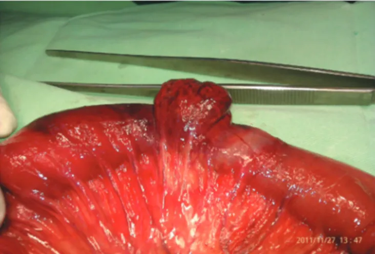
Obturator Hernia With Meckel’s Diverticulum In
Hernial Sac
Obturatorna kila sa Meckelovim divertikulumom
u kilnoj kesi
I. NOVIĆ, J. MIŠIĆ, P. LAZIĆ
83 CASE REPORT
Melkersson-Rosenthal Syndrome
Melkersson-Rosenthal sindrom
D. TADIĆ, V. ĐAJIĆ, S. MILJKOVIĆ, M. NAZALEVIĆ, LJ. POPOVIĆ
87 CASE REPORT
Ultrasonographic Assessment Of Collateral
Cerebral Circulation In Patient With Internal Carotid
Artery Occlusion
Ultrasonografsko praćenje kolateralne moždane
cirkulacije kod pacijenta sa okluzijom unutrašnje
karotidne arterije
Z. VUJKOVIĆ, S. MILJKOVIĆ, V. ĐAJIĆ, D. RAČIĆ, S. MAVIJA, S. DRAGIĆ
91
Many thanks to our present reviewers
Editor’s Letter
Dear Colleagues,
Scripta Medica is a journal of the Medical Society of the Republic of Srpska which aims to become educational magazine available to any doctor in the Republic of Srp-ska, Bosnia and beyond. Scripta Medica is the journal in which you will find ideas for your research and answers to some questions. Therefore, in the next issue, we intend to publish review papers by experts, i.e. eminent specialists in specific fields.
Reports on scientific results provide faster and easier adoption and propagation of scientific ideas, making them available to the general scientific community. Scientific production is expressed through reports about results in periodical scientific journals which, with their regular appearance and circulation, provide the most reliable options of introduction to scientific research and its results. The special mechanism of quotation is used in propagation and adoption of research results, which refers to the implemented research, data source, obtained results, the author / s, etc. The scientific community divides quotations into three groups:
1. hetero-citations. i.e. real quotations - in which authors cite other authors,
2. auto-citations, in which the authors cite their previous work and
3. co - citations - in which authors cite other authors with whom they have written some earlier papers.
The basis for the analysis of the structure of quotation and the determination of appropriate indicators are so-called quotation databases. They collect bibliographic data on periodicals and appended lists of references with the appropriate database. This allows you to find not only publications cited in specific papers in these journals but papers that they have cited as well. Afterwards, the corresponding indicators that are used more widely are based upon them. Quotation of literature is a method of referencing the used documents or publication by using bibliographic elements: author / s, title, name and numeric data of magazines, city, publisher and year of publication. It is mandatory for the data on the quoted unit (reference) to appear twice –firstly in the text, and secondly in the
bibliography. In the text, references are marked with Arabic numbers starting with 1, and bibliography, they are entered in the order in which they appear in the text. Quotation of the scientific and technical papers serves the reader to become familiar with the sources that the author used when writing his papers. It is simple, fast, efficient and totally reliable way to find a magazine of the quoted content for your research. Quotation shows that we are not alone in a scientific research. Fundamental postulate that we must respect in citing implies that there needs to be a clear distinction where do the attitudes of the author himself end, and where the disclosure of other people’s opinions starts. Otherwise, there is a possibility that the author is faced with intellectual dishonesty charges and appropriation of someone else’s copyright work, piracy, plagiarism. etc. Nowadays, it is easy to detect plagiarism with the usage of widely available softwares, such as http:// www.plagtracker.com/, Google and Yahoo search engines. Nonstandard cases all over the world, with magazines written and then erased from the SCI lists, have forced us to think of where to publish our papers. There has been an emersion of the so-called predatory magazines that present a range of tempting offers, with alleged “excellent” impact factor, rapid publication and “24-72 hours” review. All this is available for a “minimum” payment of 900-1500 USD. What is happening to scientific magazines today?
This is the case with magazines with reputable names, very similar or even exactly the same as the famous magazines that actually exist, but solely on the Internet and are not found published anywhere. They don’t have a support of any significant impact factor, certainly not a licensed pub-lisher, and peer review generally does not exist. The prob-lem is that sometimes more publishers publish a magazine under the same name. Thus, for example, Journal of Hy-pertension has seven publishers and often creates confu-sion among authors.
Publishers of “predatory” magazines do not apply review process adequately, and hence, they acquire a huge profit. With the payment of prescribed amount of money being the only criteria for publication, their profitable activity has highly negative impact on the evaluation system of sci-entific research, as in “predatory” magazines the result of scrupulously executed research and compilation of general knowledge or trivial findings are treated in the same man-ner. Therefore, the publication in these kinds of journals can have a very negative impact on the careers of young researchers.
The Board at the University of Belgrade has given a num-ber of recommendations to their teachers, researchers, collaborators, and students, regarding the preparation for publication of the results of their scientific research. They
50
Scripta MedicaVol. 45 • No 2 • October 2014. • www.scriptamedica.comare advised to check the integrity of professional journals in which they plan to publish their paper. The most pre-cise criteria for the recognition of “predatory” magazines and publishers was given by Jeffrey Beall, a librarian at the University of Colorado with a long-term engage-ment in collecting and analyzing data in these journals and publishers. He publishes current information re-garding the subject on his blog: http://scholarlyoa.com. We invite you to write your papers for Scripta Medica fol-lowing the Instructions for authors, and, if needed, the edi-torial board and reviewers will encourage you and assist in making your text fully acceptable for the criteria of Scripta
Medica. On the other hand, you are expected to give us sug-gestions to improve the magazine.
We invite all heads of departments of medical schools and all presidents of the specialists association to give us sugges-tions for an extended list of reviewers for all scientific fields. Scripta Medica is both your and our magazine, and we must do everything to make it even better.
Editor of Scripta Medica Doc. Milka Mavija
Banja Luka, RS, Bosnia and Herzegovina
Contact address:
Zoran Mavija
University Hospital Clinical Centre Banja Luka
Internal medicine clinic 12 beba bb 78000 Banja Luka Email: [email protected] Submitted: October 15th, 2013 Accepted: May 19th, 2014 Introduction
Ascites is considered to be a pathological state where fluid is accumulated in the free abdominal cavity effectuated by various factors. Its constitution may differ depending on the etiology, i.e. the cause of its development. Regardless of its causes, ascites is prevalent world-wide while its incidence marks a significant rise.1,2 Etiology of ascites may be classified into conditions in which peritoneum has not
been directly affected and those in which it has been affected by pathological process. In most cases (90%), causes of ascites are liver cirrhosis, malignoma, congestive cardiac insufficiency and tuberculosis.3 According to available scientific sources, 80% patients with ascites in the USA have liver cirrhosis. Malignant disease effectuates less than 10% of ascites causes. Cardiac insufficiency is responsible for less than 5% ascites cases. Ascites may be classified by
And Diuretics During Ascites
Treatment
ABSTRACT:Introduction
In clinical practice, ascites treatment is, in majority of cases, unsatisfactory and followed by multiple complications. During the therapy, some side effects,in relation to therapeutic method, may occur. The aim of the study was to compare the level of tolerance and effectiveness of ascites therapy in applying abdominal puncture versus diuretics between two groups of patients to establish connection and differences in applied treatments.
Patients and methods. There were 60 patients examined with ascites 3+ and 4+
divided into two equal groups. First group was treated by abdominal puncture several times a week while patients in the other group were administered diuretics either monotherapeutically or in combination. Majority of patients (86.7%) experienced no side effects after applied therapeutic protocol. 6,7% of patients experienced abdominal pain, 3.3% of them had cramps, ailment 1.7% and nausea 1.7% with no statistic difference between two groups of patients (p>0.05). Registered side effects were mild (5%) to moderate (8.3%), while only 1.7% of patients treated by abdominal puncture experienced leaking of ascitic fluid at the puncture site.
Conclusion. No major statistic difference between groups of patients was recorded
in relation to side effects and complications in applied ascites therapy (p>0.05). Abdominal puncture and diuretics were both equally well tolerated in hospital conditions. Potential risk in ascites therapy can be reduced to the smallest possible extent by intensive observation of the patient.
Key words: ascites, abdominal puncture, diuretics, effectiveness of therapy
52
Scripta MedicaVol. 45 • No 2 • October 2014. • www.scriptamedica.comits size using the following system: 1+, detected by careful examination only; 2+, easily detected, but is of relatively small volume; 3+, apparent ascites, while not tense; 4+, tense ascites.
The ascites therapy in clinical practice appears to be unsa tisfactory in most of cases and it is often followed by numerous complications.4,5 The common therapy protocol implies bed rest, sodium uptake restriction as well as diuretics, either individually or in combination thereof. After prolonged application of this medication and poor therapeutic response to it, we choose to apply abdominal puncture. Prolonged use of diuretics implies risk of occu-rrence of hepatorenal syndrome as well as electrolyte imba lance. Nevertheless, uncontrolled abdominal pun-ctu re includes plenty of risks such as infection, re nal insufficiency and encephalopathy. Numerous and diffe-rent problems may arise in ascites therapy. Therefore, controversial attitudes towards comparative advantages and disadvantages of ascites therapy are present.6,7
The objective of this study is to make comparison in clinical conditions between the level of tolerance and effectiveness of ascites therapy through application of abdominal pun-cture against diuretics in two groups of patients, in or der to establish advantages and risks between the applied therapies.
Patients and methods
The clinical tests were performed within the Department of Gastroenterology of the Clinic of Internal Medicine at the University Clinical Center of Banja Luka. The study sample constituted of a group of patients, formed on prospective principle, who were admitted for treatment due to evident ascites in stage 3+ or 4+ accompanied with significant clinical symptoms. 60 patients, who were divided into two identical groups, were tested. The first group of patients (30 patients) was treated with abdominal puncture several times a week up to the point of disappearance of ascites, while the second group (30 patients) was treated with combined application of diuretics.
Immediately upon admission, blood was taken for com-plete blood count; a detailed physical examination was performed, as well as ultrasonography and esophago-ga stro duodenoscopy. Definitive diagnosis of ascites was based on diagnostic abdominal puncture.
The examination protocol was used as the basic metho-dological instrument having provided data necessary for the clinical tests. All subjective discomforts related to the tests were recorded. Therapy tolerability and effect of therapeutic protocol was observed. Statistical data analysis was made through application of SPSS for Windows 15, 0 program (Chi-sqare test, Student’s t-test), while results were presented in tabular and graphical form.
Results
60 patients with ascites were tested. Among the examinees, there were 45 men (75%) and 15 women (25%). The ave-rage age of the patients was 56,6 years. In the group of patients who were treated with abdominal puncture, 22 were men (36, 7%), while in the group of patients treated with diuretics, there were 23 men (38, 3%). In the group of patients who were treated with abdominal puncture, 8 were women (13, 3%), while in the group of patients treated with diuretics, there were 7 women (11, 7%). No statistically significant difference was established between patients with regard to the distinction between the two groups defined by sex (p>0,05). The average age of the patients in the group treated with abdominal puncture was 59, while in the group of patients treated with diuretics, it was 58, 3. Diagnosis of liver cirrhosis was verified as the cause of ascites in almost 88, 3% of cases, while malignant disease was the cause of ascites in 11,7% of cases. In the group of patients treated with abdominal puncture, 24 examinees (40%) had ascites of cirrhosis genesis, while 29 examines (48,4%) from the group treated with diuretics had liver cirrhosis as the primary diagnosis of disease. In the group of patients treated with abdominal puncture, 6 examinees (10 %) had ascites of malignant genesis, while malignant disease was the cause of ascites in 1 examinee (1,7%). Statistically significant difference between the two groups of patients was established in relation to the cause of ascites (p>0,05). The difference is seen in the fact that, in the group of patients treated with diuretics, there was a greater number of examinees with liver cirrhosis as the cause of ascites. Furthermore, in the group of patients treated with abdominal puncture, a greater number of examinees was the one with ascites of malignant genesis.
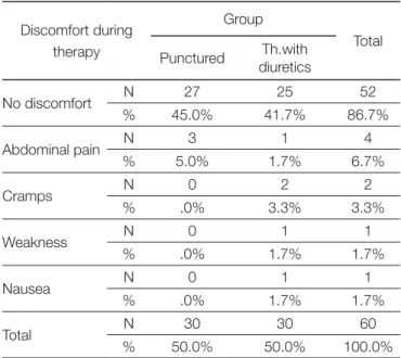
Table 1. Distribution of patients regarding discomfort during therapy.
Discomfort during therapy
Group
Total Punctured diureticsTh.with
No discomfort N 27 25 52 % 45.0% 41.7% 86.7% Abdominal pain N 3 1 4 % 5.0% 1.7% 6.7% Cramps N 0 2 2 % .0% 3.3% 3.3% Weakness N 0 1 1 % .0% 1.7% 1.7% Nausea N 0 1 1 % .0% 1.7% 1.7% Total N 30 30 60 % 50.0% 50.0% 100.0%
The largest percentage of patients underwent the therapy protocol with no discomfort (86,7%), 6,7% patients complained of having abdominal pain, 3,3% patients had leg cramps, weakness was present in 1,7% patients, and 1,7% patients complained of nausea. Table 1. demonstrates no statistically significant difference between groups of patients regarding discomfort arising during the ascites therapy (p>0,05).
The degree of discomfort described above was classified as mild to moderate. Out of the total number of examinees, 5% patients classified their discomfort as mild. Moderate discomfort was present in 8% patients, while leakage at the place of puncture was present in 2% patients. The listed discomfort, related to the applied therapy in groups of examined patients, did not require disruption of therapy or their exclusion from the study. The therapy tolerability was very good in 68% patients, it was good in 29% patients, while 3% showed poor tolerance for the applied ascites therapy. Figure 1. shows no statistically significant difference between the groups of patients (p>0,05).
RAD 02
Figure 1. Patients according to their tolerance of the therapy
The effect of the applied ascites therapy was excellent in 33% patients, good in 52%, while in 15% patients, the effect of ascites therapy was assessed as unsatisfactory. The study demonstrated there was a statistically significant difference (p<0,05) between the groups of patients in relation to the arising therapy effects (Figure 2.).
RAD 02
Figure 2. Patients according to the therapy effects
The global assessment of effectiveness of the applied the-rapy was far better for the group of patients treated with abdominal puncture. 21, 7% patients from the punctured group were assessed to have demonstrated excellent global
effectiveness, which showed significantly larger frequency than in the group of patients treated with diuretics. The groups differed in the category of weak effectiveness as well. More frequently, weak effectiveness of the applied therapy was registered in the group of patients treated with diuretics.
Discussion
Different etiological factors may cause ascites and its inci dence may vary depending on the hospital, probably reflecting different population exposure to many agents that lead to the emergence of ascites. Considerable mis-under standing of the severity of the clinical condition is present, so ascites is still being diagnosed and treated by different criteria.
Out of the total number of patients examined, men were predominantly represented while the average age of the patients was 59. Available data in scientific literature con-firm predominance of male sex and older age in patients with ascites.8
Ascites may be caused by the variety of diseases. However, the study undoubtedly demonstrated predominance of liver cirrhosis as the cause of the disease.9
In 88,3% of the examined cases, ascites was caused by liver cirrhosis. The study undoubtedly demonstrated the liver cirrhosis as the dominant cause for ascites emergence. The results of the research are in accordance with the available literature data.10
Malignant disease is unordinary cause for the ascites emergence, but most of the patients with the ascites related to malignity live only for a few weeks or months after the start of the emersion. In patients with long history of stable cirrhosis and subsequent development of ascites, the probability for the development of hepatocellular carcinoma in the place of liver cirrhosis should be considered. In 11.7% of examinees, the cause of the ascites was malignant disease. Distribution of causes of ascites would be different if the examined patients had been from the Departments of oncology, cardiology or pediatrics.11,12 Ascites represents a clinical challenge that we encounter daily. Selection of the optimal treatment of each patient depends on the circumstances that led to the ascites eme0-rgence. The therapy requires very careful monitoring of pharmacological treatment with special attention to the balance of body fluids. In this study, ascites was tre-ated by repetre-ated abdominal puncture in the first group of examinees, while the second group was treated by a combined application of diuretics (furosemide and spiro-nolactone).
Certain discomforts related with the applied treatment me thod may arise during ascites therapy. This study
54
Scripta MedicaVol. 45 • No 2 • October 2014. • www.scriptamedica.comdemonstrated no grave or undesirable effects of the applied therapy that would require disruption of therapy or exclusion from the study . Therapy tolerability was very good in 68% patients, it was good in 29% patients, while 3% patients showed poor tolerance for the applied ascites therapy. There was no statistically significant difference between the patient groups, which supports the fact that both abdominal puncture as well as diuretic therapy are well tolerated in conditions of hospitalization. However, the study demonstrated that there was statistically significant difference between the groups of patients with regard to the effectiveness of therapy. The difference reflects in the fact that the patients from the group treated with abdominal puncture showed significantly smaller percentage of the unsatisfactory effect in gradation. Furthermore, the group of patients treated with abdominal puncture demonstrated significantly larger percentage of the excellence of the effects of the therapy in gradation.
The study presented the assessment of the effectiveness of the applied therapy as superiorly better for the group of patients treated with abdominal puncture. The results of these clinical tests noted abdominal puncture as a quicker and more efficient therapy method, which was confirmed by other authors as well.13
Conclusion
There was no statistically significant difference between the groups regarding the level of discomfort and complications of the applied therapy. Abdominal paracentesis achieves an optimal clinical response in therapy of patients with ascites of stages 3+ and 4+.
Adequate knowledge of the etiology of ascites, its early diagnostics and capacities for prevention prove to be of particular importance. Observation of therapeutic reco-mmendations and their application in clinical practice facilitate a more uniform position in ascites treatment. The authors of this article have not declared any conflict of interest related to this study.
Reference
1. Runyon BA. AASLD Practice Guidelines Committee.Management of adult patients with ascites due to cirrhosis: an update. Hepatol-ogy 2009;49:2087-107.
2. Moore KP, Wong F, Gines P, et al. The management of ascites in cirrhosis: report on the consensus conference of the International Ascites Club. Hepatology 2003; 38:258-66.
3. Europian Association for the Study of the LIVER. EASL clinical practice guidelines on the management of ascites, spontaneous bacterial peritonitis and hepatorenal syndrome in cirrhosis.J Hepatol 2010;53:397-417.
4. Wong F, Gines P,Watson H, et al. Effects of a selective vasopresin V2 receptor antagonist satavaptan on ascites recurrence after paracentesis in patients with cirrhosis. J Hepatol 2010;53:283-90.
5. Sanyal AJ, Genning C, Reddy KR, et al. The North American Study for the Treatment of Refractory ascites. Gastroenter 2003; 124:634-61.
6. Arroyo V, Colmenero J. Ascites and hepatorenal syndrome in cir-rhosis: Pathophysiological basis of therapy and current manage-ment. J Hepatol 2003; 34.1242-51.
7. Franchis R, Primignani M. Natural hystori of portal hypertension in patients with cirrhosis. Clin Liver Dis 2001; 5:645-63. 8. Luca A, Garcia-Pagan JC, Bosch J, et al. Effects of ethanol
con-sumption on hepatic emodynamics in patients with alcoholic cir-rhosis. Gastroenterology 1997; 112:1284-9.
9. Abraldes JG, Tarantino I, Turnes J, et al. Hemodynamic response to pharmacologic Treatment of portal hypertension and long-term prognosis of cirrhosis. Hepatology 2003; 37:902-8.
10. Chang HC, Sang H, Do Young K, et al. Long-term clinical outcome of large volume paracentesis with intravenous albumin un patients with spontaneous bacterial peritonitis: A randomized prospective study. J Gastroenterol Hepatol 2005;(supl 8): 1215-22.
11. Alessandria C, Marzano A, Todros L, et al. Low vs standard albu-min dosages in the prevention of paracentesis induced circulary disfunction: a randomized pilot study. J Hepatol 2005;42 (suppl 2):75.
12. Gerbas AL ed. Ascites, hyponatriemia and hepatorenal syndrome: progress in treatment. Frontiers of Gastrointestinal Research. Volume 28 Basel; Karger, 2011.
13. Gines P, Guevara M. Hyponatriemia in cirrhosis: pathogenesis, clinical significance and management. Hepatology 2008;48:1002-10.
Komparacija abdominalne punkcije i diuretika tokom
terapije ascitesa
SAŽETAK
Uvod. U kliničkoj praksi terapija ascitesa je u većini slučajeva nezadovoljavajuća i često praćena mnogobrojnim komplikacijama.
Tokom terapije mogu se javiti različiti problemi u vezi sa primijenjenom terapijskom metodom. Cilj rada je bila komparacija abdominalne punkcije i diuretika kod dvije homogene grupe ispitanika, radi utvrđivanja podnošljivosti i efikasnosti primijenjenog odgovarajućeg terapijskog protokola.
Ispitanici i metode. Ispitivano je ukupno 60 pacijenata sa ascitesom u stadijumu 3+ i 4+ , koji su podijeljeni u dvije grupe.
Prva grupa je liječena abdominalnom punkcijom više puta nedeljno, dok je druga grupa ispitanika liječena diureticima bilo monoterapijski ili kombinovano. Najveći broj ispitanika (86,7%) je primijenjeni terapijski protokol podnio bez tegoba. Na bolove u trbuhu se žalilo 6,7% ispitanika, grčeve 3,3%, malaksalost 1,7% i mučninu 1,7% bez statistički značajne razlike između dvije grupe ispitanika (p>0,05). Registrovane tegobe su bile blagog (5%) do umjerenog stepena (8,3%), dok je samo u 1,7% bolesnika iz grupe liječenih abdominalnim punkcijama registrovana komplikacija u vidu curenja ascitesa na mjestu punkcije.
Zaključak. Nema statistički značajne razlike između grupa ispitanika u odnosu na stepen tegoba i komplikacije primjenjene
terapije ascitesa (p>0,05). U hospitalnim uslovima abdominalna punkcija i diuretska terapija se jednako dobro podnose. Eventualne potencijalne opasnosti od terapije ascitesa se mogu smanjiti na najmanju moguću mjeru intenzivnim praćenjem pacijenta.
Aleksandar Guzijan1, Božana Babić2, Zdenka Gojković1, Radoslav Gajanin2, Jovan Ćulum3, Davor Grahovac1
1 Oncology Clinic, University hospital Clinical center Banja Luka,
Banja Luka, Bosnia&Herzegovina 2 Institute of pathology, University hospital Clinical center Banja Luka, Banja Luka, Bosnia&Herzegovina 3 Faculty of Medicine,
University of Banja Luka, Banja Luka, Bosnia&Herzegovina
Contact:
Aleksandar Guzijan
Oncology Clinic – Breast Center University hospital Clinical center Banja Luka
Dvanaest beba 1, Banja Luka Phone: 065/568-989, 051/342-435 E-mail: [email protected]
Submitted: August 23rd, 2014 Accepted: September 22th, 2014
ORIGINAL ARTICLE
Sentinel Lymph Node Biopsy In
Breast Cancer: Validation Study And
Comparison Of Lymphatic Mapping
Techniques
ABSTRACT
Introduction: Sentinel lymph node biopsy is a standard staging procedure in
patients with early breast cancer. Aim of the study is a validation procedure of sentinel lymph node biopsy (SLNB) in our institution and comparison between two mapping techniques - isotope mapping and methylene blue dye for lymphatic mapping.
Material and methods: The study comprised 75 women with breast cancer of
clinical stage T1-2N0M0. We analyzed patients from June, 2010 to March, 2013. In 39 patients, (Group A) lymphatic mapping technique was performed by using the peritumoral injection of 37MBq activity isotope (99m Technetium NANOCIS),and in 36 patients (Group B) mapping technique was performed by using the periareolar injection of 2-4 ml blue dye (Methylene blue 1%). Axillary dissection was performed in both groups after SLNB.
Results: Out of 75 patients, sentinel lymph node was identified in 68 (90.7%).
Identification rate was similar between the groups - 89.7% (Group A), 91.7% (Group B). Accuracy rate was 97% between the groups, that is, Group A 97.1% and Group B 96.9%. In relation to the Group A (90.6%), sensitivity rate was slightly higher in the Group B - 91.6%,. False negative rate of SLNB was higher in the Group A (9.1%) in relation to the Group B (8.3%). The average number of sentinel nodes detected in both groups was 1.2.
Conclusion: The results of the study confirmed and validated both methods
of lymphatic mapping techniques in SLNB. There were no significant statistical differences (p>0.05) in accuracy, sensitivity and false negative rate between these two groups.
Keywords: breast cancer, sentinel lymph node biopsy, isotope, blue dye
DOI: 10.7251/SMD1402052G (Scr Med 2014:45:56-61)
Introduction
On the basis of numerous clinical studies carried out over many years, the procedure of sentinel lymph node biopsy (SLNB) was taken as a valid procedure in diagnosing lymph node axillary metastasis in invasive breast cancer.1-3 In clinical manuals, the following methods of marking, that is, mapping of sentinel lymph node (SLN), are reco-mmended: isotope mapping method, blue dye mapping method and a combined mapping method with blue dye and isotope.4,5 Before SLNB introduction, validation studies were conducted with the aim to determine predictive values of sentinel lymph node in relation to the status of other lymph nodes in the axilla, and, at the same time, to practice surgical teams in the procedure conducting.6-9 Practical importance of SLNB procedure can be seen in significant decreasing of postoperative comorbidity (lymphedema, parenthesis, pain, etc.) in relation to the patients who under went axillary lymphadenectomy.
The study conducted in our institution had an aim to determine procedure validity and compare the methods of SLN blue dye mapping (methylene blue) and isotope (Technetium, 99Tc).
Materials and methods
The study was conducted in Clinical Center Banja Luka in the period from June, 2010 to March, 2013. It was approved by the Ethic Council of Clinical Center Banja Luka. 75 patients who fulfilled the criteria for participating in study were analyzed in this period. Sentinel biopsy procedure was first introduced to the patients and they gave a written consent to participate in the study. The patients fulfilled the following criteria to enter the study: female patients, 30-year-olds and more, histologically verified primary invasive breast cancer, unifocal tumor in a breast, clinical axillary area without dubious palpable lymph nodes,ultrasound check upwithout visualization of dubious infiltrated lymph nodes, clinic stage T1/2NoMo, that in the past, lymph node sentinel biopsy in axillary area was not performed, that axillary area was not treated by rays and/or operated, that a patient was not pregnant, and that a patient previously did not receive neoadjuvant chemotherapy.
The patients with histologically verified invasive breast cancer were separated in two groups: patients in which sentinel lymph node mapping was performed by isotope application (Group A), and in which sentinel lymph node mapping was performed by blue dye (Group B).
In Department of Nuclear Medicine of Clinical Center Banja Luka, radioisotope technetium-nano-sulphur colloid used in sentinel lymph node mapping procedure was obtained from pharmaceutics nano-sulphur colloid (Cis Bio international Paris, France) and technetium pertechnetate
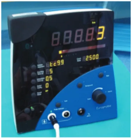
(Tc99m). Preoperatively, in the case of a palpable tumor, radioisotope was applied subcutaneously peritumoral. In a case of a non-palpable tumor, radioisotope was applied periareolar, usually four puncture spots in the amount of 0.2ml per puncture spot in a dose of 0.25mCi (9.25MBq), that is in total, 0.8ml in radioactivity dose 1mCi (37MBq). The procedure was conducted 1 to 4 hours prior to an operation. Sentinel lymph node was intra-operatively detected by mobile gamma camera. Mobile gamma camera “Europrobe” (Lyon, France) was used for detection. While detecting, one or more sentinel lymph nodes were identified.
Figure 1. Mobile „gamma“ probe
All identified sentinel nodes were analyzed histopatho-logically intra-operatively (“ex tempore“) on frozen se-ctio ns, and afterwards, on permanent paraffin molds, de pending on size and number of samples, by standard method of dyeing hematoxylin-eosine (HE).
During the operation, mobile gamma camera detected a place of greatest radiation, and after extirpation, radiation level of an extracted sentinel node was detected on a counter. As a proper parameter, a radiation detection 10 times bigger in relation to surrounding tissue was taken.2,3 After performed sentinel lymph node biopsy, axillary lymphadenectomy was performed in all patients in the group. Total number of analyzed patients in Group A was 39 women.
In the other group (Group B), sentinel lymph node ma-pping was performed by method of dyeing with blue dye (Methylene blue 1%), which is in our conditions available because of its economic acceptability. Methylene blue has smaller molecular weight in relation to patent blue and isosulphan blue. Immediately after giving anesthetic to the patient, blue dye (1% Methylene blue) was applied subcutaneously periareolary to a breast quadrant where a tumor was localized in amount of 2 to 4ml. After blue dye application, a gentle massage of a breast was done for 2-3 minutes in order to stimulate lymph drainage. In interval
58
Scripta MedicaVol. 45 • No 2 • October 2014. • www.scriptamedica.comfrom 15 to 20 minutes from dye application, incision and exploration of axillary area and visual identification of sentinel lymph node were performed. After identification and extirpation of sentinel lymph node, axillary lympha-denectomy was performed in this group of patients as well. 36 women were analyzed in Group B.
Figure 2. Sentinel lymph node mapped by blue dye
χ2 test, Fisher’s exact test, Yates correction for continuity, Mann-Whitney’s Test and Kolmogorov–Smirnov test were used in the analysis. Analytical statistical tool SPSS – version 20 was used in statistical data processing.
Results
In statistical analysis of the patients, the following cha-racteristics of examined groups were processed: age, tu-mor size, histological grade of a tutu-mor, histological type of a tumor, lymph-vascular and perineural invasion of a tumor, immune-histochemical determined hormone status of a tumor and expression of HER2 gene. In the examined groups, model of an applied surgical treatment was analyzed.
Iin the attached statistical data analysis, except for the type of operation, there is no proved statistically significant difference between examined Groups A and B, according to their general characteristics.
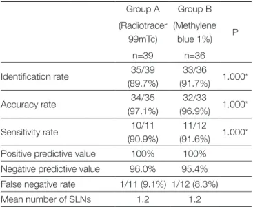
In data related to sentinel biopsy, the following are pro-cessed: identification rate, accuracy rate, sentinel biopsy sensitivity rate, false negative results rate, average number of extracted sentinel lymph nodes and presence of micro-metastasis in a sentinel lymph node and their correlation with other lymph nodes in dissection of axillary lymphatics. In Group A, where mapping was performed by isotope, identification rate in the study was 89.7%, while in group B, where mapping was performed by blue dye, the rate was 91.7 %. In Group A, accuracy rate was 97.1% and in group B 96.9%. Our results showed 90.9% sensitivity rate in Group A and 91.6% in Group B. False negative results
rate in Group A was 9.1% and in group B 8.3%. Statistically significant difference between examined groups was not determined by using Fisher’s test (p=1.000).
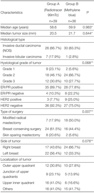
Table 1. Characteristics of examined patients and tumors
Characteristics Group A (Radiotracer 99mTc) n=39 Group B (Methylene blue) n=36 P
Median age (years) 58.6 59.9 0.983* Median tumor size (mm) 20.5 21.7 0.644* Histological type
Invasive ductal carcinoma
(NOS) 26 (66.7%) 30 (83.3%) Invasive lobular carcinoma 7 (17.9%) 1 (2.8%)
Hystological grade of tumor 0.068** Grade 1 9 (23.1%) 2 (5.6%) Grade 2 18 (46.1%) 24 (66.7%) Grade 3 12 (30.8%) 10 (27.7%) ER/PR positive 35 (89.7%) 28 (77.8%) ER/PR negative 4 (10.3%) 8 (22.2%) HER2 positive 3 (7.7%) 9 (25.0%) HER2 negative 36 (92.3%) 27 (75.0%) Type of surgery 0.007** Modified radical mastectomy 7 (17.9%) 18 (50.0%) Breast conserving surgery 24 (61.5%) 16 (44.4%) Skin sparing mastectomy 8 (20.6%) 2 (5.6%)
Side of tumor 0.076** Right breast 17 (43.6%) 24 (66.7%)
Left breast 22 (56.4%) 12 (33.3%) Localization of tumor
Outer upper quadrant 12 (30.8%) 10 (27.8%) Junction of upper
quadrants 9 (23.1%) 5 (13.9%) Upper inner quadrant 16 (41.0%) 6 (16.6%) Others 16 (41.0%) 15 (41.7%) * - Mann Whitney test, **- c-square test
Table 2. Sentinel biopsy results Group A (Radiotracer 99mTc) n=39 Group B (Methylene blue 1%) n=36 P Identification rate 35/39 (89.7%) 33/36 (91.7%) 1.000* Accuracy rate 34/35 (97.1%) 32/33 (96.9%) 1.000* Sensitivity rate 10/11 (90.9%) 11/12 (91.6%) 1.000* Positive predictive value 100% 100%
Negative predictive value 96.0% 95.4% False negative rate 1/11 (9.1%) 1/12 (8.3%) Mean number of SLNs 1.2 1.2 * - Fisher’s exact test
Discussion
So far, many conducted comparative studies showed that a percentage of sentinel lymph node identification by usage of double method was bigger in relation to mapping using only blue dye or isotope. In comparison of sentinel lymph node mapping methods with blue dye and mapping with isotope, there was no statistically significant difference related to accuracy rate, sensitivity rate and false negative results rate of a sentinel node. 10-12 Dilemma whether to use just one reagent or both in the sentinel biopsy procedure is still without consensus and for now, there are only recommendations.
Because of its acceptable price, methylene blue is a dye which is available for all the health institutions. In the case of dye validity, methylene blue dye in a sentinel biopsy procedure, in relation to the other two (patent blue, isosulphane), previously conducted studies did not confirm superiority of patent blue and isosulphane in relation to methylene blue dye. The results of these studies showed that methylene blue dye is valid in sentinel biopsy procedure. There was no statistically significant difference in efficiency of sentinel biopsy procedure in relation to the method of dyeing with methylene and isosulphane or patent blue dye.13-15 Disadvantage of the studies dealing with methylene blue dye used in sentinel biopsy procedure was in a number of analyzed patients. Those were usually single institution studies involving a relatively small number of patients. Vital methylene blue dye related to patent and isosulphane dyes has certain advantages, such as lower price and low allergy potential, but it also has some disadvantages which reflect in lower resorption in lymph flow, in relation to the other two dyes. In our study, we applied methylene blue dye in 36 patients. In all examined patients who underwent sentinel biopsy procedure with methylene blue dye, we did
not record any kind of allergic reactions. As for side effects, only mention temporary pigmentation of skin in the place where dye was injected and temporary urine discoloration is worth mentioning.
Nowadays, there is still no clear consensus and attitude about optimal spot for application of isotope and blue dye. There are two basic localizations for marker application. One technique is to apply isotope or blue dye in the tumor area, that is peritumoral, and the other one is based on applying the marker in the area of areolar complex, that is periareolar. The other dilemma concerns the depth of marker application; one option recommends superficial, subcutaneous application, and the other one recommends deeper, that is parenchyma marker application.
British study (The New Start) and French study (FRAN-SENODE) note that optimal spot for dye application is periareolar localization towards a breast quadrant of the tumor location, so as subcutaneous dye injection.7,16 We practiced methylene blue dye application in periareolar area subcutaneously towards a breast quadrant where the tumor change was localized. This type of application is also more practical at non-palpable lesions.
In more extensive studies, identification rate was from 80% - 99%.1-3,6 On the basis of the results of the studies done so far, in most cases ,double method of sentinel node mapping has the biggest identification rate. Validity and success of SLNB procedure is valued on the basis of accuracy rate. SLNB accuracy rate in more extensive studies was from 95-99%.2,3,9 Sensitivity rate in study NSABP B-32 in mapping SLN by blue dye was 87.8%, while isotope mapping was more successful with 92.2%.3 In some studies, sensitivity was greater in patients who underwent SLN mapping by blue dye.17 Many studies indicated bigger sensitivity rate in SLN mapping by double method.2,9 One of the first validation studies done at the area of former Yugoslavia showed blue dye sensitivity rate at 82% and double method at 95%.18 SLN false negative results rate in the studies varied from 2-22%.19-21 In the studies involving greater number of examined patients, false negative results rate was from 6.7%, 8.2% to 11.4%.9,22,23 Results of validation study conducted in our institution pass all criteria set by most of training programs and recommendations of oncology associations dealing with introduction of SLNB procedure in standard use.
In present micro-metastasis in SLN, without any official confirmation in the form of clinical manuals, the attitude which is becoming more and more common is not doing the axillary lymphadenectomy. On the basis of obtained results of the study IBCSG 23-01 there was no statistically significant difference in five years period without disease (DFS, Disease free survival) between a group of patients who underwent lymphadenectomy and a group of patients who did not undergo lymphadenectomy after verified
60
Scripta MedicaVol. 45 • No 2 • October 2014. • www.scriptamedica.commetastasis in SLN.24 In our study, micrometastatic deposits in SLN were verified in four patients, and observing the state of other lymph nodes in lymphadenectomy preparation, there were no verified metastatic deposits.
Recent studies (ACOSOG Z0011 trial) have given the results which are still discussed by experts, and they are related to an attitude that axillary lymphadenectomy in early invasive breast cancer should not be done, not even if macro-metastasis of breast cancer are histologically verified in sentinel lymph node.25 In such cases, axillary area would undergo radiotherapy. The results of the American study Z0011 showed that there were no statistically significant difference between examined groups of patients regarding the overall survival (OS, Overall Survival) and a period without disease (DFS) in women who underwent axillary lymphadenectomy and those who underwent radiotherapy of axillary area after histologically verified macro-metastasis in SLN.25 A study of European Oncology Institute in Milan (SOUND trial) went even further, stating that in the case of early invasive breast cancer with clinically negative axillary area, nothing more than an observation should be done.26 An attitude of experts about this is not unanimous.27 German, Austrian and Swiss Senologic Society still withhold their attitude towards this subject.28 Their recommendation is to avoid lymphadenectomy in positive SLN only in groups of patient with a low risk for disease return and clinically and ultrasonographicly negative axillary area.
Reference
1. Veronesi U, Viale G, Path FRC, Paganelli G, Zurrida S, Luini A, et all. Sentinel lymph node biopsy in breast cancer: ten-year results of a randomized controlled study. Ann Surg 2010; 251(4): 595-600.
2. Mansel RE, Fallowfield L, Kissin M, Goyal A, Newcombe RG, Dixon JM et al. Randomized multicenter trial of sentinel lymph node biopsy versus standard axillary treatment in operable breast cancer: the ALMANAC Trial. J Natl Cancer Inst 2006; 98: 599-609
3. Krag DN, Anderson SJ, Julian TB, Brown AM, Harlow SP, Ashika-ga T, et al. Technical outcomes of sentinel lymph node resection and conventional axillary lymph node dissection in patients with clinically node negative breast cancer: results from the NSABP B-32 randomized phase III trial. Lancet Oncol 2007;8(10): 881-888
4. Early and locally advanced breast cancer overview [Internet]. Manchester (UK): National institute for health and care excel-lence. 2012; [cited2013Aug30]. Available from: http://pathways. nice.org.uk/pathways/early-and-locally-advanced-breast-cancer 5. Lyman GH, Giuliano AE, Somerfield MR, Benson AB, Bodurka
DC, Burstein HJ, et al. American society of clinical oncology guideline recommendations for sentinel lymph node biopsy in early-stage breast cancer. J Clin Oncol 2005;23(30): 7703-7720 6. Krag D, Weaver D, Ashikaga T, Moffat F, Klimberg SV, Shriver C,
et al. The sentinel node in breast cancer – a multicenter validation study. N Engl J Med 1998;339:941-946
7. Keshtgar M, Aresti N, MacNeil F. Establishing axillary sentinel lymph node biopsy (SLNB) for early breast cancer in the United Kingdom: A survey of the national training program. Eur J Surg Oncol 2010;36:393-398
8. Friis E, Galatius H, Garne JP. Organized nation-wide implemen-tation of sentinel lymph node biopsy in Denmark. Acta Oncologica 2008;47: 556-560
9. Kuehn T, Vogl FD, Helms G, Pueckler SV, Schirrmeister H, Strue-ber R, et al. Sentinel node biopsy for axillary staging in breast can-cer: results from a large prospective German multi-institutional trial. Eur J Surg Oncol 2004;30:252-259
10. Hung WK, Chan CM, Ying M, Chong SF, Mak KL, Yip AWC. Ran-domized clinical trial comparing blue dye with combined dye and isotope for sentinel lymph node biopsy in breast cancer. Br J Surg 2005;92:1494-1497
11. Varghese P, Mostafa A, Abdel-Rahman AT, Akberali S, Gattuso J, Canizales A et al. Methylene blue dye versus combined dye-radio-active tracer technique for sentinel lymph node biopsy localization in early breast cancer. Eur J Surg Oncol 2007;33:147-152 12. Derossis AM, Fey J, Yeung H, Yeh SD, Heerdt AS, Petrek J et al. A
trend analysis of the relative value of blue dye and isotope local-ization in 2.000 consecutive cases of sentinel lymph node biopsy for breast cancer. J Am Coll Surg 2001;193:473-478
13. Eldrageely K, Vargas MP, Khalkhali I, Venegas R, Burla M, Gon-zalez KD, et al. Sentinel lymph node mapping of breast cancer: a case control study of methylene blue tracer compared to isosulfan blue. The american surgeon 2004;70(10):872-875
14. Kavallaris A, Camara O, Runnebaum IB. Subareolar blue dye only injection sentinel lymph node biopsy could reduce the numbers of standard axillary lymph node dissection in environments without access to nuclear medicine. J Cancer Res Clin Oncol 2008;134:667-672
15. Chintamani, Tandon M, Mishra A, Agarwal U, Saxena S. Sentinel lymph node biopsy using dye alone method is reliable and accu-rate even after neo-adjuvant chemotherapy in locally advanced breast cancer – a prospective study. World J Surg Oncol 2011;9:19 16. Rodier JF, Velten M, Wilt M, Martel P, Ferron G, Vaini-Elies V,
et al. Prospective multicentric randomized study comparing peri-areolar and peritumoral injection of radiotracer and blue dye for the detection of sentinel lymph node in breast sparing procedures: FRANSENODE trial. J Clin Oncol 2007;25:3664-3669
17. Meyer-Rochow GY, Martin RC, Harman RC. Sentinel node biopsy in breast cancer: validation study and comparison of blue dye alone with triple modality localization. ANZ J Surg 2003;73:815-818
18. Radovanovic Z, Golubovic A, Plzak A, Stojiljkovic B, Radovanovic D. Blue dye versus combined blue dye – radioactive tracer tech-nique in detection of sentinel lymph node in breast cancer. Eur J Surg Oncol 2004;30:913-917
19. Chua B, Olivotto IA, Donald JC, Hayashi AH, Doris PJ, Turner LJ, et al. Outcomes of sentinel biopsy for breast cancer in British Columbia 1996-2001. Am J Surg 2003;185(2):118-126
20. Cox CE, Bass SS, McCann CR, Ku NN, Berman C, Durand K, et al. Lymphatic mapping and sentinel lymph node biopsy in patients with breast cancer. Annu Rev Med 2000;51:525-542
21. Nano MT, Kollias J, Farchid G, Gill PG, Bochner M. Clinical impact of false-negative sentinel node biopsy in primary breast cancer. Br J Surg 2002;89(11):1430-1434
22. Harlow SP, Krag DN, Julian TB, Ashikaga T, Weaver DL, Feld-man SA, et al. Prerandomization surgical training for the National Surgical Adjuvant Breast and Bowel Project (NSABP) B-32 trial: a randomized phase III clinical trial to compare sentinel node resec-tion to convenresec-tional axillary dissecresec-tion in clinically node negative breast cancer. Ann Surg 2005;241(1):48-54
23. Mansel R, MacNeill F, Horgan K, Goyal A, Britten A, Keshtgar M, et al. Results of a national training programme in sentinel lymph node biopsy for breast cancer. Br J Surg 2013;100(5):654-661 24. Galimberti V, Cole BF, Zurrida S, Viale G, Luini A, Veronesi P,
et al. Axillary dissection versus no axillary dissection in patients with sentinel-node micrometastases (IBSCG 23-01): a phase 3 randomised controlled trial. Lancet Oncol 2013;14:297-305
25. Giuliano AE, McCall LM, Beitsch PD, Whitworth PW, Morrow M, Blumencranz PW, et al. ACOSOG Z0011: A randomized trial of axillary node dissection in women with clinical T1-2 N0 M0 breast cancer who have a positive sentinel node. J Clin Oncol 2010; 28: 18
26. Gentilini O, Veronesi U. Abandoning sentinel lymph node biopsy in early breast cancer? A new trial in progress at the European Institute of Oncology of Milan (SOUND: Sentinel node vs Obser-vation after axillary UltraSouND) The Breast 2012; 21(5): 678-681 27. Kumar A, Puri R, Gadgil PV, Jatoi I. Sentinel lymph node biopsy
in primary breast cancer: window to management of the axilla. World J Surg 2012;36(7):1453-1459
28. The Consensus Panel. German, Austrian and Swiss consensus conference on the diagnosis and local treatment of the axilla in breast cancer. Eur J Cancer 2013;49(10):2277-2283
Sentinel biopsija limfnog čvora kod karcinoma dojke:
Validaciona studija i komparacija metoda obeležavanja
sentinel čvora
SAŽETAK
Cilj: Sentinel biopsija limfnog čvora je standard u tretmanu pacijenata oboljelih od ranog invazivnog karcinoma dojke. Cilj
istraživanja je standardizacija procedure sentinel biopsije u našoj ustanovi i poređenje metode obilježavanja sentinel limfnog čvora između tehnike obilježavanja radiofarmakom i tehnike obilježavanja tkivnom bojom.
Materijal i metode: U istraživanju je učestvovalo 75 žena oboljelih od karcinoma dojke sa kliničkim stadijumom T1/2N0M0.
Ispitanice su analizirane u periodu od juna 2010. godine do marta 2013. godine. Kod 39 ispitanica (Grupa A), za obilježavanje sentinel čvora korišćen je radiofarmak Tehnecijum aplikovan peritumorski u aktivnosti od 37MBq. Kod 36 ispitanica (Grupa B), za obeležavanje je korišćena 1% tkivna boja metilen plavo koja je aplikovana periareolarno u volumenu od 2-4ml. Disekcija aksilarnih limfatika sprovedena je kod svih pacijentkinja nakon procedure sentinel biopsije.
Rezultati: Od ukupno 75 analiziranih ispitanica, sentinel limfni čvor je identifikovan kod njih 68 (90,7%). Stopa identifikacije bila
je slična između poređenih grupa - u grupi A iznosila je 89,7% , a u grupi B 91,7%. Stopa preciznosti iznosila je 97%, između poređenih grupa 97,1% (Grupa A) i 96,9% (Grupa B). Stopa senzitivnosti je bila nešto veća u grupi B (91,6%) u odnosu na grupu A (90,9%). Stopa lažno negativnih nalaza sentinel limfnog čvora bila je veća u grupi A (9,1%) u odnosu na grupu B (8,3%). Prosečan broj izvađenih sentinel čvorova iznosio je 1,2.
Zaključak: Rezultati istraživanja potvrdili su validnost obe metode obilježavanja kao i samu proceduru sentinel biopsije. Između
poređenih grupa nije bilo značajne statističke razlike (p>0,05) u odnosu na stopu identifikacije, preciznosti, senzitivnosti i lažno negativnih nalaza.
UDK 616.31-053.9
Introduction
One of the most important objectives of an oral health care is helping patients in the effort to reach a high level of satisfaction with their mouth cavity with or without teeth.1,2 The research of oral health and its impact on patients’ life quality has largely advanced in late 20th and early 21st century.3,4 In this regard, only few studies have proved interdependence among the psychological profile, oral status, denture treatments, and a patient’s quality of life. Thereby, the aim was to estimate specific forms of social
functions, behavior, and effects of patient’s oral health satisfaction, all of which are expressed via aesthetics, performance, and functions.5
Strauss and Hunt 6 inferred that dental disease and consequences of oral treatments affected patients’ ability to live and enjoy life, make social connections, be positive about their jobs, and stay positive. Oral health satisfaction can be measured by using different indicators such as taste, pain, speech, aesthetics, etc. All these affect the quality of life.7 Sanja Gnjato1 Faculty of Medicine, University of Banjaluka Contact: Sanja Gnjato
University of Banja Luka Faculty of Medicine, Save Mrkalja 14 78000 Banja Luka tel. +38751 234 113 e-mail: [email protected] Submitted: September 2nd, 2014 Accepted: October 23rd, 2014 ORIGINAL ARTICLE
Life Quality Of Patients Treated With
Fixed And Mobile Dentures
ABSTRACT
Introduction. One of the most important objectives of an oral health care is helping
patients in the effort to reach a high level of satisfaction with their mouth cavity with or without teeth.
Patients and Methods. The paper aims to establish the connection between the
satisfaction of patients treated with fixed and mobile dentures on one side, and oral health, quality of life and psychological characteristics of patients on the other. The research was conducted as a prospective study with 320 patients separated into three groups – group I (patients treated with fixed dentures), group II (patients treated with mobile dentures), and group III (patients treated with both mobile and fixed dentures). The oral related life quality was observed within five dimensions: anamneses data, symptoms of ill-functioning of stomatogenic system (chewing and speech), dental status – extraoral, dental status – intraoral and dental abilities.
Conclusion. The linear regression model was adopted for the purpose of analysis of
target markers and oral health determination. The research showed that oral-related quality of life in patients treated with fixed and mobile dentures was most affected by intraoral clinical features, psychological characteristics, and the type of denture, along with a whole range of minor factors (age, timing of denture treatment after the teeth loss, period of denture usage, order of denture implantation, mouth cavity hygiene, diet).
Key words: quality of oral health, dental abilities, oral health features
As he studied the effects of oral health on denture-treated patients’ daily performances, Wostmann8 concluded that the quality of life greatly improves upon the denture tre a tments. The improvement referred to appearance, functions, comfort, pain relief, and eating disorder. World Health Organization initialized these issues within the context of development policy in 1980s.9
Our target study resulted in an original model for the estimation of oral-related quality of life. The research covered five dimensions of dental health quality: anamneses data, symptoms of ill-functioning stomatogenic system (chewing and speech), dental status – extraoral, dental status – intraoral and dental abilities. Eventually, it confirmed our initial hypothesis on the interdependence between the quality of life on one side and psychological profile and type of denture on the other.
Having tested the model of oral-related quality of life on patients treated with fixed and mobile dentures under the clinical conditions, we assumed that the improvement of patients’ life quality should be managed in a responsible and institutional manner.
Methods and materials
The research was conducted at the Institute of stomatology in Banja Luka and it covered 360 patients singled out in three groups: group I (patients treated with fixed dentures), group II (patients treated with mobile dentures), and group III (patients treated with both mobile and fixed dentures). Each group had the identical number of patients and equal age and sex structure. All denture treatments were performed in order to establish complete mastication and other oral functions. For the purpose of the research, a special dental record was designed and the data were entered for each individual patient. The dental record included the following information: the general information with 10 markers, anamneses data with 16 markers, symptoms of ill-functioning oral system (chewing and speech) with 6 markers, clinical examination – extraoral dental status with 8 markers, clinical examination – intraoral status with 12 markers, additional clinical procedures with 5 markers, scanning of TM joints with 3 markers and a questionnaire of dental abilities with the total of 36 markers grouped in five dimensions (appearance, pain, comfort, satisfaction, and eating limitations). Each questionnaire provided three entry options: positive, neutral, and negative.
Patients had been observed for 17 months ranging from May 2012 to September 2013. The clinical dental status, dental abilities, and psychological status of each patient were estimated three times - at the beginning of the research, six months afterwards, and at the end.
The statistical methods of the research were as
follows10: statistical methodology applied at the research
result display covered four research phases – descriptive statistics, statistical analysis, inferential statistics, and the research deduction and decision making. Statistical methods were applied via relevant statistical standards (ISO 10576-1: Statistical methods – Guidelines for the evaluation of conformity with specified requirements – Part 1: General principles; ISO TS 21747: Statistical methods – Process performance and capability for measured quality characteristics; ISO 11453: Statistical interpretation of data – Tests and confidence intervals relating to proportions; ASQ Z1.4: Sampling procedures and tables for inspection by attributes) and the standard software was used (Microsoft Office: Excel 2010, www.microsoft.com/Office365).
Modeling of oral-related quality of life: Dental health
quality was observed through five dimensions - anamneses data, symptoms of ill-functioning stomatogenic system (chewing and speech), dental status – extra-oral, dental status – intraoral, and dental abilities. The quality of oral health was modeled as the linear regressive model with hierarchical weight coefficient, assuming that the adequate regressive residues were equal to zero. Thus, the index of dental health quality was presented by using the general regression equation: DS DS DSI DSI DSE DSE S S A A KDZ K *I K *I K *I K I K *I I
Slika 1. Distribucija pacijenata po kvalitetu dentalnog zdravlja u odnosu na dimenziju dentalnih sposobnosti „Izgled”
Slika 2. Distribucija pacijenata po kvalitetu dentalnog zdravlja u odnosu na dimenziju dentalnih sposobnosti “Bol”
in which IKDZ is the dependent variable, i.e. the index (score) of dental health quality; KA is regression coefficient, i.e. the hierarchical weight coefficient of the dental health quality dimension referred to as “anamneses“, IA is the independent variable, i.e. the index (score) of the dental health quality dimension referred to as “anamneses“, KS is the regression coefficient, i.e. the hierarchical weight coefficient of the dental health quality dimension referred to as “symptoms“, IS is the independent variable, i.e. the index (score) of the dental health quality dimension referred to as “symptoms“, KDSE is the regression coefficient, i.e. the hierarchical weight coefficient of the dental health quality dimension referred to as “dental status extra-oral”, IDSE is the independent variable, i.e. the index (score) of the dental health quality dimension referred to as “dental status extra-oral”, KDSI is the regression coefficient, i.e. the hierarchical weight coefficient of the dental health quality dimension referred to as “dental status intraoral”, IDSI is the independent variable, i.e. the index (score) of the dental health quality dimension referred to as “dental status intraoral”, KDS is the regression coefficient, i.e. the hierarchical weight coefficient of the dental health quality dimension referred to as “dental abilities“ and IDS is the independent variable, i.e. the index (score) of the dental health quality dimension referred to as “dental abilities“. In order to calculate the oral health quality (index), for each observed dimension and element within it, the following was set: relevance rank – hierarchical weight coefficient
64
Scripta MedicaVol. 45 • No 2 • October 2014. • www.scriptamedica.comand contribution (positive – the more the better, negative – the less the worse).
Results
The clinical research determined the dental health quality (index) with the chosen sample of 360 patients treated with fixed and mobile dentures. The result was that the oral-related life quality can be divided into five attributive categories depending on the total number of index score obtained by calculating index of dental health quality for each patient: excellent (score<38,0), good (score 38,0-51,0),satisfactory (score 51,1-65,0), unsatisfactory (score 65,1-79,0) and poor (score (>79,0).
The effect of individual dimensions of dental abilities (as a psychological category) was observed separately. We shall display the research results showing impact of five dimensions of dental abilities (appearance, pa in, conformity, satisfaction, eating limitations). Each dimen-sion of dental abilities was divided into five attributive categories as shown in pictures.
Picture 1. Distribution of patients and their oral-related life quality from the aspect of the dental ability dimension referred to as “appearance”
Picture 2. Distribution of patients and their oral-related life quality from the aspect of the dental ability dimension referred to as “pain”
Picture 1. Distribution of patients and their oral-related life quality from the aspect of the dental ability dimension referred to as “appearance”
Picture 1. displays the effect of the dental ability dimension termed “appearance” on the patients’ oral-related quality of life, i.e. it shows how patients’ satisfaction with appearance, color, and teeth distribution affect their quality of life. The aesthetic appearance of patients is very important, and it provides the patients with confidence along with the adequate therapy, teeth settings, color choice, and shape and size of the teeth. Testing proved that there was a statistically relevant difference between oral-related quality of life and the dental ability dimension referred to as “appearance” (p=0, 0029).
Picture 1. Distribution of patients and their oral-related life quality from the aspect of the dental ability dimension referred to as “appearance”
Picture 2. Distribution of patients and their oral-related life quality from the aspect of the dental ability dimension referred to as “pain”
Picture 2. Distribution of patients and their oral-related life quality from the aspect of the dental ability dimension referred to as “pain”
Picture 2. displays the effect of the dental ability dimension referred to as “pain” on the patients’ oral-related quality of life, i.e. it shows how patients’ type of pain, intensity of pain (small, moderate, strong, very strong),pain duration, spontaneous or provoked pain due to physical-chemical agents, hot food, bruxism, trauma, diet, and inflammation affect their quality of life. Testing proved that there was a statistically relevant difference between oral-related quality of life and the dental ability dimension referred to as “pain” (p=0, 0000).
2
Picture 3. Distribution of patients and their oral-related life quality from the aspect of the dental ability dimension referred to as “conformity”
Picture 4. Distribution of patients and their oral-related life quality from the aspect of the dental ability dimension referred to as “satisfaction”
Picture 3. Distribution of patients and their oral-related life quality from the aspect of the dental ability dimension referred to as “conformity”
Picture 3. displays the effect of the dental ability dimension referred to as “conformity” on the patients’ oral-related quality of life, i.e. it shows how patient’s oral-related life quality is affected by teeth and gum issues. The lack of teeth, tooth migration towards empty space within gums, change of tooth position, etc. result in IKP ratio disturbance. Patients are not satisfied with the existing condition within their mouth cavity as it results in no “conformity” while eating and drinking. Testing proved