Letter
©2018 Dustri-Verlag Dr. K. Feistle ISSN 0722-5091 DOI 10.5414/NP301093 e-pub: February 16, 2018
LETTERS TO THE EDITOR
Calf muscle hypertrophy
following S1 radiculopathy:
A stress disorder caused by
hyperactivity with variable
response to treatment
Nila Volpi1,2, Federica Ginanneschi1,2,
Alfonso Cerase1,3, Salvatore Francesco
Carbone4, Margherita Aglianò1,
Paola Lorenzoni1, Matteo Bellini3,
Sabina Bartalini2, Giovanni Di Pietro5,
and Alessandro Rossi1,2
1Department of Medical, Surgical, and
Neurological Sciences, University of Siena, 2Unit of Neurology and Clinical
Neurophysiology, 3Unit of Neuroradiology, 4Unit of Diagnostic Imaging, and 5Unit of Neurosurgery, University
Hospital Santa Maria alle Scotte, Siena, Italy
Sir, – Neurogenic focal muscle hyper-trophy (NMH) is a rare event in different instances of impaired nerve conduction: ac-quired and inherited polyneuropathies/mul-tineuropathies [1, 2], post-polio syndromes [3], spinal stenosis, spinal muscle atrophies, radiculopathies [4]. Unilateral calf hypertro-phy is an uncommon, but not unique, finding in S1 radiculopathy [5]. Muscle histology shows denervation, with atrophic as well as hypertrophic fibers, and frequent coexistence of inflammatory changes. Nevertheless, clin-ical presentation and responsiveness to treat-ment are not uniform. We report a 65-year-old man, who presented with a 2-month history of painful progressive enlargement of the right calf, which started 5 months af-ter an acute lumbago, causing pain of poste-rior right thigh, leg, and heel. The right calf was markedly enlarged (7.5 cm right > left), tense, and sensitive to touch/pressure, with no skin/subcutaneous changes, no involve-ment of the regional lymph nodes (Figure 1). Neurological examination detected weak-ness of right foot plantar flexion (3/5 MRC scale) and absence of right ankle jerk reflex; the patient complained of tingling
paresthe-sias in the right foot and continuous calf pain. Routine blood tests, including sedimentation rate, C-reactive protein, eosinophil count, thyroid hormones, liver enzymes, and cry-ocrit were within normal ranges. ANA-ENA screening and search for neurotrophic vi-ruses were negative. Serum creatine kinase (CK) was increased (520 UI/L).
Needle electromyography (EMG) showed marked abnormalities consisting of continu-ous complex repetitive discharges (CRDs), polyphasic motor unit potentials with reduced voluntary recruitment in the right gastroc-nemius muscle. EMG recordings of right tibialis anterior, quadriceps femoris, gluteus maximum, and adductor magnus muscles were unremarkable.
Magnetic resonance imaging (MRI) (Fig-ure 1) showed a volume increase of right leg posterior muscles, more evident on soleus and medial gastrocnemius, with intrafascial edema and mild hypervascularization of hy-pertrophic muscles.
Lumbosacral spinal MRI and computed tomography (Figure 1) evidenced diffuse de-generative disease of the lumbar spine and a L5-S1 right paramedian posterior disc herni-ation compressing the ipsilateral S1 radicular sheath and S1 nerve root.
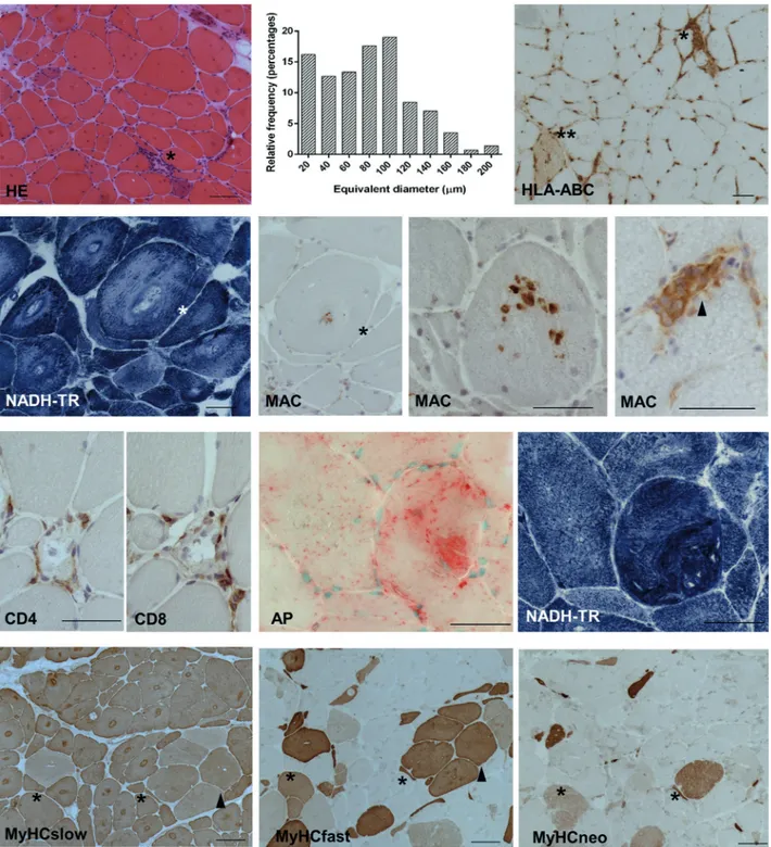
A biopsy of the medial right gastroc-nemius muscle (Figure 2) showed grossly hypertrophic fibers, grouped atrophy, and sparse myonecroses. Target lesions indicat-ed denervation in most hypertrophic fibers. Inflammation consisted of scattered foci of mononuclear cells surrounding necrotic fi-bers. MHC-I (HLA-ABC) neolocalization was restricted to necrotic/regenerating fibers. Focal increase of acid phosphatase (AP) ac-tivity and cytoplasmic patchy deposits of complement terminal complex (membrane attack complex: MAC) were detected in the largest fibers with myofibrillar disarray. Immunohistochemistry for myosin heavy chains (MyHC) showed reactivity of all fi-bers for the slow isoform, with a milder stain in fibers coexpressing fast myosin, resulting in transitional hybrid fibers (~ 40%). Recur-rent regenerating fibers coexpressed neona-tal/fast/slow myosin. By morphometric analysis, the frequency peak of fiber equiva-lent diameters was observed at ~ 100 µm, with recurrent elements being larger than 200 µm.
Steroid treatment was carried out with prednisone 50 mg/day for 3 weeks, with no clinical remission. Consequently, the patient was submitted to CT-guided percutaneous transforaminal periganglionic, and
trans-articular zygapophyseal infiltration of the right L5-S1 root with a mix of local anes-thetic (Ropivacain, 2 mg) and corticosteroid (Triamcinolon, 40 mg), with considerable clinical remission: shortly after the
proce-Figure 1. Top left: grossly hypertrophic right calf. Top right: lumbar spine neuroimaging. T1- (A) and T2-weighted (B) sagittal, T2-T2-weighted consecutive axial (C) MRI, and axial CT (D): disc and facet joints degen-erative disease at right side of L5-S1 level, including zygapophyseal osteophytes (white closed arrow) and paramedian disc herniation (open arrows) in the right S1 lateral recess, compressing ipsilateral S1 root (black closed arrow). Bottom: MRI of the legs. T1-weighted image (A): volume increase of soleus and medial gas-trocnemius muscles (white arrows). Fat-suppressed T2-weighted image: edema, with interfascial component (arrows). No fat involution was detectable by T1/T2 images. Regular popliteal artery run-off excluded vascu-lar impairment (C). Steady-state post-contrast T1-weighted image (D): muscle masses showed homoge-neous enhancement (*) because of increased interstitial spaces due to inflammatory components.
Figure 2. Medial right gastrocnemius muscle: biopsy findings. Hematoxylin-eosin: myonecrosis with mononuclear invasion (*); atrophic/hypertrophic fibers, internalized nuclei; target lesions as hypereosino-philic areas. Histogram of fiber equivalent diameter: high percentage of diameters > 100 µm; > 15% small atrophic fibers. MHC-I (HLA-ABC) IP: only necrotic (*) and sparse degenerating/regenerating (**) fibers show neolocalization. NADH-TR: target lesions as areas devoid of enzymatic activity with peripheral hy-perreactive rim and coarse architectural changes. MAC IP: patchy cytoplasmic spots on target fibers (suc-cessive sections*), contrasting with uniform cytoplasmic stain in necrotic fibers invaded by mononuclear cells (arrowhead). CD4 – CD8 IP on successive sections: only necrotic fibers present with infiltrates. Acid phosphatase hyperactivity in a grossly hypertrophic fiber, with severe disruption of myofibrillar organiza-tion, as shown in successive section stained for NADH-TR. Serial MyHCs IP: all fibers express slow MyHC, target lesions show a hyperreactive rim; variable intensity of MyHC slow stain in grouped fast fibers (arrowhead). Regenerating fibers (*) coexpress neonatal, slow, and fast isoforms. (NADH-TR = reduced nicotinamide adenine dinucleotide-tetrazolium reductase; AP = acid phosphatase; IP = immunoperoxi-dase; scale bar: 50 µm).
dure, pain and muscle tenderness decreased and within 4 weeks a 2.5-cm reduction of the right calf circumference was discern-ible. Nevertheless, 2 months later, pain and swelling recurred so that surgical discectomy was performed. Three months later, the right calf circumference, although still hypertro-phied, was 3.5 cm less. Clinical examina-tion showed full recovery of the right plantar flexion (MRC: 5), a sharp decrease of the foot paresthesia, and a reduction of the calf tension. EMG demonstrated a reduction of CRDs with improved muscular recruitment.
Coexisting inflammatory changes have led to inclusion of NMH into the domain of focal myositis (FM) [5, 6], a benign pseudo-tumor lesion within a single skeletal muscle, predominant at the lower limbs, possibly re-current or evolving into diffuse myositis [7]. Unlike the present case, histology of pure FM usually shows no neurogenic changes and a prominent inflammation [8, 9].
In most calf NMH cases, increased CK plasma levels and focal inflammation likely represent a reactive response to degeneration remodeling of denervated fibers [5]. Corre-spondingly, MHC-I upregulation and T-cell deposits were extremely limited in our pa-tient’s gastrocnemius muscle, in contrast to the usual pattern of myositides. It is notewor-thy that the pathophysiology of NMH does not seem to be univocal. In most radiculop-athy-associated NMH cases, CRDs, gener-ated by denervgener-ated fibers, are likely to act as hypertrophying agents, in analogy with increased muscle mass and fiber diameters obtained by functional muscle electrical stimulation, both experimentally [10] and in conus/cauda syndrome patients [11]. None-theless, NMH reports in absence of CRDs [5, 12] suggest coexisting pathomechanisms, such as compensatory overload; according-ly, in our case, a minority of hypertrophied “non-target” fibers, thus possibly not dener-vated, were detected.
The unusual cytoplasmic multifocal de-posits of MAC on MHC-I-negative, hyper-trophic fibers, might therefore depend on the double stress of denervation and hyperactiv-ity, with subsequent increase of intracellu-lar proteases and degenerative events, also consistent with focal AP reactivity. In our patient, steroid unresponsiveness was con-gruous with paucity of muscle inflammatory
changes. However, the varying response to steroids does not strictly correlate with in-flammation, neither in FM [9] nor in radic-ulopathy-associated NMH [6], suggesting, besides a direct action on radicular compres-sion, a steroid activity on the suppression of the membrane excitability.
Genetic factors or selective axon vul-nerability have been suggested to be re-sponsible for the uncommon occurrence of large “atrophy-resistant” fibers in muscle of subjects with spinal cord injuries [13]; yet, unrecognized motoneuron/muscle molecular features might also explain the low preva-lence of NMH associated with the relatively frequent S1 radiculopathy.
A transitional “hybrid” fiber phenotype, coexpressing different isoforms of myosin and other contractile proteins, has been re-ported in denervated muscle, with a shift to fast type 2 fibers [14], whereas a fast-to-slow shift occurs following electric stimulation or excessive exercise [15]. Our finding of an ongoing major transition to the slow phe-notype suggests that histotype shift, from continuous muscle activity, might cause the reported type 1 predominance in gastrocne-mius NMH [3]. Serial MyHC immunohis-tology for analysis of histotype transition was not, to our best knowledge, previously performed in NMH. Comparison of this fea-ture in CRD+ and CRD– cases of neurogenic muscle hypertrophy would be of interest.
In our patient, with conspicuous repeti-tive discharges causing type shift and de-generative muscle changes, only surgery led to stable improvement. The case calls for attention to the relevance of a thorough in-strumental and pathological investigation in the uncommon occurrence of NMH, with the aim of establishing the most effective thera-peutic strategy.
Funding
None.
Conflict of interest
References
[1] Pareyson D, Morandi L, Scaioli V, Marazzi R,
Boiardi A, Sghirlanzoni A. Neurogenic muscle
hypertrophy. Report of two cases. J Neurol. 1989;
236: 292-295. CrossRef PubMed
[2] Uncini A, Di Muzio A, Chiavaroli F, Gambi D,
Sabatelli M, Archidiacono N, Antonacci R, Mar-zella R, Rocchi M. Hereditary motor and sensory
neuropathy with calf hypertrophy is associated with 17p11.2 duplication. Ann Neurol. 1994; 35: 552-558. CrossRef PubMed
[3] Gutmann L. AAEM minimonograph #46: neuro-genic muscle hypertrophy. Muscle Nerve. 1996;
19: 811-818. CrossRef PubMed
[4] Rowin J, Meriggioli MN. Complex repetitive dis-charges: cause or effect of neurogenic muscle hy-pertrophy? Muscle Nerve. 1999; 22: 1603-1606.
CrossRef PubMed
[5] Lunde HM, Skeie GO, Bertelsen AK, Karlsen B,
Miletic H, Lindal S, Brautaset NJ, Bindoff LA.
Fo-cal myositis – neurogenic phenomenon? Neuro-muscul Disord. 2012; 22: 350-354. CrossRef PubMed
[6] Hemmi S, Shirakawa S, Kurokawa K, Sunada Y. Unilateral calf hypertrophy and focal myositis in-duced by S1 radiculopathy: dramatic response to steroid treatment. BMJ Case Rep. 2013; 2013: bcr2013200870. CrossRef PubMed
[7] Flaisler F, Blin D, Asencio G, Lopez FM, Combe
B. Focal myositis: a localized form of
polymyosi-tis? J Rheumatol. 1993; 20: 1414-1416. PubMed
[8] Auerbach A, Fanburg-Smith JC, Wang G, Rushing
EJ. Focal myositis: a clinicopathologic study of
115 cases of an intramuscular mass-like reactive process. Am J Surg Pathol. 2009; 33: 1016-1024.
CrossRef PubMed
[9] Toti P, Romano L, Villanova M, Zazzi M, Luzi P. Focal myositis: a polymerase chain reaction anal-ysis for a viral etiology. Hum Pathol. 1997; 28: 111-113. CrossRef PubMed
[10] Pette D, Vrbová G. The Contribution of Neuro-muscular Stimulation in Elucidating Muscle Plas-ticity Revisited. Eur J Transl Myol. 2017; 27: 6368. CrossRef PubMed
[11] Kern H, Rossini K, Carraro U, Mayr W,
Vogelau-er M, Hoellwarth U, HofVogelau-er C. Muscle biopsies
show that FES of denervated muscles reverses hu-man muscle degeneration from perhu-manent spinal motoneuron lesion. J Rehabil Res Dev. 2005; 42
(3 Suppl. 1): 43-53. PubMed
[12] Streichenberger N, Meyronet D, Fiere V, Pellissier
JF, Petiot P. Focal myositis associated with S-1
radiculopathy: report of two cases. Muscle Nerve. 2004; 29: 443-446. CrossRef PubMed
[13] Biral D, Kern H, Adami N, Boncompagni S, Protasi
F, Carraro U. Atrophy-resistant fibers in
perma-nent peripheral denervation of human skeletal muscle. Neurol Res. 2008; 30: 137-144. CrossRef PubMed
[14] Burnham R, Martin T, Stein R, Bell G, MacLean I,
Steadward R. Skeletal muscle fibre type
transfor-mation following spinal cord injury. Spinal Cord. 1997; 35: 86-91. CrossRef PubMed
[15] Gondin J, Brocca L, Bellinzona E, D’Antona G,
Maffiuletti NA, Miotti D, Pellegrino MA, Botti-nelli R. Neuromuscular electrical stimulation
training induces atypical adaptations of the
hu-man skeletal muscle phenotype: a functional and proteomic analysis. J Appl Physiol (1985). 2011;
110: 433-450. CrossRef PubMed
Correspondence to Nila Volpi, MD Department of Medical
Surgical and Neurological Sciences University of Siena
Viale Bracci 16 53100 Siena, Italy [email protected]