Open Access
Research article
Identification of a human immunodominant B-cell epitope within
the immunoglobulin A1 protease of Streptococcus pneumoniae
Francesca De Paolis
1, Elisa Beghetto
1, Andrea Spadoni
1,
Francesca Montagnani
2, Franco Felici
3, Marco R Oggioni
2and
Nicola Gargano*
1Address: 1Kenton laboratories, Kenton Srl, Rome, Italy, 2Department of Molecular Biology, University of Siena, Italy and 3Department of
Microbiology, Genetics and Molecular Biology, University of Messina, Italy
Email: Francesca De Paolis - [email protected]; Elisa Beghetto - [email protected];
Andrea Spadoni - [email protected]; Francesca Montagnani - [email protected]; Franco Felici - [email protected]; Marco R Oggioni - [email protected]; Nicola Gargano* - [email protected]
* Corresponding author
Abstract
Background: The IgA1 protease of Streptococcus pneumoniae is a proteolytic enzyme that
specifically cleaves the hinge regions of human IgA1, which dominates most mucosal surfaces and is the major IgA isotype in serum. This protease is expressed in all of the known pneumococcal strains and plays a major role in pathogen's resistance to the host immune response. The present work was focused at identifying the immunodominant regions of pneumococcal IgA1 protease recognized by the human antibody response.
Results: An antigenic sequence corresponding to amino acids 420–457 (epiA) of the iga gene
product was identified by screening a pneumococcal phage display library with patients' sera. The epiA peptide is conserved in all pneumococci and in two out of three S. mitis strains, while it is not present in other oral streptococci so far sequenced. This epitope was specifically recognized by antibodies present in sera from 90% of healthy adults, thus representing an important target of the humoral response to S. pneumoniae and S. mitis infection. Moreover, sera from 68% of children less than 4 years old reacted with the epiA peptide, indicating that the human immune response against streptococcal antigens occurs during childhood.
Conclusion: The broad and specific recognition of the epiA polypeptide by human sera
demonstrate that the pneumococcal IgA1 protease contains an immunodominant B-cell epitope. The use of phage display libraries to identify microbe or disease-specific antigens recognized by human sera is a valuable approach to epitope discovery.
Background
Streptococcus pneumoniae is a human pathogen causing
sig-nificant morbidity and mortality worldwide. It is a tran-sient member of the normal bacterial flora that colonizes
the upper respiratory tract of the host being a major cause of various diseases such as otitis media, pneumonia, sep-sis and meningitis. Despite the constant development of therapeutics, antimicrobial drugs and vaccines, pneumo-Published: 18 December 2007
BMC Microbiology 2007, 7:113 doi:10.1186/1471-2180-7-113
Received: 19 July 2007 Accepted: 18 December 2007
This article is available from: http://www.biomedcentral.com/1471-2180/7/113 © 2007 De Paolis et al; licensee BioMed Central Ltd.
This is an Open Access article distributed under the terms of the Creative Commons Attribution License (http://creativecommons.org/licenses/by/2.0), which permits unrestricted use, distribution, and reproduction in any medium, provided the original work is properly cited.
coccal infection still causes severe diseases in young chil-dren, elderly people and immunocompromised individuals [1,2]. In adults, pneumococcal infection is the most common cause of community-acquired pneumonia and otitis media and, since the introduction of vaccina-tion against Haemophilus influenzae (serotype b) and
Neis-seria meningitidis also the most frequent cause of
meningitis.
Current immunization strategies focus on the use of S.
pneumoniae polysaccharides-based vaccines, employing
the 23-valent vaccine, which protects humans from two years of age, and the 7-valent toxoid-conjugated vaccine, used in children less than two years old [3,4]. However, the available vaccines have several limitations due to the low immunogenicity of capsular polysaccharides, the high serotype variability and the genomic plasticity of this bacterium. Therefore, in the last decade there has been a great effort in developing improved vaccines to prevent pneumococcal disease.
Several surface-associated proteins, which are well con-served among the different strains of S. pneumoniae and thus represent candidates of choice for the development of novel vaccine formulations have been identified and characterized. Among this class of proteins the immu-noglobulin A1 (IgA1) protease is a promising candidate since (i) it plays a major role in pathogen's resistance to the host immune response [5,6], and (ii) it is present in all pneumococcal strains and serotypes [7,8]. The impor-tance of IgA protease is underlined by the fact that this host-specific enzyme is conserved in other pathogens of comparable disease and colonising similar niches [9-11].
The IgA1 protease is one of the two to four large zinc met-alloproteinase present in the pneumococcal genome [7,12]. The pneumococcal protease is a polypeptide of about 1900 amino acids associated to the bacterium via N-terminal anchoring [7,13-15]. It is a proteolytic enzyme that specifically cleaves human IgA1 antibodies in the hinge region of the immunoglobulin heavy chain [14,15].
Cross-inhibition experiments performed with sera from immunized rabbits have revealed considerable serological diversity of IgA1 proteases from different S. pneumoniae strains [16]. Serological analysis indicated that the sequence repeats domain of S. sanguis IgA1 protease was immunogenic in rabbits and in humans, although the antibodies recognizing this region did not inhibit enzyme activity [17]. Specific antibodies reacting with IgA1 pro-tease have been detected in sera from patients hospital-ized for pneumococcal infection [18] as well as in young children [19], highlighting the immunogenity of pneu-mococcal IgA1 protease in humans.
The aim of this work was to identify the immunodomi-nant epitopes of pneumococcal IgA1 protease involved in the human antibody response against bacterial infection.
Results
In a recent study, we isolated several antigenic regions of
S. pneumoniae proteins by challenging a pneumococcal
genome display library with antibodies from one patient infected by the bacterium [20]. In the present work a sim-ilar approach was employed by using "ad-hoc" patients' sera which displayed a low titer of antibodies against the pneumococcal antigens identified in the previous study (data not shown). Accordingly, sera from two patients hospitalized for pneumococcal pneumonia were used for a novel screening of the pneumococcal library. Phage pools were analyzed after two round of affinity selection and 30 out of 200 screened phage clones (15%) were iso-lated by immunoscreening. Among them, 9 clones (30%) contained insert encoding fragments of pneumococcal IgA1 protease and 21 clones (70%) contained insert of other bacterial proteins. At the end of the selection proce-dure, 3 distinct phage clones whose DNA inserts matched the sequence of the pneumococcal iga gene product were identified: clone SP1, with a DNA insert encoding for a protein fragment of 145 amino acids; clone SP2 encoding for a polypeptide of 133 residues; clone SP4 encoding for a polypeptide of 194 amino acids. As shown in Figure 1, all the protein inserts of the selected phages contain a common region of 72 amino acids corresponding to resi-dues 420–493 of pneumococcal IgA1 protease (strain R6).
To characterize the biochemical and immunological properties of the selected protein fragments independ-ently from the phage display context, the DNA inserts of phages SP2 and SP4 were subcloned into vector pGEX-SN [21]. This resulted in the expression of GST fusion pro-teins which were purified from E. coli cells under native conditions by 1-step affinity chromatography. The recom-binant proteins were efficiently expressed and purified in large amounts from the cytoplasm of bacterial cells, with the yields of purified GST-SP2 and GST-SP4 being 7 and 5 mg per liter of bacterial culture, respectively. Immunore-activity of recombinant antigens with immunoglobulins G (IgG) of human sera was examined. To this aim, an enzyme-linked immunoassay was employed (Rec-ELISA). The GST-SP2 and GST-SP4 fusion proteins were assayed with 30 sera collected from healthy adults. As a control, the IgG reactivity against wild-type GST protein was assessed for each serum. The criterion used to assign a spe-cific reactivity against single antigens was an OD
GST-ANTI-GEN greater than twice the ODGST. Both the SP2 and SP4
polypeptides specifically reacted with more than 80% of serum samples (data not shown).
Next, the presence of immunodominant epitopes within the selected antigen fragments was investigated. To this purpose, the common region of SP2 and SP4 protein frag-ments, corresponding to amino acids 420–493 of pneu-mococcal IgA1 protease was analyzed. Figure 2 shows the alignment of this polypeptide of strain D39 to IgA pro-teases of other pneumococci and related species. In strain D39 such the sequence contains three 17 amino acid repeats which are part of a region of the IgA protease found to show high intraspecies conservation and a high
degree of interspecies variability [22]. Pneumococcal IgA proteases fall in this region into two clusters: IgA1 pro-teases in genomes of strains of serotypes 1, 2, 3, 4 and 6B all have three repeats and a common downstream region while other strains share a common downstream region with S. mitis and have from two (type 23F), to seven (type 14), eight (type 19F) or even nine of these highly con-served 17 amino acid repeats. The sequences of IgA pro-teases of other oral streptococci such as S. sanguinis or S.
Comparison of streptococcal IgA1 protease sequences
Figure 2
Comparison of streptococcal IgA1 protease sequences. The protein region to which peptides epiA and epiB
corre-spond to amino acids 420–493 of the IgA1 protease of S. pneumoniae R6. The amino acid sequences are from S. pneumoniae type 2 strain R6 (U47687, Q54875), the type 4 strain TIGR4 (NC_003028), and the type 19F strain G54 (AL449925). The sequence of the type 6B strain 670 is from the TIGR Website [35]. BLAST comparisons to further sequences were done on the Sanger Website for the type 1 strain INV104B, the type 3 strain OXC141, the type 14 strain INV200 and the type 23F strain Spanish 23F-1 [36]. The S. mitis sequence is from GenBank AAY40355. Identical amino acid sequences are in white let-ters on a black background.
420 437 454 471 493 | | | | |
type_2 IQPELPEAVVSDKGEPE VQPTLPEAVVTDKGETE VQPESPDTVVSDKGEPE QVAPLPEYKGNIEQVKPETPVEK
type_1 IQPELPEAVVSDKGEPE VQPTLPEAVVTDKGETE VQPESPDTVVSDKGEPE QVAPLPEYKGNIEQVKPETPVEK
type_3 IQPELPEAVVSDKGVPE VQPALSEAVVTDKGETE VQPESSDTVVSDKGEPK QVAPLPEYKGNIEQVKPETPVEK
type_4 IQPELPEAVVSDKGEPE VQPTLPEAVVTDKGETE VQPESPDTVVSDKGEPE QVAPLPEYKGNIEQVKPETPVEK
type_6B IQPELPEAVVSDKGVPE VQPALSKAVITDKGETE VQPESPDTVVSDKGEPE QVAPLPEYKGNIEQVKPETPVEK
type_14 VQPELPEAVVSDKGEPA VQPELPEAVVTDKGETE VQPESPDTVVSDKGEPK QVAPLPEYTGPQASAIVEPEQVA type_19F IQPELPEAVVTDKGEPA VQPELPEAVVTDKGETE VQPESPDTVVSDKGEPK QVAPLPEYTGPQASAIVEPEQVA type_23F VQPELPEAVVSDKGVPE VQPALPEAVMTDKGDPE --- QVEPLPEYTGVQAGAIVEPEKVE
S.mitis VQPALPEAVVTEKGEPA VQPELPEAVVTDKGETE VQPESPDTVVSDKGEPE QVAPLPEYKGPQAGAIVEPEQVA |---epiA---||---epiB---|
Schematic representation of the selected phage clones
Figure 1
Schematic representation of the selected phage clones. Alignment of the selected antigen fragments with the S. pneu-moniae IgA1 protease of strain R6. The streptococcal IgA1 protease is typically organised in (i) a signal peptide (white box), (ii)
a 200 amino acid N-terminal domain with three transmembrane segments and an LPXTG anchor domain (light grey box), (iii) a 200 amino acid fibrillar domain (grey box), (iv) a region with repeat segments possibly involved in the binding of extracellular matrix components (empty box with arrows), and (v) a large central and C-terminal domain containing the active site of the protease (black box). The repeat segments in R6 are three repeats of seventeen amino acids with a conservation of 62 to 75% followed by two repeats of 78 amino acids with 64% identity. The common region of the antigen fragments in shown in diago-nal bars.
420
1
1963
NH2
566
COOH
IgA1 protease
SP1
360
493
SP2
396
590
SP4
(HEMTH) Active siteoralis can not be aligned in this region since no significant
homology was detected.
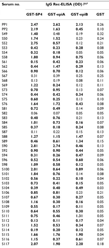
Two GST fusion proteins, respectively carrying residues 420–457 (epiA) and 458–493 (epiB) of the IgA1 protease from S. pneumoniae R6 strain were expressed. As shown in Figure 3, the recombinant GST-epiA and GST-epiB anti-gens were efficiently purified under native conditions from transformed E. coli cells. Immunoreactivity of recombinant antigens with IgG antibodies of human sera was examined and the results were compared to those obtained by use of the largest fragment of the IgA1 pro-tease (clone SP4). The GST-epiA, GST-epiB and GST-SP4 antigens were assayed by Rec-ELISA with serum samples from the two patients hospitalized for pneumococcal pneumonia (used for the library selection) and from 46 healthy individuals. Immunoreactivity of wild-type GST was also assessed for each serum. As shown in Table 1, the recombinant GST-SP4, GST-epiA and GST-epiB proteins specifically reacted with 44 (92%), 43 (89%) and 33 (69%) of the sera, respectively, highlighting a broad anti-body recognition of all antigen fragments.
Because of the broad reactivity of the epiA fragment, its recognition by antibodies present in sera from young dren was also investigated. Serum samples from forty chil-dren with an age comprised between 1 and 4 years were assayed with the recombinant GST-epiA antigen by Rec-ELISA. As shown in Fig. 4, specific IgG antibodies against the epiA polypeptide were detected in sera from the majority of subjects (27 out of 40), thus demonstrating
Table 1: Immunoreactivity of the S. pneumoniae IgA1 protease antigen fragments PaP
Serum no. IgG Rec-ELISA (OD) PbP
GST-SP4 GST-epiA GST-epiB GST PP1 2.47 2.62 2.13 0.26 PP2 2.19 1.63 0.45 0.10 S49 1.40 0.48 0.19 0.32 S50 1.74 1.52 0.23 0.24 S52 2.75 0.57 0.12 0.21 S53 0.42 0.23 0.28 0.08 S54 0.32 0.18 0.05 0.05 S58 1.80 0.94 0.75 0.06 S61 0.15 0.42 0.23 0.06 S62 0.44 1.47 0.29 0.13 S66 0.90 0.70 0.12 0.06 S67 0.31 0.39 0.25 0.25 S68 0.13 0.19 0.08 0.11 S69 1.22 0.24 0.12 0.11 S73 0.70 0.95 0.13 0.07 S74 0.44 0.42 0.34 0.06 S75 0.60 0.25 0.13 0.10 S76 1.64 1.72 0.43 0.08 S81 0.72 0.49 0.14 0.09 S82 0.06 0.07 0.05 0.05 S83 0.40 0.76 0.21 0.13 S84 1.01 0.73 0.16 0.07 S85 0.37 0.18 0.24 0.08 S87 0.11 0.22 0.15 0.13 S88 1.27 1.15 1.47 0.07 S89 0.46 0.61 1.34 0.15 S90 2.81 2.74 0.46 0.13 S92 0.90 0.90 0.44 0.05 S94 0.31 0.26 0.47 0.09 S96 0.52 0.54 0.60 0.06 S98 1.09 0.58 0.12 0.05 S100 2.01 0.64 0.89 0.06 S101 1.84 0.76 0.14 0.08 S102 0.56 0.22 0.10 0.04 S103 0.73 0.41 0.15 0.05 S104 0.39 0.40 0.49 0.03 S106 0.85 0.81 0.23 0.21 S107 0.67 0.30 0.27 0.05 S108 1.16 0.30 0.16 0.05 S109 0.55 0.17 0.11 0.05 S110 0.64 0.55 0.30 0.05 S111 0.75 0.46 1.31 0.05 S112 0.13 0.11 0.17 0.04 S113 1.52 0.53 0.34 0.07 S114 0.19 0.20 0.12 0.05 S115 1.66 1.76 1.90 0.04 S116 1.15 0.37 0.61 0.07 S117 2.07 1.90 2.20 0.18
Pa PSerum samples from patients with pneumococcal pneumonia (PP1 and PP2) and from healthy adults (S49 to S117) were analyzed by IgG Rec-ELISA with individual recombinant proteins.
Pb PThe cutoff values were determined for each serum as two times the OD readings obtained with the GST. Boldface type, OD values greater than the cutoff values.
Characterization of the recombinant fusion proteins
Figure 3
Characterization of the recombinant fusion proteins.
Purified proteins were subjected to SDS-PAGE analysis and loaded as follows: (1) and (7), molecular weight markers; (2) GST wild-type protein; (3) SP2; (4) SP4; (5) GST-epiA; and (6) GST-epiB.
119 87 46 31 24 19 -1 2 3 5 6 7 kDa 4
that 68% of children less than 4 years old had specific antibodies against this short polypeptide.
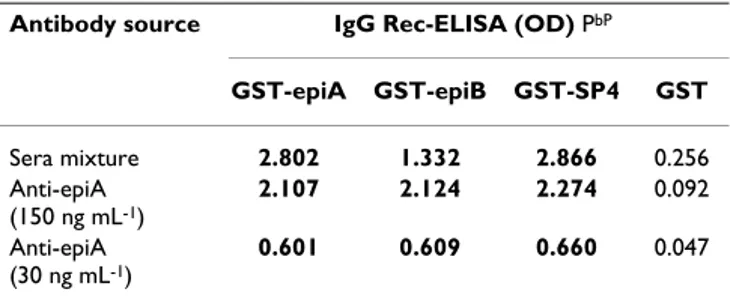
Next, a detailed epitope mapping was performed employ-ing affinity-purified antibodies. Specific immunoglobu-lins against the GST-epiA fusion protein were purified from 5 epiA-reactive sera and then assayed with the recombinant GST-epiA, GST-epiB and GST-SP4 antigens by Rec-ELISA. As a negative control, the IgG reactivity against wild-type GST protein was also assessed. As shown in Table 2, the affinity-purified anti-epiA antibodies
spe-cifically recognized both the epiA and epiB polypeptides as well as the large SP4 fragment, indicating that a com-mon epitope was present in the epiA and epiB protein sequences.
Next, to exclude non-specific immunoreactivity due either to altered conformation of the small polypeptides with respect to native IgA1 protease or to cross-reactivity of unrelated antibodies in human sera, a Western blot anal-ysis was performed. To this aim, a whole cell lysate of S.
pneumoniae was challenged with the affinity-purified
anti-epiA antibodies. Bacterial lysates from pneumococcal iga-deficient mutant strain FP174 and from Streptococcus
gor-donii were assayed as controls. As shown in Figure 5, a
unique protein band with an apparent molecular mass higher than 220 kDa specifically reacted with the anti-epiA antibodies in an unencapsulated strain of pneumo-coccus (R6). In the FP174 mutant strain, in which the iga gene was deleted, as well as in S. gordonii, such a protein band was not visible, demonstrating that the anti-epiA antibodies specifically recognized the pneumococcal enzyme.
Finally, to investigate whether the epiA polypeptide is exposed on the bacterium and is recognizable by anti-epiA antibodies, a cell-ELISA analysis was performed on whole S. pneumoniae cells. The FP174 strain was used as the negative control. The affinity-purified epiA anti-bodies reacted with intact bacterial cells (strain R6) but not with the iga-deleted FP174 strain (data not shown),
Recognition of pneumococcal iga gene product by affinity-purified anti-epiA antibodies
Figure 5
Recognition of pneumococcal iga gene product by affinity-purified anti-epiA antibodies. Western blot
analysis on whole cell lysates from S. pneumoniae R6 (lanes 1 and 4) and FP174 strains (lanes 2 and 5), and from S. gordonii (lanes 3 and 6). A homogeneous mixture of 5 human sera (lanes 1–3) or affinity-purified anti-epiA antibodies (lanes 3–6) were used. 220 130 100 1 2 3 4 5 6 7 kDa
Sera mix. Anti-epiA
Analysis of the B cell reactivity of the epiA peptide in healthy adults and children
Figure 4
Analysis of the B cell reactivity of the epiA peptide in healthy adults and children. Immunoreactivity of the
recombinant GST-epiA protein analyzed by IgG Rec-ELISA with serum samples from 48 blood donors and 40 children is shown. The values were calculated as the ratio of the OD readings obtained with the GST-epiA to twice the corre-sponding OD readings obtained with the GST.
0 1 10 100 OD GST-ANTI GEN /2 x (O DGS T ) Children 8-12 months Children 1-2 years Children 2-3 years Children 3-4 years Adults cutoff
Table 2: Cross-reactivity of epiA and epiB polypeptides with affinity-purified anti-epiA antibodies PaP
Antibody source IgG Rec-ELISA (OD) PbP
GST-epiA GST-epiB GST-SP4 GST Sera mixture 2.802 1.332 2.866 0.256 Anti-epiA (150 ng mL-1) 2.107 2.124 2.274 0.092 Anti-epiA (30 ng mL-1) 0.601 0.609 0.660 0.047
Pa PRecombinant antigens were PPanalyzed by IgG Rec-ELISA with a homogeneos mixture of human sera (diluted 1:50) or with affinity-purified anti-epiA antibodies
Pb PThe cutoff values for the IgG Rec-ELISA with the recombinant GST-epiA, GST-epiB, and GST-SP4 proteins were calculated as two times the OD readings obtained with the GST. Boldface type, OD values greater than the cutoff values.
indicating that the epiA epitope within the tandem repeats of the pneumococcal enzyme is indeed exposed in the native state. Noteworthy, our results are in close agree-ment with a previous work where it was demonstrated that the tandem repeats of S. sanguis IgA1 protease contain immunodominant epitopes exposed in the denatured and native state [16].
Discussion
Bacterial IgA1 proteases are highly specific endopepti-dases that are secreted by a small number of bacteria asso-ciated with humans [23,24]. IgA1 protease-producing bacteria include the mucosal pathogens N. meningitidis,
N. gonorrhoeae, H. influenzae, S. pneumoniae, U. urealyticum
and some members of pharyngeal microfloras such as
Prevotella, Gemella haemolysans, S. sanguis, S. oralis and S. mitis [14,22]. The importance of this bacterial enzyme in
the interaction with the host is evidenced by the fact that most of these enzymes are specific for human IgA only, and that their impact on the host is so great as to become one of the few recognised factors of positive evolutionary selection on the human genome [25]. For the bacterial pathogens colonizing mucosal surfaces, IgA1 protease production represents a virulent factor and contributes to the pathogenesis of invasive infection by cleaving the Fc receptor from secreted immunoglobulins A1, thus pre-venting opsonophagocytosis [5]. IgA1 proteases have been shown to be targets of enzyme-neutralizing antibod-ies in serum and secretions [26], which may be induced in a state of bacterial carriage as well as during invasive infec-tion [27].
We previously demonstrated the potential of phage-dis-play technology in identifying B-cell epitopes recognized by the human immune response against Toxoplasma gondii and S. pneumoniae infection [20,28-30], even though such a technique usually favors the identification of linear epitopes whereas conformational epitopes are often over-looked. Nevertheless, the present work allowed the iden-tification of antigenic regions of the pneumococcal IgA1 protease that are broadly recognized by human antibod-ies. Challenging a lambda-display library of S. pneumoniae with sera from patients hospitalized for pneumococcal pneumonia enabled the selection of three distinct clones harboring a common region of the IgA1 protease. All clone sequences matched the pneumococcal enzyme encompassing amino acids 360–590. The immunoreac-tivity of the selected fragments, expressed as GST fusion products, was assessed; overall, IgG antibodies in serum samples from more than 90% of healthy adults reacted with at least one antigen fragment, which emphasizes the broad recognition of pneumococcal IgA1 protease by the human B cell response.
Because of the broad reactivity of the selected IgA1 pro-tease fragments, a detailed characterization of their com-mon region, composed by 72 amino acids, was performed. This lead to classify the sequence correspond-ing to residues 420–457 (epiA peptide) of the iga gene product as the most reactive polypeptide, which was rec-ognized by antibodies present in sera from 90% of healthy adults and 68% of children with an age comprised between 1 and 4 years, thus suggesting that the human antibody response against IgA1 protease occurs during childhood. Noteworthy, our data are in agreement with a previous study showing that IgG antibodies from the majority of children less than two years old specifically reacted with pneumococcal antigens, including the IgA1 protease [19].
The evidence that affinity-purified anti-epiA antibodies also reacted with the epiB fragment (residues 458–493) indicated that a common epitope within polypeptides was recognized by human sera. Notably, a repeated 9-mer sequence VVTDKGEPE is present twice in epiA and once in epiB fragments (Fig. 1 and 2). This duplication might account for the larger reactivity of the epiA peptide with respect to the epiB peptide.
The IgA1 protease of S. pneumoniae is a major antigen expressed in all known pneumococcal strains [7]. Despite the pneumococcal enzyme has a peculiar structure among bacterial proteases, homology searches have indicated that the iga gene of S. pneumoniae is also found in other streptococci and related organisms [8,14,15,22]. As already noted by others, the repeats region shows high conservation within the different pneumococcal strains and between S. pneumoniae and the most closely related S.
mitis (90% identity), while homology to IgA proteases
from other streptococci is significantly lower in this seg-ment when compared to the rest of the molecule [8,22]. The epiA peptide identified in this work is conserved in all pneumococci and in two out of three S. mitis strains [22], while it is not conserved in other oral streptococci sequenced so far. The fact that the domain covered by epiA is not shared with oral streptococci other than S.
mitis suggests that this domain is probably not
inter-changeable, which reduces the risk of escape mutants by horizontal gene transfer. In other words, this would indi-cate that any reservoir of divergent IgA1 proteases in non pneumococcal species should not be imported readily by recombination into pneumococcus.
We have shown here that an immunodominant polypep-tide (epiA peppolypep-tide), located within the sequence repeats domain of pneumococcal IgA1 protease, is well conserved among several serotypes and strains of S. pneumoniae but not to the corresponding regions of the iga gene products from other streptococci such as S. oralis, S. sanguis and S.
sanguinis. However, we also found a very high sequence
homology of this protein region between S. pneumoniae and S. mitis (see Fig. 2), suggesting that the observed high rate of seropositivity against pneumococcal IgA1 protease might be due to early colonization with bacteria species other than pneumococcus.
Conclusion
The broad and specific recognition of the epiA polypep-tide by human sera demonstrates that the pneumococcal IgA1 protease contains an immunodominant B-cell epitope. In perspective, it will be of great interest to inves-tigate the ability of the anti-epiA antibodies in conferring protective immunity against S. pneumoniae infection in animal models. The use of phage display libraries to iden-tify microbe or disease-specific antigens recognized by the human sera is a promising and valuable approach to epitope discovery.
Methods
Bacterial strainsBacteria were grown in tryptic soy broth (TSB) in a 5% COB2B-enriched atmosphere at 37°C. Where necessary,
streptomycin and kanamycin were used at a final concen-tration of 500 μg mLP-1P. Streptococcus pneumoniae R6 [31],
FP174 (ΔSP1154/iga) [12] and S. gordonii [32] strains were used for this study.
Affinity selection of the S. pneumoniae display library and serum samples
Serum samples from two patients with a respective age of 2 and 64 years old, collected during patient's hospitaliza-tion for acute pneumococcal pneumonia, were used for the affinity selection of a whole genome lambda-display library of S. pneumoniae D39 strain [20]. The construction of the pneumococcal library and the selection of the library with human sera were performed as previously described [33]. Briefly, magnetic beads linked to Protein G (Dynal, Norway) were incubated for 40 min at room temperature with 10 μL of serum. Beads were then incu-bated with 5 × 10¹⁰ plaque forming units (PFU) of the
phage library (complexity of the library, 2 × 10⁷
independ-ent clones) for 3 hours at room temperature. Escherichia
coli cells were infected with phage pools by mixing the
beads and the bacterial cells, and recombinant phages were amplified by using standard procedures [34]. After two rounds of affinity selection, single phage clones were isolated from phage pools by immunoscreening [28].
Forty-eight serum samples collected from 48 blood donors were anonymously collected and then used for the analysis of selected antigen fragments' immunoreactivity. Forty sera from 40 children born to mothers with congen-ital infections in pregnancy (i.e. Toxoplasma gondii and human cytomegalovirus infection) and referred for
post-natal follow up of congenital diseases were also included in the study. Sera were collected from children with asymptomatic diseases with an age comprised between 1 and 4 years, and the serum samples were assayed in a blinded fashion.
Cloning and recombinant protein expression and purification
DNA inserts of phage clones SP2 and SP4 were amplified by PCR, digested with Spe I and Not I endonucleases and subcloned into the bacterial vector pGEX-SN [21]. DNA fragments encoding for amino acids 420–457 and amino acids 458–493 of the S. pneumoniae IgA1 protease (Gen-Bank Q54875) were PCR amplified from phage clone SP4 by using oligonucleotides K708 (5'-ACGACTAGTGCAAT-TCAGCCTGAGT TGCCC-3') and K709 (5'-GGTGCG-GCCGCTCACTCTGGTTGAACCTCAGTCTC-3'), and oligonucleotides K710 (5'-CGAACTAGT TCGCCAGA-TACTGTGGTAAG-3') and K711 (5'-GGTGCGGCCGCT-CACTTCTCAACCGGAGTTTCAC-3'), respectively. PCR products were digested with Spe I and Not I enzymes and then cloned into pGEX-SN plasmid. Competent
Escherichia coli cells (AD202 strain) [34] were transformed
with recombinant plasmids and single clones were iso-lated.
Recombinant proteins produced in E. coli as fusion pro-teins with glutathione S-transferase (GST) were purified from the cytoplasm of bacterial cells by affinity chroma-tography, as previously described [29]. Briefly,
recom-binant E. coli was induced with
isopropyl-thiogalactopyranoside (IPTG), centrifuged and suspended in 10 mM Tris-HCl (pH 8), 150 mM NaCl, 100 μg mL-1 of
lysozyme and protease inhibitors (Boheringer, Germany). The mixture was sonicated, and Triton-X100 was added to a final concentration of 1%. After centrifugation at 10,000 × g for 30 min at 4°C, the supernatant was incubated with Glutathione-Sepharose (Amersham-Pharmacia Biotech, Sweden) and GST-proteins were eluted by following the manufacturer's instructions. Finally, protein purity and content were assessed by sodium dodecyl sulfate-polyacri-lamide gel electrophoresis (SDS-PAGE) and by Bradford assays, respectively.
Recombinant protein enzyme-linked immunoassays (Rec-ELISA)
Maxisorb multiwell plates (Nunc) were adsorbed with recombinant proteins (GST-SP2, GST-SP4, GST-epiA, GST-epiB and GST) at a concentration of 5 μg mL-1 in 50
mM NaHCOB3B, pH 9.6. After incubation overnight at 4°C, the plates were blocked with 5% non-fat dry milk and 0.05% Tween-20 in PBS (blocking buffer) and incu-bated for 1 h at 37°C with serum samples diluted 1:50 in blocking buffer. The plates were washed with 0.05% Tween-20 in PBS, and anti-human IgG horseradish
perox-idase-conjugated antibodies (Sigma-Aldrich, USA) were then added to each well. Finally, incubating the plates with the chromogenic substrate tetramethylbenzidine (TMB; Sigma Aldrich) revealed the enzymatic activity. The results were recorded as the difference between the absorbance (optical density, OD) at 450 and at 620 nm, as detected with an automated ELISA reader (Labsystem Multiskan, Finland). Assays were performed in duplicate, and average values were calculated. GST carrier protein was used as the negative control in every assay.
Purification of anti-epiA antibodies from human sera
The GST-epiA fusion protein was immobilized to a NHS-activated-agarose column (Amersham-Pharmacia Bio-tech, Sweden) in accordance with the manufacturer's instructions. A mixture of sera from five blood donors (sera S90, S117, S115, S62 and S76) were allowed to flow through the column overnight to permit the binding of specific antibodies to the recombinant protein. After extensively washing the column with PBS and then with a solution containing 0.5 M NaCl in PBS, the antigen-spe-cific antibodies were eluted with a solution of 50 mM gly-cine (pH 3) containing 100 mM NaCl. The antibody concentration was determined by Bradford assay.
Western blot analysis
S. pneumoniae strains R6 and FP174 and S. gordonii strain
Challis [32] were grown in 15 mL of Tryptic Soy Broth (TSB, BD Biosciences) in standard conditions. Cells were collected by centrifugation at 3000 g for 15 min at 4°C and washed with PBS. Bacteria were suspended in a final volume of 0.5 mL of loading buffer (10 mM Tris-HCl, 1 mM EDTA, 1% SDS, 10 mM dithiothreitol, 10% glycerol, 0.01% bromophenol blue), heated for 15 min at 95°C and then subjected to SDS-PAGE. Proteins were trans-ferred onto nitrocellulose membranes (BioTrace NT, Pure Nitrocellulose Blotting Membrane, Pall Life Science), which were subsequently blocked with 5% non-fat dry milk and 0.05% Tween-20 in PBS (blocking buffer). The filters were incubated overnight at 4°C with the affinity-purified anti-epiA antibodies used at a concentration of 150 ng mL-1. After being extensively washed with 0.05%
Tween-20 in PBS, filters were incubated with anti-human IgG alkaline phosphatase-conjugated antibodies (Sigma-Aldrich, USA). Finally, protein bands were visualized by using nitroblue tetrazolium (NBT; Sigma Aldrich) and 5-bromo-4-chloro-3-indosyl phosphate (BCIP; Sigma Aldrich) as choromogenic substrates.
Authors' contributions
FDP carried out affinity selection of the phage display library, purification of recombinant proteins and Rec-ELISA analysis. EB performed bacterial growth and West-ern blot analysis. AS performed the characterization of anti-epiA antibodies purified from human sera. FM
pro-vided clinical samples of pneumococcal pneumonia. MRO provided bio-informatics support and continuous help and discussions during the conduct of the work. FF provided helpful discussion and critical reading of the manuscript. NG designed the study and wrote the script. All authors read and approved the final manu-script.
Acknowledgements
Preliminary sequence data was obtained from The Institute for Genomic Research (TIGR) and from the Sequencing Group at The Sanger Institute. We acknowledge Enza Piccolella and Paola Del Porto for very helpful sug-gestions and advice, and Luca Bruno for skillful technical assistance.
Work carried out in Siena was supported in part by the European Commis-sion grant LSHM-CT-2005-512099 to M.R.O.
References
1. Ejstrud P, Hansen JB, Andreasen DA: Prophylaxis against pneu-mococcal infection after splenoctomy: a challenge for hospi-tals and primary care. Eur J Surg 1997, 163:733-738.
2. Bogaert D, De Groot R, Hermans PW: Streptococcus pneumoniae colonization: the key to pneumococcal disease. Lancet Infect
Dis 2004, 4:144-154.
3. Bogaert D, Sluijter M, De Groot R, Hermans PW: Multiplex opsonophagocytosis assay (MOPA): a useful tool for the monitoring of the 7-valent pneumococcal conjugate vaccine.
Vaccine 2004, 22:4014-4020.
4. Mangtani P, Cutts F, Hall AJ: Efficacy of polysaccharide pneumo-coccal vaccine in adults in more developed countries: the state of the evidence. Lancet Infect Dis 2003, 3:71-78.
5. Kilian M, Reinholdt J, Lomholt H, Poulsen K, Frandsen EV: Biological significance of IgA1 proteases in bacterial colonization and pathogenesis: critical evaluation of experimental evidence.
APMIS 1996, 104:321-338.
6. Weiser JN, Bae D, Fasching C, Scamurra RW, Ratner AJ, Janoff EN: Antibody-enhanced pneumococcal adherence requires IgA1 protease. Proc Natl Acad Sci USA 2003, 100:4215-4220.
7. Camilli R, Pettini E, Grosso MD, Pozzi G, Pantosti A, Oggioni MR: Zinc metalloproteinase genes in clinical isolates of
Strepto-coccus pneumoniae : association of the full array with a clonal
cluster comprising serotypes 8 and 11A. Microbiol 2006, 152:313-321.
8. Poulsen K, Reinholdt J, Jespersgaard C, Boye K, Brown TA, Hauge M, Kilian M: A comprehensive genetic study of streptococcal immunoglobulin A1 proteases: evidence for recombination within and between species. Infect Immun 1998, 66:181-190. 9. Bricker J, Mulks MH, Plaut AG, Moxon ER, Wright A: IgA proteases
of Haemophilus influenzae : cloning and characterization in E.
coli K12. Proc Natl Acad Sci USA 1983, 80:2681-2685.
10. Koomey JM, Falkow SS: Nucleotide sequence homology between the immunoglobulin A1 protease genes of Nesseria
gonorrhoeae, N. meningitidis and Haemophilus influenzae. Infect Immun 1984, 43:101-107.
11. Gilbert JV, Plaut AG, Wright A: Analysis of the immunoglobulin A protease gene of Streptococcus sanguis. Infect Immun 1991, 59:7-17.
12. Chiavolini D, Memmi G, Maggi T, Iannelli F, Pozzi G, Oggioni MR: The three extra-cellular zinc metalloproteinases of Streptococcus
pneumoniae have a different impact on virulence in mice. BMC Microbiol 2003, 3:14-21.
13. Bender MH, Weiser JN: The atypical amino-terminal LPNTG-containing domain of the pneumococcal human IgA1-spe-cific protease is required for proper enzyme localization and function. Mol Microbiol 2006, 61:526-543.
14. Poulsen K, Reinholdt J, Kilian M: Characterization of the
Strepto-coccus pneumoniae immunoglobulin A1 protease gene (iga)
and its traslation product. Infect Immun 1996, 64:3957-3966. 15. Wani JH, Gilbert JV, Plaut AG, Weiser JN: Identification, cloning
and sequencing of the immunoglobulin A1 protease gene of
Publish with BioMed Central and every scientist can read your work free of charge "BioMed Central will be the most significant development for disseminating the results of biomedical researc h in our lifetime."
Sir Paul Nurse, Cancer Research UK Your research papers will be:
available free of charge to the entire biomedical community peer reviewed and published immediately upon acceptance cited in PubMed and archived on PubMed Central yours — you keep the copyright
Submit your manuscript here: BioMedcentral
16. Gilbert JV, Ramakrishna JP, Wright A, Plaut AG: Streptococcal IgA1 protease tandem repeat influences antigenicity but not activity. J Dent Res 2003, 72:327.
17. Lomholt H: Evidence of recombination and an antigenically diverse Immunoglobulin A1 protease among strains of
Strep-tococcus pneumoniae. Infect Immun 1995, 63:4238-4243.
18. Romanello V, Marcacci M, Dal Molin F, Moschioni M, Censini S, Cov-acci A, Baritussio AG, Montecucco C, Tonello F: Cloning, expres-sion, purification and characterization of Streptococcus
pneumoniae IgA1 protease. Protein Expr Purif 2006,
45(1):142-149.
19. Adrian PV, Bogaert D, Oprins M, Rapola S, Lahdenkari M, Kilpi T, de Groot R, Kayhty H, Hermans PW: Development of antibodies against pneumococcal proteins alpha-enolase, immunoglob-ulin A1 protease, streptococcal lipoprotein rotamase A, and putative proteinase maturation protein A in relation to pneumococcal carriage and Otitis Media. Vaccine 2004, 22:2737-2742.
20. Beghetto E, Gargano N, Ricci S, Oggioni M, Garufi G, Peppoloni S, Pozzi G, Felici F: Discovery of a novel Streptococcus pneumoniae antigen by screening a whole genome lambda-display library.
FEMS Microbiol Lett 2006, 262:14-21.
21. Minenkova O, Pucci A, Pavoni E, DeTomassi A, Fortugno P, Gargano N, Cianfriglia M, Barca S, DePlacido S, Martignetti A, Felici F, Cortese R, Monaci P: Identification of tumor-associated antigens by screening phage-displayed human cDNA libraries with sera from tumor patients. Int J Cancer 2003, 106:534-544.
22. Takenouchi-Ohkubo N, Mortensen LM, Drasbek KR, Kilian M, Poulsen K: Horizontal transfer of the immunoglobulin A1 pro-tease gene (iga) from Streptococcus to Gemella haemoly-sans. Microbiol 2006, 152:2171-2180.
23. Kilian M, Mestecky J, Kulhavy R, Tomana M, Butler WT: IgA1 pro-teases from Haemophilus influenzae, Streptococcus
pneumo-niae, Neisseria meningitidis, and Streptococcus sanguis:
comparative immunochemical studies. J Immunol 1980, 124:2596-2600.
24. Plaut AG: The IgA1 proteases of pathogenic bacteria. Ann Rev
Microbiol 1983, 37:603-622.
25. Vallender EJ, Lahn BT: Positive selection on the human genome.
Hum Mol Genet 2004, 13 Spec No 2():R245-R254.
26. Gilbert JV, Plaut AG, Longmaid B, Lamm ME: Inhibition of micro-bial IgA proteases by human secretory IgA and serum. Mol
Immunol 1983, 20:1039-1049.
27. Brooks GF, Lammel CJ, Blake HS, Kusecek B, Achtman M: Antibod-ies against IgA1 proteases are stimulated both by clinical dis-ease and asymptomatic carriage of serogroup A Nesseria
meningitidis. J Infect Dis 1992, 166:1316-1321.
28. Beghetto E, Pucci A, Minenkova O, Spadoni A, Bruno L, Buffolano W, Soldati D, Felici F, Gargano N: Identification of a human immu-nodominant B-cell epitope within the GRA1 antigen of
Tox-oplasma gondii by phage display of cDNA libraries. Int J Parasitol 2001, 31:1659-1668.
29. Beghetto E, Spadoni A, Buffolano W, Del Pezzo MA, Minenkova O, Pavoni E, Pucci A, Cortese R, Felici F, Gargano N: Molecular dissec-tion of the human B-cell response against Toxoplasma gondii infection by lambda display of cDNA libraries. Int J Parasitol 2003, 33:163-173.
30. Di Cristina M, Del Porto P, Buffolano W, Beghetto E, Spadoni A, Guglietta S, Piccolella E, Felici F, Gargano N: The Toxoplasma
gon-dii bradyzoite antigens BAG1 and MAG1 induce early
humoral and cell-mediated immune responses upon human infection. Microbes Infect 2004, 6:164-171.
31. Hoskins J, Alborn WE, Arnold J, Blaszczak LC, Burgett S, DeHoff BS, Estrem ST, Fritz L, Fu DJ, Fuller W, Geringer C, Gilmour R, Glass JS, Khoja H, Kraft AR, Lagace RE, LeBlanc DJ, Lee LN, Lefkowitz EJ, Lu J, Matsushima P, McAhren SM, McHenney M, McLeaster K, Mundy CW, Nicas TI, Norris FH, O'Gara M, Peery RB, Robertson GT, Rockey P, Sun P, Winkler ME, Yang Y, Young-Bellido M, Zhao G, Zook CA, Baltz RH, Jaskunas S, Rosteck PR Jr, Skatrud PL, Glass JI: Genome of the bacterium Streptococcus pneumoniae strain R6. J Bacteriol 2001, 183:5709-5717.
32. Oggioni MR, Pozzi G: A host-vector system for heterologous gene expression in Streptococcus gordonii. Gene 1996, 169:85-90.
33. Beghetto E, Nielsen HV, Del Porto P, Buffolano W, Guglietta S, Felici F, Petersen E, Gargano N: A combination of antigenic regions of
Toxoplasma gondii microneme proteins induces protective
immunity against oral infection with parasite cysts. J Infect Dis 2005, 191:637-645.
34. Sambrook J, Russell DW: Molecular cloning: a laboratory man-ual. 3rd edition. Cold Spring Harbor Laboratory Press; 2001. 35. The Institute for Genomic Research [http://www.tigr.org/] 36. The Sanger Institute