International PhD Program in Neuroscience
XXIX Cycle
Nanocarriers for nose-to-brain delivery:
a novel strategy for the treatment of CNS
disorders
Ph.D Thesis
Angela Bonaccorso
Coordinator: Prof. Salvatore Salomone Tutor: Prof. Giovanni Puglisi
Co-Tutor: Dr. Teresa Musumeci Co-Tutor: Prof. Ijeoma F. Uchegbu
2016
Department of Biomedical and Biotechnological Sciences.
This work was supported with a fellowship by the International PhD Program in Neuroscience from the University of Catania, School of Medicine (Catania, Italy). This work was partially supported by the Italian grant number PRIN 2010/2011 [grant number: 2010H834LS_005].
The work for this thesis was carried out in the laboratories of:
Home Institute Guest Institute
Professor Giovanni Puglisi and Professor Ijeoma F.Uchegbu Dr. Teresa Musumeci Pharmaceutics,
Department of Drug Sciences, School of Pharmacy, Catania, University of Catania University College London
Viale Andrea Doria, 6 29-39 Brunswick Square, 95125 Catania London WC1N 1AX,
Italy United Kingdom
To my family and Andrea
“Perplexity is the beginning of knowledge”
Khalil Gibran
“I have not failed 700 times. I have not failed once. I have succeeded in proving that those 700 ways will not work. When I have eliminated the ways that will not work, I will find the way that will work.”
1
ACKNOWLEDGEMENTS………...4
ABSTRACT……….6
LIST OF ABBREVIATIONS………...10
CHAPTER I GENERAL INTRODUCTION……….14
1. NEURODEGENERATIVE DISEASES AND BBB………15
1.1. Blood brain barrier, general concept and mechanisms of passage……….……18
2. NANOTECHLOGY-BASED DRUG DELIVERY SYSTEMS………...21
2.1. Polymers used in preparation of nanoparticles………...….25
2.2. Polymeric nanoparticles………...30
3. NANOPARTICLES FOR BRAIN TARGETING: CROSSING OR BYPASSING THE BLOOD-BRAIN-BARRIER……….…..34
3.1. Crossing the blood-brain-barrier………..35
3.2. Bypassing the blood-brain-barrier………38
4. NOSE-TO-BRAIN...38
4.1. Pathways and mechanisms……… ..41
4.2. Free molecules delivery via nose-to-brain…...44
5. EPILEPSY………..49
5.1. Oxcarbazepine……….54
2
6.1. RNAi Mechanism……….57
6.1.1. siRNA delivery………..59
6.1.2. Viral vectors………..60
6.1.3. Non-viral vectors……….………..60
7. POLYMERS SELECTED IN THIS PROJECT TO ENHANCE NANOPARTICLES BRAIN TARGETING………...62
7.1. PLGA……….………...63
7.2. PLGA-PEG………...64
7.3. Chitosan………66
8. AIM OF THE STUDY………...69
CHAPTER II Revisiting the role of sucrose in PLGA-PEG nanocarrier for potential intranasal delivery………...72
CHAPTER IIINose-to-brain delivery: localization and time-course of polymeric nanocarriers on different brain regions of brain rats………...110
CHAPTER IVNose-to-brain delivery of DiR–loaded PLGA nanoparticles (in preparation)………... 149
CHAPTER VControlling seizures by intranasal delivery of Oxcarbazepine-loaded nanoparticles in rats (in preparation)………....155
CHAPTER VI Nose-to-brain delivery and chitosan derivatives nanocomplexes for siRNA delivery to the brain (in preparation)………...161
CHAPTER VII GENERAL DISCUSSION AND CONCLUSION………....………168
CHAPTER VIII REFERENCESANDSITOGRAPHY……….177
3
1. List of pubblications and scientific contributions……….……196 1.1. Publications……….196 1.2. Conference proceedings………..196
4
ACKNOWLEDGEMENTS
I would like to thank many people, without whom my work would not be possible: I would like to express my gratitude to Prof. Filippo Drago and to the Coordinator of the International PhD program in Neuroscience Prof. Salvatore Salomone for giving me the opportunity to take part to this prestigious PhD program that represented an important moment for my scientific growth.
I would like to thank my supervisor Prof. Giovanni Puglisi for his excellent supervision, constructive comments and discussion. It has been an honour working in his research group which has been a source of knowledge as well as good advice and collaboration.
I would like to thank my co-tutor Dr. Teresa Musumeci for her guidance in my research and for her support and encouragement during my PhD studies. With her great experience and knowledge, she taught me how to approach “Research”.
Sincerely thank to Prof. Ijeoma F. Uchegbu, welcoming me in her group at UCL School of Pharmacy, in London, and leading me working on exciting project. It was a great pleasure and honor to work in such inspiring scientific environment. It has been an enriching experience for me both personally and professionally for which I will always be grateful.
I would like to express my special appreciation and thanks to Dr Rosalia Pellitteri (Institute of Neurological Sciences, CNR, Section of Catania) and Professor Francesca Serapide (Department of Biomedical and Biotechnological Science, University of Catania) for stimulating scientific discussions and for their precious collaboration and contribution in this thesis for the in vivo studies in chapter III and V.
5 I wish to thank also Professor Giulio Sancini (Department of Health Sciences, University of Milan-Bicocca) and Dr Roberta Dal Magro (Department of Health Sciences, University of Milan-Bicocca) for their collaboration regarding the in vivo study described in chapter IV.
I thank my PhD colleagues and all the members of the PhD Program with which I shared this experience pleasantly. I would also like to thank all the people I met during my PhD and, with which I spent these years learning and enjoying research.
Special thanks go to my “Londoners” collegues, Uchechukwu Odunze, Xian Weng Jiang, Sunish Patel, Lisa Godfrey, Fion O’Brien, Cindy Chen, Paeng Jittrawan,
Georgios Kasparis, Ambra Bertoni, Paula Mendez, Nick Hobson, Abdullah Alamoudi, Ramesh Soundararajan, for their infinite kindness and for the friendly atmosphere in our workgroup. With them I learned techniques that I had never even thought of doing! Having lived and worked in London surely was a unique and unforgettable experience and the networking and friends I have made during this time, are the most valuable outcome of my experience abroad. It was great to meet lots of interesting people from all over the world which, through their stories and shared experiences, and advices have consistently enriched me.
Last but no least, thanks go to my parents for their endless love, support and encouragement and to Andrea for his love, patience and help anytime dispite the distance.
6
ABSTRACT
The number of neurodegenerative diseases is estimated to be a few hundred. Despite the high prevalence and incidence, central nervous system (CNS) disorders are still incurable. The current therapeutic approach is based on the administration of symptomatic drugs which reduce the signs and symptoms of CNS diseases but not its causes. The development of effective preventive or protective therapies has been impeded by the difficult to deliver therapeutic agent to the brain.
The blood-brain-barrier (BBB) precludes the delivery of drugs to the brain, preventing the therapy of a number of neurological disorders.In the last 20 years, intranasal (IN) administration has gained great attention in research and has been investigated extensively with regard to its feasibility to serve as a direct drug transport route to the CNS. Drugs can be transported directly from the nasal cavity to the brain through the olfactory epithelium by trigeminal nerve systems and olfactory nerve pathways thereby bypassing the BBB. Even though some studies have demonstrated the transport of therapeutic agents to the brain via nasal route, the quantities of drug transported to the brain are very low, normally less than 0.1%, which is less than the therapeutically effective dose.
The incorporation of drugs into nanoparticles (NPs) might be a promising approach to improve the amount of pharmaceuticals delivered to the CNS, protecting them from the enzymatic activity in the nasal cavity and enhancing their transport across the biological barriers. Taking into account these considerations, the goal of my PhD thesis is to assess the effective molecule delivery to the brain by using a new approach: IN administration combined with the nanotechnology-based carriers.
7 In particular my aim was to investigatewhether polymeric NPs can end up the brain after IN administration; which region of the brain can be reached; how does surface property affect NPs transport.This thesis focuses on designing and exploring novel and different polymeric nanosystems with aim to improve nose-to-brain delivery.
Once NPs translocation to the brain via this route was determined, our nanosystems have been formulated to study their potential application in important neurological conditions: epilepsy and brain cancer. The promising nanosystems were successfully loaded with Oxcarbazepine and the model siRNA. In particular, we investigate PLGA NPs, unmodified and surface modified. PLGA is an FDA-approved biodegradable polymer, it allows the preparation of NPs able to encapsulate both hydrophobic and hydrophilic compounds.
Surface modification of PLGA carrier, such as PEGylation or chitosan coating, would serve as one of the excellent approaches to manage drug delivery properties of formulations by interaction of surface coating with a biological system and to enhance brain delivery.
In Paper I we deeply studied PEGylated PLGA NPs, this work was based on several technological analyses aimed at obtaining PEGylated NPs with simple composition and long-term storage suitable for nose-to-brain delivery. To achieve this purpose a screening to select the degree of PEGylation of PLGA (5-10-15%) was performed and the effects of the double function of sucrose as surfactant-like and cryoprotectant agent was evaluated. Mucoadhesive evaluations between NPs and mucin were assessed by the mucin particle method and differential scanning calorimetry. Preliminary in vitro evaluation of cytotoxicproperties of PEGylated systemswas also performed.
8 Our results suggest the use of sucrose for its double effect. We support the use of PEG 5% to confer a sufficiently hydrophilic and uncharged surface to minimize effectively mucin-NPs adhesive interactions, allowing particles to diffuse rapidly through human mucus and cross respiratory epithelium. Our nanosystems did not show any cytotoxic effects. Therefore, in the present work we propose a new formulation for IN drug delivery.
In Paper II we looked at the in vivo fate of PLGA NPs and PLGA NPs surface modified with Chitosan (CS) after IN administration in rats. These formulations have been optimized in terms of mean size and stability by photon correlation spectroscopy and Turbiscan AGS, and tested in vivo. Both NPs systems were loaded with Rhodamine B and in vitro release study was evaluated by dyalisis bag technique. Biodistribution studies were carried out in healthy rats after IN administration of NPs at different time intervals. Fluorescent microscopy was conducted to value the localization of NPs in the CNS. Our results contribute to the understanding that compounds encapsulated in NPs may have a direct access to the CNS following IN administration. Our findings led us to hypothesize that different pathways were involved in the transport of unmodified and modified NPs, suggesting the involvement of the olfactory transport and the trigeminal nerve pathway respectively.
Furthermore, additional experiments, (Paper III, in preparation), were partially reported to confirm our results. In particular, the investigation of DiR-loaded PLGA NPs biodistribution and bioavailability to the brain after IN administration in living healthy mice by Fluorescence Molecular Tomography system.
Once established that our NPs reach different brain areas, we aim to investigate whether NPs canenhance the efficacy of the drugs loaded.
9
Paper IV (in preparation) is focused on the encapsulation of Oxcarbazepine in PLGA
NPs aiming at direct nose-to-brain delivery to improve epileptic therapy, the possibility of using less daily drug amounts to reduce undesirable interactions and toxic effects and to evaluate the possible neuroprotection of this drug against the seizures and brain damage induced by Pentylentetrazole administration.
We also investigate nose-to-brain delivery by using NPs system for gene therapy in
Paper V (in preparation). The partial negative charge and the susceptibility to
degradation by nucleases have hampered nucleic acid use in a naked form. In this study, we investigate the use of “homemade” polymers as potential delivery carriers of siRNA in order to protect it from instability and degradation. The polymer bind to siRNA through electrostatic interaction to form complexes in a non-covalent manner. The nanocomplexes were characterized in terms of size, zeta potential and stability. Cell cytotoxicity of the nanocomplex was determined in A431 cell line. Transfection and silencing efficiency were evaluated in vitro and in vivo after IN administration in rats by using Western Blot. Our nanocomplexes present mean diameter less than 300 nm, positive surface charge and good stability under destabilizing conditions. Our systems show good in vitro transfection and down regulation of the model protein in vivo.
10
LIST OF ABBREVIATIONS
AEDs Antiepileptic drugs AGO 2 Argonaute 2 AUC Area under the curve BBB Blood brain barrier
BBBD Blood brain barrier disruption Cmax Maximum concentration
CNS Central Nervous System CS Chitosan
CSF Cerebrospinal fluid CPP Cell-penetrating peptide D Daltons
DDS Drug delivery systems DiR DiOC18(7)
DNA Deoxyribonucleic acid dsRNA Double stranded RNA
DSC Differential scanning calorimetry DTX Docetaxel
EMA European Medicine Agency FDA Food and Drug Administration FMT Fluorescence Molecular Tomography
11
G Gram
GRAS Generally recognized as safe h Hour
IC50 The concentration of an inhibitor that is required for 50-percent inhibition of an
enzyme in vitro
I.N. Intranasal delivery
I.P. Intraperitoneal administration I.V. Intravenous administration kD Kilodaltons
Kel elimination rate constant
mg Miligram
mg.mL-1 Miligram per mililiter min Minute
mM Milimole
miRNA Micro RNA
MPSMonuclear phagocyte system
mRNA Messenger RNA
MRP-1 Multidrug Resistance Protein-1 MRT mean residence time
MTT 3-[4,5-dimethylthiazol-2-yl]-2,5-diphenyltetrazolium bromide thiazolyl blue-indicator dye
12
Mw Molecular weight NPs nanoparticles
N/P ratio Nitrogen to phosphate ratio NT Nucleotide
OX Oxcarbazepine
PAMAM Poly(amidoamine) PASA Poly(aspartic acid) PBS Phosphate buffer saline PCL Poly(ε-caprolactone) PDI Polydispersity Index PEG Poly ethylene glycol PEI Poly(ethylenimine) PGA Poly(glycolic acid) P-gp P-glycoprotein PLA Poly(lactic acid)
PLGA Poly(lactic-co-glycolic acid) PS Polystyrene
PTZ Pentylentetrazole
RISC RNA-Induced Silencing Complex
RNA Ribonucleic acid
13
ROI Region of interest rpm Rotations per minute RSC Racine’s convulsive scale s Second
SEM Scanning electron microscopy siRNA Small interfering RNA
TEM Transmission Electron Microscopy Tmax Time of maximum concentration observed
TQ Thymoquinone TJs Tight junctions
TSI Turbiscan stability index US Ultrasound
UV Ultra-violet V Volt
w/v Weight per volume μg Microgram
μL Microliter
μg.μL-1 Microgram per microliter
ºC Celsius degree % Percent
14
CHAPTER I
15
1. NEURODEGENERATIVE DISEASES AND BBB
The term neurodegeneration is a combination of two words "neuro," referring to nerve cells (i.e., neurons), and "degeneration," which refers to, in the case of tissues or organs, a process of losing structure or function. Thus, in the strict sense of the word, neurodegeneration corresponds to any pathological condition primarily affecting neurons (Przedborski et al., 2003). In practice, neurodegenerative diseases represent a large group of neurological disorders with heterogeneous clinical and pathological expressions affecting specific subsets of neurons in specific functional anatomic systems; they arise for unknown reasons and progress in a relentless manner (Nguyen et al., 2014). The number of neurodegenerative diseases is currently estimated to be a few hundred, and, among these, many appear to overlap with one another clinically and pathologically, rendering their practical classification quite challenging.
Increasing age is the main risk factor for developing a neurodegenerative disorder. Over the past century, the growth rate of the population aged 65 has far exceeded that of the population as a whole. Thus, it can be anticipated that, over the next generations, the proportion of persons suffering from some kind of neurodegenerative disorder will double. This prediction is at the center of growing concerns in the medical community, for the increasing magnitude of emotional, physical, and financial burdens on patients, caregivers and society that are related to these disabling diseases (Przedborski et al., 2003). Despite many studies in this research field, patients suffering from debilitating central nervous system (CNS) disorders, such as brain tumors, HIV encephalopathy, epilepsy, cerebrovascular diseases and neurodegenerative disorders, far outnumber those dying of all types of systemic cancer or heart disease (Nasreen et al., 2015).
16 There are currently no therapies available to cure neurodegeneration. For each of the diseases, medication can only alleviate symptoms and help to improve patients’quality of life. Furthermore, the chronic use of several drugs is often associated with debilitating side effects, and none seems to stop the progression of the degenerative process.
In keeping with this, the development of effective preventive or protective therapies has been impeded by the limitations of our knowledge of the causes and the mechanisms by which neurons die in neurodegenerative diseases and the difficult to deliver therapeutic agent to the CNS (Przedborski et al., 2003). The brain is a delicate organ and evolution built very efficient ways to protect it. The CNS has developed a series of barriers to protect itself from invading pathogens, neurotoxic molecules and circulating blood cells. These structures with diverse degrees of permeability include the blood-cerebrospinal fluid (CSF) barrier, the blood-brain barrier (BBB), the blood-retinal barrier and the blood-spinal cord barrier (Saraiva et al., 2016). Unfortunately, the same mechanisms that protect it against nosy and inappropriate substances can also hinder therapeutic interventions. Many existing pharmaceuticals are rendered ineffective in the treatment of brain diseases due to inability to effectively deliver and sustain them within the brain (Misra et al., 2003). The clinical failure of potentially effective therapeutics is often not due to a lack of drug potency but rather to shortcomings in the method by which the drug is delivered. This is a major impeding factor to progress in the field. Hence, several active compounds may have been abandoned because sufficient drug levels in the brain cannot be achieved via the blood. In response to the insufficiency in conventional delivery mechanisms, new strategies are being developed and investigated in order to more effectively deliver active compounds to the CNS. Unfortunately, most of them are
17 still invasive and lack the target specificity (Ladola et al., 2014). These strategies generally fall into the following categories: non-invasive technique, invasive technique and finding alternative routes for drug delivery (Fig.1).
The non-invasive techniques include:
-Chemical approaches rely on chemical structure transformation of drugs to improve their unsatisfactory physicochemical properties (such as solubility or membrane penetration) and therefore change their functionalities (i.e. Lipophilic analogs, Prodrugs);
-Colloidal drug carriers, (i.e. Liposomes, Nanoparticles, Micelles, Dendrimers ect.), can be effectively transported across various in vitro and in vivo BBB models by endocytosis and/or transcytosis, and have demonstrated early preclinical success for the management of CNS conditions. Particle size, surface affinity, and stability in circulation are the important factors influencing the brain distribution of colloidal particles;
-Biological approaches primarily emanate from the understanding of the physiological and anatomical differences of the BBB transportation (i.e. Receptor/vector-mediated delivery of chimeric peptides, Cell-penetrating peptide (CPP)-mediated drug delivery, Viral vectors).
The invasive techniques cover the disruption of the BBB or administration of the drug
directly into the brain tissue (i.e. Intracerebral implants,
Intraventricular/intrathecal/interstitial delivery, Convection-enhanced delivery, Osmotic BBB disruption (BBBD) strategy, Biochemical strategy, Ultrasound (US)-mediated BBBD strategy.
18
-Intranasal delivery which provides a practical, non-invasive method of bypassing the
BBB to deliver therapeutic agents to the brain and spinal cord. This is possible because of the unique connections that the olfactory and trigeminal nerves provide between the brain and external environment;
-Iontophoresis is a method to deliver ionized molecules across the BBB by using an externally applied electric current (Lu et al., 2014).
Figure 1. Schematic representation of current strategies for CNS drug delivery.
1.1. Blood brain barrier, general concept and mechanisms of passage
With the endothelium as its central unit, the BBB is a complex multicellular structure separating the CNS from the systemic circulation. The BBB is the most extensive and exclusive barrier among those that CNS has developed to protect itself from invading
19 pathogens, neurotoxic molecules and control the entry of compounds into the brain, reflecting the brain’s critical roles in cognition, regulating metabolism and coordinating the functions of peripheral organs (Pardridge, 2005). Because communicating this information depends on fine control of electrical and chemical signals between neurons, the brain requires a precise and balanced microenvironment. Neuroinflammation and neurodegeneration may develop as a consequence of failure in maintaining any of these components resulting in the breakdown of this specialized multicellular structure.
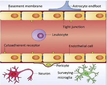
Microvascular endothelium, basement membrane, and glial cells such as astrocytes and pericytes work together to form the BBB (Fig. 2).
Figure 2. BBB composition. The BBB is mainly composed of vascular endothelial cells, highly
connected by adherens and tight junctions (TJs), and a sparse layer of pericytes. A basement membrane and a layer of astrocyte end-foot processes surround the endothelium. Neurons and surveying microglia are also important mediators of BBB integrity in physiological conditions. Adapted from Saraiva, 2016.
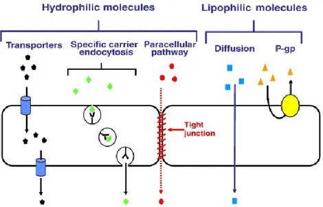
To sustain this robust barrier, CNS endothelial cells have properties distinct from endothelial cells in other tissues: the presence of BBB-specific transporter and receptor proteins to control entry and exit of metabolites across cells (transcellular transport); high electrical resistance tight junctions (TJs) to limit movement between adjacent cells
20 (paracellular transport); low levels of transcytotic vesicles compared to peripheral endothelia and an absence of fenestrae (small pores that allow rapid passage of molecules in peripheral endothelial cells) (Keaney et al., 2015).
The relative impermeability of the BBB results from tight junctions between capillary endothelial cells which are formed by cell adhesion molecules. Brain endothelial cells also possess few alternate transport pathways (e.g., fenestra, transendothelial channels, pinocytotic vesicles), and express high levels of active efflux transport proteins, including P-glycoprotein (P-gp), Multidrug Resistance Protein-1 (MRP-1), and breast cancer resistance protein. The BBB also has additional enzymatic aspects which serve to protect the brain (Gabathuler, 2010). Some small molecules with appropriate lipophilicity, molecular weight (Mw) and charge will diffuse from blood into the CNS. However, the majority of small molecules (mwN500 daltons, D), proteins and peptides do not cross the BBB. It has been reported that approximately 98% of the small molecules and nearly all large molecules (mwN1 kD, kilodaltons), such as recombinant proteins or gene-based medicines do not cross the BBB (Pardridge, 1998). Therefore, to reach the brain, most molecules must cross the BBB through interactions with specific transporters and/or receptors expressed at the luminal (blood) side of the endothelial cells. Crossing the BBB remains a key obstacle in the development of drugs for brain diseases despite decades of research. The minimal BBB transport of the majority of all potential brain therapeutic agents, leads predictably to the current situation, which is that there are few effective treatments for the majority of CNS (Pardridge, 2005).
A schematic representation of different mechanisms used to cross the BBB is shown in Fig. 3.
21
Figure 3.Schematic representation of the transport of molecules across the BBB.
From Gabathuler, 2010.
2. NANOTECHLOGY-BASED DRUG DELIVERY SYSTEMS
“I want to build a billion tiny factories, models of each other, which are manufacturing simultaneously. . . The principles of physics, as far as I can see, do not speak against the possibility of maneuvering things atom by atom. It is not an attempt to violate any laws; it is something, in principle, that can be done; but in practice, it has not been done because we are too big” (Feynman, 1960).
This quote is part of a lecture titled “There’s plenty of room at the bottom” by physicist Richard Feynman in 1959, that introduced the concept of nanotechnology as an important field for future scientific researches (Feynman, 1960). Feynman described a process in which scientists would be able to manipulate and control individual atoms and molecules. Over a decade later, Professor Norio Taniguchi coined the term nanotechnology (Nikalje, 2015). Nanotechnology is the study and application of extremely small things and can be used across all the science fields, such as chemistry, biology, physics, materials science, engineering, medicine and pharmaceutics.
22 Nowadays scientists are finding a wide variety of ways to make materials at the nanoscale to take advantage of their enhanced properties such as higher strength, lighter weight, increased control of light spectrum, and greater chemical reactivity than their larger-scale counterparts (Nano.gov http://www.nano.gov/nanotech-101).
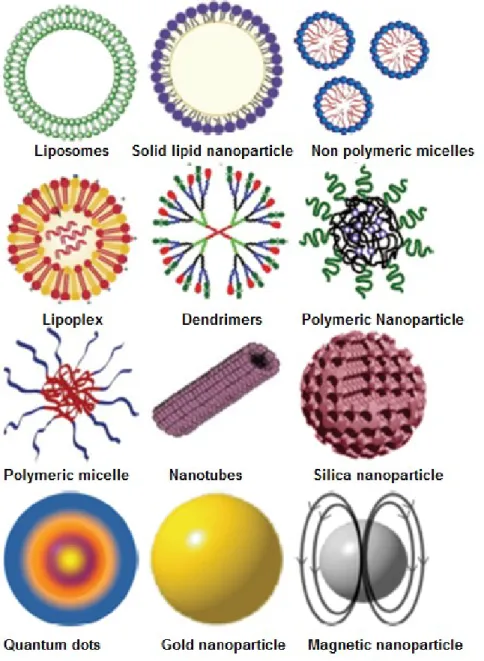
Nanotechnology has been used in medicine for the targeted delivery and/or controlled release of therapeutic agents and the development of treatments for a variety of diseases (Safari et el., 2014). Unfortunately, many drugs, even those discovered using the most advanced molecular biology strategies, have unacceptable side effects due to off-target adverse effects (modulation of unintended targets). Consequently, optimal design of medications for many diseases such as cancer, neurodegenerative and infectious diseases is limited. Drug delivery systems (DDS) present indubitable benefits such as control the rate at which a drug is released and the location in the body where it is released reducing dosing frequency and improving shelf life by enhancing its in vivo stability. Several types of nano-sized carriers, such as polymeric nanoparticles, solid lipid nanoparticles, nanostructure lipid carriers, nanocrystals, ceramic nanoparticles, magnetic nanoparticles, polymeric micelles, polymer-drug conjugates, lipid drug conjugates, nanotubes, nanoshells, nanowires, nanocages and dendrimers are being developed for various drug-delivery applications (Mukherjee, 2013; Mostafavi et al., 2013). In figure 4 are shown the most commonly used NPs for biomedical applications.
23
Figure 4.The types of NPs that are more popular for biomedical applications.
The DDS may encourage the use of therapeutic agents that were previously unsafe for disease treatment and targeted carriers may also help to address multi-drug resistant diseases.
This contributes to increase safety, efficacy, patient compliance, extending shelf life of drugs (Saha et al., 2015). Development of a novel drug-carrier system requires considerations of multiple factors. For example, after a drug is selected, a suitable
24 delivery route, drug release mechanism and kinetics, and proper materials selection have to be taken into account (Park, 2014). The use of biodegradable polymers for biomedical applications is continually increasing and evolving especially their application in nanotechnology systems for drug delivery (Bret et al., 2011; Numata et al., 2013). The main advantages of nanotechnological systems are reported in table 1.
Table 1.Main advantages of nanotechnological systems.
ADVANTAGES OF NANOTECHNOLOGICAL SYSTEMS Advantages
Particle size and surface characteristics of nanosystems can be easily manipulated to
achieve both passive and active drug targeting
Deliver/transport relevant drugs to the brain overcoming the presence of blood–brain barrier
Selective localization in specific tissues
Control and sustain release of the drug during the transportation and at the site of localization, altering pharmacokinetic profile of the active compound, increasing therapeutic efficacy and reducing the
side effects
Control release and particle degradation characteristics by choosing the constituents of the matrix
Site-specific targeting by attaching the target ligands to the carrier surface or by using magnetic guidance
Administration by various routes such as oral, nasal, parenteral, intra-ocular etc
Better transmission and retention of the drug in the tumors and inflamed tissues
Nanotechnology has the potential to revolutionize the medical area with new tools for the molecular treatment of diseases, and rapid disease detection. It advances materials with a nanodimension and provides several means for innovative design of nanosize drug delivery systems to overcome biological barriers (Athar et al., 2014).
25
2.1. Polymers used in preparation of nanoparticles
Nanocarriers based on different engineering hyperbranched polymer types, dendrimers, micelles, hydrogel are a growing area of present-day pharmaceutical research, due to their unique properties and large potential in drug delivery (Athar et al., 2014; Chen et al., 2016). For using polymers in drug delivery, a polymer must exhibit essential characteristics such as biocompatibility, biodegradability, flexibility, minimal side effectsand should improve drug release kinetics, as a result of its erosion or degradation in addition to drug diffusion through the polymeric material (Liechty et al., 2010). Polymer must meet specific quality criteria as reported by Safari and collegues: a. Biocompatibility backbone of the polymer and its degradation products. b. Mechanical strength sufficient to meet the needs of specific applications.
c. Degradability with degradation kinetics matching a biological process such as wound healing.
d. Processibility using available equipment. e. Solubility in various solvents.
f. Chemical, structural and application versatility. g. Economically acceptable shelf life.
h. Approval by European Medicine Agency (EMA) or Food and Drug Administration (FDA), USA (Safari et al., 2014).
The use of polymers as biomaterials has greatly impacted the advancement of modern medicine. Polymeric biomaterials that are biodegradable provide the significant advantage of being able to be broken down and removed after they have served their function. Their main advantage is that the products of degradation are not toxic/ non-harmful or/and are completely and easily eliminated from the body by natural metabolic
26 pathways with minimal side effects (Marin et al., 2013). These degradation products define the biocompatibility of a polymer. For example, polymers derived from glycolic acid and from D,L-lactic acid enantiomers are presently the most attractive compounds because of their biocompatibility and their resorbability through natural pathways. Degradation of PLA or PLGA occurs by autocatalytic cleavage of the ester bonds through spontaneous hydrolysis into oligomers and D,L-lactic and glycolic acid monomers (Liu et al., 2006). Lactate converted into pyruvate and glycolate enter the Krebs’ cycle to be degraded into CO2 and H2O. Bazile and collegues found that after intravenous administration of 14C-PLA radiolabeled nanoparticles to rats, 90% of the recovered 14C was eliminated within 25 days, among which 80% was as CO2. As reported by the authors, the elimination of the 14C was quick on the first day (30% of the administered dose) but then slowed down. In fact, if the metabolism of the PLA proceeds to lactic acid which is rapidly converted into CO2 (80% of the total excretion was fulfilled by the lungs), anabolism from the lactic acid may also have taken place leading to long-lasting radioactive remnants, by incorporation of 14C into endogenous compounds (Bazile et al., 1992).
Several parameters influence the degradation rate, including: hydrolysis rate constant (correlated with the molecular weight, the lactic/glycolic ratio, and the morphology), amount of water absorbed, diffusion coefficient of the polymer fragments through the polymer matrix, and solubility of the degradation products in the surrounding aqueous medium. In turn, all these parameters are influenced by temperature, additives (including drug molecules), pH, ionic strength, buffering capacity, size and processing history, steric hindrance etc. (Olivier et al., 2005).
27 The major mechanisms of degradation for polymers are hydrolysis, oxidation, or enzymatic reactions (Untereker et al., 2009). Numerous natural and synthetic polymers, (Fig.5), have been investigated as candidate for biomedical applications and new materials have been developed to meet new challenges (Ulery et al., 2013).
While natural polymers have been used in the medical field for thousands of years, research into biomedical applications of synthetic degradable polymers is relatively new, starting in the 1960s. In the fifty years since, successes have been numerous, but grand challenges still exist in both the basic and translational elements of biomaterial design.
Natural polymers, materials of both plant and animal origin, are the first option in biomedicine, Since they occur in nature, are often presumed to exhibit enhanced biocompatibility (Russell et al., 2014). However some problem could arise due to batch-to-batch variations in properties or risk of viral infections as reported by Hino et al., showing the transmission of parvovirus B19 by blood products such as fibrin which is widely used as a surgical adhesive, hemostatic agent, and sealant (Hino et al., 2000). Synthetic polymers, on the other hand, have attracted researchers’ interest because of their manufacturing flexibility and reproducibility and can be produced using many synthetic methods. Synthesis determines molecular structure with the purpose of achieving product with lower level of impurity. Several of the reactions involved in synthesis of these polymers, include ring opening, polycondensation, bulk synthesis, dehydrative coupling, transesterification, and polymerization.
28
Figure 5. Examples of natural and synthetic polymers.
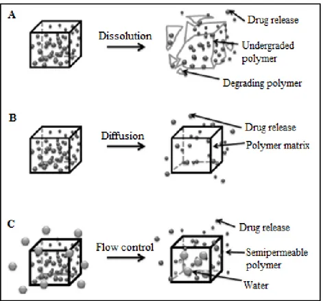
Controlled degradation of polymers helps to maintain drug levels within a suitable therapeutic window (Siegel et al., 2012). The degradation mechanism of a polymer is essential to achieve an efficient drug delivery, the polymeric matrix has to degrade under physiologic conditions in a controlled manner to allow sustained release of the drug as temporal drug delivery systems(Fig. 6).
29
Figure 6. Mechanisms for temporal controlled-release drug systems (A) Dissolution of a polymer with
slow break-down that delays exposure of drug to water from the environment of the delivery system. (B) Drug diffusion-controlled release through gaps in insoluble polymeric devices. (C) Controlled flow using osmotic forces on a semi-permeable polymer matrix. Adapted from Marin, 2013.
A delivery system promotes the continued release of drug in a specific period of time, which allows to maintain drug concentration in blood or target tissues at the therapeutic level (Uhrich et al., 1999; Lee et al., 2015). As a consequence the frequency of administration can be reduced. Temporal release delays diffusion of the molecule out of the polymeric matrix, inhibiting diffusion or controlling drug flow through the matrix. These strategies involve manipulation of some physicochemical properties of the polymers, e.g., copolymerizing or blending of polymers in order to change the degradation behavior.
30
2.2. Polymeric NPs
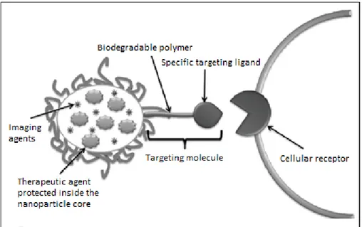
Nanoparticles (NPs) are the solid colloidal particles that can vary from 1 to 1000 nm in size, utilized as career for drug delivery (Saraiva et al., 2016). What makes NPs even more attractive for medical applications is the possibility of conferring on them features such as high chemical and biological stability, feasibility of incorporating both hydrophilic and hydrophobic compounds (in relation to the preparation method), and the ability to be administered by a variety of routes (i.e. oral, nasal, and parenteral). Moreover, NPs can be functionalized by covalent conjugation to various ligands to target specific tissues. Nanosized drug delivery systems can increase drug accumulation in specific tissue and/or reduce drug elimination via passive or active targeting. NPs provide massive advantages regarding drug targeting, delivery and release, and with their additional potential to combine diagnosis and therapy, emerge as one of the major tools in nanomedicine. In figure 7 is reported a strategy to create targeted drug delivery systems.
Figure 7. Strategy to create targeted drug delivery systems. Therapeutic tools like genes, proteins, and
small drug molecules, as well as imaging tools such as fluorescent probes or magnetic contrast agents are encapsulated inside the nanoparticle core. In parallel, targeting molecules like specific antibodies or recognition peptides are located on the nanoparticle surface. Adapted from Marin, 2013.
31 A targeted drug delivery system is based on the delivery of a certain amount of drug for a prolonged period of time to a targeted diseased area within the body. This helps maintain the required plasma and tissue drug levels in the body; therefore avoiding any damage to the healthy tissue via the drug and drug loss due to natural distribution in the body (Rani et al., 2014).
NPs can have a natural or synthetic origin. Synthetic NPs may be prepared from polymeric materials such as ethylenimine (PEI), alkylcyanoacrylates, poly-amidoamine dendrimers (PAMAM), poly-ε-caprolactone (PCL), poly-lactic-co-glycolic acid (PLGA), polyesters such as poly-lactic acid (PLA), or from inorganic materials such as gold, silicon dioxide (silica), among others. Inorganic NPs offer advantages over polymeric NPs in terms of control over size and shape and simplicity of preparation and functionalization but also have disadvantages because they might not be degraded or present undesired toxicity (e.g. carbon nanotubes and fullerenes may lead to lipid peroxidation and oxygen radical formation). On the other hand, natural NPs are produced from natural polymers, such as polysaccharides (chitosan, alginate), amino acids (poly-lysine), poly-aspartic acid (PASA), or proteins (gelatin, albumin).
Natural NPs have the advantage of providing biological interaction with specific receptors/transporters expressed by endothelial cells but they have the disadvantage of batch-to-batch variability, and poor tracking capacity by imaging platforms.
These carriers can transport drugs which may be bound in form of a solid solution or dispersion or be adsorbed to the surface or chemically attached (Tiwari et al., 2012). Depending on the method of preparation, nanocapsules or nanospheres can be obtained differing in their composition and properties such as the ability to encapsulate, deliver and release the active compound (Guterres et al., 2007). Nanocapsules are systems in
32 which the drug is confined to a cavity surrounded by a unique polymer membrane, while nanospheres are matrix systems in which the drug is physically and uniformly dispersed (Velavan et al., 2015). Knowledge of the physicochemical properties of the drug is crucial in order to select the best method of preparation and starting materials to prepare the carrier system with desired shape, diameter and surface properties and with good entrapment efficiency of the drug.
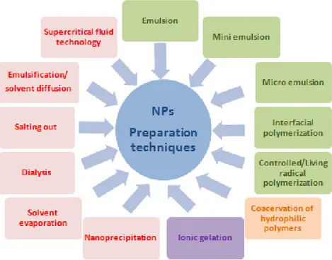
The essential aspects that NPs preparation methods should have are the use of less toxic reagents, the simple final composition, (minimal number of components/eccipients in the formulation), simplification of the procedure to achieve production scale-up and optimization to improve yield and entrapment efficiency (Nagavarma et al., 2012) . Preparation techniques are classified according to the initial state of the polymer into two main categories, NPs obtained from polymerization of a monomer or NPs obtained from a macromolecule (Fig. 8).
Figure 8. NPs techniques of preparation: methods for preparation of NPs from dispersion of preformed
polymer (red); methods for preparation of NPs from polymerization of monomers (green); other methods commonly used (violet; orange).
33 It is possible to obtain NPs with the desired properties for a particular application by combining the selection of raw materials and the preparation technique (Dinda et al., 2013). When formulating drug loaded NPs, it should always be kept in mind that in most cases, drug contents are 5-10%wt/wt of NPs weights or even less.
Therefore, generally about 90% of the material administered is NPs excipients with their potential toxicity. Thus, drugs with high intrinsic pharmacological activities should be preferred to avoid the administration of massive dose of NPs material (Olivier, 2005). Other important factors to be considered in designing a successful nanoparticulate system are the drug release and the polymer biodegradation.
The release profile is strictly related to drug solubility, desorption of the surface bound/adsorption of the drug and the diffusion of the drug through the polymeric matrix, degradation rate or erosion profile of the polymer matrix, and combination of erosion/diffusion process (Meena et al., 2011). Moreover, a successful nanoparticulate system should be physically stable without aggregation for prolonged period of time during storage and stable during in vivo administration.
Summarizing, the formulation must be scalable and follow cost effective manufacturing process; should be amenable to load small molecules, peptides, proteins, or nucleic acids; should be able to withstand minimal nanoparticle-excipient induced drug alteration, chemical degradation and protein denaturation and should have high drug loading capacity to reduce the quantity of the materials to be needed for its matrix formation and able to interact and overcome with biological barriers.
34
3.
NANOPARTICLES FOR BRAIN TARGETING: CROSSING OR
BYPASSING THE BLOOD-BRAIN-BARRIER
Nowadays the development of innovative approaches and new effective treatment for brain diseases is one of the primary goal for pharmaceutical companies and academic research and it is also the most expensive. The process of discovery and development of new drug for CNS disorders, is time-consuming and very expensive.
The average cost of getting a drug onto the market is ever increasing and now approaching US$1 billion before reaching the consumer (Tsaioun et al., 2009). Findings and advances in the field of nanomedicine have generated strategies that improve drug transport across/bypass the BBB, such as the use of NPs, currently under intensive investigation (Saraiva et al., 2016). The current challenges are to design and formulate drug delivery carriers, which must be able to deliver the drug to the brain safely and effectively.
Among different delivery systems, NPs seem to be efficient systems in delivery of conventional drugs, recombinant proteins, peptides, vaccines as well as nucleotides. These last molecules may be advantageously formulated in brain-targeted nanocarriers in order to be protected from their poor stability in biological fluids, rapid enzymatic degradation, unfavorable pharmacokinetic properties, and lack of diffusion toward the CNS. Moreover, the small dose requested for therapeutic activity could easily fit the loading capacity of NPs and would not require the administration of large amount of potentially toxic excipients (Olivier, 2005).
35
3.1. Crossing the Blood-brain-barrier
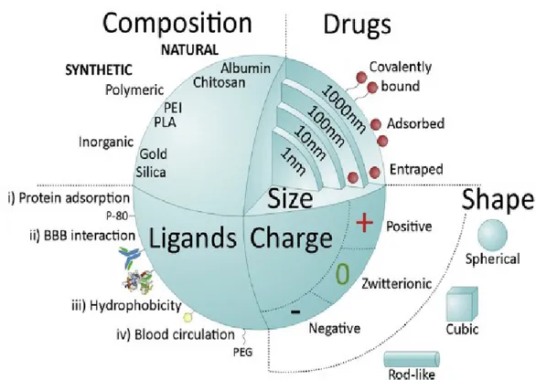
NPs are exciting systems for brain drug delivery due to the possibility to modulate them in terms of composition, shape, size, hydrophobicity, coating, chemistry, surface charge and ligands (Fig.9) (Lahkar et al., 2013). Control over these features can enhance the ability of NPs to improve the therapeutic agent stability in biological environment, to control the cargo release into the desired target site, to enhance BBB penetration efficiency and to escape the reticuloendothelial system.
Figure 9. Main NPs features influencing systemic delivery and BBB passage. From Saraiva, 2016.
NPs may be because of its size and functionalization characteristics able to penetrate, overcome and facilitate the drug delivery through the barrier (Tosi et al., 2013). There are different mechanisms and strategies found to be involved in this process, which are based on the type of nanomaterials used and its combination with therapeutic compounds. The use of these nanosystems is expected to reduce the need for invasive
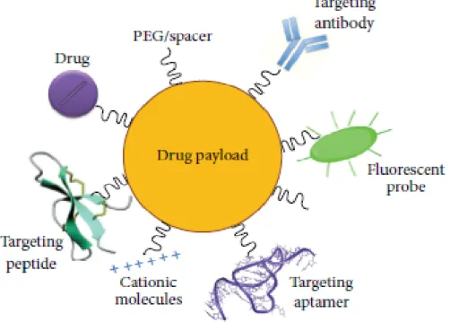
36 procedures for delivery of therapeutics to the CNS (Dinda et al., 2013). Many strategies are under investigation in order to enhance drug delivery to the brain such as NPs functionalization with different types of ligands. (Fig. 10).
Figure 10. Multifunctionalized NPs. Graphical representation of surface-modified NPs with drugs
(incorporated within the core of NPs or coniujated to the surface), targeting molecules (antibodies, peptides, aptamers, and cationic molecules) for brain drug delivery, with PEG for stealthiness and with fluorescent probe as a tracer.From Masserini, 2013.
NPs could play this role at least in two ways: (i) by increasing the drug concentration inside, or at the luminal surface of BBB cells, establishing a local high concentration gradient between blood an brain, higher than that obtainable after administration of the free drug. The gradient should then favor the enhanced passive diffusion of the drug; (ii) by moving themselves into the CNS, together with their drug cargo (Masserini, 2013). Ligands are distributed into four major categories: i) capable of mediating protein adsorption (e.g. poly-sorbate 80, P-80); ii) able to interact directly with the BBB (e.g. transferrin proteins, antibody or peptides); iii) capable of increasing hydrophobicity (e.g. amphiphilic peptides); and iv) able to improve blood circulation (e.g. poly-ethylene
37 glycol, PEG). NPs can assume different shapes and charges (negative, zwitterionic, positive).
The shape of NPs also influences body distribution and cellular uptake. The shape of NPs can vary from spherical, cubic, rod-like, among other forms (Fig. 9).
It has been well demonstrated that the size, coating and surface charge of NPs have a crucial impact on the intracellular uptake process as demonstrated by Shilo and collegues which investigated the effect of NPs size on the probability to cross the BBB, using the endothelial brain cell model and found that intracellular uptake of gold NPs is strongly dependent on gold NPs size (Shilo et al., 2015). Another study performed by Georgieva and collegues demonstrated that surface modifications of NPs, (of a fixed sizes), including charge and protein ligands, affect their mode of internalization by brain endothelial cells and thereby their subcellular fate and transcytotic potential. They found that the coupling of a ligand or charge at the surface of a nanoparticle of a given size modifies its entry pathway and processing in human BBB endothelial hCMEC/D3 cells. Careful analyses suggest that uncoated NPs do not enter in an all-or-nothing or exclusive pathway but following surface modification show preference for a specific pathway(s) and as a consequence NPs are delivered to intracellular compartments that are distinct with regard to their ultrastructural morphology and composition (Georgieva et al., 2011). Therefore, several parameters influence the transport of NPs through the BBB at different extents. So far, NPs conjugated with ligands able to interact with BBB receptors at a relatively low density have the best performance.
NPs brain delivery improvement might require systems that target and cross efficiently the BBB but also systems that are slowly clear from the bloodstream.
38 Many studies have demonstrated that the surface charge and the morphology of the NPs have a very important effect in the clearance. Neutral and zwitterionic NPs have a longer circulation time after intravenous administration, in contrast to negatively and positively charged NPs (Arvizo et al., 2011). Moreover, as reported by Huang and collegues, short-rod NPs are preferentially retained in the liver and present a rapid clearance rate, while long-rod NPs are caught in the spleen and have a lower clearance rate. If the surface is modified with PEG, retention increases in lung for both formulations (Huang et al., 2011).
3.2. Bypassing the Blood-brain-barrier
Because of the difficulty for drugs or particles to cross the BBB, alternative ways should be considered. “Bypass” rather than “cross” could be one of these.
There is a route to deliver drugs or NPs directly from the nasal cavity to the brain, which is intranasal delivery. This route is considered a promising strategy in brain-targeted drug delivery, it provides a non-invasive method of bypassing BBB to deliver therapeutics into the brain (Illum, 2000).
Delivery of the molecules occurs mainly through olfactory and trigeminal nerve systems in the nasal epithelium to the olfactory bulb and brainstem and further to different parts of the brain. This topic is widely described in section 4. Nose-to-brain.
4. NOSE-TO-BRAIN
Commonly, the nasal route has been used to administer topically acting molecules to treat local diseases, anti-allergic drugs and nasal decongestants are the most typical examples (Djupesland, 2013). During the last decades, intranasal (IN) administration
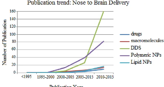
39 has gained great attention in research and has been investigated extensively with regard to its feasibility to serve as a direct drug transport route to the CNS as demonstrated by the increasing number of publications in this field (fig.11) (Kozlovskaya et al., 2014). To identify scientific publications reporting data on drugs, macromolecules, DDS and more specifically polymeric and lipid nanoparticles to the brain via the nasal route, searches in the PubMed database (http://www.ncbi.nlm.nih.gov/pubmed/) have been performed. Screening of the publications was performed, (based on their abstract, and subsequently on their full text), to identify the publications that were suitable for this analysis.
Figure 11. The publication trends in the field of drugs, macromolecules, DDS, polymeric and lipid
nanoparticle for brain delivery and targeting via the nasal route. 2015 PubMed database, keys words: intranasal administration to brain macromolecules, drug delivery systems (DDS), polymeric nanoparticles, lipid nanoparticles; Temporal range: From 1995 to 2015.
As shown in Fig.11 the intranasal route has gained interest during the last decade, above all the studies regarding DDS. Among these, polymeric NPs seem to be excellent candidates for brain delivery.
40 The widespread interest in IN route for therapeutic purposes arises from the particular anatomical, physiological and histological characteristics of the nasal cavity.
“The nose is the only natural corridor where the brain meets the outside world” (cit. by
Dr. Gerallt Williams) (Djupsland, 2014).
The olfactory neuroepithelium located inside the nasal cavity is the only area of the body in direct contact with both the CNS and the external environment, which opens up for therapeutic treatments (Chang et al, 2014 ; Sveinbjorn, 2012).
Furthermore, IN administration avoids the gastrointestinal and hepatic metabolism, enhancing drug bioavailability and allowing a lower therapeutic drug dose and fewer systemic side effects (Shabana et al., 2015). Additionally, it also offers several practical advantages either from the viewpoint of patients (non-invasiveness, essentially painless, ease drug delivery and favorable tolerability profile) and pharmaceutical industry (i.e. unnecessary sterilization of nasal preparations) (Pires et al., 2009). Table 2 reports some advantages and limitations of nose-to-brain delivery. Hence, it seems to be an encouraging route for the treatment of acute and also chronic conditions requiring considerable drug exposure.
Although IN route to improve access to the systemic circulation (due to the highly vascularized mucosa) is important for some applications, it is the potential for circumventing the systemic circulation and delivering drugs directly into the brain that represents a particularly novel, attractive and little understood application of IN delivery. Direct transport of active molecule along the olfactory and trigeminal nerves is increasingly considered a promising route whereby drugs delivered to the nose can access the CNS in therapeutic concentrations (Djupsland, 2014).
41
Table 2. Advantages and limitations of nose-to-brain drug delivery.
ADVANTAGES AND LIMITATIONS OF NOSE TO BRAIN DRUG DELIVERY
Advantages Disadvantages
Rapid, safe, non-invasive and convenient method
Rapid elimination of drug substances from nasal cavity due to mucocilliary clearance Avoids drug degradation in gastrointestinal
tract, first-pass metabolism and gut-wall metabolism of drugs, allowing enhanced
bioavailability
Absorption enhancers used in formulation may create mucosal toxicity
Reduction of systemic exposure of drugs and systemic side effects
Variability in the concentration attainable in different regions of brain and spinal cord Bioavailability for low molecular weight
drugs
Nasal congestion due to cold or allergic condition may interfere with this technique
of drug delivery Rapid drug absorption via highly
vascularized mucosa
Suitable for potent drugs since only a limited volume can be sprayed into the
nasal cavity Ease of administration
(self-administratiom), Improved convenience, Better patient compliance
Frequent use of this route leads to mucosal damage irritation of nasal mucosa Convenient route when compared with
parenteral route for long term therapy
Mechanical loss of the dosage form could occur due to improper technique of
administration Bioavailability of larger drug molecules
can be improved by means of absorption enhancer or other approach
Mechanisms of drug transport are still unclear
4.1. Pathways and mechanisms
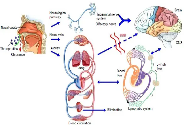
Although the exact mechanisms underlying nose-to-brain delivery are not entirely understood yet, an accumulating body of evidences demonstrates that pathways involving nerves connecting the nasal passages to the brain and spinal cord are important (Fig.12). Moreover, pathways involving the vasculature, CSF and lymphatic
system have been employed in transport of molecules from nasal cavity to the CNS. It is possible that a combination of these pathways is responsible, even if one pathway may
42
predominate, depending on the properties of active compounds, the characteristics of formulations and the delivery device used.
Figure 12. Olfactory and trigeminal pathways to the CNS. From Cui-Tao Lu et al., 2014.
The delivery from the nose to the CNS may occur via olfactory neuroepithelium and may involve paracellular, transcellular and/or neuronal transport. Paracellular pathway through tight junctions between sustentacular cells or the so-called clefts between sustentacular cells and olfactory neurons. This is slow and passive route and it is responsible for transport of hydrophilic drugs and it shows rate dependency on the molecular weight of a drug (Belgamvar et al., 2013). Transcellular process is responsible for the transport of lipophilic drugs that show a rate dependency on their lipophilicity.Expecially across the sustentacular cells, most likely by receptor-mediated endocytosis, fluid phase endocytosis or by passive diffusion. It is mediated rapidly and at a high rate (Pavuluri et al., 2015). Neuronal transport in which drug is taken up into
43 the neuronal cell by endocytosis or pinocytosis mechanisms and transported by intracellular axonal transport to the olfactory bulb (Mustafa et al., 2016).More recently, the contribution made by the trigeminal pathway to IN delivery to the CNS has also been recognized, especially to caudal brain regions and the spinal cord. The first researchers that clearly demonstrated the involvement of this pathway wereThorne and colleagues which assessed the potential of delivering insulin-like growth factor-I (125I-IGF-I), directly into the CNS following IN administration and they elucidated the mechanisms involved in the transport (Thorne et al., 2004). They found high levels of radioactivity in the trigeminal nerve branches, trigeminal ganglion, pons, and olfactory bulbs, consistent with delivery along both trigeminal and olfactory nerves.
Trigeminal nerve innervates the respiratory and olfactory epithelium of nasal cavity and enters the CNS in the pons and represents another important pathway connecting nasal cavity to the CNS. Interestingly, a small portion of trigeminal nerve also terminates in the olfactory bulbs. The trigeminal nerve-mediated transport pathway also plays a key role in the distribution of intranasally administered drugs to brain areas distant from the olfactory bulbs.
The ophthalmic and maxillary branches of trigeminal nerve are important for nose to brain drug delivery as neurons from these branches pass directly through the nasal mucosa. A unique feature of the trigeminal nerve is that it enters the brain from the respiratory epithelium of the nasal passages at two sites: i) through anterior lacerated foramen near the pons and ii) through the cribriform plate near olfactory bulb, creating entry points into both caudal and rostral brain areas following IN administration. Because one portion of the trigeminal neural pathway enters the brain through the cribriform plate alongside the olfactory pathway, it is difficult to distinguish whether
44 intranasally administered drugs reach the olfactory bulb and other rostral brain areas via the olfactory or trigeminal pathways or if both are involved.
In addition to these direct pathways, transport may also occur via blood vasculature, lymphatics, and cerebrospinal fluid present in the nasal mucosa tissue (Kozlovskaya et al., 2014). The nasal mucosa is highly vascularized, the relative density of blood vessels is greater in the respiratory mucosa compared to the olfactory mucosa making the region an ideal site for absorption into the blood. The blood vessels with continuous and fenestrated endothelium allow passage of both small and large molecules to enter the systemic circulation following nasal administration (Dhuria et al., 2010). The drug that has been absorbed into the systemic circulation has to cross the BBB in order to reach the CNS. However, many problems may arise with the systemic delivery due to drug elimination via hepatic and renal mechanisms, and some other limiting factors such as: the BBB, drug binding to plasma proteins, degradation by plasma proteases, and potential peripheral side effects (Alavijeh et al.,2005).
4.2. Free molecules delivery via nose-to-brain
Several investigations have been reported concerning the transport of free drug from the nasal cavity to the CNS. Considering the large number of compounds that have been shown in animal models to be directly transferred from the nasal cavity to the olfactory bulb there should be no doubt that this type of transfer occurs (Mathison et al., 1998; Illum, 2000). The question of whether this form of transfer is sufficiently extensive to result in therapeutically effective concentrations at the site of action in humans, however, remains to be answered.
45 Very interesting experiments were performed by Serrailhero and collegues, using an antiepileptic drug, carbamazepine. They assessed the pharmacokinetics of carbamazepine administered via the IN and intravenous (IV) routes to mice, and investigate whether a direct transport of the drug from nose to brain could be involved. The similar pharmacokinetic profiles obtained in all matrices following both administration routes indicate that, after IN delivery, carbamazepine reaches quickly and extensively the bloodstream, achieving the brain predominantly via systemic circulation.
However, the uneven biodistribution of carbamazepine through the brain regions with higher concentrations in the olfactory bulb and frontal cortex following IN instillation, in comparison with the homogenous brain distribution pattern after IV injection, strongly suggests the involvement of a direct transport of carbamazepine from nose to brain. Regarding the mean residence time parameter (MRT), higher values were attained for plasma and brain after IN administration comparatively to IV administration, in contrast with the liver, where the highest MRT value was assigned to the IV route (Serralheiro et al., 2014). Westin and collegues investigated whether morphine can be transferred along the olfactory pathway to the CNS. In their study the autors found [3H]-morphine in the CNS surrounding the olfactory bulbs by autoradiography in rats within 5 minutes of IN administration (Westin et al., 2007). Morphine was found in the olfactory bulb ipsilaterally to the side of the nasal cavity that was administered with the dose after 60 min and a gradient of radioactivity was found also in the brain when higher level of morphine were found closer to the cribriform plate. However, no significant penetration of the radioactivity was detected in deeper brain areas.
46 Many scientific publications shown that there are evidence of drug directly delivered to the brain after the IN administration but the key finding from all these studies is that the amount of the therapeutic agent into the CNS is normally minimal, less than 1% of the drug administered very low compared to the dose administered (about 0.12% of the administered dose as reported by Jansson) (Charlton et al., 2007; Jansson, 2004; Sakane et al., 2004; Sakane et al., 1991). The optimization of nasal administration using DDS represents a possible strategy to overcome this problem. Moreover, drug should have specific properties to be administered intranasally, as reported in table 3.
Table 3.Summary of drug properties required for nasal delivery.
SUMMARY OF DRUG PROPERTIES REQUIRED TO IMPROVE NASAL DELIVERY
Given the low volume of nasal cavity, the drug's solubility in water must be high enough to accommodate the necessary dose
For high bioavailability, a drug must be resistant to metabolizing enzymes in the nasal environment
Drug residence time in contact with the mucosal membrane is an important factor influencing drug absorption
Potential local toxicities need to be considered in parallel with benefits
Nasal solutions with tonicities ranging from0.6–1.8% NaCl equiv. are well tolerated, 0.9% NaCl equiv. being isotonic.
The nasal cavity can accommodate only a low solution volume, necessitating highly concentrated nasal drug solutions
Drug delivery technologies represent a good strategy to improve drug properties for nasal delivery and can also modify drug release profile, absorption, distribution and elimination for the benefit of improving product efficacy and safety, as well as patient convenience and compliance (Mittal et al., 2014; Kapoor et al., 2016).
Furthermore, DDS can protect the encapsulate compound from degradation because the nasal mucosa retains some enzymatic activity (Mistry, 2009). The pharmacokinetic