Alma Mater Studiorum – Università di Bologna
DOTTORATO DI RICERCA IN
Scienze Veterinarie
Ciclo XXIX
Settore Concorsuale di afferenza: 07/H3 – MALATTIE INFETTIVE E PARASSITARIE DEGLI ANIMALI
Settore Scientifico disciplinare: VET/05 – MALATTIE INFETTIVE DEGLI ANIMALI DOMESTICI
TITOLO TESI
Finfish and human pathogens in bivalve molluscs
Presentata da: dott. Enrico Volpe
Coordinatore Dottorato
Relatore
Prof.ssore Arcangelo Gentile
Dott.ssa Sara Ciulli
Contents
Preamble 1
CHAPTER 1 3
1.1 ABSTRACT 3
1.2 INTRODUCTION 4
1.3 MATERIALS AND METHODS 5
1.3.1 Bivalve mollusc and viruses 5 1.3.2 RNA extraction, RT-PCR and nested PCR 7 1.3.3 Sequencing and phylogenetic analysis 7
1.4 RESULTS 8
1.4.1 Virus detection 8
1.4.2 Sequencing and phylogenetic analysis 8
1.5 DISCUSSION 14 1.6 CONCLUSIONS 15 1.7 REFERENCES 16 CHAPTER 2 19 2.1 ABSTRACT 19 2.2 INTRODUCTION 20
2.3 MATERIALS AND METHODS 21
2.3.1 Clam maintenance and RGNNV screening 21 2.3.2 Cell culture maintenance and virus propagation 22 2.3.3 Culture analysis of clam hepatopancreas, faecal matter and water samples 22 2.3.4 Detection limit of TCID50 endpoint dilution assay in RGNNV-inoculated
clam hepatopancreas homogenates
23
2.3.5 RGNNV clam exposure trial 23 2.3.6 Clam RGNNV shedding trials 24
2.3.7 Statistics 24
2.4 RESULTS 25
2.4.1 Clam maintenance and VNNV screening 25 2.4.2 Detection limit of TCID50 endpoint dilution assay in RGNNV-inoculated
clam hepatopancreas homogenates
25
2.4.3 RGNNV clam exposure trial 26 2.4.4 Clam RGNNV shedding trial 27
2.5 DISCUSSION 30 2.6 CONCLUSIONS 32 2.7 REFERENCES 32 CHAPTER 3 37 3.1 ABSTRACT 37 3.2 INTRODUCTION 38
3.3 MATERIALS AND METHODS 40
3.3.1 Disinfectant and experimental design 40 3.3.2 Bactericidal activity assay 41 3.3.3 Virucidal activity assay 41
3.3.4 Depuration assay 42
3.3.5 Artificial contamination with VNNV, Betanodavirus and specific depuration assay
44
3.3.6 Biochemical analyses 44
3.3.7 Cortisol measurement 45
3.3.8 Histological examination 45 3.3.9 Sodium dodecylbenzenesulfonate (LAS12) quantification 46 3.3.10 Statistical analysis 46
3.4 RESULTS 47
3.4.1 Bactericidal activity 47
3.4.2 Virucidal activity 48
3.4.3 Depuration assay 50
3.4.4 Artificial contamination with VNNV, Betanodavirus and specific depuration assay
51
3.4.5 Biochemical analyses 52
3.4.6 Cortisol analyses 54
3.4.7 Histopathological analyses 54 3.4.8 Sodium dodecylbenzenesulfonate (LAS12) quantification 55
3.5 DISCUSSION 56
3.6 CONCLUSIONS 59
3.7 REFERENCES 59
Final considerations 66
1
Preamble
Bivalve molluscs are an important food source for living beings, including humans. Aquaculture provides 89.6% of global bivalve mollusc production, which plays an important role in ensuring food and employment to the world population (FAO 2013).
Bivalve molluscs are obligated filter feeders, that feed on microalgae, bacteria and organic particles present in the aquatic environment. Accordingly, they could accumulate chemical compounds, marine biotoxins, bacteria and viruses, including human and animal pathogens (Molloy et al., 2013; Serratore et al., 2014).
The interaction between microorganisms and bivalve molluscs, both in natural and artificial environments, such as integrated multitrofic aquaculture, might influence the epidemiology of animal and human infectious diseases (Skår & Mortensen, 2007).
Some studies, investigating the interaction between bivalve molluscs and fish pathogens, show that
Infectious salmon anemia virus (ISAV) is inactivated from Atlantic mussels (Mytilus edulis)
(Molloy et al. 2014), whereas Infectious pancreatic necrosis virus (IPNV) could be transmitted from contaminated Atlantic mussels to Atlantic salmon (Salmo salar) (Molloy et al. 2013).
Previous studies, performed by the virology research group of the DIMEVET, have shown a wide presence of betanodaviruses in bivalve molluscs including Manila clam (Ruditapes philippinarum), mussels (Mytilus galloprovincialis) and oysters (Crassostrea gigas) (Ciulli et al., 2010). Betanodaviruses are small ssRNA viruses of the genus Betanodavirus, family Nodaviridae (Thiéry et al., 2012) responsible of viral encephalopathy and retinopathy (VER), otherwise known as viral nervous necrosis (VNN), one of the most threatened disease in marine aquaculture worldwide. The Ph.D thesis, arranged in three chapters, deals with finfish and human pathogens in bivalve molluscs and focus on betanodavirus presence in bivalve molluscs, on their interaction with the
Redspotted grouper nervous necrosis virus (RGNNV), a viral species of the genus Betanodavirus,
and the development of a novel method to mitigate bacterial and viral contaminations of bivalve molluscs.
The first chapter reports a research on the molecular detection and phylogenetic analysis of betanodaviruses in bivalve molluscs collected from 2008 to 2015, different European countries and three species. In this study, detected viruses have been analyzed genetically to find out whether bivalve molluscs could be a source of genetically close related betanodaviruses to finfish. In fact, the finding of betanodaviruses in bivalve molluscs strictly related to finfish betanodaviruses could pose a possible risk of inter-specific transmission.
2 The second chapter focused on the fate of RGNNV in experimentally challenged Manila clam to investigate the potential role of clams as an RGNNV reservoir and the potential risks posed by RGNNV-contaminated molluscs.
The third study deals with sea water disinfection to complement and improve the microbial depuration of Manila clams. A novel sea water disinfection process was tested on Manila clam by employing a potassium peroxymonosulfate (MPS)-based product. The biocidal activity of the disinfection process was evaluated against both bacterial and viral contaminants of bivalve molluscs. Particularly, bactericidal activity was evaluated against the Vibrio spp. population naturally associated with sea water that include several human and finfish pathogens; virucidal activity was assessed against VNNV, the most threatening among the viral pathogens of marine finfish.
3
CHAPTER 1
Molecular detection and phylogenetic analysis of betanodaviruses in bivalve molluscs
Volpe Enrico1, Grodzki Marco1, Serratore Patrizia1, Guercio Annalisa2, Ciulli Sara1
1
Department of Veterinary Medical Science, Alma Mater Studiorum, University of Bologna, Italy;
2
Istituto Zooprofilattico Sperimentale della Sicilia, Palermo, Italy
1.1 ABSTRACT
Betanodaviruses are small ssRNA viruses of the family Nodaviridae responsible of viral encephalopathy and retinopathy (VER) otherwise known as viral nervous necrosis (VNN) in marine fishes worldwide. They can be transmitted both vertically and horizontally and invertebrates, where they have been detected sporadically, have been suspected to be a source of the virus.
This is the first study focusing on betanodavirus in bivalve molluscs. Twenty seven new betanodavirus strains were detected in bivalve molluscs of different species, European countries and year of collection and genetically characterized.
Betanodaviruses detected in mollusc bivalve and in finfish are very closely related to betanodaviruses previously detected in finfish in Southern Europe from 2000 to 2009. However, also a new betanodavirus strain not belonging to any of the already known betanodavirus genotypes was detected.
Such a massive and variegate presence of betanodaviruses in bivalve molluscs greatly stresses the risks of transmission previously feared for other invertebrates. Molluscs bivalve reared in the same area of farmed and wild finfish could act as a reservoir of virus. Furthermore, the marketing of alive bivalve mollusc and the relaying activity, allowed by the European regulation, can pose also a real risk of spreading betanodavirus between different geographical areas.
4
1.2 INTRODUCTION
Betanodaviruses are small ssRNA viruses of the genus Betanodavirus, family Nodaviridae (Thiéry et al., 2012) responsible of viral encephalopathy and retinopathy (VER), otherwise known as viral nervous necrosis (VNN), in several fish species worldwide. Betanodavirus genome consist of two segments named RNA 1 (3.1 KB) and RNA 2 (1.4 kb) coding respectively for the RNA-dependent RNA polymerase and the coat protein. Moreover, during virus replication, a subgenomic transcript called RNA3 is originated from the 3’ terminus of RNA1 (Iwamoto et al., 2005; Thiéry et al., 2012). Based on phylogenetic analysis of the T4 variable region within the RNA 2 segment, betanodaviruses have been historically divided into four genotypes, currently accepted as official species of this genus: Striped jack nervous necrosis virus (SJNNV), Tiger puffer nervous necrosis
virus (TPNNV), Barfin flounder nervous necrosis virus (BFNNV) and Redspotted grouper nervous necrosis virus (RGNNV) (Nishizawa et al. 1997; Thiéry et al., 2012). Demarcation of species is
mainly based on genetic characterization, however, more genotypes than that recognized as species has been described such in the case of Atlantic cod nervous necrosis virus (ACNNV) (Gagné et al., 2004) and the turbot nodavirus (TNV) (Johansen et al., 2004). Although, genotyping was based on RNA 2 phylogenetic analysis (Nishizawa et al. 1997), RNA 1 phylogenetic analysis added further information showing the presence of reassortant strains (Olveira et al., 2009; Toffolo et al., 2007). As a matter of fact, the presence of reassortant betanodaviruses SJNNV/RGNNV has been previously described in sea bass (Dicentrarchus labrax) caught in Italy and Croatia, in the form of a genetic variant containing the RNA 1 segment deriving from the SJNNV genotype and the RNA 2 molecule originating from the RGNNV-type (Toffolo et al., 2007). Later on, a new reassorted betanodavirus, in the form of a RGNNV/SJNNV genetic variant, has been detected in sea bream (Sparus aurata), common sole (Solea solea) and Senegalese sole (Solea senegalensis) farmed in Portugal, Italy and Spain (Olveira et al., 2009; Panzarin et al., 2012).
Viral encephalopathy and retinopathy is observed mainly in farmed fish, however severe outbreaks were observed in wild fish affecting mainly groupers (Gomez et al., 2009; Vendramin et al., 2013). Furthermore, asymptomatic betanodavirus infection is often detected in wild fish (Barker et al., 2002; Gomez et al., 2004; Baeck et al., 2007; Ciulli et al., 2007a; Gomez et al. 2008a; Panzarin et al., 2012; Liu et al., 2015). Moreover. sporadic presence of betanodaviruses in invertebrates was shown in the Mediterranean Sea, South Korea and Japan (Gomez et al., 2006; Gomez et al., 2008b; Gomez et al. 2010; Ciulli et al. 2010; Panzarin et al., 2012; Fichi et al., 2015). Particularly, considering bivalve mollusc betanodavirus presence was reported in two mussels (Mytilus
galloprovincialis) collected in Korea and one clam (Ruditapes philippinarum) in Italy (Gomez et
5 since 2010 and some preliminary results were presented to the 14th International Biotechnology Symposium and Exhibition (Ciulli et al., 2010).
Actually, most of the genetically characterized betanodaviruses detected in bivalve mollusc/invertebrates were included in RGNNV genotype. However, a reassortant RGNNV/SJNNV strain was found in Artemia salina and Opistobranchia (Gomez et al., 2008b; Gomez et al., 2008c; Ciulli et al., 2010; Panzarin et al., 2012). Overall, a very limited number of studies have been made on this topic. Betanodavirus can be transmitted by both vertical and horizontal transmission. In addition, interspecies transmission is possible and genetically related viruses are often detected in different species. For these reasons wild fish have been supposed reservoir for the virus (Gomez et al., 2006; Gomez et al., 2008a; Doan et al., 2016). Similarly, it was hypothesized that Betanodavirus can be transmitted to finfish trhough trash fish composed of both marine vertebrates and invertebrates (Gomez et al., 2010). However, several factors can affect real risk of betanodavirus transmission from invertebrate to finfish, including the prevalence of the virus in invertebrates populations and the similarity of the virus found in invertebrates with those of finfish. In this study, we examined bivalve molluscs reared in different European countries for the presence of betanodaviruses; detected viruses have been analyzed genetically to find out whether these animals could be a source of genetically close related betanodaviruses to finfish. The finding of betanodaviruses in molluscs strictly related to virus found in fish could pose a possible risk of spreading the virus into new areas.
1.3 MATERIALS AND METHODS
1.3.1 Bivalve mollusc and viruses
Betanodaviruses characterized in this study were obtained through a preliminary survey conducted in 2009 to investigate the betanodavirus presence in three bivalve mollusc species (Ciulli et al., 2010). In the survey, a total of 57 lots (19 for each species) of retail bivalve molluscs were analyzed including a species reared on the seabed such as clam (Ruditapes philippinarum) and species usually farmed in the water column such as oysters (Crassostrea gigas) and mussels (Mytilus
galloprovincialis). Each species was equally represented in the sampling. Each sample was
composed of 30 clams, 10 mussels or 6 oysters. Bivalve mollusc lots were collected directly from the market in 2009 and originated from France (oysters), Italy (clams and mussels) and Spain (mussels). This survey allowed to collect one betanodavirus strain from mussel, six from oysters and eight from clams (Table 1).
6 Further diagnostic activity focused on Italian clam was conducted from September 2012 to May 2015 consisting in the betanodavirus screening of further 36 lots of clams. This activity allowed to collect further 12 betanodavirus strains. A strain detected in mussels collected in Sicily in 2008 at the IZS (Istituto Zooprofilattico Sperimentale della Sicilia) was also included in the analysis (Table 1).
Table 1. Details of betanodavirus strains detected in bivalve molluscs and used for phylogenetic analysis.
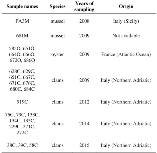
Sample names Species Years of
sampling Origin
PA3M mussel 2008 Italy (Sicily)
681M mussel 2009 Not available
585O, 651O, 664O, 666O, 672O, 686O
oyster 2009 France (Atlantic Ocean)
628C, 629C, 651C, 667C, 671C, 676C, 680C, 684C
clams 2009 Italy (Northern Adriatic)
919C clams 2012 Italy (Northern Adriatic)
76C, 79C, 133C, 134C, 135C, 229C, 271C,
272C
clams 2014 Italy (Northern Adriatic)
7
1.3.2 RNA extraction, RT-PCR and nested PCR
The mollusc hepatopancreas were homogenized and treated with proteinase K (Sigma, St. Louis, USA), then RNA was extracted according to the manufacturer’s instructions with NucleoSpin® RNA II (Macherey-Nagel, Düren, Germany). RNA samples were stored at -80 °C until use.
Betanodavirus presence was investigated by an RT-nested PCR method using primers previously described targeting the viral RNA 2 (Ciulli et al., 2007b). Briefly, the first amplification step was conducted through a one-step RT-PCR assay with primers S6-S7 (Ciulli et al., 2006) using the SuperScript III One-Step RT-PCR System (Invitrogen, Carlsbad, USA). The reaction mixture consisted of 15 µl of reaction mix containing 3 µl RNA, 7.5 µl 2X Reaction Mix, 0.8 µM of each primer and 0.3 µl Superscript III/Platinum Taq enzyme mix. The optimal thermal cycling conditions were 45 °C for 30 min, 95 °C for 2 min, followed by 40 cycles of 94 °C for 60 s, 58 °C for 60 s and 72 °C for 60 s. A final extension was performed at 72°C for 7 min. Nested PCR was conducted with primers F2-R3 (Nishizawa et al., 1994) using the Platinum Taq DNA polymerase (Invitrogen). The reaction mixture for the nested PCR had a total volume of 25 µl and contained 2.5 µl of 10X PCR buffer, 1.5 mM MgCl2, 0.25 µM of each primer, 1 µl of cDNA diluted 1:100, 1.25 units of Platinum Taq DNA polymerase (Invitrogen) and nuclease free water. The thermal cycle for nested PCR consisted of 95°C for 5 min and 40 cycles of 94°C for 30 sec, 56°C for 30 sec and 72°C for 30 sec. A final extension was performed at 72°C for 7 min. To avoid cross contamination, negative controls were run along with all reactions. The results of all RT-PCR and nested PCR analyses were checked by agarose gel electrophoresis of PCR products along with a 100 bp DNA molecular marker (Invitrogen, Carlsbad, USA).
Nine betanodaviruses detected between 2012 and 2015 were further analyzed for an RNA 1 fragment using primers previously described (Toffolo et al., 2007). RT-PCR was performed with the SuperScript III One-Step RT-PCR System (Invitrogen, Carlsbad, USA) using primers VNNV 5-6. Eminested PCR was conducted with primers VNNV 6-7 using the Platinum Taq DNA polymerase (Invitrogen).
1.3.3 Sequencing and phylogenetic analysis
PCR products were purified using the High Pure PCR Product Purification Kit (Roche, Mannheim, Germany) and then sequenced through the Bio-Fab Sequencing Service (Rome, Italy). Amino acid sequences were predicted by the BioEdit software (http://bioedit.software.informer.com/). Sequences were then aligned and compared with betanodavirus sequences previously obtained from
8 strains isolated from farmed and wild finfish and with betanodavirus reference genotypes strains (Thiery et al., 2012) available in GenBank (www.ncbi.nlm.nih.gov) using Clustal W in BioEdit software (http://bioedit.software.informer.com/). Particularly, a selection of betanodavirus sequences collected in southern Europe and used for a previous extensive and comprehensive phylogenetic study was used (Panzarin et al., 2012). Percentage of similarity of pairwise distances was calculated with BioEdit software. Phylogenetic analysis was carried out using the MEGA version 6 software (www.megasoftware.net) and employing the general time-reversible (GTR) model of nucleotide substitution. A phylogenetic tree was constructed using the maximum-likelihood method. Bootstrap analysis was carried out on 1000 replicates.
1.4 RESULTS
1.4.1 Virus detection
All samples collected in the 2009 survey resulted negative to RT-PCR, while 15 lots out of 57 resulted positive to nested PCR (26.3 %). Only one positive lot was found out of 19 lots of mussels (M. galloprovincialis), whereas clams (T. philippinarum) and oysters (C. gigas) resulted highly positive to betanodavirus, with 42.1 % (8/19) and 31.6% (6/19) of lots positive to betanodavirus respectively. All the positives except one (585/2009) were collected from June to September.
1.4.2 Sequencing and phylogenetic analysis
The maximum likelihood phylogenetic trees inferred for the RNA 1 and RNA 2 genes of the viruses collected from shellfish from 2008 to 2015 revealed that all betanodaviruses detected in this study except one (681M/2009) felt within RGNNV genotype (Figs. 1 and 2). The virus 681M/2009 clustered in a group by itself, outside all RGNNV subgroups (Fig. 2). Unfortunately, for this virus, as well as for all viruses detected before 2012 was not possible to carry out RNA 1 sequencing due to the failure storing of samples.
The topology of the RNA 1 and RNA 2 phylogenetic trees confirmed the genetic clustering obtained in a previous study that analyze several betanodavirus sequences collected in southern Europe (Panzarin et al., 2012) (Figs. 1 and 2) therefore corroborating the validity of the analysis based on the partial fragment of RNA 1 (position 678-1097 of SJNNV AB056571) and the T4 variable region of the RNA 2 (position 635-923 of SJNNV AB056572).
9 RNA 1 and RNA 2 phylogenetic trees identified 11 and 7 well supported monophyletic genetic subgroups inside RGNNV genotype (bootstrap values >70%) (Figs. 1 and 2). For a better comparison clusters were designated with names used in the study of Panzarin et al. (2012).
Analysis of RNA 1 showed that betanodaviruses detected in Italian clams clustered in 3 RGNNV subgroups previously identified and named II, IV and X (Panzarin et al., 2012). On the basis of the comparison with viruses previously included in these groups, some of the 2014 and 2015 clam samples clustered with viruses isolated several years ago (1996-2000) from finfish (cluster X). Clam viruses clustered together with both Italian and other European countries viruses, regardless of host origin, farmed or wild status.
Fig. 2. Phylogenetic tree constructed with RNA 1 fragment of betanodaviruses detected in bivalve molluscs combined with reference strains and finfish betanodavirus sequences retrieved from Genbank.
10 Analysis of RNA 2 showed that betanodaviruses detected in bivalve molluscs clustered in 2 subgroups (B, E) out of the seven previously identified (Panzarin et al., 2012). Both subgroups included viruses with high variability respect to year of detection and host status (wild/farmed). Moreover, subgroup E included viruses isolated in different European countries, including a virus detected in a French oyster. Two further subgroups were identified in RNA 2 phylogenetic tree. One subgroup (named H) was represented by viruses detected in four French oysters and in one Italian mussel (Sicily). They clustered together with a wild Italian Epinephelus spp. virus that did not fall in any subgroups in the previous analysis (Panzarin et al., 2012). The other subgroup (named I) consists of betanodavirus strains detected in seven Italian clams in 2014 and 2015 and European viruses detected in farmed finfish that did not fall in any subgroups in the previous analysis (Panzarin et al., 2012). All genetic clusters were well supported by bootstrap analysis (bootstrap values >70%).
11 Fig. 3. Phylogenetic tree constructed with RNA 2 fragment of betanodaviruses detected in bivalve molluscs combined with reference strains and finfish betanodavirus sequences retrieved from Genbank.
12 Betanodaviruses detected in bivalve mollusc in this study showed nucleotide and amino acid identities between each other higher than 88.9% and 86.0% respectively except with strain 681M/2009 that has a nucleotide and amino acid identity always lower than 75.0% and 81.7% respectively.
The percentage of pairwise nucleotide and aminoacid similarity with the RNA 2 of the four betanodavirus genotypes are reported in Table 2.
All the viruses detected in bivalve molluscs except strain 681M/2009 showed the highest nucleotide and amino acid identities with RGNNV genotype ranging between 89.6 and 99.6% and between 87.0 % and 100.0% respectively, whereas the nucleotide identities with other genotypes were lower than 76.8, 65.8 and 65.5% for BFNNV, SJNNV and TPNNV respectively and the amino acid identity was lower than 84.9, 68.4 and 70.5% for BFNNV, SJNNV and TPNNV respectively. Strain 681M/2009 showed similar nucleotide identities with RGNNV (75.0% nucleotide identity and 80.6% amino acid identity) and BFNNV (72.2% nucleotide identity and 79.5% amino acid identity) genotype, these values were lower than those between RGNNV and BFNNV genotypes (76.1% nucleotide identity and 84.9% amino acid identity).
13 Table 2. Comparisons of nucleotide and amino acid sequences of a RNA 2 fragment including the variable region of betanodaviruses detected in bivalve molluscs with reference betanodavirus genotypes (RGNNV: AY32487; BFNNV: EU826138; SJNNV: AB056572; TPNNV: EU236149; Thiery et al., 2012). Percentage of similarity of pairwise distances are shown.
RGNNV BFNNV SJNNV TPNNV nt aa nt aa nt aa nt aa 38C/2015 99.6 100 76.5 84.9 65.5 67.3 64.8 69.4 39C/2015 99.6 100 76.5 84.9 65.5 67.3 64.8 69.4 58C/2015 99.6 100 76.5 84.9 65.5 67.3 64.8 69.4 76C/2014 99.6 100 76.5 84.9 65.5 67.3 64.8 69.4 79C/2014 91.1 89.2 71.8 82.7 64.4 68.4 64.8 70.5 133C/2014 98.5 100 76.8 84.9 65.5 67.3 64.8 69.4 134C/2014 98.9 98.9 76.1 83.8 65.1 66.3 64.4 68.4 135C/2014 90.7 89.2 71.5 82.7 65.1 68.4 64.8 70.5 229C/2014 99.6 100 76.5 84.9 65.5 67.3 64.8 69.4 271C/2014 99.6 100 76.5 84.9 65.5 67.3 64.8 69.4 272C/2014 90.7 89.2 71.5 82.7 65.1 68.4 64.8 70.5 919C/2012 98.5 100 76.8 84.9 64.8 67.3 64.8 69.4 585O/2009 93.2 92.4 72.9 81.7 64.4 64.2 60.6 66.3 628C/2009 89.6 87 71.1 79.5 64.8 68.4 63.7 69.4 629C/2009 90.7 89.2 71.5 82.7 65.1 68.4 64.8 70.5 651O/2009 98.9 100 76.5 84.9 65.1 67.3 65.1 69.4 651C/2009 98.9 100 76.5 84.9 65.1 67.3 65.1 69.4 664O/2009 92.8 91.3 72.5 80.6 64.1 63.1 60.6 65.2 666O/2009 92.5 91.3 72.2 81.7 64.4 64.2 60.6 65.2 667C/2009 98.9 100 76.5 84.9 65.1 67.3 65.1 69.4 671C/2009 98.5 100 76.1 84.9 65.5 67.3 64.8 69.4 672O/2009 98.5 100 76.8 84.9 64.8 67.3 65.5 69.4 676C/2009 90.7 89.2 71.5 82.7 65.1 68.4 64.8 70.5 680C/2009 98.9 100 76.5 84.9 65.1 67.3 65.1 69.4 681M/2009 75.0 80.6 72.2 79.5 65.8 68.4 65.1 68.4 684C/2009 98.9 100 76.5 84.9 65.1 67.3 65.1 69.4 686O/2009 93.2 92.4 72.9 81.7 64.4 64.2 60.6 66.3 PA3M/2008 93.5 94.6 73.6 81.7 63.7 65.2 62.0 67.3
14
1.5 DISCUSSION
This is the first study focusing on betanodavirus in bivalve molluscs. Twenty seven new betanodavirus strains were detected in bivalve molluscs of different species, European countries and year of collection and genetically characterized.
The sporadic presence of betanodaviruses in marine invertebrate has been previously detected (Gomez et al., 2008b; Ciulli et al., 2010; Gomez et al., 2010; Panzarin et al., 2012).
In our study, the finding of betanodavirus in bivalve molluscs was evidenced in samples collected for a long period from 2008 to 2015, from different Euopean countries and three species showing a consistent presence of this virus in these hosts. Particularly, clams collected in the North-eastern Italy in a time span of 7 years resulted constantly positive for betanodavirus. Moreover, a different prevalence was shown in bivalve mollusc species; particularly, clams seem to be more frequently contaminated than others species such as oyster and mussel. The presence of the virus in bivalve mollusc might be a natural consequence of their biology. Bivalve molluscs are obligate filter feeders and can accumulate particles including viruses from the surrounding water (Serratore et al., 2014). The fossorial behaviour of clams could favorite the virus-host contact and virus retention, compared to suspended farming methods used for oysters and mussels. However, also the geographical origin could have influenced the different prevalence evidenced among bivalve mollusc species in our study.
Phylogenetic analysis of both RNA 1 and RNA 2 fragments of betanodaviruses detected in bivalve molluscs showed a wide range of strains mainly belonging to RGNNV genotype, the most reported genotype in Europe.
However, the RNA 2 genetic analysis showed the presence of one atypical betanodavirus (681M/2009), placing definitively it outside of all genotypes officially recognized of the genus
Betanodavirus. Particularly, the virus 681M/2009 seems to be more similar to RGNNV and
BFNNV genotypes than to SJNNV and TPNNV genotypes, but it did not cluster with any of them. Pairwise percentage analysis confirms this result, with a nucleotide and amino acid sequence identity to each genotype lower than that evidenced between different genotypes.
Phylogenetic analysis of viruses detected in bivalve mollusc showed no correlation with their host species and geographical origin clustering together viruses detected in Italian clams, mussels and French oysters.
The comparison of the viruses detected in bivalve molluscs with several betanodaviruses isolated from finfish in Southern Europe (Panzarin et al., 2012) shows the circulation of similar viruses in finfish and in bivalve molluscs. In analogy to a previous study, several subgroups were identified in RNA 1 and RNA 2 phylogenetic trees inside RGNNV genotype. Most of these clusters include both
15 bivalve molluscs and finfish viruses with different geographic origin, year of isolation and host status (wild/farmed). Accordingly, the bivalve mollusc betanodaviruses did not cluster separately from finfish viruses, but rather they reflect the epidemiological patterns of betanodavirus circulating in finfish in Southern Europe.
Furthermore, some recent bivalve mollusc viruses clustered with finfish viruses detected several years ago (1996-2000) showing the persistent circulation of these viruses.
Previous study revealed the presence of a betanodavirus closely related to RGNNV in marine invertebrate (Japanese common squid Todarodes pacificus) and showed that it has high pathogenicity to finfish causing severe mortalities after i.m. challenge (Gomez et al., 2010). Similarly, the high genetic identy of viral strains detected in bivalve molluscs to those found in finfish highlights the inter-specific exchange of betanodaviruses.
1.6 CONCLUSIONS
The finding of this study show that betanodaviruses are widespread also in bivalve molluscs, maybe more than we could expect.
Phylogenetic analysis of these viruses shows that strains detected in mollusc bivalve and in finfish are very closely related and that betanodaviruses detected in bivalve molluscs in different European countries from 2008 to 2015 mimic the epidemiological patterns of betanodaviruses previously detected in finfish in Southern Europe from October 2000 to November 2009.
Moreover, the nucleotide and amino acid sequence analysis of the strain 681M/2009 show the existence of a new betanodavirus strain not belonging to any of the already known betanodavirus genotypes.
Such a massive and variegate presence of betanodaviruses in bivalve molluscs greatly stresses the risks of transmission previously feared for other invertebrates. Molluscs bivalve reared in the same area of farmed and wild finfish could act as a reservoir of virus. Furthermore, the marketing of alive bivalve mollusc and the relaying activity, allowed by the European regulation, can pose also a real risk of spreading betanodavirus between different geographical areas.
16
1.7 REFERENCES
Baeck G.W., Gomez D.K., Oh K.S., Kim J.H., Choresca C.H., Park S.C. 2007. Detection of piscine nodaviruses from apparently healthy wild marine fish in Korea. Bulletin of the European
Association of Fish Pathologists, 27 (3): 116–122.
Barker, D.E., MacKinnon, A.M., Boston, L., Burt, M.D.B., Cone, D.K., Speare, D.J., Griffiths, S., Cook, M., Ritchie, R., Olivier, G., 2002. First report of piscine nodavirus infecting wild winter flounder Pleuronectes americanus in Passamaquoddy Bay, New Brunswick, Canada.
Diseases of Aquatic Organisms, 49: 99–105.
Ciulli S., Gallardi D., Scagliarini A., Battilani M., Hedrick R.P., Prosperi S. 2006. Temperature-dependency of Betanodavirus infection in SSN-1 cell line. Diseases of Aquatic Organisms, 68: 261-265.
Ciulli S., Galletti E., Grodzki M., AlessiA., Battilani M., Prosperi S. 2007a. Isolation and Genetic Characterization of Betanodavirus from Wild Marine Fish from the Adriatic Sea. Veterinary
Research Communications, 31 Suppl. 1: 221-224.
Ciulli S., Galletti E., Grodzki M., Alessi A., Battilani M., Scagliarini A., Prosperi S. 2007b. Spread of Betanodavirus in wild marine fish from Adriatic Sea: use of cell culture isolation or nested RT-PCR? Abstract book, 13th International EAFP Conference on “Diseases of fish and shellfish” p125.
Ciulli S., Grodzki M., Bignami G., Serratore P., Prosperi S. 2010. Molecular detection and genetic analysis of Betanodaviruses in bivalve molluscs. Journal of Biotechnology, 150, Supplement, Page 4.
Doan Q.K., Vandeputte M., Chatain B., Morin T., Allal F. 2016. Viral encephalopathy and retinopathy in aquaculture: a review. Journal of Fish Diseases, doi: 10.1111/jfd.12541.
Fichi G., Cardetti G., Perrucci S., Vanni A., Cersini A., Lenzi C., De Wolf T., Fronte B., Guarducci M., Susini F. 2015. Skin lesion-associated pathogens from Octopus vulgaris: first detection of Photobacterium swingsii, Lactococcus garvieae and betanodavirus. Diseases of Aquatic
Organisms, 115: 147- 156.
Gagné N, Johnson SC, Cook-Versloot M, MacKinnon AM, Olivier G. 2004. Molecular detection and characterization of nodavirus in several marine fish species from the northeastern Atlantic. Diseases of Aquatic Organisms, 62(3): 181–189.
17 Gomez D.K., Sato J., Mushiake K., Isshiki T., Okinaka Y., Nakai T., 2004. PCR-based detection of betanodaviruses from cultured and wild marine fish with no clinical signs. Journal of Fish
Diseases, 27: 603-608.
Gomez, D.K., Lim, D.J., Baeck, G.W., Youn, H.J., Shin, N.S., Youn, H.Y., Hwang, C.Y., Park, J.H., Park, S.C., 2006. Detection of betanodaviruses in apparently healthy aquarium fishes and invertebrates. Journal of Veterinary Science, 7 (4): 369–374.
Gomez DK, Baeck GW, Kim JH, Choresca CH Jr, Park SC 2008a. Molecular detection of Betanodavirus in wild marine fish populations in Korea. Journal of Veterinary
Diagnostic Investigation, 20: 38-44.
Gomez D.K., Baeck G. W., Kim J. H., Choresca C. H. Jr., Park S. C. 2008b. Molecular detection of betanodaviruses from apparently healthy wild marine invertebrates. Journal of Invertebrate
Pathology, 97: 197–202.
Gomez D.K., Baeck G.W., Kim J.H., Choresca C.H.J., Park S.C. 2008c. Genetic analysis of betanodaviruses in subclinically infected aquarium fish and invertebrates. Current
Microbiology, 56: 499 – 504.
Gomez D.K., Matsuoka S., Mori K., Okinaka Y., Park S.C., Nakai T. 2009. Genetic analysis and pathogenicity of Betanodavirus isolated from wild redspotted grouper Epinephelus akaara with clinical signs. Archives of Virology, 154: 343–346.
Gomez D.K., Mori K., Okinaka Y., Nakai T. & Park S.C. 2010. Trash fish can be a source of betanodaviruses for cultured marine fish. Aquaculture, 302: 158-163.
Iwamoto T., Mise K., Taked A., Okinaka Y., Mori F., Arimoto M., Okuno T., Nakai T. 2005. Characterization of Striped jack nervous necrosis virus subgenomic RNA3 and biological activities of its encoded protein B2. Journal of General Virology, 86: 2807– 2816.
Johansen R., Sommerset I., Tørud B., Korsnes K., Hjortaas M.J., Nilsen F., Nerland A.H., Dannevig B.H. 2004. Characterization of nodavirus and viral encephalopathy and retinopathy in farmed turbot Scophthalmus maximus (L). Journal of Fish Diseases, 27: 591– 601.
Liu X.D., Huang J.N., Weng S.P., Hu X.Q., Chen W.J., Qin Z.D., Dong X.X., Liu X.L., Zhou Y., Asim M., Wang W.M., He J.G., Lin L. 2015. Infections of nervous necrosis virus in wild
18 and cage-reared marine fish from South China Sea with unexpected wide host ranges.
Journal of Fish Diseases, 38: 533-540.
Nishizawa, T., K. Mori, T. Nakai, I. Furusawa, and K. Muroga. 1994. Polymerase chain reaction (PCR) amplification of RNA of striped jack nervous necrosis virus (SJNNV). Diseases of
Aquatic Organisms, 18: 103–107.
Nishizawa T., Furuhashi M., Nagai T., Nakai T., Muroga K. 1997. Genomic classification of fish nodaviruses by molecular phylogenetic analysis of the coat protein gene. Applied and
Environmental Microbiology, 63 (4): 1633-1636.
Olveira J.G., Souto S., Dopazo C.P., Thiéry R., Barja J.L., Bandín I. 2009. Comparative analysis of both genomic segments of betanodaviruses isolated from epizootic outbreaks in farmed fish species provides evidence for genetic reassortment. Journal of General Virology, 90: 2940– 2951.
Panzarin V, Fusaro A, Monne I, Cappellozza E, Patarnello P, Bovo G, Capua I, Holmes EC, Cattoli G. 2012. Molecular epidemiology and evolutionary dynamics of betanodavirus in southern Europe. Infection, Genetics and Evolution, 12: 63–70.
Serratore, P., Ciulli, S., Piano, A., Cariani, A. (2014). Criticism of the purification process of bivalve shellfish. Literature review and our industrial research experiences. In R.M. Hay (Ed.), Shellfish Humam consumption, health implications and conservation concerns. Nova Science Publishers, Inc., New York, USA. pp. 1-50. ISBN 9781633211964
Thiéry R., Johnson K.L., Nakai T., Schneemann A., Bonami J.R., Lightner D.V. 2012. Family Nodaviridae. In: King AMQ, Adams MJ, Carstens EB, Lefkowitz EJ (ed) Virus Taxonomy, Ninth Report of the International Committee on Taxonomy of Viruses. Elsevier Academic Press, California, pp 1061-1067.
Toffolo V., Negrisolo E., Maltese C., Bovo G., Belvedere P., Colombo L., Dalla Valle L. 2007. Phylogeny of betanodaviruses and molecular evolution of their RNA polymerase and coat proteins. Molecular Phylogenetics and Evolution, 43: 298–308.
Vendramin N., Patarnello P., Toffan A., Panzarin V.,Cappellozza E., Tedesco P., Cattoli G. 2013. Viral Encephalopathy and Retinopathy in groupers (Epinephelus spp.) in southern Italy: a threat for wild endangered species. BMC Veterinary Research, 9: 20.
19
CHAPTER 2
Fate of Redspotted grouper nervous necrosis virus (RGNNV) in experimentally challenged Manila clam Ruditapes philippinarum
Volpe Enrico1, Pagnini Niko2, Serratore Patrizia1, Ciulli Sara1.
1
Department of Veterinary Medical Sciences, University of Bologna, 47042 Cesenatico, FC, Italy
2
Cesena Campus, University of Bologna, 47042 Cesenatico, FC, Italy
2.1 ABSTRACT
Redspotted grouper nervous necrosis virus (RGNNV), genus Betanodavirus, family Nodaviridae, is
the causative agent of Viral encephalopathy and retinopathy (VER), otherwise known as Viral nervous necrosis (VNN), and is a virus that could infect more than 70 fish species worldwide. Betanodaviruses, including RGNNV, are very resilient in the aquatic environment and their presence has already been reported in several wild marine species including invertebrates. In order to investigate the interaction between the bivalve mollusc Ruditapes philippinarum and the RGNNV a culture-based method was optimised. The bioaccumulation of the pathogenic RGNNV by R. philippinarum and the potential shedding of viable RGNNV from RGNNV-exposed clams was evaluated through a culture-based method. R. philippinarum clearly accumulate viable RGNNV in the hepatopancreas tissue and are able to release viable RGNNV via faecal matter and filtered water into the surrounding environment. The role of clams as bioaccumulators and shedders of viable RGGNV could put at risk susceptible cohabiting cultured fish. The presence of RGNNV-contaminated molluscs could behave as RGNNV reservoirs and may modify the virus epidemiology.
20
2.2 INTRODUCTION
Redspotted grouper nervous necrosis virus (RGNNV), a virus of the genus Betanodavirus, family Nodaviridae, is responsible for Viral encephalopathy and retinopathy (VER), otherwise know as
Viral nervous necrosis (VNN), a disease with nervous signs and mortality in more than 70 fish species worldwide (Doan et al. 2016). Betanodaviruses are small, icosahedral viruses that contain two segments of positive-sense single-stranded RNA. The RNA1 (3.1 kb) and the RNA2 (1.4 kb) encode a RNA-dependent RNA polymerase of 100 kDa and a major coat protein of 42 kDa, respectively (Mori et al., 1992; Guo et al., 2003). Based on a partial nucleotide sequence of coat protein gene, betanodaviruses are divided into four species: Striped jack nervous necrosis virus (SJNNV), Tiger puffer nervous necrosis virus (TPNNV), Barfin flounder nervous necrosis virus (BFNNV) and Redspotted grouper nervous necrosis virus (RGNNV) (Thiéry et al., 2011). Viral nervous necrosis virus (VNNV) is frequently isolated during outbreaks of VER in several farmed fish species, including European sea bass (Dichentrarchus labrax) in the Mediterranean Sea (Panzarin et al., 2012; Vendramin et al., 2013). Moreover, VNNV has also been detected in numerous wild marine fish species and invertebrates in the Mediterranean Sea, South Korea, China and Japan (Gomez et al., 2004, 2008a; Ciulli et al., 2007; Liu et al., 2015).
Betanodavirus infection is transmitted horizontally, either directly through the introduction
of infected fish, or indirectly by contaminated water and equipment, as well as vertically, through reproduction (Munday et al., 2002).
Currently, no successful therapies or commercial vaccines, apart from one in Japan (OIE 2013), are available to enable adequate control of VER, so disease prevention is based mainly on maintaining proper sanitary procedures, screening activities and correct farm management (Costa & Thompson, 2016; Doan et al., 2016).
Recent studies have also demonstrated that a certain population of apparently healthy wild marine fish carries betanodaviruses, and suggested that these wild fish can be a persistent potential source of virus for cultured fish and the breeding environment (Ciulli et al., 2007; Gomez et al., 2008a; Vendramin et al., 2013).
Moreover, a recent finding suggests that trash fish/molluscs can be a source of betanodaviruses for cultured fish and that they pose a serious risk for outbreaks of VER in susceptible cultured fish (Gomez et al., 2010).
It is well known that pathogenic agents may be spread via water masses, wild carriers, or vectors and restrictions do not fully ensure the control of disease spread by these routes (Mortensen, 2000; Mortensen et al., 2006). Water is both a dilution and a transport medium, and the fate of pathogenic agents shed into the water is dependent upon a series of factors, including dilution, inactivation by
21 UV light or other physical and chemical factors, particle bonding and uptake in filter-feeding organisms or particle-feeding plankton (Noble & Fuhrman 1997; Sinton et al., 2002; Wilhelm et al., 2003). Accordingly, aquatic organism interaction, both in the case of natural or artificial environments, such as integrated multitrophic aquaculture (IMTA), can greatly affect the epidemiology of fish infectious diseases. In fact, there is evidence indicating that, when placed closely together, bivalves may act either as bio-filters or as reservoirs for finfish pathogens, as a consequence of their ability to bioaccumulate microorganisms (Mortensen et al., 1992; Mortensen, 1993; Paclibare et al., 1994; Skår & Mortensen, 2007; Molloy et al., 2011; Pietrak et al., 2012; Wangen et al., 2012). However, the outcome of the interaction may differ on the basis of the morphology and physiology of the pathogen, which influences whether the pathogen remains viable in bivalve mollusc tissues and it is shed back into the environment, or is inactivated by the molluscs (Skår & Mortesen 2007; Molloy et al., 2013).
The presence of VNNV in invertebrates and particularly bivalve molluscs has already been reported (Ciulli et al., 2007; Gomez et al., 2008a) including the Manila clam (Ruditapes philippinarum) in the Mediterranean Sea (Ciulli et al., 2007).
Some studies have investigated the role of wild aquatic organisms such as bivalve molluscs in the interaction with fish pathogens. These studies showed that the infectious salmon anaemia virus (ISAV) is inactivated by blue mussels (Mytilus edulis) (Molloy et al., 2014). In contrast, infectious pancreatic necrosis virus (IPNV) can be transmitted from IPNV-exposed mussels to Atlantic salmon (Salmo salar) (Molloy et al., 2013).
The aim of this study was to examine the bioaccumulation of a pathogenic RGNNV by Manila clam and to investigate the potential shedding of viable RGNNV from RGNNV-exposed clams through a culture-based method. The potential role of clams as an RGNNV reservoir and the potential risks posed by RGNNV-contaminated molluscs are pointed out in this study.
2.3 MATERIALS AND METHODS
2.3.1 Clam maintenance and RGNNV screening
Batches of market-size Manila clam (Ruditapes philippinarum), hereafter referred to as clams, were obtained from a commercial clam trader and were reared in an artificial recirculation system (RAS) (Adriatic Sea) supplied with natural seawater, collected from the Adriatic Sea and thermostated at 15°C. Batches of clams were acclimated for 24 h in order to start filtration. Trials were conducted in a static system consisting of 5 l plastic tanks supplied with 2 l natural seawater, aeration and thermostated at 15°C.
22 Prior to all trials, 30 clams from each batch were screened for presence of VNNV-RNA via an RT-PCR assay followed by a nested RT-PCR performed according to methods previously described (Ciulli et al., 2006; Nishizawa et al., 1994).
2.3.2 Cell culture maintenance and virus propagation
Striped snakehead fish cells (SSN-1) were maintained in Leibovitz-15 medium (L-15) (Gibco) supplemented with 1% L-glutamine (Gibco), 1% antibiotic–antimycotic solution (Gibco) and 7.5% foetal bovine serum (FBS) (Gibco) at 25°C. For virus isolation assays, SSN-1 cells were harvested, counted and transferred to 96-well culture plates at a density of 7 × 10⁴ cell/cm2. Cells were allowed to attach and acclimate for 24 h at 25°C in order to achieve 80% confluence.
The previously characterised (Ciulli et al., 2006) It/351/Sb isolate of RGNNV was propagated in SSN-1 cells grown at 25°C in L-15 medium containing 2% FSB. When the cells demonstrated a 75% cytopathic effect (CPE), the cells and supernatant were centrifuged at 500 g for 10 min and the supernatant was stored at -80°C until use. The titre of the stock was determined by 50% tissue culture infectious dose (TCID50) end point analysis in SSN-1 cells. The TCID50 was calculated according to Spearman-Karber method (Hierholzer & Killington 1996).
2.3.3 Culture analysis of clam hepatopancreas, faecal matter and water samples
RGNNV presence was detected and quantified by performing TCID50 analysis in SSN-1 cells in hepatopancreas tissue, faecal matter and water samples. Water samples were centrifuged at 3000 g for 5 min and the supernatant was filtered through 0.20-μm-poresize filters and incubated with 1% antibiotic–antimycotic solution (Gibco) at 4°C overnight. Samples were diluted 10-fold in L-15 with 2% FBS (Gibco). If samples reported negative results, a 2-fold dilution of the supernatants was performed and tested.
Hepatopancreas tissue was weighed, diluted 1:9 (wt/vol) with L-15 containing 2% FBS (Gibco) and homogenised before centrifuging at 3000 g for 5 min. The supernatant was diluted 10-fold in L-15 with 2% FBS (Gibco).
Faecal matter samples were centrifuged at 3000 g for 5 min, the faecal pellets were weighed, diluted 1:9 (wt/vol) with L-15 containing 2% FBS (Gibco) and incubated with 1% antibiotic–antimycotic solution (Gibco) at 4°C overnight. The supernatant was diluted 10-fold in L-15 with 2% FBS (Gibco).
23 For viral titration assays, each dilution was added in 100 μl volumes to five wells of a 96-well plate containing 24 hours-old SSN-1 cells. Negative control wells consisting of L-15 with 2% FBS (Gibco) were included for each plate. The inoculum from wells receiving samples were removed after 1 h viral adsorption period at 25°C to prevent cell cytotoxicity before the addition of 100 μl of L-15 fresh medium containing 2% FBS. The plates were incubated at 25°C and observed daily for visible CPE for 7 days. The titre referred to water samples was expressed as TCID50 ml-1
. For hepatopancreas tissue and faecal matter samples, culture analysis TCID50 values were normalised to (g of hepatopancreas tissue or faecal matter)-1 and hereafter referred to as TCID50 g-1.
2.3.4 Detection limit of TCID50 endpoint dilution assay in RGNNV-inoculated clam
hepatopancreas homogenates
Hepatopancreas from seven VNNV-RNA-negative clams were weighed, diluted 1:9 (wt/vol) with L-15 containing 2% FBS (Gibco) and homogenised before centrifuging at 3000 g for 5 min. Serial 10-fold dilutions of stock RGNNV, ranging in titre from 7.5 to 2.5 log TCID50 ml-1, were prepared in L-15 cell culture medium. Each virus dilution was added in 100 μl volumes to six of the seven hepatopancreas homogenates and thoroughly mixed to achieve predicted titres ranging from 6.7 to 1.7 log TCID50 ml-1. L-15 containing 2% FBS was added to the seventh homogenised sample, which served as a negative control for the TCID50 assays. RGNNV-inoculated hepatopancreas homogenates were processed for TCID50 analysis in SSN-1 as described above.
2.3.5 RGNNV clam exposure trial
In order to measure RGNNV uptake in clams, three independent exposure trials were performed. In each trial, 60 mussels were placed in 5 l plastic tanks containing 2 l of seawater thermostated at 15°C. An air-lift pump circulated the water and provided aeration. RGNNV suspension in L-15 cell culture medium was then added up to the final virus concentration in the tanks was 5 log TCID50 ml–1. The clams were left for 24 h to bioaccumulate the virus and then removed. Ten ml of water and random triplicate clam samples were collected at 3, 6 and 24 h post-exposure (hpe). Culture analysis of clam hepatopancreas and water samples was carried out in SSN-1 cells as described above.
24 Hepatopancreas results were expressed as the mean ± standard deviation (SD) of positive samples obtained from the three trials. The samples of faecal matter and water were analysed in two repeats and the results were shown as the mean of the positive repeats ± SD.
2.3.6 Clam RGNNV shedding trials
The clam’s ability to shed viable RGNNV in water through faecal matter was evaluated with two subsequent trials.
Trial 1. The shedding trial 1 was carried out in the same manner as the exposure trial with the
following modifications. After 24 hpe, the shell of each clam was surface disinfected with a 1% Virkon®S (DuPont) solution, followed by running tap water and transferred to a clean static system supplied with fresh seawater. During the depuration, triplicate clam samples were collected at 1, 2, 5, 6 and 7 days post-depuration (dpd) for culture assays. Furthermore, after 7 dpd, 10 ml of water and a sample of faecal matter were collected for culture assays.
Trial 2. The shedding trial 2 was carried out in the same manner as the trial 1 with the following
modifications. After the transfer, the clams were moved daily to a clean static system supplied with 100% fresh seawater until 7 dpd. Prior to the daily placements, the shell of each clam was surface disinfected with 1% Virkon®S (Dupont, Suffolk, UK) solution, followed by running tap water. Ten ml of water, faecal matter and triplicate clam samples were harvested for culture assays prior to each daily movement.
Hepatopancreas results of shedding trials were reported as the mean of positive samples ± SD. The samples of faecal matter and water were analysed in two repeats and the results were shown as the mean of the positive repeats ± SD.
2.3.7 Statistics
Data obtained from the detection limit assay were analysed by a simple linear regression analysis (Prism version 6.0 software, GraphPad Software), considering predicted values as a predictor and measured values as dependent variables. The level for accepted statistical significance was p < 0.05. Positive data of culture assays, after testing for normality, were analysed by one-way ANOVA followed by Tukey’s tests to determine statistically the differences among virus titres detected in samples (Prism version 6.0 software, GraphPad Software). The level for accepted statistical significance was p < 0.05.
25
2.4 RESULTS
2.4.1 Clam maintenance and VNNV screening
The VNNV screening showed that all the batches involved in the trials were negative for VNNV-RNA presence. During all trials, no mortality was recorded in batches of clams used.
2.4.2 Detection limit of TCID50 endpoint dilution assay in RGNNV-inoculated clam
hepatopancreas homogenates
The detection limit for viable RGNNV isolation by culture analysis was 1.7 log TCID50 ml-1. Viable RGNNV was detected by culture analyses in hepatopancreas homogenates with predicted titres of log 6.7 to 2.7 TCID50 ml-1
(Fig. 1). Titres measured in SSN-1 cells decreased in a linear trend as predicted titres decreased. Linear regression analysis showed a significant association between measured and predicted values (p = 0.001). In particular, a decrease of predicted value was associated with a decrease of the measured value (R2 = 0.96, F(1.4) = 91.64, y = 1.206x – 1.562). However, the determined titres were lower than the predicted titres by a mean of 0.5 ± 0.2 log TCID50 ml-1. The most dilute sample in which virus was detected had a predicted titre of 2.7 log TCID50 ml-1 although the measured titre was 1.7 log TCID50 ml-1. For samples at predicted titre of 1.7 log TCID50 ml-1 and lower, no virus was detected by culture assays.
Fig. 1. Measured (•) and predicted (–) log TCID50 ml -1
of RGNNV-inoculated clam hepatopancreas homogenates determined in SSN-1 cells.
26
2.4.3 RGNNV clam exposure trial
Uptake of clams of viable RGNNV in the hepatopancreas tissues was shown as early as 3 hpe (Fig. 2). No statistically significant difference was observed among mean viable virus titres detected in clam hepatopancreas collected at the same time points during the three clam exposure trials (data not shown). Accordingly, results are expressed as the mean ± SD of all positive samples obtained from the three trials.
Eight of the nine replicate clams were positive by virus isolation at 3 hpe, with a mean titre of 4.0 ± 0.2 log TCID50 g-1
(n = 8). At 6 hpe six of the nine replicate clams were positive by virus isolation with a mean titre of 4.3 ± 0.4 log TCID50 g-1
(n = 6). After 24 hpe, all sampled clams were positive at virus isolation with a mean titre of 4.4 ± 0.5 log TCID50 g-1 (n = 9). During the exposure trials the amount of viable RGNNV increased from 4.0 ± 0.2 to 4.4 ± 0.5 log TCID50 g-1
with no statistical significance (Fig. 2).
Moreover, the RGNNV loads measured at different time points in water samples showed no statistical significance; nevertheless virus titres decreased from 3.5 ± 0.3 to 2.8 ± 0.2 logTCID50 ml-1 (Fig. 2).
Fig. 2 Graph represents the log TCID50 ml -1
or g-1 of RGNNV in clam hepatopancreas and water samples over time.
27
2.4.4 Clam RGNNV shedding trial
Trial 1. Viable RGNNV was isolated from all the clams sampled (Fig. 3). The RGNNV mean titre
was 5.0 ± 0.2 log TCID50 g-1
; no statistical significances were shown between viable RGNNV amounts at different time points in hepatopancreas samples. After 7 dpd, RGNNV-exposed clams released viable RGNNV into water and through faecal matter (Fig. 3). The titres of viable RGNNV detected in faecal matter and water were 3.5 log TCID50 g-1 and 1.5 log TCID50 ml-1, respectively; these values were statistically lower (p < 0.05) than viable RGNNV found in hepatopancreas tissues (5 ± 0.2 log TCID50 g-1).
Fig. 3. Graph represents the log TCID50 ml -1
or g-1 of RGNNV in clam hepatopancreas , water and faecal matter samples over time. The asterisks indicate statistically significant different values from Manila clam values.
28
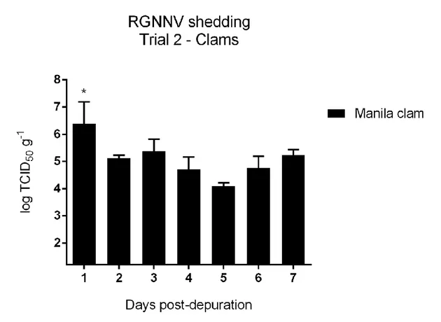
Trial 2. Viable RGNNV was isolated from all the hepatopancreas tissues analysed with a mean titre
of 5.1 ± 0.2 log TCID50 g-1. RGNNV titre in hepatopancreas at 1 dpd was statistically higher than the titres at 2, 4, 5 and 6 dpd (p < 0.05) (Fig. 4).
Fig. 4. Graph represents the log TCID50 g-1 of RGNNV in clam hepatopancreas samples. The asterisk indicates a statistically significant different value from 2, 4, 5, 6 dpd samples.
Viable RGNNV was also isolated from water samples at 1, 2, 3 and 4 dpd with a mean titre of 1.3 ± 0.3 log TCID50 ml-1. At 2 dpd only one repeat of the water sample reported viable RGNNV. No statistically significant differences were revealed among virus titres detected at different time points (Fig. 5). In faecal matter, viable RGNNV was isolated from both repeats of all the samples with a mean titre of 3.9 ± 0.5 log TCID50 g-1 except from one repeat of the 7 dpd sample. The titre values in faecal matter samples showed variable amounts of viable RGNNV during the trial; in particular RGNNV titration at 1 dpd was statistically higher than the titres at 2 and 6 dpd (p < 0.05). No statistical differences were shown among other time points (Fig. 6).
In water samples, the titrations showed statistically lower values than in the hepatopancreas tissues and then in the faecal matter samples at all tested time points (p < 0.05).
29 Fig. 5. Graph represents the log TCID50 ml
-1
of RGNNV in water samples over time.
Fig. 6. Graph represents the log TCID50 g -1
of RGNNV in faecal matter samples. The asterisks indicate statistically significant different values from 1 dpd sample.
30
2.5 DISCUSSION
Bivalve molluscs are well known bioaccumulators and may serve as reservoirs or as natural barriers for important finfish pathogens (Molloy et al., 2013, 2014).
In order to understand the fate of RGNNV in virus-exposed clams, a culture assay method using the SSN-1 fish cell line for quantification of viable virus in clam hepatopancreas tissue, faecal matter and water samples was optimised. Through this culture assay, we were able to determine whether or not clams bioaccumulate viable RGNNV after experimental exposure to the virus, and to determine their proficiency to shed viable RGNNV particles into the surrounding environment.
Previous studies aimed at investigating virus persistence in bivalve molluscs used both cell culture and molecular methods to evaluate the viral load in bivalve tissue (Skår & Mortensen 2007; Molloy et al., 2013, 2014). However, due to the presence of PCR inhibitors in bivalve tissues and the inability of molecular methods to distinguish viable from nonviable virus, the most sensitive techniques to evaluate the viral load in bivalve tissues is virus isolation on cell culture (Molloy et al., 2013).
The physiology and morphology of pathogen microorganisms influence the ability of the bivalve molluscs to inactivate or to accumulate and then shed viable microorganisms (Molloy et al., 2013, 2014). As a matter of fact, mussels (Mytilus edulis) are capable of bioaccumulating finfish viral pathogens, such as infectious salmon anaemia virus (ISAV) and infectious pancreatic necrosis virus (IPNV). In particular, ISAV is inactivated by M. edulis; therefore viable viral particles are not shed into the water. Conversely, viable IPNV shed by IPNV-exposed mussels may infect cohabitating Atlantic salmon (Salmo salar) (Molloy et al., 2013, 2014).
In this study, Manila clam had clearly accumulated viable RGNNV in the hepatopancreas tissue. During the 24 h exposure trials, time did not show a statistically significant effect on the RGNNV load in clam tissues. However, the viral load and the number of positive clams at virus isolation increased progressively during the exposure trials. Significantly, the decrease of viable virus in water during the exposure trials suggests the bioaccumulator role of clams and their ability to remove viable RGNNV from the water column. However, the RGNNV loads in clam tissues was not significantly higher than RGNNV levels in the water, indicating that clams do not concentrate RGNNV in their tissues.
A previous study, observing IPNV uptake by mussel during a 120 h trial, showed that mussels significantly accumulate viable IPNV in their digestive gland tissues over time (Molloy et al., 2013). However, this study also showed that IPNV particles were not efficiently removed from the water column. Authors hypothesised that the small particle size of IPNV (60 nm) may contribute to the inefficiency of particle uptake by the mussel (Molloy et al. 2013). However, bivalve molluscs
31 can concentrate virus as small as RGNNV (25 nm), such as Hepatitis A (27 nm) (Wolf 1988; Enriquez et al., 1992). Viral uptake and concentration ability of bivalve molluscs can vary from one virus to another, indicating the presence of different factors contributing to virus uptake (Molloy et al., 2013; Bosch et al., 1995).
RGNNV-exposed clams were able to release viable RGNNV via faecal matter and filtered water. RGNNV was detected in faecal matter and water up to 7 and 4 days post-depuration, respectively. Moreover, Trial 2 showed the amount of viable virus shed daily into the surrounding environment by RGNNV-exposed clams, and the persistence in the clam tissue. The shedding by clams of viable RGNNV after daily 100% water changes stresses the persistence of viable virus in hepatopancreas tissues.
This work, together with previous studies of Molloy (2013, 2014) and Skår & Mortensen (2007) seems to demonstrate that the inactivation of viruses is influenced by their morphology. In particular, nonenveloped viruses such as IPNV and RGNNV can be bioaccumulated by bivalve molluscs and be released alive into the water column (Molloy et al., 2013). In contrast, mussels act as a barrier for enveloped viruses such as ISAV (Molloy et al., 2014). Accordingly, our study, showing the persistence and shedding of viable RGNNV by clams, supports this hypothesis.
Actually, the fate of a microbe in bivalve tissue will be determined by a balance between uptake rate, digestion and depuration (Skår & Mortensen, 2007).
The finding of viable RGNNV shed through faecal matter and filtered water after 1 dpd suggests the potential of some filtered RGNNV particles to bypass the digestive system and be released back into the environment as viable particles entrapped in pseudofaecal pellets, as already hypothesised for other viral particles (Molloy et al., 2013).
The role of clams as bioaccumulators and shedders of viable RGNNV could put at risk susceptible cohabitating fish in an analogous way to that demonstrated by Molloy et al. (2013) for IPNV and Atlantic salmon. However, while virus shed into the water column in a fish farm during an outbreak is diluted by the water current, laboratory challenges are normally performed with high doses of pathogens in static or semi-static systems (Skår & Mortesen, 2007). Hence, it is difficult to predict whether wild or cultured clams near to farms of susceptible species might act as the cause of new outbreaks.
Betanodaviruses, including RGNNV, are very resilient in the aquatic environment and their presence has already been reported in wild marine invertebrates, especially molluscs and other invertebrates used as live fish food, including Artemia sp. nauplii, copepods (Tigriopus japonicas) and shrimps (Acetesinte medius) (Gomez et al., 2008b, 2008c; Chi et al. 2003; Costa & Thompson 2016). Furthermore, a recent study has shown that trash fish can be a source of betanodaviruses for
32 cultured marine fish (Gomez et al., 2010). Similarly, the presence of natural RGNNV-contaminated invertebrates, including Manila clam, close to susceptible cultured fish species, both in a natural marine environment and in artificial systems (live feed), could behave as RGNNV-reservoirs and be a source of viruses, posing a serious risk of outbreaks of VNN in susceptible cultured fish.
2.6 CONCLUSIONS
Finally, the cell culture method set up in this study has allowed an understanding of the fate of RGNNV in experimentally challenged Manila clam Ruditapes philippinarum. Clams are able to take up and then shed viable RGNNV into the surrounding environment through faeces and filtered water.
The persistence of viable RGNNV in clam tissues and the shedding of virus into the surrounding environment presents a serious risk for susceptible cohabitant fish species.
Further studies could establish whether the viral transmission from RGNNV-contaminated molluscs to finfish may be a result of viral release into the water or even a result of direct consumption of molluscs by fish. According to the results of this study, there is little doubt that the placing of contaminated molluscs into a fish farm, without proper control, could represent a serious risk for farmed fish.
2.7 REFERENCES
Bosch A., Pinto R.M., Abad F.X. 1995. Differential accumulation and depuration of human enteric viruses by mussels. Water Science and Technology, 31: 1–4.
Chi S.C., Shieh J.R., Lin S.J. 2003. Genetic and antigenic analysis of betanodaviruses isolated from aquatic organisms in Taiwan. Diseases of Aquatic Organisms, 55: 221–228.
Ciulli S., Gallardi D., Scagliarini A., Battilani M., Hedrick R.P., Prosperi S. 2006. Temperature-dependency of Betanodavirus infection in SSN-1 cell line. Diseases of Aquatic Organisms, 68: 261– 265.
Ciulli S., Galletti E., Grodzki M., Alessi A., Battilani M., Prosperi S. 2007. Isolation and genetic characterization of Betanodavirus from wild marine fish from the Adriatic sea. Veterinary
33 Costa J.Z., Thompson K.D. 2016. Understanding the interaction between Betanodavirus and its host for the development of prophylactic measures for viral encephalopathy and retinopathy. Fish
and Shellfish Immunology, 53: 35–49.
Doan Q.K., Vandeputte M., Chatain B., Morin T., Allal F. 2016. Viral encephalopathy and retinopathy in aquaculture: a review. Journal of Fish Disease, doi: 10.1111/jfd.12541.
Enriquez R., Froesner G.G., Hochstein-Mintzel B.V., Riedemann S., Reinhardt G. 1992. Accumulation and persistence of hepatitis A virus in mussels. Journal of Medical Virology, 37: 174–179.
Gomez D.K., Sato J., Mushiake K., Isshiki T., Okinaka Y., Nakai T. 2004. PCR-based detection of betanodaviruses from cultured and wild marine fish with no clinical signs. Journal of Fish
Diseases, 27: 603–608.
Gomez D.K, Baeck G.W., Kim J.H., Choresca Jr. C.H., Park S.C. 2008a. Molecular detection of betanodavirus in wild marine fish populations in Korea. Journal of Veterinary Diagnostic
Investigation, 20: 38–44.
Gomez D.K, Baeck G.W., Kim J.H., Choresca Jr. C.H., Park S.C. 2008b. Molecular detection of betanodaviruses from apparently healthy wild marine invertebrates. Journal of Invertebrate Pathology, 97: 197–202.
Gomez D.K., Baeck G.W., Kim J.H., Choresca Jr. C.H., Park S.C. 2008c. Genetic analysis of betanodaviruses in subclinically infected aquarium fish and invertebrates. Current
Mircrobiology, 56: 449–504.
Gomez D.K, Mori K.I., Okinaka Y., Nakai T., Park S.C. 2010. Trash fish can be a source of betanodaviruses for cultured marine fish. Aquaculture, 302: 158–163.
Guo Y.X., Wei T., Dallmann K., Kwang J. 2003. Induction of caspase-dependent apoptosis by betanodaviruses GGNNV and demonstration of protein a as an apoptosis inducer. Virology, 308: 74–82.
Hierholzer J.C., Killington R.A. 1996. Virus isolation and quantitation. In: Mahy BWJ, Kangro HO, (eds) Virology Methods Manual. Academic Press Limited, London, UK.
Liu X.D., Huang J.N., Weng S.P., Hu X.Q., Chen W.J., Qin Z.D., Dong X.X., Liu X.L., Zhou Y., Asim M., Wang W.M., He J.G., Lin L. 2015. Infections of nervous necrosis virus in wild