Characterization of a Globin-coupled Oxygen Sensor with a
Gene-regulating Function
*
□SReceived for publication, July 6, 2007, and in revised form, September 27, 2007 Published, JBC Papers in Press, October 9, 2007, DOI 10.1074/jbc.M705541200
Liesbet Thijs‡, Evi Vinck§1, Alessandro Bolli¶, Florin Trandafir§, Xuehua Wan储, David Hoogewijs**,
Massimiliano Coletta‡‡, Angela Fago§§, Roy E. Weber§§, Sabine Van Doorslaer§, Paolo Ascenzi¶ ¶¶,
Maqsudul Alam储, Luc Moens‡, and Sylvia Dewilde‡2
From the Departments of‡Biomedical Sciences and§Physics, University of Antwerp, Universiteitsplein 1, B-2610 Antwerp, Belgium, the¶Dipartimento di Biologia and Laboratorio Interdipartimentale di Microscopia Elettronica, Universita` Roma Tre, Viale G. Marconi 446, 00146 Roma, Italy, the储Department of Microbiology, University of Hawaii, Honolulu, Hawaii 96822,
the **Department of Biology and Centre for Molecular Phylogeny and Evolution, Ghent University, K .L. Ledeganckstraat 35, 9000 Ghent, Belgium, the‡‡Dipartimento di Medicina Sperimentale e Scienze Biochimiche, Universita` di Roma
Tor Vergata, Via Montpellier 1, 00133 Roma, Italy,§§Zoophysiology, Department of Biological Sciences, University of Aarhus, DK-8000 Aarhus C, Denmark, and¶¶Istituto Nazionale per le Malattie Infettive Lazzaro Spallanzani, Via Portuense 292, 00149 Roma, Italy
Globin-coupled sensors (GCSs) are multiple-domain trans-ducers, consisting of a regulatory globin-like heme-binding domain and a linked transducer domain(s). GCSs have been described in both Archaea and bacteria. They are generally assumed to bind O2(and perhaps other gaseous ligands) and to transmit a conformational change signal through the transducer domain in response to fluctuating O2levels. In this study, the heme-binding domain, AvGReg178, and the full protein, AvGReg of the Azotobacter vinelandii GCS, were cloned, expressed, and purified. After purification, the heme iron of AvGReg178 was found to bind O2. This form was stable over many hours. In contrast, the predominant presence of a bis-histi-dine coordinate heme in ferric AvGReg was revealed. Differences in the heme pocket structure were also observed for the deoxygenated ferrous state of these proteins. The spectra showed that the deoxy-genated ferrous derivatives of AvGReg178 and AvGReg are charac-terized by a penta-coordinate and hexa-coordinate heme iron, respectively. O2binding isotherms indicate that AvGReg178 and AvGReg show a high affinity for O2with P50values at 20 °C of 0.04 and 0.15 torr, respectively. Kinetics of CO binding indicate that AvGReg178 carbonylation conforms to a monophasic process, comparable with that of myoglobin, whereas AvGReg carbonyla-tion conforms to a three-phasic reaccarbonyla-tion, as observed for several proteins with bis-histidine heme iron coordination. Besides sens-ing ligands, in vitro data suggest that AvGReg(178) may have a role in O2-mediated NO-detoxification, yielding metAvGReg(178) and nitrate.
Microorganisms often have at their disposal sensors that enable them to detect fluctuations in O2, CO, and NO levels and induce signal cascades as specific adaptive responses. Key regulators in this mechanism are the “heme-based sen-sors” (1–7). Some well known examples of this class are the FixLJ system (8), EcDos (9), AxPDEA1 (10), CooA (11), and the HemATs (12). A common feature of FixL, EcDos, and AxPDEA1 is the N-terminal heme-binding PAS (Per-ARNT-Sim) domain. In brief, the PAS folding consists of a set of ␣-helices (C␣, D␣, E␣, and F␣) and a network of five antipa-rallel-strands (A, B, G, H, and I) with the heme-linked proximal histidine (F␣ 3 histidine) as the most con-served residue. PAS domains are characterized by a predominantly hydrophobic heme distal pocket (7). FixL, EcDos, and AxPDEA1 specifically sense changes in environ-mental O2levels even though the response differs as a con-sequence of the different linked enzymatic domain(s). In FixL, a drop in O2tension leads to activation of the kinase domain that catalyzes a phosphoryl transfer from ATP to FixJ. FixJ-P acts as a transcription factor that induces gene expression involved in nitrogen fixation (13). The signaling domains of AxPDEA1 and EcDos are very similar. This region includes two domains, the GGDEF (domain with the Gly-Gly-Asp-Glu-Phe motif) and the EAL domain (domain with a conserved Glu-Ala-Leu motif) (14). The GGDEF domain catalyzes the formation of c-di-GMP3from two mol-ecules of GTP, whereas the EAL domain has phosphodies-terase activity and hydrolyzes c-di-GMP. In AxPDEA1, phosphodiesterase activity is observed under hypoxic condi-tions. Because of the degradation of c-di-GMP, which is an
*This work was supported by the Fund for Scientific Research of Flanders, the Danish Natural Science Research Council, the Novo Nordisk Foundation, and National Science Foundation Grant MCB0446431. The costs of publi-cation of this article were defrayed in part by the payment of page charges. This article must therefore be hereby marked “advertisement” in accord-ance with 18 U.S.C. Section 1734 solely to indicate this fact.
□S The on-line version of this article (available at http://www.jbc.org) contains supplemental text, Tables S1–S2 and Figs. S1–S5.
1Research assistant of the Fund for Scientific Research of Flanders. 2Postdoctoral fellow of the Fund of Scientific Research Flanders. To whom
correspondence should be addressed: Dept. of Biomedical Sciences, University of Antwerp, Campus Drie Eiken, Universiteitsplein 1, B-2610 Antwerp, Belgium. Tel.: 32-3-8202392; Fax: 32-3-8202339; E-mail: [email protected].
3The abbreviations used are: c-di-GMP, cyclic diguanosine monophos-phate; GCS(s), globin-coupled sensor(s); HS, high spin; LS, low spin; Mb, myoglobin; Ngb, neuroglobin; RR, resonance Raman; SVD, singular value decomposition; AvGReg, A. vinelandii Greg; AvGReg⫹, open fast form of the full molecule AvGReg; AvGReg#, closed slow form of the full molecule AvGReg; AvGReg178⫹, open fast form of the heme-binding domain of AvGReg; AvGReg178#, closed slow form of the heme binding domain of AvGReg; flavoHb, flavohemoglobin; trHb, truncated hemo-globin; legHb, leghemohemo-globin; DTT, dithiothreitol; Tricine, N-[2-hy-droxy-1,1-bis(hydroxymethyl)ethyl]glycine; CW, continuous wave.
THE JOURNAL OF BIOLOGICAL CHEMISTRY VOL. 282, NO. 52, pp. 37325–37340, December 28, 2007 © 2007 by The American Society for Biochemistry and Molecular Biology, Inc. Printed in the U.S.A.
at BIABLIOTECA AREA BIOMEDICA on December 27, 2007
www.jbc.org
allosteric activator of cellulose synthase (15, 16), cellulose is not produced. Recently, EcDos has also been found to func-tion as a c-di-GMP-specific phosphodiesterase (9). A special heme-binding protein is CooA with its b-type heme. CooA is a CO-sensing transcription factor that governs the oxidation of CO to CO2(11).
The most recently discovered subclasses of heme-based sensors in prokaryotes are the “globin-coupled sensors” (GCS), with the HemATs as the best studied. As suggested by the name “GCS,” the sensor domain has a typical␣-helical globin fold. Hou and co-workers (12) first reported that the sensor domain of the HemATs of Bacillus subtilis and Halobacterium salinarium shows myoglobin (Mb)-like absorption spectra and selectively binds O2. This selectivity is probably related to specific H-bonding between O2and key amino acid residues (17, 18). The second domain con-sists of a methyl-accepting domain, which triggers the aero-tactic response. In general, however, other functions are also hypothesized for the signaling domains of GCS. These domains may be involved in gene-regulating functions, in protein-protein interactions, or in yet unknown functions (19).
In this study, we report the characterization of the GCS of Azotobacter vinelandii(AvGReg). A. vinelandii is a free-liv-ing aerobic N2-fixing bacterium commonly found in soil, where CO and NO are typically found in trace amounts (20). Soil is the most important biological sink for CO in nature. Although there is no evidence for metabolizing CO as a car-bon source in A. vinelandii, Youn and co-workers (21) iden-tified CooA homologues by searching the data base of diverse microorganisms. Additionally, NO is a naturally occurring soil component that is produced and consumed by diverse microorganisms. NO, which can diffuse out of the cell, is an intermediate of the denitrification cycle, convert-ing nitrogen to dinitrogen and thereby completconvert-ing the nitro-gen cycle (22).
AvGReg is a soluble heme-binding protein consisting of 472 amino acids. The globin domain contains 178 amino acids and is homologous to Mb (Fig. 1A, see also supplemen-tal Fig. S1and Table S1). Based on sequence alignment, it has been proposed that the second domain of AvGReg is a GGDEF signaling domain of 170 amino acids (Fig. 1B, see also supplemental Table S2). As mentioned above, this GGDEF domain is predicted to be an enzyme in regulating second messenger levels (c-di-GMP formation) (14, 23, 24). Moreover, c-di-GMP not only regulates the expression of cellulose, as in AxPDEA1, but also stimulates the expression of adhesive curli and represses various modes of motility in
Salmonella enterica(25, 26).
To gain insight into the molecular mechanism of signal transduction of the AvGReg protein and its sensor domain (AvGReg178), both the spectroscopic and ligand (O2, CO, and NO) binding properties of these proteins have been investigated.
EXPERIMENTAL PROCEDURES
Cloning and Expression of AvGReg178 and AvGReg—The A.
vinelandiistrain was purchased from American Type Culture
Collection (ATCC 12518). Genomic DNA was isolated using the GNOME DNA isolation kit (Qbiogene, Morgan Irvine, CA). The gene fragments of AvGReg178 (codons 1–178) and
AvGReg (codons 1– 472) were amplified by PCR. The PCR products were cloned into the pCR4Blunt-TOPO vector and then subcloned into pET3a. Expression of both proteins in
Escherichia coliBL21(DE3)pLysS was performed as described previously (27).
Purification of AvGReg178—Harvesting of the cells and puri-fication of AvGReg178 were mainly based on a procedure described previously (28). The expressed protein was purified from inclusion bodies. Inclusion bodies were boiled for 5 min in 50 mMTris, pH 7.5, 6Mguanidine-HCl, 72 mMDTT,
centri-fuged (10,700⫻ g, 20 min at 4 °C), refolded by adding a 1.4M
excess of hemin, and dialyzed against 5 mMTris-HCl, pH 8.5, at
4 °C. Final purification was performed by gel filtration using a Sephacryl S200 column equilibrated in 50 mMTris, pH 8.5, 150
mMNaCl, 0.5 mMEDTA. The fractions were pooled, dialyzed
against 5 mMTris-HCl, pH 8.5, and concentrated. The samples
were stored at⫺20 °C.
Purification of AvGReg—E. coli cells were harvested as described (27). Alternatively, the cells were resuspended in 50 mMTris-HCl, pH 8.0, 5 mMEDTA, 1 mMphenylmethylsulfonyl
fluoride. The cells were then exposed to three freeze-thaw steps and were sonicated until completely lysed. Inclusion bodies were washed two times with 50 mMTris-HCl, pH 8.0, 5 mM
EDTA, 2% sodium deoxycholate, washed once with pure water, and solubilized by incubation in 6Mguanidine-HCl, 100 mM
Tris-HCl, pH 8, 72 mMDTT at a concentration of 100 mg/ml
for 1 h at room temperature. After centrifugation (10,700⫻ g, 20 min., 4 °C), AvGReg was refolded by adding a 1.4Mexcess of
hemin. After an incubation time of 10 min at room tempera-ture, the solution was diluted into 4 volumes of refolding buffer (100 mMTris-HCl, pH 8.0, 0.2MKCl, 0.4Marginine, 5 mMDTT,
2% glycine) and dialyzed against the refolding buffer at 4 °C (29). Final purification was performed as described for
AvGReg178 and checked by SDS-PAGE.
UV-visible Spectroscopy—Optical measurements were done with a Varian Cary-5 UV-visible near-infrared spectrophotom-eter. All UV-visible spectra were measured in the range from 200 to 800 nm.
Resonance Raman Spectroscopy—Resonance Raman (RR) measurements were carried out on a Dilor XY-800 Raman scat-tering spectrometer consisting of a triple 800-mm spectro-graph, operating in low dispersion mode and a liquid nitrogen-cooled CCD detector. The spectra were recorded at room temperature. The excitation source was a Kr⫹ion laser (Spectra Physics 2020) at 413.1 nm. The protein solution was stirred at 600 rpm to avoid local heating and photochemical decomposi-tion in the laser beam. Five to 10 spectra (120 –300-s recording time each) were acquired to allow the removal of cosmic ray spikes. This was done by eliminating the lowest and highest data points for each frequency value and averaging the remain-ing values. Laser powers in the range of 1– 40 milliwatts were used, as specified in the figure legends (Fig. 3 and Fig. 4).
EPR Spectroscopy—The X-band continuous wave (CW)-EPR experiments were performed on a Bruker ESP300E spectrome-ter (microwave frequency 9.45 GHz) equipped with a gas flow
at BIABLIOTECA AREA BIOMEDICA on December 27, 2007
www.jbc.org
cryogenic system, allowing operation from room temperature down to 2.5 K. The magnetic field was measured with a Bruker ER035M NMR gaussmeter. The CW-EPR spectra were recorded at 10 K, a microwave power of 2 milliwatts, a modu-lation amplitude of 1 millitesla, and a modumodu-lation frequency of 100 kHz. The CW-EPR simulations were done with the EasySpin program, a Matlab toolbox developed for EPR simu-lations (30).
Carbonylation of AvGReg178 and AvGReg—Ferrous deoxy-genated AvGReg and AvGReg178 (final concentration, 2.0⫻ 10⫺6to 6.0⫻ 10⫺6M; 0.1Mphosphate buffer, pH 7.0) was obtained by reduction of the ferric derivative with a slight
excess of sodium dithionite (31). The concentrations of ferrous penta-coordinate AvGReg178 and of hexa-coordinate AvGReg were estimated using the extinction coefficients of sperm whale Mb (penta-coordinate globin) and human neuroglobin (Ngb; hexa-coordinate globin), respectively (27, 30, 31).
Kinetics of CO binding to AvGReg178 and AvGReg were determined by rapidly mixing the hemoprotein solution (final concentration,⬃3.0 ⫻ 10⫺6M) with the CO solution (final con-centration from 1.3⫻ 10⫺5to 4.2⫻ 10⫺4M), at pH 7.0 (0.1M phosphate buffer) and T⫽ 20.0 °C. The CO solution was pre-pared by keeping in a closed vessel distilled water under CO at p⫽ 760.0 torr, anaerobically (T ⫽ 20.0 °C). The solubility of CO FIGURE 1. Alignment of the GCS of A. vinelandii with reference molecules. A, alignment of the GCS globin domain. Reference molecules are the globin domains of sperm whale Mb (SwMb, lA6M), human Hb chain A (hHb chain A, 1IRD_A), human Hb chain B (hHb chain B, 1IRD_B), B. subtilis GCS (B. subtilis, NP 389038), and Erwinia carotovora GCS (E. carotovora, YP_049782). B, alignment of the GGDEF domain. The NCBI numbers of the reference molecules are provided in supplemental data (Tables S1and S2).
at BIABLIOTECA AREA BIOMEDICA on December 27, 2007
www.jbc.org
in water is 1.03⫻ 10⫺3Mat p⫽ 760.0 torr and T ⫽ 20.0 °C (31). Then different aliquots of the CO stock solution were added to the phosphate buffer solution (final concentration, 0.1 M; pH 7.0) to obtain CO solutions containing different ligand concen-trations (final concentration, 1.3⫻ 10⫺5Mto 4.2⫻ 10⫺4 M). Kinetic progress curves were monitored between 390 and 500 nm (wavelength interval ⫽ 3 nm) using the rapid mixing SX.18MV stopped-flow apparatus equipped with the PDA.1 photodiode array accessory (Applied Photophysics, Salisbury, UK). Absorbance spectra were recorded between 3 ms and 100 s on a logarithmic time base. For each CO concentration, binding kinetics were collected (at least) in triplicate and averaged.
Deoxygenation Kinetics of AvGReg178-O2 and AvGReg-O2— Kinetics of O2 dissociation from oxygenated AvGReg and AvGReg178 were obtained by mixing the deoxygenated hemo-protein solutions (final concentration 3.0⫻ 10⫺6 M), in the presence of sodium dithionite (final concentration 10 mg/ml), with oxygenated pH 7 phosphate buffer solution (final concen-tration 0.1M). The reaction was recorded between 3 ms and 50 s on a logarithmic time scale and monitored between 390 and 500 nm (wavelength interval⫽ 3 nm).
Thermodynamics of AvGReg178 and AvGReg Oxygenation— O2equilibrium experiments were carried out with both tono-metric and thin layer optical methods (28, 29).
In the tonometric method, ferrous oxygenated AvGReg and AvGReg178 (final concentration, 1–3⫻ 10⫺6 M; 0.1 M phosphate buffer, pH 7.0) was prepared by reducing the heme-Fe atom with sodium dithionite. The excess of dithio-nite and by-products was removed by passing the protein solution through a Sephadex G-25 gel filtration column (Amersham Biosciences) equilibrated in air with 0.1M phos-phate buffer, pH 7.0, at 20.0 °C (31). The enzymatic Met-reducing system (32) was added to the protein solutions in the amounts detailed previously (33). O2-binding curves were recorded at 20.0 °C by monitoring absorbance changes between 390 and 450 nm.
In the thin layer optical method, ferric AvGReg and AvGReg178 samples were reduced under anaerobic conditions by dialysis against CO-equilibrated 50 mMHepes buffer, 0.5 mM EDTA, pH 7.6, containing 2.0 mg/ml sodium dithionite, and 1.0 mg/ml DTT (33) and stored at⫺80 °C as the CO derivative. Samples were thawed shortly before measurements and kept on ice until needed. Oxygen equilibrium curves of 3-l samples were recorded at 20.0 °C by monitoring absorbance at 436 nm using a thin layer equilibration chamber fed by cascaded Wo¨sthoff gas mixing pumps that deliver a constant flow of pre-cise mixtures of humidified air or O2 and ultrapure N2 (⬎99.998%) (34). The samples were dissolved in 0.1MHepes buffer, 0.5 mM EDTA at a protein concentration of 0.3 mM heme and contained the enzymatic Met-reducing system (32) in the amounts specified previously (33). Before determination of oxygen equilibria, CO was removed from heme by repeated cycles of N2/O2equilibration of the sample within the equili-bration chamber until the absorbance remained constant.
Denitrosylation of AvGReg-NO and AvGReg178-NO—The AvGReg-NO (final concentration, 2.2⫻ 10⫺6 M) and AvGReg178-NO solutions (final concentration, 1.9⫻ 10⫺6M)
were prepared by reductive nitrosylation, i.e. by keeping over-night the buffered ferric AvGReg and AvGReg178 solutions (final concentration, 4.4⫻ 10⫺6Mand 3.8⫻ 10⫺6M, respec-tively) (pH 8.3, 5.0⫻ 10⫺2MTricine buffer) under purified NO (p⫽ 760.0 mm Hg), at 10.0 °C, under anaerobic condi-tions. Note that reductive nitrosylation of ferric hemepro-teins is facilitated at alkaline pH values. The gaseous NO was then gently pumped off, and the AvGReg-NO and AvGReg178-NO solutions were stored without gaseous phase (35, 36) (for nitrosylation details see supplemental data S2). NO was purchased from Aldrich and purified by flowing through an NaOH column to remove acidic nitrogen oxides. The NO solution was prepared by keeping in a closed vessel the 5.0⫻ 10⫺2 M Tricine buffer solution (pH 8.3)
under NO at p⫽ 760.0 mm Hg, anaerobically (T ⫽ 20.0 °C). The solubility of NO in the aqueous buffered solution is 2.05⫻ 10⫺3M, at p⫽ 760.0 mm Hg and 20.0 °C (31, 35). All
the other chemicals were purchased from Sigma and Merck. All chemicals were of analytical grade and used without puri-fication unless stated.
Values of the first-order rate constant for NO dissociation from AvGReg-NO and of AvGReg178-NO were obtained by mixing the AvGReg-NO or AvGReg178-NO (final concentra-tion 2.2⫻ 10⫺6Mand 1.9⫻ 10⫺6M, respectively) solution with
the CO-dithionite (final concentration, 1.0⫻ 10⫺4 M to
5.0⫻ 10⫺4Mand 1.0⫻ 10⫺2M, respectively) solution under
anaerobic conditions, at pH 8.3 (5.0⫻ 10⫺2MTricine buffer)
and T⫽ 20.0 °C. No gaseous phase was present. Kinetics was monitored spectrophotometrically between 360 and 460 nm (37).
NO-mediated Oxidation of AvGReg-O2and AvGReg178-O2— Values of the pseudo-first-order rate constant for NO-medi-ated oxidation of AvGReg-O2 and AvGReg178-O2 were
obtained by rapidly mixing the AvGReg-O2or AvGReg178-O2 (final concentration 4.8⫻ 10⫺7M and 4.7⫻ 10⫺7 M,
respec-tively) solution with the NO (final concentration, 2.0⫻ 10⫺6M
to 1.6⫻ 10⫺5 M) solution, at pH 8.3 (5.0⫻ 10⫺2 M Tricine
buffer) and T⫽ 20.0 °C. The dead time of the SFM-400 rapid-mixing stopped-flow apparatus (Bio-Logic SAS, Claix, France) was 1.4 ms. No gaseous phase was present. Kinetics was moni-tored between 360 and 460 nm (35).
Data Analysis—Data were analyzed with the program Mat-lab 6.1 (MathWorks, Inc., South Natick, MA) and singular value decomposition (SVD) analysis. Data were fitted to Schemes 1– 6 using the program GEPASI 3.30 (38 – 40).
The time courses for CO binding to AvGReg178 were fitted to the minimum reaction mechanism depicted in Scheme 1 (31), AvGReg178⫹ CO ¢O¡ konCO koffCO AvGReg178-CO SCHEME 1 employing Equation 1,
SobsCO⫽ St⫽ ⬁⫾ ⌬S ⫻ exp共⫺kobsCO⫻ t兲 (Eq. 1)
where SobsCOis the observed absorbance signal; St⫽∞ is the
at BIABLIOTECA AREA BIOMEDICA on December 27, 2007
www.jbc.org
absorbance signal at the end of the observed process;⌬S is the absorbance change (⫾ symbol refers to the fact that signal can either decrease or increase); t is time, and kobsCOis the observed first-order rate constant (calculations are provided in supple-mental Equation S3).
Values of konCOand koffCOwere obtained according to
Equa-tion 2,
kobsCO⫽ konCO⫻关CO兴 ⫹ koffCO (Eq. 2)
From the dependence of the total amplitude of carbonylation kinetics (proportional to the molar fraction of carbonylated
AvGReg178; YCO) on [CO], the value of the equilibrium
con-stant for CO binding to AvGReg178 (i.e. KCO⫽ koffCO/konCO) was obtained according to Equation 3,
YCO⫽关CO兴/共KCO⫹关CO兴兲 (Eq. 3)
The time courses for CO binding to AvGReg could not be fitted with a single exponential, as from Equation 1a; therefore, progress curves have been fitted with the following multiexpo-nential Equation 3,
Sobs⫽ St⫽ ⬁⫾
冘
i⫽ 1
i⫽ n
⌬Si⫻ exp共⫹ikobs⫻ t兲 (Eq. 4) where different symbols have the same meaning as in Equation 1a. The best fitting has been obtained with n⫽ 3; therefore, we can describe the CO binding process to AvGReg by three con-secutive reversible reactions according to the minimum reac-tion mechanism depicted in Scheme 2 (41),
AvGReg# ¢ O¡ k1 k⫺1 AvGReg* ¢O¡ koffHis konHis AvGReg⫹ AvGReg⫹⫹ CO ¢O¡ konCO koffCO AvGReg⫹-CO SCHEME 2
where AvGReg#is the closed hexa-coordinate form of AvGReg,
AvGReg* is the open hexa-coordinate form of AvGReg, and
AvGReg⫹ is the open penta-coordinate form of AvGReg. In particular, the different observed rate constants are defined by equations 5–7:
1k
obs⫽ k1⫹ k⫺1 (Eq. 5) 2k
obs⫽ konHis⫹ koffHis (Eq. 6) 3k
obs⫽ konCO⫻关CO兴 ⫹ koffCO (Eq. 7)
Therefore,3k
obscorresponds to the same event described by
kobsin Equation 2, and it might display similar values for the
rate constants (under the assumption that the active site of the sensor domain has the same binding features when it is isolated and when it is assembled in the whole molecule). On the other hand, the accurate determination of values for individual rate constants of Equations 3 and 4 is not possible, and their values are reported in Table 1 only as estimates.
The nonlinear least square fitting of time courses of O2
dis-sociation from AvGReg178-O2and AvGReg-O2was also car-ried out employing Equation 4, and the best fitting was obtained with n⫽ 2. Therefore, in both cases we can describe the O2
dissociation process according to the minimum reaction mech-anism depicted in Scheme 3 (31),
AvGReg178⫹-O2 O¡ k⫹offO2 AvGReg178⫹⫹ O2 AvGReg⫹-O2 O¡ k⫹offO2 AvGReg⫹⫹ O2 AvGReg178#-O 2 O¡ k# offO2 AvGReg178#⫹ O 2 AvGReg#-O 2 O¡ k# offO2 AvGReg#⫹ O 2 SCHEME 3
where AvGReg178⫹and AvGReg⫹represent the “open fast” O2
dissociation species, whereas AvGReg178# and AvGReg# are
the “closed slow” O2dissociation species. In this case, because sodium dithionite does not allow the reverse leftward reactions in Scheme 3, k⫹offO2and k#
offO2represent the dissociation rate
constants of AvGReg178#-O
2 (or AvGReg#-O2) and
AvGReg178⫹-O2(or AvGReg⫹-O2) species, respectively.
The dependence of the O2molar saturation fraction (YO2) of
AvGReg178 (or AvGReg) on the ligand concentration (i.e. [O2])
was fitted according to the minimum mechanism (Scheme 4) (31): AvGReg178⫹ O2¢O¡ KO2 AvGReg178-O2 AvGReg⫹ O2 ¢O¡ KO2 AvGReg-O2 SCHEME 4 according to Equation 8,
YO2⫽关O2兴/共KO2⫹关O2兴兲 (Eq. 8)
where KO2(⫽ koffO2/konO2⫽ K#⫽ k#offO2/k#onO2) is the
equi-librium constant for O2binding to AvGReg178.
Values of P50(i.e. oxygen tension at half-saturation) and n50
(i.e. Hill coefficient at half-saturation) were interpolated from the zero intercept and the slope, respectively, of Hill plots, log(Y/(1⫺ Y)) versus logPO2.
The time course of NO dissociation from AvGReg-NO and
AvGReg178-NO was fitted to a two-exponential process according to the minimum reaction mechanism represented by Scheme 5 (31),
at BIABLIOTECA AREA BIOMEDICA on December 27, 2007
www.jbc.org
AvGReg⫹-NO⫹ CO O¡ k⫹offNO AvGReg⫹-CO⫹ NO AvGReg178⫹-NO⫹ CO O¡ k⫹offNO AvGReg178⫹-CO⫹ NO AvGReg#-NO⫹ CO O¡ k# offNO AvGReg#-CO⫹ NO AvGReg178#-NO⫹ CO O¡ k# offNO AvGReg178#-CO⫹ NO SCHEME 5
where AvGReg⫹-NO (or AvGReg178⫹-NO) and AvGReg#-NO (or AvGReg178#-NO) represent the open fast and the closed slow ferrous nitrosylated species, respectively, and AvGReg⫹-CO (or AvGReg178⫹-CO) and AvGReg#-CO (or AvGReg178#-CO) represent the open fast and the closed slow ferrous carbonylated derivatives, respectively.
Values of k⫹offNO and k#
offNO have been determined from data analysis, according to Equations 9 and 10 (31):
关AvGReg⫹-NO兴t⫹关AvGReg#-NO兴t⫽关AvGReg⫹-NO兴i ⫻ e⫺koffNO⫹⫻ t⫹关AvGReg#-NO兴i⫻ e⫺koffNO#⫻ t
(Eq. 9) 关AvGReg178⫹-NO兴t⫹关AvGReg178#-NO兴t
⫽关AvGReg178⫹-NO兴i⫻ e⫺koffNO⫹⫻ t⫹关AvGReg178#-NO兴i ⫻ e⫺koffNO#⫻ t
(Eq. 10) The time course of NO-mediated oxidation of AvGReg-O2 and AvGReg178-O2was fitted to a two-exponential process according to the minimum reaction mechanism represented by Scheme 6 (31), AvGReg⫹-O2⫹ NO O¡ hon⫹ metAvGReg⫹⫹ NO3⫺ AvGReg178⫹-O2⫹ NO O¡ hon⫹ metAvGReg178⫹⫹ NO3⫺ AvGReg#-O 2⫹ NO O¡ hon # metAvGReg#⫹ NO 3 ⫺ AvGReg178#-O 2⫹ NO O¡ hon# metAvGReg178#⫹ NO 3 ⫺ SCHEME 6
where metAvGReg⫹ and metAvGReg178⫹, and metAvGReg# and metAvGReg178#represent the open fast and the closed slow ferric derivatives, respectively, and honrepresents the rate of oxidation inM⫺1s⫺1.
Values of h and h#
have been determined from data analysis, according to Equations 11 and 12 (31),
关AvGReg⫹-O
2兴t⫹关AvGReg#-O2兴t⫽关AvGReg⫹-O2兴i⫻ e⫺h⫹ ⫻ t ⫹关AvGReg#-O 2兴i⫻ e⫺h# ⫻ t (Eq. 11) 关AvGReg178⫹-O 2兴t⫹关AvGReg178 #-O 2兴t⫽关AvGReg178⫹-O2兴i ⫻ e⫺h⫹ ⫻ t⫹关AvGReg178#-O 2兴i⫻ e⫺h# ⫻ t (Eq. 12) Values of the second-order rate constant for NO-mediated oxidation of AvGReg⫹-O2or AvGReg178⫹-O2(i.e. h⫹on) and of AvGReg#-O
2 or AvGReg178#-O2 (i.e. h#on) were estimated according to Equations 13 and 14 (31):
h⫹⫽ hon⫹⫻关NO兴 (Eq. 13) h#⫽ h#
on⫻关NO兴 (Eq. 14)
All the experiments were carried out at least in triplicate. The results are given as mean values of at least three experiments plus or minus the corresponding standard deviation.
Sequence Alignment—The globin domain sequence of the GCS of A. vinelandii and a selection of globin sequences were manually aligned, as described previously (42). The glo-bin sequences used for the alignment are described in sup-plemental Table S1. Sequence alignment of the GGDEF domain of the GCS of A. vinelandii together with a selection of GGDEF sequences was performed with MUSCLE version 3.6 (43) and manually adjusted using Genedoc version 2.6 (44). The GGDEF sequences used for the alignment are described in supplemental Table S2.
RESULTS
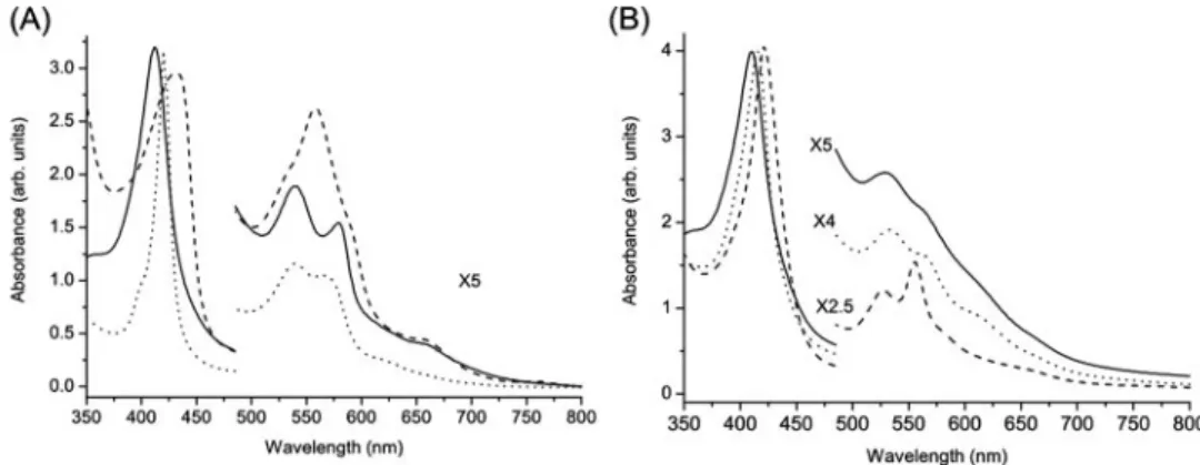
UV-visible Spectroscopy—The optical absorption spec-trum of the purified form of AvGReg178 shows the Soret maximum at 412 nm and␣ and  maxima at 579 and 540 nm, respectively (Fig. 2A). This is typical of oxygenated globins. Similar values were observed for the oxygenated derivative of Paramecium Hb (45) and sperm whale Mb (31). The absorption spectrum of the freshly purified form of AvGReg exhibits the Soret maximum at 410 nm and␣ and  maxima at 567 and 531 nm, respectively, typical for six-coordinate, low spin (LS) ferric proteins (46) (Fig. 2B). A very weak marker line at⬃619 nm suggests that a small high spin (HS) ferric component is present. Upon addition of dithionite to
AvGReg178, the absorbance maxima appear at 430 and⬃560 nm. This indicates that the deoxygenated protein is in the five-coordinate, HS ferrous heme state (Fig. 2A) (31). The spectrum of ferrous deoxygenated AvGReg shows the Soret maximum at 421 nm and the␣ and  maxima at 555 and 526 nm, respectively, which is indicative of a six-coordinate LS ferrous heme iron (Fig. 2B). Similar UV-visible spectra were found for ferrous unliganded nonsymbiotic barley Hb (47), nonsymbiotic rice Hb (46), Ngb (27), and cytoglobin (5). The spectra are typical of globins exhibiting bis-histidine heme-Fe atom coordination. Upon CO binding to ferrous
AvGReg178 and AvGReg, the Soret maximum is shifted to 421 and 416 nm, respectively. The ␣ and  maxima of
at BIABLIOTECA AREA BIOMEDICA on December 27, 2007
www.jbc.org
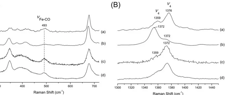
AvGReg-CO are 534 and 566 nm (LS). A small band at 616 nm indicates a small fraction of an HS ferric form. The␣ and  maxima of AvGReg178-CO are situated at 540 and 570 nm. Resonance Raman Spectroscopy—It is well established that the high frequency region of the RR spectra is composed of porphyrin in-plane modes that are sensitive to the oxidation, spin, and coordination states of the heme iron;2and3are linearly correlated with the distance between the central Fe atom and a nitrogen atom of the porphyrin ring (coordination and spin state), whereas4is sensitive to the oxidation state. In the spectrum of the as-isolated sensor domain AvGReg178-O2, 4,3, and2are located at 1372, 1502, and 1575 cm⫺1, respec-tively, which are typical values of oxygenated globins (Fig. 3B, trace a). In the RR spectrum of the freshly purified AvGReg, two 4bands at 1375 and⬃1360 cm⫺1and two3bands at 1506 and 1494 cm⫺1were observed, which are indicative of a mixture of ferric and ferrous LS AvGReg (Fig. 3B, trace b). The relative intensity of these bands depends on the laser power, i.e. the intensity of the band at 1360 cm⫺1 (because of the ferrous heme) increases with the laser power (see supplemental Fig.
S4). Therefore, this component can be assigned to the photo-dissociated form of the protein. This indicates that a small frac-tion of the protein is in the oxygenated form (the4and3
frequencies of oxygenated and LS ferric hemeproteins are undistinguishable).
The deoxygenated ferrous form of AvGReg178 displays the 4,3, and2bands at 1354, 1470, and 1562 cm⫺1, respectively,
typical of penta-coordinate HS ferrous hemeproteins (Fig. 3B,
trace c). TheFe-Hisstretching frequency is found to be⬃220
cm⫺1. This value is similar to the Fe-His stretching mode observed for deoxygenated Mb (220 cm⫺1) (48). In contrast, the Fe-His stretching frequency of deoxygenated Bacillus subtilis HemAT (HemAT-Bs) was reported at a higher frequency (i.e. 225 cm⫺1) (49), a feature that was related to a decrease in strain imposed on the Fe-His bond. The RR spectrum of ferrous deox-ygenated AvGReg is clearly different, showing the4,3, and2
bands at 1361, 1495, and 1592 cm⫺1, respectively, which are characteristic of a six-coordinate LS ferrous heme iron (Fig. 3B,
trace d). Accordingly, no Fe-His stretching frequency is observed in the low frequency region, confirming the hexa-coordinate LS character of ferrous deoxygenated AvGReg. These find-ings reinforce the interpretation of the absorption spectroscopic data.
In the low frequency region of the RR spectra of the deoxygenated derivative, the bending mode of the heme propionate, ␦(CCcCd), is
sensitive to electrostatic interaction on the heme propionates, such as hydrogen bonds and salt bridges (50). Thus, it is recognized that the stronger the electrostatic interac-tion between the heme propionate and the surrounding residues, the higher is the frequency of the ␦
FIGURE 2. A, absorption spectra of AvGReg178. Oxygenated form (obtained after protein purification) (contin-uous line), deoxygenated form (dashed line), and carbonylated form (dotted line) are shown. B, absorption spectra of AvGReg. Ferric form (obtained after protein purification) (continuous line), deoxygenated form (dashed line), and carbonylated form (dotted line) are shown.
FIGURE 3. Low frequency (A) and high frequency (B) RR spectra of purified AvGReg178 (oxygenated form) (trace a), purified AvGReg (ferric form) (trace
b), ferrous AvGReg178 (trace c), and ferrous AvGReg (trace d). The spectra were recorded at laser powers of 17 milliwatts (traces a, c, and d) and 40 milliwatts
(trace b). The peaks indicated by * are plasma lines from the krypton laser.
at BIABLIOTECA AREA BIOMEDICA on December 27, 2007
www.jbc.org
(CCcCd) band. The␦ (CCcCd) bands were observed around ⬃374, ⬃379, and ⬃376 cm⫺1 in ferrous unliganded AvGReg178 (Fig. 3A, trace c). This is comparable with the cor-responding form of Mb (371, 377, and 378 cm⫺1, respectively), for which moderate hydrogen bonds exist on heme propionate 7 (51). The vinyl mode,␦ (CCcCb2,4), was observed at 413 cm⫺1. In contrast to AvGReg178, the propionate and vinyl modes of AGReg fall together in a broad band (Fig. 3A, trace d). This indicates that the heme-globin contacts change signif-icantly upon reconstitution of the full protein.
The RR spectrum of the carbonylated derivative of ferrous AvGReg178 reveals two4bands as follows: a dominant peak at 1376 cm⫺1with a lower frequency shoulder (1359 cm⫺1) (Fig. 4B, traces a and b). The shoulder at 1359 cm⫺1stems from the HS ferrous state formed by photo-dissociation of CO. A similar, albeit smaller, photo-dissociation effect was observed for car-bonylated AvGReg (Fig. 4B, traces c and d). In agreement with carbonylated HemAT-Bs (49) and Paramecium Hb (45), the Fe-COband of AvGReg178 and AvGReg was observed at 493 cm⫺1(Fig. 4A, trace d). This frequency suggests an open con-formation of the heme pocket.
EPR Spectroscopy—CW-EPR provides an excellent tool to analyze ferric hemeproteins (52). In the X-band (9.5 GHz) CW-EPR spectrum of as-isolated AvGReg, three different contribu-tions could be observed stemming from one HS and two LS ferric heme forms (Fig. 5). The HS component is characterized by the principal g values, gz⫽ 6.4, gy⫽ 5.85, and gx⫽ 1.995, it can be ascribed to a penta-coordinate ferric form or the aquomet derivative of the sensor (53). The EPR spectra of the two LS ferric components are characterized by a highly rhom-bic g tensor. The principal g values of the dominant LS ferric component (LS1) are gz⫽ 2.94, gy⫽ 2.28, and gx⫽ 1.52. These gvalues can be directly related to different ligand field param-eters, such as the tetragonal splitting⌬ and the rhombic split-ting V, which in turn give insight in the type of axial ligands coordinating to the heme-Fe atom (52). For the dominant LS1 component in ferric AvGReg, this treatment leads to the
follow-ing values: V/ ⫽ 1.9, ⌬/ ⫽ 3.21, and V/⌬ ⫽ 0.59, where is the spin-orbit coupling. These values fall within the B region of the so-called “truth tables” of Blumberg and co-workers (54, 55), which is typical for hemeproteins with a bis-histidine heme-Fe atom coordination. This indicates that in ferric AvGReg, the heme iron appears to be bound to both the proximal His(F8) and distal His(E7) residues. The fact that the gzvalue of LS1 is smaller than 3, combined with the value of V/ ⫽ 1.9, hints that the two His axial ligands may be co-planar (56, 57). Comparison of g values obtained here with those reported by Quinn et al. (57), who related x-ray and CW-EPR data for five LS Fe(III) porphyrin systems with co-planar imidazoles, suggests that the angle between the projection of the His axial ligand normal on the porphyrin plane and the nearest Np-Fe-Npaxis is⬃22°.
Furthermore, a minor contribution because of a second LS ferric form (LS2) could be observed with gz⫽ 2.51, gy⫽ 2.28, FIGURE 4. Low frequency (A) and high frequency (4region) (B) RR spectra of the carbonylated form of AvGReg 178, laser power 0. 5 milliwatt (trace
a), AvGReg178, laser power 1 milliwatt (trace b), AvGReg, laser power 1 milliwatt (trace c), and AvGReg, laser power, 20 milliwatts (trace d).
FIGURE 5. CW-EPR spectra of ferric AvGReg. Experimental (continuous line, upper curve) and simulations of HS1, LS1, LS2, and total (dashed line, upper curve).
at BIABLIOTECA AREA BIOMEDICA on December 27, 2007
www.jbc.org
and gx⫽ 1.91. A component with similar principal g values (gz⫽ 2.52, gy⫽ 2.31, and gx⫽ 1.86) was observed in Chlamydomonas chloroplast Hb (58) and was assigned to heme-Fe atom coordi-nation by the Tyr(B10) residue. The ratio LS1/LS2 is 1/2. Note that it is difficult to estimate the ratio of the HS and LS ferric species from CW-EPR spectra because of the different satura-tion behavior of the HS and LS EPR signals at low temperature. The absorption spectra indicate a dominance of the LS compo-nent (see Fig. 2B). Note that attempts to oxidize the heme iron of as-isolated AvGReg178 failed even after long exposure to air. This clearly marks a difference between the isolated sensorial domain and the full-length protein.
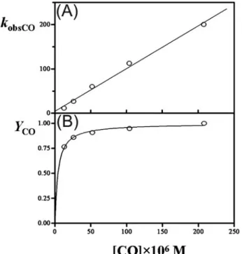
AvGReg178 and AvGReg Carbonylation Kinetics—SVD anal-ysis of CO binding to AGReg178 showed that between 390 and 500 nm only one optical transition accounts for more than 97% of the total absorbance changes (data not shown). Moreover, over the whole CO concentration range explored, the time course of the AvGReg178 carbonylation conforms to a single-exponential decay for more than 95% of its course, being in pseudo-first-order conditions (Fig. 6). Therefore, the minimum reaction mechanism depicted by Scheme 1 was employed to determine kinetic parameters for CO binding to AvGReg178. Data analysis carried out with fitting procedures allowed deter-mination of the following kinetic parameters: konCO⫽ (1.0 ⫾ 0.2)⫻ 106
M⫺1s⫺1and koffCO⫽ (4.0 ⫾ 1.0) s⫺1(pH 7.0 and T⫽ 20.0 °C). They are in good agreement with the estimates from the plot of observed pseudo-first-order rate constant (kobsCO) as a function of CO concentration (Fig. 7A). Values of the molar fraction of carbonylated AvGReg178 (YCO⫽ Aobs/Amaxat each wavelength) are wavelength-independent. The plot of YCO ver-sus[CO] is hyperbolic with KCO⫽ (4.5 ⫾ 1.0) ⫻ 10⫺6M(Fig. 7B; see Equation 3), which is in excellent agreement with the value of KCO(⫽ koffCO/konCOfor a penta-coordinate form)⫽ (4⫾ 2) ⫻ 10⫺6Mcalculated from values of konCOand koffCO, as expected from Scheme 1.
For AvGReg, SVD analysis showed that between 390 and 500 nm only one optical transition accounts for more than 90% of the total absorbance changes (data not shown). However, over
the whole CO concentration range explored, the time course for AvGReg carbonylation does not conform to a single-expo-nential decay but rather to a multiphase process (Fig. 8). The minimum reaction mechanism describing the experimental data demands three consecutive reversible reactions (see Scheme 2). If we assume that the active site of the globin domain of the full molecule has the same binding features of AvGReg178, FIGURE 6. CO binding to AvGReg178. Best fit of the time courses of CO
binding to AvGReg178 at different ligand concentrations. Over the whole CO concentration range explored (trace 1, [CO]⫽ 1.3 ⫻ 10⫺5M; trace 2, [CO]⫽ 2.6⫻10⫺5M; trace 3, [CO]⫽ 5.2⫻10⫺5M; trace 4, [CO]⫽ 1.04 ⫻ 10⫺4M; trace 5, [CO]⫽ 2.08 ⫻ 10⫺4M) the time course of AvGReg178 carbonylation conforms to single-exponential decay for more than 95% of their courses. Data have been analyzed according to Equation 1.
FIGURE 7. CO binding to AvGReg178. A, dependence of the pseudo-first-order constant (kobsCO) of CO binding to the AvGReg178 protein on the ligand concentration. The value of the second-order rate constant for the CO bind-ing to the AvGReg178 protein can be calculated from the slope of the regres-sion line (konCO⫽ (1.0 ⫾ 0.2) ⫻ 10
6
M⫺1s⫺1), whereas the y intercept can give an estimate of the first-order dissociation constant (koffCO⫽ (4.0 ⫾ 1.0) s⫺1). B, molar fraction of carbonylated AvGReg178 (relative amplitude YCO) as a function of the ligand concentration. From data analysis the value of the dissociation equilibrium constant for CO binding to the AvGReg178 protein was estimated (KCO⫽ (4.5 ⫾ 1.0) ⫻ 10⫺6M). Open circles represent experi-mental data whereas the straight line (A) and hyperbolic (B) represent the theoretical curves according to a simple CO-binding mechanism (see Scheme 1). Data in A have been analyzed according to Equation 2. Data in B have been analyzed according to Equation 3.
FIGURE 8. CO binding to AvGReg. Best fit of the time course of CO binding to AvGReg at different ligand concentrations (trace 1, [CO]⫽ 1.3 ⫻ 10⫺5M; trace 2, [CO]⫽ 2.6 ⫻ 10⫺5M; trace 3, [CO]⫽ 5.2 ⫻ 10⫺5M; trace 4, [CO]⫽ 4.2 ⫻ 10⫺4 M). The continuous lines were obtained from the fitting procedure carried out with GEPASI on the basis of the Scheme 2 (for details see text). The percentage of the fast, middle, and slow phase was 33⫾ 4%.
at BIABLIOTECA AREA BIOMEDICA on December 27, 2007
www.jbc.org
then data fitting analysis can be carried out fixing values of konCO and koffCOto those obtained from AvGReg178 for the bimolecular CO binding rate constant (see Schemes 1 and 2). This allowed us to determine the remaining kinetic parameters (see Scheme 2), for some of which (namely k⫺1and k⫺2) only the upper limiting values were obtained, i.e. k1⫽ 0.1 ⫾ 0.01 s⫺1, k⫺1ⱕ 0.01 s⫺1, koffHis⫽ 2.8⫾ 0.2 s⫺1, and konHisⱕ 0.5 s⫺1(pH⫽ 7.0 and T ⫽ 20.0 °C).
Values of kinetic and thermodynamic parameters for AvGReg178 and AvGReg carbonylation are shown in Table 1, where they are compared with those of globins. A striking observation is the fact that for both AvGReg178 and AvGReg the CO dissociation constant koffis⬃200-fold faster than for sperm whale Mb, whereas the CO association rate constants are similar (see Table 1).
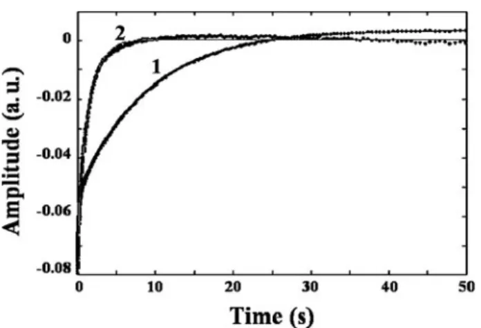
AvGReg178 and AvGReg Oxygen Dissociation Kinetics—SVD analysis concerning kinetics of O2dissociation from oxygen-ated AvGReg178 and AvGReg showed that between 390 and 500 nm only one optical transition accounts for more than 98 and 90%, respectively, of the total absorbance changes. The time course of O2 dissociation from AvGReg178-O2 and AvGReg-O2was fitted with a two-phase exponential decay (see Fig. 9). The biphasic time course of AvGReg178 and of AvGReg represents O2dissociation from the open fast (AvGReg⫹and AvGReg178⫹) and closed slow (AvGReg# and AvGReg178#) species (see Scheme 3).
The data fitting analysis was carried out fixing the value of the fast phase (i.e. open fast) of the time course of deoxygen-ation kinetics (i.e. koffO2) of AvGReg178-O2and AvGReg-O2 (see Scheme 3). Values of the dissociation constants of the open fast (k⫹offO2) and of the closed slow (k#offO2) oxygen-ated derivatives of AvGReg178 are k⫹offO2⫽ 10.6 ⫾ 0.7 s⫺1 and k#
offO2⫽ 0.13 ⫾ 0.04 s⫺1and of AvGReg are k⫹offO2⫽ 10.6⫾ 0.8 s⫺1and k#
offO2⫽ 0.73 ⫾ 0.04 s⫺1. Values of kinetic parameters k⫹offO2and k#
offO2for AvGReg178 and AvGReg deoxygenation are shown in Table 1.
AvGReg178 and AvGReg Oxygen Equilibrium Measurements— Fig. 10 shows the isotherms for O2binding to AvGReg178 and FIGURE 9. Time courses of O2dissociation from AvGReg178-O2(trace 1) and AvGReg-O2(trace 2). Points represent experimental data. Continuous lines were obtained by the two independent exponential data analysis according to Scheme 3. Values of koffO2and k
#
offO2for AvGReg178 and AvGReg are koffO2⫽ 10.6 ⫾ 0.7 s⫺1, k
#
offO2⫽ 0.13 ⫾ 0.04 s⫺1, koffO2⫽ 10.6 ⫾ 0.8 s⫺1, and k#
offO2⫽ 0.73 ⫾ 0.04 s⫺1, respectively. The percentages of the fast and slow phases were 31 and 69% for AvGReg178, and 48 and 52% for AvGReg, respectively. Both time courses start at⫺0.08 absorbance units (a.u.). TABLE 1 Rate and equilibrium constants for ligand binding His CO O2 P50 Ref. konHis koffHis KHis k1 kⴚ 1 koffHis konHis konCO koffCO KpentaCO a konO2 koffO2 KpentaO2 a KhexaO2 b measured s ⫺ 1 s ⫺ 1 s ⫺ 1 s ⫺ 1 s ⫺ 1 s ⫺ 1 M ⫺ 1s ⫺ 1⫻ 10 6 s ⫺ 1 MM ⫺ 1s ⫺ 1⫻ 10 6 s ⫺ 1 M M torr SwMb wild type 0.5 0.019 0.038 5.3 10 1.887 0.51 103, 104 Av GReg178 1 4 4.5 424 10.6 0.025 0.04 This study 4 5.2 0.13 c 0.025 Av GReg ⱕ 0.5 2.8 ⱖ 5.6 0.1 ⱕ 0.01 2.8 ⱕ 0.5 1 4 4 10.6 0.12 0.15 This study 0.73 c 0.12 HemAT-Bs (sensor domain) 0.43 0.07 0.16 19 1800 94 to 74 105 50 HemAT-Bs 0.34 0.067 0.20 19 1900 100 0.1 105 87 HNgb 2000 4.5 0.002 65 0.014 0.00022 250 0.8 0.003 0.70 2.6 33 MNgb 1000 0.5 0.0005 55 200 0.4 0.002 0.25 2.2 83 Rice Hb 7.2 0.001 0.00014 68 0.038 0.001 46 Human cytoglobin ⫹ DTT 200 2 0.01 5 27 0.9 0.033 0.30 83 Paramecium Hb 27.7 0.328 11.8 30.1 25.2 0.837 0.45 45 aValues of the dissociation constants (koff /kon ). bK hexaO2 was calculated as follows: KhexaO2 ⫽ (K O2dis ⫺ 1/(1 ⫹ KHis ⫺ 1)). cValues of the closed slow conformation.
at BIABLIOTECA AREA BIOMEDICA on December 27, 2007
www.jbc.org
AvGReg as obtained by the tonometric and the thin layer opti-cal methods. Both proteins display a high affinity for O2(KO2⫽ K#
O2⫽ (2.5 ⫾ 1.3) ⫻ 10⫺8Mfor AvGReg178 and KO2⫽ K#O2⫽ (1.2⫾ 0.4) ⫻ 10⫺7Mfor AvGReg) with P50values of 0.04 torr for AvGReg178 and 0.15 torr for AvGReg.
Values of KO2for AvGReg178 and AvGReg oxygenation are an average of tonometric and thin layer results. Binding iso-therms for both AvGReg178 and AvGReg could be fitted satis-factorily employing a single equilibrium binding constant, although experiments on AvGReg178 by the thin layer method could be better fitted implying slight apparent negative cooper-ativity (n⬵ 0.8), which appears consistent with the functional heterogeneity observed in oxygen dissociation kinetics. In any event, this result clearly demonstrates that for the two proteins, the open fast (characterized by K⫹O2) and the closed slow spe-cies (characterized by K#
O2) display a similar binding affinity. Therefore, the biphasic O2dissociation kinetics (i.e. k⫹offO2⫽ k#
offO2) implies also a biphasic O2 association process (i.e. k⫹onO2⫽ k#onO2), which partially compensates, resulting in closely similar affinity (i.e. K⫹O2⬵ K#O2). As a consequence, values of k⫹onO2 and k#
onO2 were roughly estimated for AvGReg178 from values of K⫹O2, k⫹offO2, and k#
offO2, accord-ing to Scheme 4. However, for AvGReg, a hexa-coordinate glo-bin, this extrapolation cannot be done because the affinity for the internal histidine cannot be ignored. Values of thermody-namic parameters for AvGReg178 and AvGReg oxygenation are shown and compared with those reported for sperm whale Mb in Table 1. It clearly emerges that the closed slow conformation of AvGReg178 displays a value for k#
onO2similar to the associ-ation rate constant for sperm whale Mb, whereas the dissocia-tion rate constants k#
offO2 are much slower than for sperm whale Mb. On the other hand, in the case of the open fast con-formation the opposite is found, whereby a close similarity with sperm whale Mb is observed for the dissociation rate constant koffO2, whereas a much faster O2association rate constant is observed for AvGReg178 (Table 1).
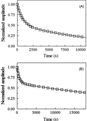
AvGReg-NO and AvGReg178-NO Denitrosylation—Over the wavelength and [CO] ranges explored, the time course for NO dissociation from AvGReg-NO and AvGReg178-NO conforms to a two-exponential process for more than 90% of its course (Fig. 11). Values of the first-order rate constant for NO disso-ciation from AvGReg⫹-NO (k⫹offNO ⫽ 1.2 ⫻ 10⫺3 s⫺1) and AvGReg#-NO (k#
offNO ⫽ 8.6 ⫻ 10⫺5 s⫺1) as well as from AvGReg178⫹-NO (k⫹offNO ⫽ 1.7 ⫻ 10⫺3 s⫺1) and AvGReg178#-NO (k#
offNO ⫽ 2.5 ⫻ 10⫺5s⫺1), at pH 8.3 and 20.0 °C, are wavelength- and [CO]-independent in the presence of dithionite excess (data not shown).
Values of the first-order rate constant for NO dissociation from heme-NO-proteins (i.e. koffNO) span over 3 orders of mag-nitude (Table 2) (59 – 66), reflecting structurally different sta-bilization mode(s) of the heme-bound NO by heme distal residue(s).
NO-mediated Oxidation of AvGReg-O2and AvGReg178-O2— NO induces the oxidation of hemoproteins, and this process is postulated to depend on the superoxide character of the heme-bound O2(35, 67–74). Over the wavelength range explored, the FIGURE 10. O2equilibrium measurements carried out at 20.0 °C with the
tonometric method (squares) and the thin layer optical method (circles) to AvGReg178 (filled symbols) and to AvGReg (open symbols). Data
obtained with the two methods were analyzed as a unique data set on the basis of Equation 8. Values of KO2⫽ K
#
O2for AvGReg178 and AvGReg oxygen-ation are (2.5⫾ 1.3) ⫻ 10⫺8M) and (1.2⫾ 0.4) ⫻ 10⫺7M), respectively. Values of P50 for AvGReg178 and AvGReg oxygenation are 0.04 and 0.15 torr, respectively.
FIGURE 11. NO dissociation from AvGReg-NO and AvGReg178-NO. A, nor-malized averaged time courses of NO dissociation from AvGReg⫹-NO and AvGReg#-NO. The time course analysis according to Equation 9 allowed deter-mination of k⫹offNO⫽ 1.2 ⫻ 10⫺3s⫺1and k#offNO⫽ 8.6 ⫻ 10⫺5s⫺1. B, normal-ized averaged time courses for NO dissociation from AvGReg178⫹-NO and AvGReg178#-NO. The time course analysis according to Equation 9 allowed us to determine k⫹offNO⫽ 1.7 ⫻ 10⫺3s⫺1and k#offNO⫽ 2.5 ⫻ 10⫺5s⫺1. Spectra were collected every 5 min. The CO and dithionite concentrations were 5.0⫻ 10⫺4 and 1.0⫻ 10⫺2 M, respectively. The AvGReg-NO and AvGReg178-NO concentrations were 2.2⫻ 10⫺6and 1.9⫻ 10⫺6M, respec-tively. All data were obtained at pH 8.3 (5.0⫻ 10⫺2MTricine buffer) and T⫽ 20.0 °C.
at BIABLIOTECA AREA BIOMEDICA on December 27, 2007
www.jbc.org
time course for NO-mediated oxidation of AvGReg-O2 and AvGReg178-O2 conforms to a two-exponential process for more than 90% of its course, at [NO] ⫽ 2.0 ⫻ 10⫺6 M and 4.0⫻ 10⫺6M(Fig. 2A). At [NO]ⱖ 8.0 ⫻ 10⫺6M, kinet-ics was essentially mono-exponential, because the time course of NO-mediated oxidation of AvGReg⫹-O2 and AvGReg178⫹-O2 was lost in the dead time of the rapid-mixing stopped-flow apparatus, and the observed trace only reflects NO-mediated oxidation of AvGReg#-O
2(Fig. 12A) and AvGReg178#-O
2 (data not shown). Values of the pseudo-first-order rate constant for NO-mediated oxidation of AvGReg⫹-O2 and AvGReg178⫹-O2 (i.e. h⫹) and of AvGReg-O2and AvGReg178-O2(i.e. h#) are wavelength-in-dependent at fixed [NO]. The plot of h⫹and h#versus[NO] is linear with a y intercept at 0, the slope corresponding to values of the second-order rate constant for NO-mediated oxidation of AvGReg⫹-O2 and AvGReg178⫹-O2 (hon⫹ ⫽ 3.2⫻ 107
M⫺1s⫺1and 4.6⫻ 107M⫺1s⫺1, respectively) and AvGReg#
-O2and AvGReg178
#
-O2(hon#⫽ 2.5 ⫻ 106M⫺1s⫺1 and 7.2⫻ 105
M⫺1s⫺1, respectively) (Fig. 12B and Table 3). As already reported for Glycine max legHb, horse heart Mb, human Hb, murine Ngb, Mycobacterium tuberculosis trHbN and trHbO, and E. coli flavoHb (35, 75– 80), NO-mediated oxidation of AvGReg#-O
2and AvGReg178#-O2appears to be limited by the formation of the heme-Fe(III)-peroxynitrite intermediate, which does not accumulate. In fact, the first-order process (i.e. independent of [NO]) representing disso-ciation and isomerization of peroxynitrite, which otherwise would follow the pseudo-first-order step (35), is undetect-able in NO-mediated oxidation of AvGReg#-O
2 and AvGReg178#-O
2(see Fig. 12A). NO-mediated oxidation of Mycobacterium leprae trHbO(II)-O2 represents an excep-tion, the dissociation and isomerization of peroxynitrite rep-resenting the rate-limiting step (81).
DISCUSSION
Both recombinant full-length AvGReg and its sensor do-main AvGReg178 were successfully expressed in E. coli BL21(DE3)pLysS cells. There was no evidence that AvGReg
and the products of this protein were toxic for the cells, although Ryjenkov et al. (24) proposed that the products of the enzymatic reaction of the GGDEF domain (i.e. c-di-GMP) are cytotoxic. This can be explained by the fact that AvGReg was FIGURE 12. NO-mediated oxidation of AvGReg-O2and AvGReg178-O2. A, normalized averaged time courses for NO-mediated oxidation of AvGReg⫹-O2and AvGReg
#-O
2. The time course analysis according to Equa-tion 10 allowed the determinaEqua-tion of h⫹⫽ 6.4 ⫻ 101s⫺1and h#⫽ 4.8 s⫺1 at [NO]⫽ 2.0 ⫻ 10⫺6M(circles), h⫹⫽ 1.3⫻102s⫺1and h#⫽ 9.9 s⫺1at [NO]⫽ 4.0 ⫻ 10⫺6M(squares), h#⫽ 1.9 ⫻ 101s⫺1at [NO]⫽ 8.0 ⫻ 10⫺6M (triangles), and h#⫽ 4.1 ⫻ 101s⫺1at [NO]⫽ 1.6 ⫻ 10⫺5M(diamonds). The time course of NO-mediated oxidation of AvGReg⫹-O2was essentially undetectable at [NO]ⱖ 8.0 ⫻ 10⫺6M. The AvGReg-O2concentration was 4.8⫻ 0⫺7 M. B, dependence of h⫹ and h# values (squares and circles, respectively) on the NO concentration for NO-mediated oxidation of AvGReg-O2(open symbols) and AvGReg178-O2(filled symbols). Data anal-ysis according to Equations 11 and 12 allowed us to determine the follow-ing values of hon⫹⫽ 3.2 ⫻ 10
7
M⫺1s⫺1and hon
#⫽ 2.5 ⫻ 106
M⫺1s⫺1for NO-mediated oxidation of AvGReg-O2, and of hon⫹⫽ 4.6 ⫻ 10
7 M⫺1s⫺1 and hon#⫽ 7.2⫻105
M⫺1s⫺1for NO-mediated oxidation of AvGReg178-O2. The AvGReg-O2and AvGReg178-O2concentration was 4.8⫻ 10⫺7and 4.7⫻ 10⫺7M, respectively. All data were obtained at pH 8.3 (5.0⫻ 10⫺2M Tricine buffer) and T⫽ 20.0 °C.
TABLE 2
Values of koffNOfor denitrosylation of heme-Fe(II)-NO proteins Heme-Fe(II)-NO
protein koffNO(sⴚ1) Conditions Ref.
AvGReg⫹ 1.2⫻ 10⫺3 pH 8.3 and 20.0 °C This study
AvGReg# 8.6⫻ 10⫺5 pH 8.3 and 20.0 °C This study
AvGReg178⫹ 1.7⫻ 10⫺3 pH 8.3 and 20.0 °C This study
AvGReg178# 2.5⫻ 10⫺5 pH 8.3 and 20.0 °C This study
G. maxlegHb 2.0⫻ 10⫺5 pH 7.0 and 20.0 °C 64
SwMb 1.0⫻ 10⫺4 pH 7.0 and 20.0 °C 63
Horse heart Mb 1.0⫻ 10⫺4 pH 7.4 and 20.0 °C 65
Human soluble guanylyl cyclase
6.0⫻ 10⫺4 pH 8.3 and 24.0 °C 66
Mouse Ngb 2.0⫻ 10⫺4 pH 7.5 and 25 °C 62
Rabbit hemopexin-heme 9.1⫻ 10⫺4 pH 7.0 and 10.0 °C 60
Human Hb, R-state, ␣-chains 1.6⫻ 10 ⫺4 pH 7.2 and RTa 61 -chains 8.0⫻ 10⫺5 pH 7.2 and RT 61 Human Hb, T-state, ␣-chains 4.4⫻ 10 ⫺3 pH 7.2 and RT 61 -chains 9.4⫻ 10⫺5 pH 7.2 and RT 61
M. lepraetrHbO 1.3⫻ 10⫺4 pH 7.0 and 20.0 °C 59
E. coliflavo-Hb 2.0⫻ 10⫺4 pH 7.0 and 20.0 °C 76 a
RT indicates room temperature.
TABLE 3
Values of konfor NO-mediated oxidation of heme-Fe(II)-O2proteins
Heme-Fe(II)-O2-protein hon(Mⴚ1sⴚ1) Conditions Ref.
AvGReg⫹ 3.2⫻ 107
pH 8.3 and 20.0 °C This study
AvGReg#
2.5⫻ 106
pH 8.3 and 20.0 °C This study
AvGReg178⫹ 4.6⫻ 107
pH 8.3 and 20.0 °C This study
AvGReg178#
7.2⫻ 105
pH 8.3 and 20.0 °C This study
G. maxlegHb 8.2⫻ 107 pH 7.3 and 20.0 °C 78 Horse heart Mb 4.4⫻ 107 pH 7.0 and 20.0 °C 77 Mouse Ngb ⬎7.0 ⫻ 107 pH 7.0 and 20.0 °C 75 Human Hb 8.9⫻ 107 pH 7.0 and 20.0 °C 77 M. tuberculosistrHbN 7.5⫻ 108 pH 7.5 and 23.0 °C 79 M. tuberculosistrHbO 6.0⫻ 105 pH 7.5 and 23.0 °C 80 M. lepraetrHbO 2.1⫻ 106 pH 7.3 and 20.0 °C 81 E. coliflavoHb ⱖ6 ⫻ 108 pH 7.0 and 20.0 °C 76
at BIABLIOTECA AREA BIOMEDICA on December 27, 2007
www.jbc.org
expressed in inclusion bodies, and there was probably no for-mation of c-di-GMP. Reconstitution of the globin domain had to be completed in vitro by the addition of 1.4M excess of hemin. No additional cofactors were added for the refolding of the second domain based on procedures found in the literature (82). In contrast with Hecht et al. (82), MgCl2was not added although it is important for the activation of the GGDEF domain.
Physiological Significance—The heme in AvGReg is thought to play an important role for sensing O2or other ligands. The nature of the ligand, the interaction between the heme-Fe-bound ligand and heme distal amino acid residues, and/or the heme geometry could be responsible for triggering the signal transduction pathway upon ligand binding to the heme in AvGReg. A study of the heme-pocket structure is therefore important to elucidate the mechanism of ligand binding and signal triggering. Absorption and RR spectroscopy observa-tions show that the heme coordination number of ferrous AvGReg178 changes upon ligand binding (five-to-six-coordi-nation). In contrast, the heme coordination number of ferrous full-length protein AvGReg does not change upon binding of an effector molecule, as it remains hexa-coordinate. This indicates that, unlike in the globin domain AvGReg178, binding of an exogenous ligand to the full-length protein brings about the dissociation and displacement of the endogenous heme distal His residue; this event may trigger conformational changes in the protein. More subtle processes, such as hydrogen bonding formation between the heme-bound effector molecule and the surrounding amino acid residues, as proposed for O2-bound HemAT-Bs (but not for CO- and NO-bound HemAT-Bs) (18), may play a critical role.
Interestingly, ferrous carbonylated AvGReg178 and AvGReg exhibit theFe-COband at 493 cm⫺1(Fig. 4A, traces c and d), indicating that the environment of the heme-bound CO is sim-ilar in the full protein and in the globin domain. Indeed, this could justify our assumption that the intrinsic bimolecular CO binding rate constant (i.e. konin Schemes 1 and 2) and the CO dissociation rate constant (i.e. koffin Schemes 1 and 2) are the same for both AvGReg178 and AvGReg (Table 1). The differ-ence between the sensor domain and the full protein resides then in the pathway for the ligand access to the reaction center, which is quite open for the sensor domain, whereas in the full protein the additional domain(s) hide(s) the heme pocket and the access of the ligand is permitted only through a series of conformational changes. The observed stretching frequency is typical of a conformation in which CO has very little polar interaction with the surrounding amino acids (open conforma-tion of the heme pocket). This suggests that the displaced heme distal His(E7) in AvGReg does not interact with the Fe-bound CO. This contrasts with the observations for the bis-histidine coordinate heme-Fe atom in Ngb (84) and cytoglobin (5), for which CO ligation induces a mixture of open and closed con-formations. On the other hand, these spectroscopic features are similar to those displayed by the CO-ligated forms of HemAT-Bs (18) and Paramecium Hb (45) but differ from observations concerning mammalian Mb (85).
Unlike the Fe-CO frequencies, those corresponding to the heme vinyl and propionate modes show significant differences
between AvGReg178 and AvGReg (Fig. 3A). Because the fre-quency of the propionate bending mode␦(CCcCd) is indica-tive of hydrogen bonding between the heme-7-propionate group and the surrounding amino acid residues, low frequen-cies observed for AvGReg178 indicate weak (or no) heme-7-propionate hydrogen bonding for the globin domain. However, the propionate and vinyl groups of the full-length protein fall together in a broad band, suggesting that the conformation of the heme group changes significantly upon reconstitution of the full-length protein. This can mean that the activation of the second domain depends on hydrogen bonding with heme peripheral groups. Indeed, it accounts for the different struc-ture of the heme pocket of the sensor domain depending on whether the second domain is present (as in AvGReg) or not (as in AvGReg178). The comparison of the globin domain and the whole molecule clearly shows differences, and as such, only the whole molecule is physiologically relevant.
Selective Ligand Binding—The data reported here indicate that AvGReg178 displays Mb-like spectroscopic properties. However, AvGReg178 and AvGReg display different functional behaviors. In fact, AvGReg178 follows Mb-like functional prop-erties (86) (see Scheme 1), whereas AvGReg displays three-pha-sic reaction kinetics reminiscent of that reported for hexa-co-ordinate hemoproteins (41) (Scheme 2). Thus, the low reactivity of the AvGReg hemoprotein may reflect the conver-sion of the AvGReg§ closed, hexa-coordinate species to the AvGReg* open, hexa-coordinate form that in turn converts to the AvGReg open, penta-coordinate species. Only the last-mentioned species reacts with CO leading to AvGReg⫹-CO (Scheme 2). The data analysis indicates that AvGReg178 and AvGReg could share the same intrinsic reactivity toward CO (i.e. konCO⫽ (1.0 ⫾ 0.2) ⫻ 106M⫺1s⫺1and koffCO⫽ (4.0 ⫾ 1.0) s⫺1), as also suggested by the closely similar spectroscopic fea-tures of the CO-bound form (see above and Fig. 4A). Therefore, the non-heme domain of AvGReg lowers the CO reactivity, stabilizing the hexa-coordinate species of the heme domain (see Scheme 2). The CO binding behavior of AvGReg mirrors the close similarity between the absorption spectrum in the Soret region of the deoxygenated derivative of the AvGReg hemopro-tein (Fig. 2B) and that of hexa-coordinate bis-histidyl adducts (27, 87, 88). A simple mechanism involving only the hexa- to penta-coordination of the heme-Fe atom step preceding the AvGReg hemoprotein carbonylation process does not fit the experimental data (data not shown), because it cannot account for the very slow process, characterized by k1and k⫺1(Fig. 8). However, more complex reaction mechanisms (including both parallel and crossing reactions) depicting CO binding to the AvGReg hemoprotein can in principle not be excluded. A very unusual feature is the relatively fast rate constant for CO disso-ciation (i.e. koffCO⫽ 4 s⫺1, Table 1), which is 10 –100-fold faster than in both mammalian hemoproteins and those from unicel-lular species (45). This result suggests that the fast CO dissoci-ation rate constant for AvGReg178 is likely related to the elec-tronic distribution of the heme and/or to a stereochemical distortion of the Fe-His proximal bond in the CO-bound form, which have been proposed to play an important role in the CO dissociation rate constant (89, 90). On the other hand, the closely similar CO association rate constant for the bimolecular
at BIABLIOTECA AREA BIOMEDICA on December 27, 2007
www.jbc.org