Mucositis in the treatment of haematological malignancies
Abstract
Background. Mucositis is a highly significant and sometimes dose-limiting complication of many treatments given for blood-related malignancies.
Aim and Methods. This review formed the basis of an invited educational lecture at the 2008 Annual conference of the European Society of Medical Oncology (ESMO). Based on a review of the most recent literature and on authors’ experiences, a summary of the current knowledge on mucositis has been provided together with the accepted recommendations on management. Results. The incidence and severity of mucositis are influenced by patient and treatment variables, being particularly high in the setting of high dose chemotherapy and haematopoietic stem cell transplantation. A five-stage model has been identified in the pathophysiological process of mucositis. Although some interventions have been shown to be potentially effective, in many cases the reported clinical trials and the available data are inadequate to support the recommendation of the majority of agents. Thus, the only proven measures in the prevention of mucositis are cryotherapy and palifermin. Conclusions and perspectives. Further intensive research is necessary, through well-structured clinical trials, in order to obtain the best scientific evidence for agents to prevent and/or treat mucositis in the setting of haematological malignancies.
Keywords: mucositis, haematological malignancies, haematopoietic stem cell
transplantation, pain, palifermin, graft versus host disease
Introduction
Mucositis is a pathological process characterised by mucosal damage, affecting one or more parts of the alimentary tract, from the mouth to the anus, as a consequence of radiation therapy and/or chemotherapy. Other mucosae, with the exception of bladder mucosa after alkylating agents and the conjunctiva after high doses of cytarabine1, do not seem to be so broadly affected, but this could be under investigation as well as to local protection. Mucositis is a devastating and debilitating complication of cytotoxic therapy given for haematological malignancies2. Indeed, it can negatively impact the patient’s quality of life and disease outcome, portraying clinical and economic consequences, such as pain3-5, life-threatening infections, the need for total parenteral nutrition use and analgesic therapy, additional nursing care and longer hospitalisation2.
Epidemiology
Cytotoxic damage to oral and/or gastrointestinal (GI) mucosa results from a complex, dynamic, multi-factorial process, to which treatment-induced and patient-related
factors contribute. The toxicity of each drug depends on its dosage and the exposure time as well as its intrinsic properties. Comorbidities, infections, poor oral hygiene and prolonged treatment with steroids are some patient-related factors1,6. Furthermore, differences in drug absorption, metabolism, distribution, and excretion, due to the genetic variants of several families of enzymes, seem to have pronounced effects, causing significant differences in the severity of mucositis among patients treated with the same chemotherapy regimens. For example, the genetic polymorphisms for the 5,10-methylenetetrahydrofolate reductase and for thiopurine S-methyltransferase are major factors responsible for large individual variations in both the toxicity and therapeutic effect of metotrexate (MTX) and thiopurine respectively. While all drug classes can cause mucositis, they do it to different degrees. The most highly mucotoxic agents are: thymidine synthetase inhibitors, such as MTX, topoisomerase II inhibitors (etoposide, irinotecan); pyrimidine analogs (cytarabine); purine analogs (6-mercaptopurine and 6-thioguanine); high doses of alkylating agents (busulfan, melphalan and cyclophosphamide); and intercalating drugs (idarubicin, doxorubicin, daunorubicin). When these agents are administered in multiple cycles, the risk of mucositis probably increases at each course. Following a standard dose-dense chemotherapy regimen for non-Hodgkin’s lymphomas (NHL), such as the CHOP (cyclophosphamide, doxorubicin, vincristine and prednisone) , an incidence of oral mucositis (OM) ranging from 2% to 10% has been reported1,7. The addition of rituximab and a shorter interval of administration (CHOP-14 regimen) has not been associated with an higher incidence of mucositis, which raises questions of the relationship between dose interval and mucosa recovery, accuracy of reporting, and interactions between mucositis and other toxicities. In the setting of Hodgkin’s lymphoma, the reported incidence of mucositis was 3% in patients who received the standard ABVD (doxorubicin, bleomicine, vinblastine and dacarbazine) regimen. Finally, the mucosal toxicity associated with intensified salvage regimens for lymphoma patients is generally mild and manageable. Patients affected by acute myeloid leukaemia (AML) treated with standard anthracycline-based regimens develop profound myelosuppression and OM (10–15% of cases). In this setting, liposomal daunorubicin seems to reduce the incidence of mucositis, unlike other aggressive protocols: the FLAG (fludarabine, cytarabine, G-CSF) regimen induces mucosal damage in 50% of patients, a rate that rises to 70% in those treated with idarubicin-containing FLAG. In patients with acute promyelocytic leukaemia treated with trans-retinoic acid (ATRA), which can cause mucosal dryness, and idarubicin (AIDA protocol), the incidence of OM is about 10%. Hydroxyurea is used as a pre-induction, palliative or mild myelosuppressive drug in AML and has not been associated with mucosal injury when used at standard doses. On the contrary, among the oral agents available for the treatment of the disease, 6-mercaptopurine is strongly mucotoxic.
Very few epidemiological data on the mucosal injury from targeted anti-cancer and biological modifier agents, which are increasingly used in oncohematology, have been reported. Tyrosine kinases inhibitors were not initially associated with mucositis, but with increasing use mucositis has become more evident, especially in the form of diarrhoea. Among monoclonal antibodies, gemtuzumab-ozogamicin, a
compound targeting the CD33 antigen on blast membranes, was thought no effect on the mucosa, but OM occurs in about 4% of patients treated. Rituximab and alemtuzumab do not have a mucotoxic effect. However, the full mucotoxic potential of these agents is yet to be elucidated, and work into pathobiology of targeted therapy-induced OM and diarrhoea is ongoing.
Recent advances have led to the use of radioimmunotherapy in patients affected by advanced NHL; Yttrium 90 ibritumomab tiuxetan has lower mucosal toxicity than standard chemotherapy. Lastly, the frequent watery diarrhoea and abdominal cramps following bortezomib administration are probably due to intestinal neuropathy rather than to mucositis. However, as recently claimed8, we believe that in the clinical trials exploring the efficacy and the safety of these agents for haematological malignancies, the provision of a regular mucosal assessment is mandatory in order to attain more firm information about their mucotoxic potential.
The amount of chemotherapy administered, the previous exposure to some drugs (e.g. anthracyclines, vinca alkaloids, cyclophosphamide, fludarabine, platinum analogs and methotrexate), female gender and the type of disease have been associated with risk of mucositis following autologous SCT 1,6. Furthermore, radiotherapy, a diagnosis of NHL and etoposide administration as part of the stem cell mobilising regimen have been associated with worse mucositis. With regard to conditioning regimens, those containing busulfan and melphalan or radiotherapy, play a crucial role in the development of mucositis. The BEAM schedule (BCNU, etoposide, cytarabine and melphalan) is currently used as a conditioning regimen for NHL patients and is responsible for severe mucositis in 75% of cases. The association of idarubicin with busulfan for autologous SCT in AML patients caused profound mucosal derangement in 82% of patients. High doses of melphalan (200 mg/m2), given prior to autologous SCT for multiple myeloma, caused mucosal injury in about 35%; intermediate doses (100 mg/m2) significantly reduced the incidence of mucositis to 23%, as reported in a study including patients over 70 years old. In the allogeneic SCT setting, the incidence of mucositis reaches 75 to 100%, depending on the type of disease, procedure and conditioning regimen; moreover, true ulcerations in the mouth have been reported in 76% of cases. Risk factors for mucosal damage in allogeneic SCT are a pre-transplant body mass index higher than 25 as well as the use of total body irradiation (TBI) as part of the conditioning regimen. However, since much chemotherapy is given based on body surface area calculations, there is a possibility that obese patients are relatively over-dosed compared with normal weight patients. Reduced myeloablative regimens for allogeneic SCT result in a low incidence of gastrointestinal toxicity.
Pathobiology
In the pathophysiological process of mucositis, including GI forms9, a five-stage model, which comprises initiation, upregulation and message generation, signalling and amplification, ulceration and finally healing phases has been identified (Table
1)6. Although this model is described in a linear way, injury occurs quickly and simultaneously in all mucosal tissues. At the beginning DNA damage, generation of reactive oxygen species (ROS), and the coincident activation of other pathways occur. During the upregulation and message generation phase, transcription factors, such as nuclear factor-B (NF-B) are activated to upregulate genes in the endothelium, fibroblasts, macrophages, and epithelium; this process is followed by the production of pro-inflammatory cytokines, such as tumor necrosis factor (TNF), interleukin-1 ß (IL-1ß), interleukin-6 (IL-6), which mediate a series of biological events leading to apoptosis and amplification of the injury, loss of epithelial integrity and the development of ulceration. At this stage, bacteria colonize the ulcer’s surface and increase the injury by shedding of cell-wall products and, in case of granulocytopenia, may lead to bacteremia and sepsis. Ultimately, spontaneous healing occurs. The lesions in the mouth mainly involve the non-keratinised part that becomes susceptible to overinfection, whereas in the GI tract the cytopathic effect is more severe in the ileum than in the colon. GIM develops through multiple mechanisms including induction of crypt cell death (apoptosis) and cytostasis10. Although the molecular control of these events throughout the GI tract has yet to be fully elucidated, p53, members of the Bcl-2 family, and caspases have been reported to be involved11. An increase of apoptosis can be observed by 24 hours after the administration of therapy, followed by a reduction in the length of the intestinal villi causing mucosal flattening around the 3rd day. From the 5th day, hyperplasia of the intestinal mucosa leads to the recovery of the GI barrier.
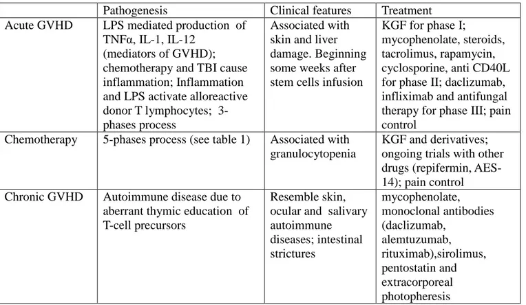
GVHD can affect the whole GI tract, the mouth being involved in 80% of the cases. Some GVHD-related clinical features may resemble mucositis. The comparison between mucositis due to chemotherapy and GVHD-related mucosal lesions is shown in table 2.
Clinical features and diagnostic tools
The main symptom of OM is pain which may be mild or severe, together with nausea, sialorrhea, sometimes profuse, and infections. The pain syndromes can range from a sense of burning in the initial phases up to severe forms and are caused by a mixture of different types of pain3,5,12. The main component is nociceptive pain, mediated by C fibers and relievable by opioids, and incidental pain, caused by movement and contact with the mucosal surface, mediated by the fast-conducting A-fibres. The latter component is insensitive to analgesics. The symptoms of GI mucositis are visceral pain (ranging from mild pain to projected abdominal wall pain), hypermotility with diarrhoea, starting from the 3rd day after the beginning of the treatment and resolving by the 7th day, coinciding with the full clinical flare of the OM. In patients undergoing treatment including high doses of cytarabine, the diarrhoea generally develops between the 5th and 8th day after starting chemotherapy and persists over the second week1. In addition, GI mucositis may be complicated by gastrointestinal obstruction, perforation, and infection.
practice due to the lack of standard diagnostic criteria established by controlled studies. The principal instruments used to assess OM and GI mucositis have been recently reviewed and elsewhere reported1,6,13. A critical aspect in the management of these patients, particularly those with OM, is the regular assessment of the pain for which validated tools are described elsewhere3,5.
Therapeutic approach
Despite its clinical significance, there is still no standard approach to prevent or treat mucositis. Interventions have been limited to the use of palliative measures, barrier protectants, topical antimicrobials, ice, and analgesics. Although some interventions have been shown to be potentially effective, in many cases the reported clinical trials and the available data are inadequate to support the recommendation of the majority of agents. The Multinational Association of Supportive Care in Cancer produces guidelines for the management of mucositis, based on the current literature7. Basic oral hygiene, periodic control of dental health and comprehensive patient education are important components of the care of any patient with haematological malignancies at risk of OM1. Effective approaches for the prevention and management of OM include oral cryotherapy and low-level laser therapy for patients undergoing SCT6,7. Cryotherapy seems to be effective in limited areas of the oral mucosa, as well as a treatment for melphalan-induced mucositis, or mucositis induced by other drugs with a short plasma half-life. Antibiotic prophylaxis, although considered a reasonable measure in subjects undergoing myelosuppression, is ineffective in reducing the colonizing microbes on the mucosal surface during autologous SCT. The topical application of chlorhexidine, GM-CSF, the salivary production stimulator pilocarpine, and histamine gel are not recommended for the prophylaxis of OM given the reported lack of efficacy of these agents7,14; but chlorhexidine does have a role in the prevention of dental caries. Although some favourable results have been reported in the setting of SCT 15,16, the amino acid glutamine cannot be recommended as a proven measure in the prevention of mucositis7. due to a recent report of its increasing mortality7. The partial efficacy of other interventions, such as benzydamine17, clarithromycin, and amifostine18 has been reported in some studies but none was powered to provide acceptable evidence of efficacy19. A consensus has recently been reached on the use of sulfasalazine to prevent GI mucositis in patients undergoing radiotherapy, while octreotide is considered useful for reducing the frequency and volume of diarrhea14.
Low level laser therapy (LLLT) has been used with some suggestion of benefit in the prevention of OM in patients undergoing HSCT with chemotherapy or chemoradiotherapy conditioning, but the evidence is not strong20,21. Among the latest approaches, the most promising is palifermin, a human recombinant keratinocyte growth factor (KGF).Upon activation of the transcription factor Nrf2, which encodes for other genes playing a role in detoxifying ROS, palifermin exerts its effects on
keratinocytes, fibroblasts and endothelial cells. Moreover, KGF can attenuate the effects of TNF and the expression of adhesion molecules. In clinical trials palifermin, compared to placebo, significantly reduced the incidence and duration of severe OM after myeloablative therapy in cancer patients22,23. Moreover, palifermin has shown some benefit in the prevention of mucosal damage induced by GVHD24. Therefore, palifermin could pave the road to a targeted approach to the prevention of mucositis. Velafarmin, another member of the fibroblast growth factor family was undergoing evaluation for the treatment or prevention of mucositis, however trials have been discontinued currently22. Supportive therapy and control of symptoms are critical aspects of the management of patients with mucositis, who generally receive TPN and analgesics. Analgesic therapy is an essential measure that, besides relieving pain, can allow the resumption of oral alimentation and reduce the time spent in hospital. However, the only measure to control the incidental pain related to mastication and swallowing is to exclude oral feeding and institute TPN or intravenous fluid therapy1. Topical analgesics and anaesthetics have been proposed to control the nociceptive pain component. Nevertheless, the mainstay of analgesic therapy in patients with OM is parenteral administration of opioids: tramadol can be employed to treat mild to moderate pain, while intravenous morphine is the recommended first-line therapy to relieve more severe pain5,14. This can be administered using a system of patient-controlled analgesia (PCA), which is associated with lower doses and a shorter duration of opioid therapy, when compared with a continuous infusion system. Little experience exists on the use of transdermal buprenorphine in the setting of SCT, while conflicting results have been reported on the efficacy of transdermal fentanyl as a pain reliever in patients undergoing autologous SCT.
Conclusions
Our understanding of the biological basis of mucosal barrier injury induced by antitumor therapies continues to evolve, opening the promising perspective of a possible pathogenetic-based approach to the prophylaxis and treatment of mucositis. The mucosal response to cytotoxic insults appears to be controlled by both global factors (gender, underlying systemic disease and race) and tissue-specific factors (epithelial type, local microbial environment and function). Interactions between these elements, coupled with underlying genetic influences, most likely govern the risk, course and severity of regimen-related mucosal injury. Thus far, there is no predictive model of the risk of mucositis, at the beginning of therapy. However, by exploiting molecular diagnostic methods, pharmacogenomics will eventually allow routine determination of a patient’s genotype, enabling the physician to tailor the drug and dosage to every patient.
Acknowledgements
We are indebted to Carolina and Tommaso Niscola for the provided intellectual contribution and editorial assistance
References
1. Niscola P, Romani C, Cupelli L et al. Mucositis in patients with hematologic malignancies: an overview. Haematologica. 2007 Feb;92(2):222-31.
2. Sonis ST, Oster G, Fuchs H et al. Oral mucositis and the clinical and economic outcomes of hematopoietic stem-cell transplantation. J Clin Oncol 2001;19:2201-5.
3. Niscola P, Arcuri E, Giovannini M et al. Pain syndromes in haematological malignancies: an overview. Hematol J. 2004;5(4):293-303.
4. Niscola P, Romani C, Cartoni C et al. Epidemiology of pain in hospital haematological setting: An Italian survey. Leuk Res. 2008 Jan;32(1):197-8.
5. Niscola P , Romani C, Scaramucci L et al. Pain syndromes in the setting of haematopoietic stem cell transplantation for haematological malignancies. BMT, 2008, in press
6. Sonis ST, Elting LS, Keefe D et al. Mucositis Study Section of the Multinational Association for Supportive Care in Cancer; International Society for Oral Oncology. Perspectives on cancer therapy-induced mucosal injury: pathogenesis, measurement, epidemiology, and consequences for patients. Cancer 2004;100 Suppl_9: 1995-2025.
7. Keefe DM, Schubert MM, Elting LS et al. Updated clinical practice guidelines for the prevention and treatment of mucositis. Cancer. 2007 Mar 1;109(5):820-31.
8. Keefe DM, Gibson RJ. Mucosal injury from targeted anti-cancer therapy. Support Care Cancer. 2007 May;15(5):483-90.
9. Keefe DM. Gastrointestinal mucositis: a new biological model. Support Care Cancer. 2004 Jan;12(1):6-9.
10.Keefe DM. Intestinal mucositis: mechanisms and management. Curr Opin Oncol. 2007
11.Bowen JM, Gibson RJ, Cummins AG, Keefe DM. Intestinal mucositis: the role of the Bcl-2 family, p53 and caspases in chemotherapy-induced damage. Support Care Cancer 2006;14:713-31.
2001;(29):37-40.
13.Quinn B, Potting CM, Stone R et al. Guidelines for the assessment of oral mucositis in adult chemotherapy, radiotherapy and haematopoietic stem cell transplant patients. Eur J Cancer. 2008 Jan;44(1):61-72.
14.Rubenstein EB, Peterson DE, Schubert M et al. Clinical practice guidelines for the prevention and treatment of cancer therapy-induced oral and gastrointestinal mucositis. Cancer 2004;100 Suppl 9: 2026-46.
15.Kuskonmaz B, Yalcin S, Kucukbayrak O et al. The effect of glutamine supplementation on hematopoietic stem cell transplant outcome in children: A case-control study. Pediatr Transplant. 2008 Feb;12(1):47-51.
16.Aquino VM, Harvey AR, Garvin JH et al. A double-blind randomized placebo-controlled study of oral glutamine in the prevention of mucositis in children undergoing hematopoietic stem cell transplantation: a pediatric blood and marrow transplant consortium study. Bone Marrow Transplant. 2005 Oct;36(7):611-6. 17.Sironi M, Milanese C, Vecchi A et al. Benzydamine inhibits the release of tumor necrosis factor-alpha and monocyte chemotactic protein-1 by Candida albicans-stimulated human peripheral blood cells. Int J Clin Lab Res 1997;27:118-22.
18.Capelli D, Santini G, De Souza C et al. Amifostine can reduce mucosal damage after high-dose melphalan conditioning for peripheral blood progenitor cell autotransplant: a retrospective study. Br J Haematol 2000;110:300-7.
19.Worthington HV, Clarkson JE, Eden OB. Interventions for preventing oral mucositis for patients with cancer receiving treatment. Cochrane Database Syst Rev. 2007 Oct 17;(4):CD000978.
20.Schubert MM, Eduardo FP, Guthrie KA et al. A phase III randomized double-blind placebo-controlled clinical trial to determine the efficacy of low level laser therapy for the prevention of oral mucositis in patients undergoing hematopoietic cell transplantation. Support Care Cancer. 2007 Oct;15(10):1145-54.
21.Posner MR, Haddad RI. Novel agents for the treatment of mucositis. J Support Oncol. 2007 Oct;5(9 Suppl 4):33-9.
22.Spielberger R, Stiff P, Bensinger W et al. Palifermin for oral mucositis after intensive therapy for hematologic cancers. N Engl J Med 2004;351:2590-8.
23.Stiff PJ, Emmanouilides C, Bensinger WI et al. Palifermin reduces patient-reported mouth and throat soreness and improves patient functioning in the
hematopoietic stem-cell transplantation setting. J Clin Oncol. 2006 Nov 20;24(33):5186-93.
24.Nasilowska-Adamska B, Rzepecki P, Manko J et al. The influence of palifermin (Kepivance) on oral mucositis and acute graft versus host disease in patients with hematological diseases undergoing hematopoietic stem cell transplant. Bone Marrow Transplant. 2007 Nov;40(10):983-8.
Table 1. Pathobiological phases of mucositis6
Biological phase Description and comments
Phase 1: Initiation RT or CT causes DNA damage in basal epithelial cells and generates ROS, which further on damage cells and blood vessels in the submucosa.
Phase 2: Signalling RT or CT and ROS induce apoptosis and upregulate inflammatory
cytokines genes in cells
Phase 3: Amplification Inflammatory cytokines produce further tissue damage, amplifying
signalling cascades and the injury process.
Phase 4: Ulceration Loss of mucosal integrity produces extremely painful lesions, providing
portals of entry for bacteria, viruses, and fungi.
Phase 5: Healing Proliferation, differentiation, and migration of epithelial cells to restore
the integrity of the mucosa.
The presence of mucositis is associated with decreasing absolute neutrophil count (ANC) levels, given that neutrophils and mucosal basal cells are actively reproducing cells which recover in parallel. Although healing of mucosal tissue is not dependent on the recovery of ANC, these lesions tend to resolve when the ANC return to normal, indicating normal mitotic activity of basal cells.
Table 2. Comparison between mucositis due to chemotherapy and GVHD
Pathogenesis Clinical features Treatment Acute GVHD LPS mediated production of
TNFα, IL-1, IL-12 (mediators of GVHD); chemotherapy and TBI cause inflammation; Inflammation and LPS activate alloreactive donor T lymphocytes; 3-phases process
Associated with skin and liver damage. Beginning some weeks after stem cells infusion
KGF for phase I;
mycophenolate, steroids, tacrolimus, rapamycin, cyclosporine, anti CD40L for phase II; daclizumab, infliximab and antifungal therapy for phase III; pain control
Chemotherapy 5-phases process (see table 1) Associated with granulocytopenia
KGF and derivatives; ongoing trials with other drugs (repifermin, AES-14); pain control Chronic GVHD Autoimmune disease due to
aberrant thymic education of T-cell precursors
Resemble skin, ocular and salivary autoimmune diseases; intestinal strictures mycophenolate, monoclonal antibodies (daclizumab, alemtuzumab, rituximab),sirolimus, pentostatin and extracorporeal photopheresis