Biochemistry PhD Programme
Cycle XXXIII (2017-2020)
Anti-trypanosomatidal drug discovery:
a challenge for structural biology
Supervisor
Coordinator
Dr. Gianni Colotti
Prof. Stefano Gianni
Prof. Francesco Malatesta
PhD Candidate
Biochemistry PhD Programme
Cycle XXXIII (2017-2020)
Anti-trypanosomatidal drug discovery:
a challenge for structural biology
Supervisor
Coordinator
Dr. Gianni Colotti
Prof. Stefano Gianni
Prof. Francesco Malatesta
PhD Candidate
I
INDEX
1. INTRODUCTION ... 1
-1.1. Drug Discovery ... - 1 -
1.1.1. The drug discovery process ... - 2 -
1.1.2. Drug target identification and validation ... - 4 -
1.1.3. Approaches in drug discovery: from hit identification to lead optimization ... - 6 -
1.1.4. High-throughput screening ... - 9 -
1.1.5. Fragment-based screening ... - 14 -
1.1.6. A rational approach: structure-based drug design ... - 15 -
1.2. The biological problem: neglected tropical diseases ... - 17 -
1.2.1. Trypanosomatidal neglected diseases: an overview ... - 18 -
1.2.2. Redox homeostasis in Trypanosomatids: the trypanothione metabolism ... - 33 -
1.3. The drug target: trypanothione reductase ... - 41 -
1.3.1. Trypanothione reductase, from structure to function ... - 41 -
1.3.2. Trypanothione reductase inhibitors ... - 44 -
2. AIM OF THE STUDY ... 53
-3. RESULTS AND DISCUSSION ... 55
II
3.1.1. HTS assay optimization ... - 55 -
3.1.2. Hit identification and confirmation ... - 56 -
3.1.3. Hit compound binding to TbTR ... - 63 -
3.1.4. Compound 1 activity in Trypanosoma brucei in vitro culture ... - 65 -
3.1.5. X-ray crystal structure of TR in complex with Compound 1 ... - 67 -
3.1.6. Discussion ... - 74 -
3.2. From LeishBox toward a drug against all Kinetoplastids ... - 76 -
3.2.1. Determination of inhibiting capacity of LeishBox compounds against LiTR ... - 76 -
3.2.2. Selected inhibitors interact directly with LiTR ... - 81 -
3.2.3. Structural analysis of the most selective inhibiting compounds .... - 82 -
3.2.4. SPR competitive binding experiments ... - 83 -
3.2.5. Docking of LiTR and hGR with compounds A1/7, C5/7, C10/7 and F1/7 and molecular basis of inhibition specificity ... - 84 -
3.2.6. X-ray crystal structure of TR in complex with compound A1/7 ... - 88 -
3.2.7. Preliminary studies on A1/7 derivatives ... - 89 -
3.2.8. Discussion ... - 95 -
3.3. Preliminary crystallographic studies for Fragment based screening ... ... - 97 -
4. MATERIALS AND METHODS ... 99
-4.1. High-throughput screening ... - 99 -
III
4.1.2. Expression and purification of trypanothione reductase from T. brucei .
... - 100 -
4.1.3. Enzymatic assays ... - 100 -
4.1.4. Competition assay ... - 101 -
4.1.5. Surface plasmon resonance experiments ... - 101 -
4.1.6. T. brucei growth inhibition assay ... 102
-4.1.7. T. brucei lysate thiol formation assay ... 103
-4.1.8. X-ray structure determination of TbTR-Compound 1 complex .. - 103 -
4.2. LeishBox compounds identification ... - 104 -
4.2.1. Expression and purification of trypanothione reductase from L. infantum ... 104
-4.2.2. Enzymatic assays ... - 105 -
4.2.3. Surface plasmon resonance experiments ... - 106 -
4.2.4. Docking of compounds in the active site of TR ... - 107 -
4.2.5. X-ray structure determination of TbTR-compound A1/7 complex ... ... - 107 -
4.3. Preliminary crystallographic studies for Fragment based screening ... ... - 108 -
5. CONCLUSIONS AND FUTURE PERSPECTIVES ... 110
-6. REFERENCES ... 113
-7. ACKNOWLEDGEMENTS ... 132
-8. LIST OF PUBLISHED PAPERS ... 133
-- 1 --
1. INTRODUCTION
1.1. Drug Discovery
Drug discovery and development is a lifelong challenge; disease has been recognized as an enemy of humankind since civilization began, and plagues of infectious diseases appeared as soon as humans began to congregate in settle-ments about 5000 years ago 1. This ancient struggle against the diseased condition has pushed humanity toward the conceptualization of “health state” and the implementation of drugs as “weapons” to tackle adverse, unhealthy states. Therapeutics evolved simultaneously with society; despite the emphasis on herbal remedies in early medical concepts, and growing scientific interest in their use as medicines from the 18th century onwards, it was only in the mid-19th century that chemistry and biology advanced sufficiently to give a scientific basis to drug therapy, and it was not until the beginning of the 20th century that this knowledge actually began to be applied to the discovery of new drugs 1. It is assessed that the foundation of modern, target-directed drug discovery relies on Ehrlich’s (1854 – 1915) catchphrase Corpora non agunt nisi fixata, whose belief paved the way for a series of striking improvements and achievements in the field of chemotherapy and, more in general, drug based-therapy.
Grounding on Ehrlich’s statement, research approaches progressed and developed thus far, focusing on specific, “druggable” molecular targets, generally proteins, rather than pathophysiological and/or biochemical mechanisms, like inflammation, blood pressure regulation etc. (the so-called “premolecular era”); in the last quarter of the 20th century technologic boost broke out, outlining trends like genomics as an approach to identifying new
- 2 -
drug targets, informatics to store and interpret data, high-throughput screening (HTS) of large compound libraries, based on target-specific assays, as a source of chemical leads and computational/automatic chemistry as a means of systematically synthesizing collections of compounds with drug-like properties in a rational manner 1.
1.1.1. The drug discovery process
As partially anticipated, drug discovery is nowadays focused on the identification of compounds active against a defined molecular target, moving away from the “premolecular”, multivariable approach. It is feasible thanks to the development of molecular technologies and target-directed tools, which define each step of the drug discovery process.
The creation of a new drug goes through different phases, which can be didactically divided in:
Drug discovery – from a therapeutic concept applied to a biological problem to the identification of a molecule or a subset of molecules; Drug development – from the molecule to registered product;
Commercialization – from the product to therapeutic application and sales 2.
It is fundamental to keep in mind that these stages are not independent and/or strictly subsequent the one to the other, but must be considered as a research continuum, in which every step can affect and mold, both positively and/or negatively, the previous or the following one. This is the reason why the research teams involved in the project must convey and coordinate, in order to puzzle out all the data and information to gain a successful candidate drug.
- 3 -
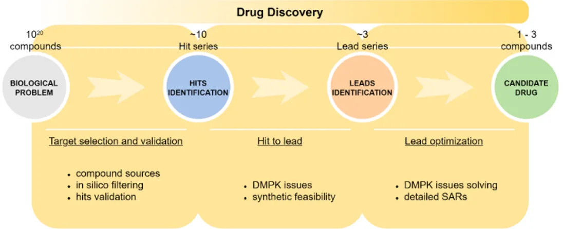
Figure 1 exemplifies the workflow that leads from the identification of a drug target to sorting out a valuable candidate drug.
Figure 1. The drug discovery process.
The very first step is, as already mentioned, the identification and validation of a molecular target, responsible for pathophysiological mechanisms (this topic will be broadly discussed in the next section); generally, in silico screening aids in filtering a huge number of compounds (from a theoretical number of 1020 up to >107 molecules, available from online data bank like ZINC, ChEMBL, PubChem databases), constituting a starting screening library, to a workable number of compounds (combinatorial libraries are constituted). Readily available “in-house” or “focused” libraries are also employed as a starting point.
HTS is then used to identify “hits”, namely compounds which show a promising activity in the chosen screen 2. Here resides one of the numerous bottlenecks of the entire process: the nature and reliability of the HTS assay and the size/quality of the library are crucial as the result is the rejection of up to hundreds or thousands of compounds.
- 4 -
Validation of hits is necessary to eliminate fictitious outcomes, and this may comprise repeating the screening of the hits by exploiting an alternative assay for proving, as well as resynthesis and retesting of the hit compounds 2. Simultaneously, structure-activity relationships (SAR) within the library are addressed in order to identify a “hit series” – a family of structurally related compounds – which represents a reasonable starting point for further chemistry 2.
In the lead identification stage, the validated hits are subjected to additional investigation, particularly in respect of their pharmacokinetic profile (DMPK issues) and toxicity as well as their feasibility for a synthetic chemistry programme. The “hit series” is consequently reduced to one or a few “lead series”.
Synthetic chemistry then begins; “lead optimization” usually involves parallel synthesis to generate derivatives of the lead series, which are consequently screened and profiled with respect to pharmacology, pharmacokinetics and toxicology, in order to obtain a small number of “drug candidates” (often a single compound), suitable for further development, at which point they are taken into preclinical development 2.
1.1.2. Drug target identification and validation
It is extensively proven that the feasibility and likelihood of success of a drug discovery project depends on the selection of an appropriate drug target; commonly drug candidates fail in development and clinical trials mainly because of low potency/specificity and/or unexpected toxicity 3,4.
The broad term “target” includes a range of biological entities such as proteins, genes and RNA, which, to be efficacious, require specific properties, i.e.
- 5 -
clinical relevance and “druggability”. A “druggable” target is accessible to the putative drug molecule, be that a small molecule or larger biologicals, and upon binding, elicit a biological response which may be measured both in vitro and in vivo 5.
Strictly dependent is target’s “assayability”, a fundamental characteristic in target-based drug discovery; the likelihood of discovering a novel modulating molecule depends on the reliability of the screening assay used for investigation. Target characteristics are summarized in Table 1.
WHAT MAKES A GOOD DRUG TARGET?
Clinical relevance Disease-modifying and/or proven function
in the pathophysiology of a disease
Druggability Susceptible to binding and modulation in
terms of activity
Assayability Favorable HTS configuration
3D structure Structure based druggability assessment
Phenotype data Prediction of potential side effects
Specific expression Target is unequally expressed throughout
districts and/or organisms
Biomarker Target/disease-specific biomarkers exist to
monitor therapeutic efficacy
- 6 -
The idea for a target can come from a variety of sources including academic, clinical research and the commercial sector. Overall, identifying novel drug targets, mainly proteins that are critically involved in the development and/or progression of a disease, is a multistep endeavor involving various disciplines, including large-scale expression profiling and bioinformatics, structural biology, traditional cell biology, and ultimately functional in vivo studies. It may take many years to build up a body of supporting evidence before selecting a target for a costly drug discovery programme 5,6.
Good target identification and validation enables increased confidence in the relationship between target and disease and allows to explore whether target modulation will lead to mechanism-based side effects 5.
1.1.3. Approaches in drug discovery: from hit identification to lead optimization
Over the last 25 years remarkable technological progress has been achieved in bioinformatics, high-throughput medicinal chemistry and screening techniques, leading to new approaches in hit identification. Hit identification stage aims at discover molecules able to interact with the validated molecular target in an initial screen, generally performed at a high concentration of compounds, to produce a quantifiable response in a robust, reproducible assay 7. Counterscreens have also been developed to eliminate false positives caused by assay interference or aggregation: compounds active in the primary identification assay are retested against another member of the target family under identical assay conditions. In case of observed activity, the hit is likely to be either promiscuous or false positive 8.
- 7 -
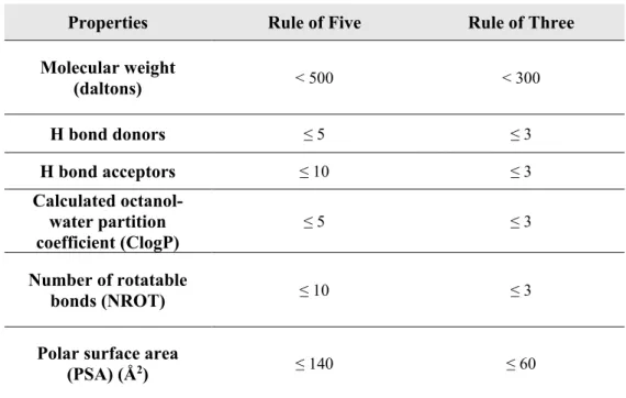
Several strategies have been adopted so far, in order to identify early hits, including existing drugs, natural ligands and products chemical evaluation, rational structure-based design, fragment-based screening, virtual screening and high-throughput screening (their application will be further discussed). These techniques can be subdivided considering the nature of the compounds used to interrogate the target: drug-like compounds in compliance with Lipinski’s Rule of Five 9 (RO5), or fragment-like molecules for which the Rule of Three10 (RO3) has been put in forth 8. ‘Drug-likeness’ is intended as the overall profile of biophysiochemical properties, determining molecules effective interaction with the target, in a biologically relevant and safe concentration for sufficient duration to produce a therapeutic effect 7. In the hit compounds chemical space, the drug-likeness concept may be reduced, according to RO3, to a “lead-likeness” definition, being hits the basis for medicinal chemistry and required to display improvable features with the aim to sprout in lead series. Thus, one could infer that an apparent disappointing success rate of compounds screening can be related to the lack of ‘drug-likeness’ in the collections; in order to avoid this condition, virtual or knowledge-based filtering of screening collections can exclude compounds which do not provide the desired drug-like physicochemical attributes (Ghose filter 11). Overall, these drug-/lead-like properties, together with hit clustering over singletons (hit series construction), play a pivotal role in the “hit to lead” (H2L) stage, as prioritization of the confirmed compounds is carried out accounting for selectivity, chemical tractability, binding mechanism, pharmacokinetic properties and patentability rather than potency per se 8.
- 8 -
Properties Rule of Five Rule of Three
Molecular weight (daltons) < 500 < 300 H bond donors ≤ 5 ≤ 3 H bond acceptors ≤ 10 ≤ 3 Calculated octanol-water partition coefficient (ClogP) ≤ 5 ≤ 3 Number of rotatable bonds (NROT) ≤ 10 ≤ 3
Polar surface area
(PSA) (Å2) ≤ 140 ≤ 60
Table 2. RO5 and RO3; Lipinski’s RO5 were implemented with NROT and PSA parameters by Veber
12.
The H2L process might involve techniques such as hit evolution, (bio)isosteric replacement and hit fragmentation, or any combination of these 8. Hit evolution provides analogues of the original hits, synthesized with different substitution patterns. Initial SAR data drive exploratory medicinal chemistry efforts, in order to achieve compounds with an improved lead-like profile. (Bio)isosteric replacements, if applicable, are useful for improving the hit profile while maintaining potency; hit fragmentation, relevant when the scaffold hits are large molecules, basically consists of structural decomposition producing promising fragments (which can be used in turn for a fragment-based screening) or minimalistic pharmacophores. Additionally, minimal core fragments represent a new starting point for fragment expansion; fragmentation can be followed by the combination of moieties coming from different hits (fragment linking or merging).
- 9 -
Figure 2. Hit to lead process: major techniques employed for hit optimization.
1.1.4. High-throughput screening
HTS was firstly introduced in the early 1990s and is now routinely employed in the hit identification step; disease-relevant validated targets are incorporated in massive parallel biochemical or cell-based assays and exposed to large numbers of compounds 7.
- 10 -
Typically, in HTS, a “primary” screen is performed resulting in the identification of numerous bioactive compounds (“primary hits” or “positives”, usually multiple members of a similar chemical core or chemical series). These undergo through successive rounds of screening (“secondary” screens) to confirm their activity, potency and, if possible, gain an early measure of specificity. Consequently, these hits enter the H2L process, during which specific hit-related compounds are synthesized for testing, in order to develop a rational understanding of SAR of the underlying chemical series 7. Major concepts regarding HTS setup deal with quality criteria of the library to be tested (drug-/lead-likeness, chemical clustering), assay robustness and reproducibility together with parallelization, miniaturization and automation. The principal goal in HTS assay development is ensuring a fast and reliable identification of positives, thus implying the choice of measurable output (e.g. inhibition, stimulation or binding), handling and cost-effectiveness.
In order to identify hits with confidence, only small variations in signal measurements can be tolerated; statistical parameters generally adopted to determine the applicability of an assay for HTS are the calculation of standard deviations (SD), the coefficient of variation (CV), signal-to-noise (S/N) ratio or signal-to-background (S/B) ratio. Inner drawbacks of S/N and S/B criteria is that neither consider the dynamic range of the output signal (expressed as difference between the background, or low control, and the maximum signal, high control) nor the unevenness of the sample and reference control measurements 7. Assay quality is generally rated with the Z’-factor equation, instead 7,13:
- 11 -
𝑍𝑍′ = 1 −�3(𝑆𝑆𝑆𝑆 𝑜𝑜𝑜𝑜 𝐻𝐻𝐻𝐻𝐻𝐻ℎ 𝐶𝐶𝑜𝑜𝐶𝐶𝐶𝐶𝐶𝐶𝑜𝑜𝐶𝐶) + 3(𝑆𝑆𝑆𝑆 𝑜𝑜𝑜𝑜 𝐿𝐿𝑜𝑜𝐿𝐿 𝐶𝐶𝑜𝑜𝐶𝐶𝐶𝐶𝐶𝐶𝑜𝑜𝐶𝐶)�
|𝑀𝑀𝑀𝑀𝑀𝑀𝐶𝐶 𝑜𝑜𝑜𝑜 𝐻𝐻𝐻𝐻𝐻𝐻ℎ 𝐶𝐶𝑜𝑜𝐶𝐶𝐶𝐶𝐶𝐶𝑜𝑜𝐶𝐶 − 𝑀𝑀𝑀𝑀𝑀𝑀𝐶𝐶 𝑜𝑜𝑜𝑜 𝐿𝐿𝑜𝑜𝐿𝐿 𝐶𝐶𝑜𝑜𝐶𝐶𝐶𝐶𝐶𝐶𝑜𝑜𝐶𝐶|
Equation 1. Z'-factor equation.
SD are the standard deviations, while low and high controls represent the minimum and the maximum signal to be measured in the assay, respectively. Alternatively to other statistical parameters, the Z’-factor includes, in assay quality assessment, the variability of high and low controls and the separation band between them, which represent the signal window in which the assay must be reliable, sensible and specific to produce an accurate outcome (Figure 3).
Figure 3. Data variability of high and low controls are taken into accout in the Z’-factor, differently from other statistical parameters; separation between them must be high in order to obtain the largest signal window possible and minimize false positive/negative.
Z’-factor fluctuates between 0 and 1, according to the sharpness of controls peaks; a value greater than 0.5 is generally accepted for HTS biochemical
- 12 -
assays, while is decreased to 0.4 in cell-based screenings. From the Z’-factor, a Z-value can be derived, whereby average signal and SD of test set are compared to reference controls 7:
𝑍𝑍 = 1 −�3(𝑆𝑆𝑆𝑆 𝑜𝑜𝑜𝑜 𝑇𝑇𝑀𝑀𝑇𝑇𝐶𝐶 𝑆𝑆𝑀𝑀𝐶𝐶) + 3(𝑆𝑆𝑆𝑆 𝑜𝑜𝑜𝑜 𝑅𝑅𝑀𝑀𝑜𝑜𝑀𝑀𝐶𝐶𝑀𝑀𝐶𝐶𝑅𝑅𝑀𝑀 𝐶𝐶𝑜𝑜𝐶𝐶𝐶𝐶𝐶𝐶𝑜𝑜𝐶𝐶)�|𝑀𝑀𝑀𝑀𝑀𝑀𝐶𝐶 𝑜𝑜𝑜𝑜 𝑇𝑇𝑀𝑀𝑇𝑇𝐶𝐶 𝑆𝑆𝑀𝑀𝐶𝐶 − 𝑀𝑀𝑀𝑀𝑀𝑀𝐶𝐶 𝑜𝑜𝑜𝑜 𝑅𝑅𝑀𝑀𝑜𝑜𝑀𝑀𝐶𝐶𝑀𝑀𝐶𝐶𝑅𝑅𝑀𝑀 𝐶𝐶𝑜𝑜𝐶𝐶𝐶𝐶𝐶𝐶𝑜𝑜𝐶𝐶|
Equation 2. Z-value equation.
Due to the intrinsic variability of test set compounds, the Z-value is generally lower than Z’ score; overall, the simplicity of the equations describing these parameters make them the primary assessment for HTS.
A broad assortment of assays configurations can be outlined in the HTS scenario, even though all fall in two main categories namely biochemical and cell-based assays. Biochemical assays are cell-free in vitro systems imitating the biochemistry of cellular processes. These systems provide information on the nature of the molecular interaction (e.g. kinetic data), ranging from simple enzyme/substrate reactions or protein-protein interactions, to more complex systems like in vitro transcription systems. Additionally, with respect to cell-based assays, are endowed with a higher solvent tolerance thereby allowing the use of higher compound screening concentration when required. The main limitation is innate in the assay itself, as lack of the cellular context leads to assay being insensitive towards properties like membrane permeability, which governs the effect of compounds on cells 7.
Cell-based assays represent a simplified in vivo system and are elective for unraveling effects towards those targets unsuitable for screening in simple
- 13 -
systems, such as those involving signal transduction pathways, membrane transport, cell division, cytotoxicity or antibacterial actions 7. Through cell-based assays multiple parameters can be inspected: growth, transcriptional activity, changes in cellular metabolism or intracellular levels of messengers like cAMP or calcium, variations in membrane potential. Major issues involving these assays are the complexity of HTS protocols set up (cell cultivation and dedicated automated systems to maintain physiological conditions during screening are required) and frequent off-target effects of test compounds affecting the readout 7. Major techniques employed for screening are reported in Figure 4.
- 14 - 1.1.5. Fragment-based screening
Fragment-based screening (FBS) has simultaneously grown with the other screening technologies, becoming a concrete alternative to more traditional hit identification methods. FBS approach relies on two central guidelines, which set it apart from HTS: (1) the concept that chemical space can be more efficiently probed by handling small fragments (molecules satisfying RO3) instead of large molecules (according to RO5) and (2) the principle that, being these fragments small in size (MW < 300 Da), they should bind with lower affinity to targets with respect to larger molecules but displaying a binding efficiency at least as high as for drug-like compounds 14.
In Fragment-based drug discover (FBDD), screening methods must be much more sensitive than HTS bioassays (biochemical, biophysical, cell-based assays). The elective strategies are structural biology techniques like NMR or X-ray crystallography, able to detect low affinity binding and to be informative on the fragment-protein interaction. Recent advances in cryo-electron microscopy made it a valuable alternative for FBDD 15; in any case X-ray crystallography still remains the primary method for FBDD, mainly thanks to dramatic improvements in automatization and therefore time-reduction in obtaining co-crystals structures 14. Also X-ray data analysis and model building have been improved, with the implementation of automated structure solving pipelines.
Efficient fragment screening requires the soaking of cocktails of fragments into preformed crystals of the target protein; fragment-hits are then subjected to intense SAR studies and undergo chemical optimization through the use of hit optimization techniques discussed in section 1.1.3.
- 15 -
A reliable concept widely used for comparing hits across different series and the effectiveness of compound optimization is “ligand efficiency” (LE). The term LE is defined as the free energy of binding (ΔG) of a ligand for a specific protein averaged for each “heavy atom” (or nonhydrogen atom). The number of heavy atoms is named “heavy atom count” (HAC),
𝐿𝐿𝐿𝐿 = −HAC ≈ −ΔG 𝑅𝑅𝑇𝑇ln(IC50)HAC
LE is expressed as (kcal/mol)·heavy atom-1 and has been extrapolated that a minimum LE of 0.3 is required in a hit or lead for it to be useful 14. A recent advance of LE is “group efficiency” (GE) which allows the estimation of an individual chemical group’s contribution toward the overall free energy of binding. This analysis requires the comparison of matched pairs of compounds with the result of an additional, fast and informative insight that can direct hit chemistry and SAR studies. ΔG values can be converted into GE similarly to LE:
𝐺𝐺𝐿𝐿 = − HAC ΔG
where HAC is the number of non-hydrogen atoms in a specific group. GE ≥ 0.3 suggest that the substituent produces an acceptable contribution to the compound’s potency overall, ensuring maintenance of good drug-like properties 14.
1.1.6. A rational approach: structure-based drug design
Structure-based drug design (SBDD) represents a comprehensive, multidisciplinary approach, aiming at overtaking the experimental and economic bottlenecks of other hit/lead identification processes. Actually, the
- 16 -
most recent advances in computer-aided structure predictions and interaction simulations are combined with experimental data, in a knowledge-based fashion, in order to increase the rate of success of focused compound series. In other words, availability of 3D-structures (obtained commonly by X-ray crystallography, NMR but also through homology modeling) helps in the identification of binding sites and interaction determinants which, in turn, guide the selection of possible active molecules and their efficacy improvement.
Once the target is structurally assessed, identification of active compounds can be carried out through three main virtual approaches: ligand-based drug design (LBDD), virtual screening and de novo generation 16. LBDD relies on known target’s interactors, like substrates or cofactors, which can be further modified in order to increase affinity; in virtual screening, instead, databases of available small molecules are interrogated and tested against the target while in de novo generation small fragments are positioned in the characterized binding site. These two last approaches are based on docking techniques, which asses a score and a ranking to each compound, predicting the binding affinity for the target. In LBDD, instead, computational tools like quantitative structure-activity relationship (QSAR) or 3D-QSAR are used to investigate the chemical features that determine binding and to be improved. Descriptors generally taken into account when characterizing active compounds are: binding scores (affinity/specificity), balance between hydrophilicity/lipophilicity, ADMET profile, biodegradation, metabolites. On the basis of these data, compounds improvement and evolution design is generally carried out in silico and designed derivatives are again computationally evaluated before being experimentally confirmed via biophysical assays or structural characterization; SBDD is, therefore, an iterative process, starting from the identification of
- 17 -
scaffolds and culminating in a rational design, synthesis and testing of derivatives.
1.2. The biological problem: neglected tropical diseases
Various criteria could be applicable when a new drug discovery project begins, regarding scientific and technical issues, strategic and operational concerns (summarized in Figure 5) 17.
Figure 5. Issues to be taken into account when planning a drug discovery project.
Balance between these features raises possibilities of successfully creating and managing a drug discovery project and must be considered in the initial project assessment. A perfect balance is obviously unlikely, and some issues can be more significant than other; in this perspective most projects are oriented toward well-known and high morbidity diseases, with a conceivable potential
- 18 -
impact on the market while other campaigns are focused on neglected diseases, where the medical need and the freedom to operate represent the driving force of the plan.
The latter is the case of tropical diseases (globally named as “neglected tropical diseases”, NTDs) representing a social plague in the poorest countries worldwide. Research interest is not sufficiently involved because of costs and unfavorable profits, as these diseases generally sweep along with poverty and critical socio-economic conditions. Particularly, parasitic diseases induced by Trypanosomatids represent a large portion of this group and will be discussed in the following sections.
1.2.1. Trypanosomatidal neglected diseases: an overview
Trypanosomatidae represent the broadest family of protozoa belonging to the class of Kinetoplastids 18; all members are exclusively parasitic, found primarily in insects while a few genera have life-cycles involving a secondary host, which may be a vertebrate, invertebrate or plant 18.
The three major human diseases induced by Trypanosomatids are caused by two genera of dixenous parasites, namely Leishmania and Trypanosoma.
Leishmania spp.
At least 20 species – of which the main are reported in Table 2 – of Leishmania (L.) are responsible for inducing vector-borne parasitic diseases in humans, transmitted between mammalian hosts by female sandflies 19 (parasite life cycle is outlined in Figure 6). Distinct species of Leishmania cause different clinical manifestations, ranging in severity from self-curing cutaneous lesions to life-threatening visceral disease. The outcome is determined by the interplay
- 19 -
of the following: parasite characteristics, vector biology, and host factors, with immune responses taking central stage among the host factors 19.
Figure 6. Leishmania spp. life cycle 19.
Visceral Leishmaniasis (VL) – caused by L. donovani in Asia and Africa and L. infantum in the Mediterranean Basin, the Middle East, central Asia, South America, and Central America – is the most severe, systemic form that is usually fatal unless treated. Post-kala-azar dermal leishmaniasis (PKDL) is a skin manifestation that occurs in otherwise healthy people after treatment of visceral leishmaniasis.
- 20 -
Species Clinical disease forms
Natural
progression reservoirs Main
High- burden countries or regions Estimated annual worldwide incidence L. donovani VL and PKDL VL is fatal within 2 years; PKDL lesions self-heal in up to 85% of cases in Africa Humans India, Bangladesh, Ethiopia, Sudan and South Sudan
50 000 – 90 000 VL cases
L. tropica CL, LR and rarely VL CL lesions often self-heal within 1 year Humans but zoonotic foci exist Eastern Mediterranean, Middle East, and northeastern and southern Africa 200 000 – 400 000 CL L. aethiopica DsCL, and CL, DCL, oronasal CL Self-healing, except for DCL, within 2–5 years
Hyraxes Ethiopia and Kenya 20 000 – 40 000 CL
L. major CL Self-healing in >50% of cases within 2–8 months; multiple lesions slow to heal, and severe
scarring Rodents Iran, Saudi Arabia, north Africa, Middle East, central Asia, and
west Africa 230 000 – 430 000 CL L. infantum VL and CL VL is fatal within 2 years; CL lesions self-heal within 1 year; confers individual immunity
Dogs, hares, and humans China, southern Europe, Brazil, and South America for VL and CL; Central America for CL 6200 – 12 000 cases of OW VL and 4500 – 6800 cases of NW VL
L. mexicana CL, DCL, DsCL healing within 3 Often self-– 4 months
Rodents and
marsupials South America
Cases included in 187 200 – 300 000 cases of
NW CL
L.
amazonensis CL, DCL, and DsCL described Not well Possums and rodents South America
Cases included in 187 200 – 300 000 cases of NW CL L. braziliensis CL, MCL, DCL, and LR Might self-heal within 6 months; 2 – 5% of cases progress to MCL Dogs, humans, rodents, and
horses South America
Majority of 187 200 – 300 000 cases of NW CL
L.
guyanensis CL, DsCL, and MCL within 6 months Might self-heal Possums, sloths, and anteaters South America
Cases included in 187 200 – 300 000 cases of
NW CL
Table 3. Clinical and epidemiological characteristics of the main Leishmania species 19. VL = visceral
leishmaniasis, PKDL = post-kala-azar dermal leishmaniasis, CL = cutaneous leishmaniasis, LR = leishmaniasis recidivans, DCL = diffuse cutaneous leishmaniasis, DsCL = disseminated cutaneous leishmaniasis, MCL = mucocutaneous leishmaniasis, OW = Old World leishmaniasis, NW = New World leishmaniasis.
- 21 -
Cutaneous leishmaniasis (CL) is usually limited to an ulcer that self-heals over 3-18 months, but can also lead to scarring, disfigurement, and stigmatization as disability outcomes. Depending on parasite species, up to 10% of cutaneous leishmaniasis cases progress to more severe manifestations. These are known as mucocutaneous leishmaniasis (MCL), diffuse cutaneous leishmaniasis (DCL), disseminated cutaneous leishmaniasis (DsCL), and leishmaniasis recidivans (LR) 19.
For decades, VL was treated by pentavalent antimonial monotherapy, available in two formulations: sodium stibogluconate and meglumine antimoniate. Increasing non-response in India led to increased dosage recommendations, currently up to 20 mg/kg per day either by intramuscular injection or intravenous infusion over the course of 28-30 days.
Figure 7. Chemical structure of pentavalent antimonials.
Aside from being painful if administered intramuscularly, antimonials have proven to be cardiotoxic and to induce arrhythmias 19,20, particularly evident in HIV-VL co-infection 19,21. In the Indian subcontinent, sodium stibogluconate
- 22 -
is no longer recommended because of drug resistance 19,22,23. Clinical research has then focused on shorter regimens and avoiding resistance 23,24. The first breakthrough was oral miltefosine (MF), registered in India in 2002, followed by the injectable paromomycin (PM) in 2006, administered in several short combination regimens, and single-dose liposomal amphotericin B (LAMB).
- 23 -
MF was adopted in 2005, as the first-line regimen by the Asian elimination initiative. However, after more than a decade of use there is evidence of reduced effectiveness in both VL and PKDL, while the existing linear dosing recommendations in children are likely to result in under-dosing and treatment failure 25. In east Africa, 20 mg/kg per day of sodium stibogluconate, in combination with intramuscular PM (15 mg/kg) over 17 days is the treatment of choice 23,26. In general, evidence in this region is limited to end-of-treatment outcomes (rather than the standard 6 months) because of high follow-up loss from migration. In east African patients with complicated VL, or those who are elderly or pregnant, treatment with LAMB is recommended because of its better safety profile 19. However, routine use of LAMB monotherapy in non-severe patients has not proven to be as effective as in Asia and is not recommended. For all VL caused by L. infantum and for VL caused by L. donovani in Asia, sodium stibogluconate has been superseded by LAMB as first-line treatment. To date, the only LAMB formulation approved by the US Food and Drug Administration (FDA) and WHO for the treatment of VL is AmBisome (Gilead Sciences, Dimas, CA, USA) 19,27. Since 2010, for VL caused by L. donovani in the Indian subcontinent during the attack phase of elimination, WHO recommends 10 mg/kg of LAMB as a single dose by infusion or 3-5 mg/kg per daily dose infusion over 3-5 days up to a total dose of 15 mg 19,28.
An alternative to the LAMB regimen is one of several combination regimens (LAMB + PM, LAMB + MF, LAMB + antimonials), whose use is planned to be adopted once elimination targets have been reached to mitigate the risk of resistance developing to LAMB 19.
- 24 -
Trypanosoma spp.
The genus Trypanosoma (T.) includes up to 30 relevant species of unicellular parasitic flagellate protozoa. Human-specific pathogens are: T. brucei, including T. brucei gambiense and T. brucei rhodesiense subspecies, and T. cruzi, causative agents of Human African trypanosomiasis (HAT, or sleeping sickness) and Chagas disease (or American trypanosomiasis), respectively.
Trypanosoma brucei – Human African trypanosomiasis
HAT occurs in sub-Saharan Africa, within the distributional limits of the vector, the tsetse fly (Diptera, genus Glossina). Two forms of the disease exist: the slow-progressing form, caused by T. brucei gambiense, which is endemic in western and central Africa; and, the fast-progressing form, caused by T. brucei rhodesiense, found in eastern and southern Africa 29,30.
Figure 9. T. brucei life cycle: A) Metacyclic trypanosomes are injected into the skin of a mammalian host, during a blood meal. B) In the mammalian host, trypanosomes transform into dividing long slender forms that, via lymph and blood, infiltrate tissues and organs. C) A tsetse fly is infected by taking blood from a human being or other mammal that contains stumpy trypanosomes. D) After about 2 weeks, trypanosomes might have colonized the salivary glands, and can then be transmitted to the next mammalian host 29.
- 25 -
Commonly, both forms of the disease are lethal if untreated; even if, for T. brucei gambiense disease, healthy carriers and self-cure have been reported in literature 29,31. The disease goes through two stages: a haemolymphatic stage followed by a meningoencephalitic stage in which trypanosomes cross the blood-brain barrier (BBB) and invade the central nervous system (CNS). T. brucei rhodesiense disease is generally acute, progressing to second stage within a few weeks, and death in less than 6 months 29,32,33. T. brucei gambiense disease follows a chronic progressive course, with a mean duration estimated at 3 years 29,33. Neurological disturbances, including sleep disorder, are typical of second-stage disease while the majority of signs and symptoms (fever, lymphadenopathy, hepatosplenomegaly, endocrine dysfunction) are common to both stages 29.
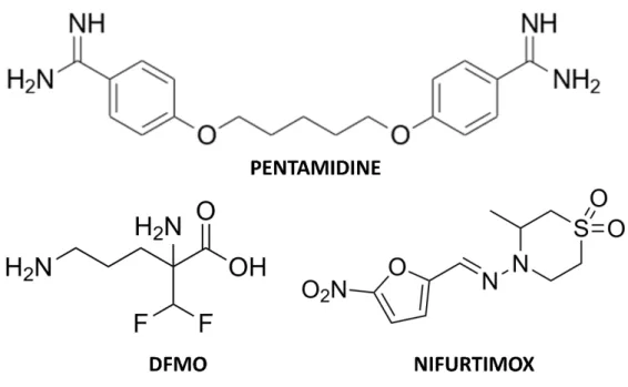
To date, five drugs are available (as monotherapies or in combination regimens) for the treatment of HAT: pentamidine and suramin for the first-stage disease treatment, and melarsoprol, eflornithine (or DFMO), and nifurtimox for second-stage disease 29. A successful recovery rate from HAT strictly depends on promptness of intervention (the earlier, the better in terms of tolerability and cure) while the choice of treatment relies on the causative agent and disease stage. As a general, and proper, rule, drugs for the treatment of first-stage disease are not recommended for the second-stage disease (and viceversa), as the latter compels drugs able to cross the BBB, which tend to be more toxic and complex to administer than first-stage drugs 29.
- 26 -
Figure 10. T. brucei gambiense treatment: chemical structures of pentamidine, eflornithine and nifurtimox.
Pentamidine’s efficacy against T. brucei gambiense disease (95-98%) has been stable for decades; in endemic areas, the most common route of administration is a daily intramuscular injection, for 7 days, but can also be delivered as an intravenous infusion in saline over 2 h. Pentamidine administration should follow the ingestion of sugar (10-20 g) to prevent hypoglycaemia, and has to be succeeded by rest in the supine position for 1-2 h to prevent hypotension. The intramuscular injection causes pain and transient swelling, while additional adverse effects are abdominal pain and gastrointestinal problems 29,34. Suramin is used only in the treatment of T. brucei rhodesiense disease, in complex treatment schedules lasting up to 1 month. A test dose is generally administered before treatment because of the risk of acute hypersensitivity reactions. Adverse effects are frequent, even if mostly mild and reversible, and
- 27 -
include pyrexia, nephrotoxicity, peripheral neuropathy, agranulocytosis and thrombocytopenia 29.
The first-line treatment for second-stage T. brucei gambiense disease is nifurtimox-eflornithine combination therapy (NECT). In 2009, NECT was included in the WHO Essential Medicines List; has higher cure rates (95-98%), lower fatality rates (<1%), less severe adverse events, simpler administration, and is believed to avoid causing drug resistance of the parasite, in comparison to melarsoprol and eflornithine monotherapy. As nifurtimox is not licensed for African trypanosomiasis (but only for Chagas disease) it can only be used to treat patients with African trypanosomiasis off label, subject to express authorization and acceptance of responsibility by national authorities. NECT consists of oral administration of nifurtimox together with intravenous eflornithine. With 14 infusions, instead of the 56 used in eflornithine monotherapy, NECT is easier to administer, demanding fewer hospital resources and reducing costs. Even though eflornithine requires at least four daily infusions for a constant trypanostatic effect, infusions every 12 hours result to be highly effective in combination with oral nifurtimox. The most common adverse events reported for NECT are abdominal pain, vomiting and headache 29,35–38. The toxicity profile replicates that of nifurtimox and eflornithine monotherapies, but with lower frequency and severity, mostly because of the shorter drug exposure. Eflornithine (α-difluoromethylornithine or DFMO) as a monotherapy is an option for T. brucei gambiense disease treatment only when nifurtimox is unavailable or contraindicated and is delivered as intravenous infusion for 14 days (generally 56 infusions in total) 29. Frequent adverse effects are fever, pruritus, hypertension, nausea, vomiting, diarrhoea, abdominal pain and myelosuppression but eflornithine is, with a
- 28 -
fatality rate below 2%, safer than melarsoprol, whose use is restricted to the treatment of second-stage T. brucei rhodesiense disease 29,39.
Figure 11. T. brucei rhodesiense treatment: chemical structures of suramin and melarsoprol.
Trypanosoma cruzi – Chagas disease
According to WHO, and in common with other neglected tropical diseases, “Chagas disease is a proxy for poverty and disadvantage: it affects populations with low visibility and little political voice, causes stigma and discrimination, is relatively neglected by researchers, and has a considerable impact on morbidity and mortality” 40,41; it is an anthropozoonosis caused by T. cruzi (whose life cycle is reported in Figure 12), which is mainly transmitted by various species of three genera of blood-sucking triatomine insects (Triatoma, Panstrongylus and Rhodnius, also known as “kissing bugs”) 40. Additional routes of transmission are described, which have a major role in non-endemic countries. Mother-to-child transmission rate is estimated about 4-7% while
- 29 -
transmission via blood products settles around 10-25% perinfected blood unit. Solid organ transplantation has a role in transmission, particular rates of infection are: 0-19% for kidney recipients, 0-29% for liver recipients and 75-100% for heart recipients 42–46.
- 30 -
Chagas disease is endemic in 21 continental Latin American countries, from southern USA to the north of Argentina and Chile, generally confined to poor, rural areas of Central and South America, where vectorial transmission is the main route of contagion 40.
The clinical course of American trypanosomiasis generally begins with an acute phase which can chronicize if untreated, in the second phase. Acute infection occurs at any age and is asymptomatic in most cases. When present, symptoms include fever, local inflammation at the inoculation site (inoculation sore), unilateral palpebral oedema (the so-called Romaña sign, in those cases when the conjunctiva is the inoculation site), lymphadenopathy, and hepatosplenomegaly 40. The acute phase generally lasts up to 8 weeks, with parasitaemia levels considerably decreasing from 90 days onwards. Severe acute disease occurs in less than 1-5% of patients and including manifestations as acute myocarditis, pericardial effusion and meningoencephalitis 40,47–49. The acute phase generally heals spontaneously but patients commonly evolve to a chronic infection, if untreated; the largest part of patient affected never develop symptoms or visceral involvement, undergoing to an indeterminate form of Chagas disease characterized by seropositivity for T. cruzi, absence of clinical signs and symptoms of cardiac and digestive involvement, and normal chest radiography and electrocardiography 40,50. It is estimated that around 30-40% of chronically infected patients can develop organ involvement, mostly cardiomyopathy and megaoesophagus and/or megacolon, in 10-30 years after the acute phase. Cardiac implication, primarily affecting myocardium and conduction system, is the most frequent and severe type of organ involvement, occurring in 14-45% of chronically infected patients. Recent studies have demonstrated that progression to cardiac involvement is around 1.4-5% per
- 31 -
year 40,50–57. Sudden death is the main cause of death in patients with Chagas heart disease, followed by refractory heart failure and thromboembolism 40,58. Only benznidazole and nifurtimox are licensed for the treatment of Chagas disease to date. Both have been the pillars of parasiticidal treatment for almost 50 years, even though their safety and efficacy profile are far from ideal, with their effectiveness seeming to decrease with time from the primary infection 40. In this perspective, early detection and intervention are essential requirements for a successful therapy.
Figure 13. American trypanosomiasis treatment: chemical structure of benznidazole.
Nifurtimox is administered orally, in three to four doses for 60-90 days, with recovery rates in the chronic indeterminate phase ranging from 86% in children younger than 14 years to 7-8% in adults 51,59–62. The frequency of adverse effects with nifurtimox is quite high (43-97.5%); the most common are anorexia, weight loss and neurological disorders as insomnia, disorientation, paresthesias, and peripheral neuropathy, nausea, vomiting, and, occasionally, fever and rash 40,61–63. Benznidazole shows, instead, a better tolerability profile and, possibly, efficacy. It is administered orally in two or three doses usually
- 32 -
for 60 days. Higher doses of up to 15 mg/kg are recommended in cases of meningoencephalitis 40,53. Additionally, 30 days of treatment seems to be useful for chronically infected adults 40,53. Luckily, benznidazole has a noticeable activity in the acute and early phases of T. cruzi infection: serological cure is achieved in up to 100% of patients with congenital disease treated during the first year of life and in 76% of patients with acute disease 40,64–66. In the chronic phase, conversely, success rates are much lower: 60-93% in children aged up to 13 years and 2-40% in adults with late chronic disease 40,53,66–70. The most common adverse effects involve hypersensitivity, mainly in the form of skin rash, digestive intolerance, and general symptoms such as anorexia, asthenia, headache and sleeping disorders.
The intent of this limited overview on trypanosomatid induced diseases is to shed light on the social and clinical relevance of these conditions, and how pharmacological therapy is still at its early days, features common to all NTDs. Epidemic loci are spread all around the world, and the burden of these plagues weighs enormously on some of the poorest societies worldwide, leading more than 100 000 people to death every year, comprehensively 71. It is clear how urgent is the need of new drugs to contrast these diseases; vaccines are unavailable while therapeutic agents used so far are old, endowed with a low tolerability profile and insufficient efficacy. As already discussed in the first part of the introduction, the modern approach in drug discovery is a target-based process, focused on the identification of compounds, active against a specific biomolecule, whose function and essentiality in the development of the disease is generally known; the uniqueness (in the sense of its natural absence in physiological conditions in the patience) of the target plays a critical
- 33 -
role in drug specificity and in avoiding toxic side effects. Trypanosomatidal biology includes feasible targets with these characteristics within a trypanosomatids-specific, unique cellular pathway, i.e. the trypanothione metabolism, whose function is the production and recycling of the spermidine-based dithiol trypanothione (T(SH)2), a glutathione analogue, necessary, among multiple functions, for redox equilibrium and host immune system resistance. Next sections will deepen the rationale behind choosing this pathway as elective mechanism for antitrypanosomatidal therapy and will focus on one of the enzymes involved in it, the trypanothione reductase (TR), whose ideality as a molecular target, critical activity and structural features will be discussed.
1.2.2. Redox homeostasis in Trypanosomatids: the trypanothione metabolism
Historically, the enigmatic thiol-dependent redox system of trypanosomatids has been puzzled out by the pioneering work of Fairlamb and Cerami 72, in 1985. Ten years after the discovery and characterization of an unusual thiol-polyamine conjugate, namely mono(glutathionyl)-spermidine (Gsp) in E. coli 73, they firstly identified and put at the crossroads of a novel, trypanosomatids-specific thiol-based redox system, the trypanothione molecule (T(SH)2); Figure 14), a bis(glutathionyl)-spermidine, almost exclusively found in the order of Kinetoplastida and there representing the principal low molecular mass thiol 74–76.
- 34 -
Figure 14. Chemical structures of trypanothione in both the reduced and the oxidized state.
T(SH)2 utilization required the evolutionary specialization of key redox enzymes, leading to a progressive suppression of NADPH-reductases glutathione reductase (GR) and thioredoxin reductase (TrxR) genes, whose products represent the backbone for redox systems among Eukaryota but are absent in all known trypanosomatidal genomes 77–79. The reconfiguration of a new redox system appears to have gradually occurred in the biological evolution of Trypanosomatids, as TrxR and GR genes are still present in the oldest kinetoplastid ancestor Euglena gracilis 77,80,81.
Overall, in most living organisms, cellular reducing power and oxidative stress control is mainly governed by GR and TrxR in the glutathione/glutaredoxin
- 35 -
(GSH/Grx) and the thioredoxin (Trx) pathways respectively. Both pathways include the activity of peroxiredoxins for oxidant species neutralization namely glutaredoxin peroxidase (GPx), in the GSH/Grx system, and peroxiredoxin (Prx), in the Trx pathway 74,82 (Figure 15).
Figure 15. The Trx and the GSH/Grx pathways represent the most common antioxidant systems.
Conversely, Trypanosomatids rely on a minimalistic system composed of trypanothione reductase (TR), T(SH)2 and the kinetoplastid-specific thioredoxin-like tryparedoxin (TXN), coupled to the tryparedoxin peroxidase (TXNPx) 77.
Being trypanothione a spermidine-based dithiol, its synthesis is closely bound to the polyamine (PAs) metabolism (Figure 16). In brief, L-Arginine (L-Arg), the precursor of PAs, is converted in ornithine by the arginase (ARG) enzyme within the very first reaction of the PAs biosynthetic pathway. Ornithine is then converted by the ornithine decarboxylase (ODC) in putrescine. Spermidine (Spd) is synthesized by the enzyme spermidine synthase (SpdS) by addition of
- 36 -
the aminopropyl group of the decarboxylated S-adenosylmethionine (dAdoMet) to putrescine 83.
Figure 16. The PAs metabolism represent the very first step of trypanothione synthesis; two molecules of glutathione (in ochre) are condensed to one molecule of spermidine (in blue), to synthesize trypanothione whose chemical-physical properties and functions are summarized in the upper-right table.
Trypanothione is synthesized in a double step reaction by the bifunctional ATP dependent-enzyme trypanothione synthetase-amidase (TryS). The X-ray
- 37 -
crystal structure of TryS from L. major was solved by Fyfe and collaborators in 2008 (PDB code: 2VOB, 2VPM, 2VPM) and set the basis for the understanding of the synthetase activity. TryS is a 75 KDa monomeric enzyme presenting a N-terminal amidase domain (residues 1-215, 634-652), by which it is believed to maintain balanced the level of intracellular T(SH)2, and a C-terminal synthetase domain (216-233), containing a roughly triangle shaped active site 84. Here, the three vertices represent three binding subpockets S1, S2, S3, able to bind ATP, GSH and Spd, respectively. The current accepted mechanism for the synthetase activity is the following: in the first step of the condensation reaction, the GSH carboxyl group is activated by ATP-dependent phosphorylation, generating an acylphosphate intermediate, stabilized by contributions from Arg328 and two Mg2+ ions. The Spd amine carries out nucleophilic attack on the anionic intermediate, which collapses to produce an amide linkage, resulting in the formation of Gsp, with release of ADP and phosphate. The Gsp remains located at S3, with the terminal amine directed toward the active site; S1 and S2 are occupied again by ATP and GSH. The second step takes place in the same manner as the first, with T(SH)2 released from S3 as final product 84,85.
In order to pursue its antioxidant role, T(SH)2 must be maintained in the reduced state. TR is the designate enzyme for this purpose; via NADPH acting as electron donor, a cycle of reduction and charge transfer between FAD, two catalytic cysteines and oxidized trypanothione (TS2) starts at the active site of the oxidoreductase, from where trypanothione is released in the reduced state 86. The molecular events that characterize TR’s mechanism will be further discussed in the following dedicated sections.
- 38 -
The enzymatic system linking T(SH)2 to the oxidant species reduction step consists of two protein partners, namely TXN, a thiol disulfide oxidoreductase, and TXNPx, a 2-cysteines (2-cys) peroxiredoxin, which are analogues of thioredoxin and thioredoxin peroxidase, respectively 85. In trypanosomatids two isoforms of TXN exist, namely TXN-1 and TXN-2, sharing the same genetic locus but endowed with different biochemical and biological properties, and cellular localization (TXN-1 is cytosolic while TXN-2 is mitochondrial) 85,87; tryparedoxins show a WCPPC motif at their catalytic center replacing the WCG/APG motifs found in thioredoxins 87 and their role is to provide reducing power to TXNPx, as a consequence of T(SH)2 reduction. TXNPx is a typical 2-cys peroxiredoxin and forms an obligate homodimer, whose active sites are constituted by the N-proximal peroxidatic cysteine (Cp) from one subunit and a C-proximal resolving cysteine from the other (Cr’) 87,88. TXN and TXNPx partake in two distinct reactions. The first takes place upon the formation of a disulfide bridge between the N-terminal Cys40 of TXN and Cr’ of TXNPx, following the resolution of the intersubunit disulfide bridge (Cp -Cr’) 87,89. This inter-protein disulfide bond subsequently undergoes nucleophilic attack by the second cysteine of TXN, in order to leave TXNPx Cr’ as a thiol or thiolate. TXN returns to the oxidized state to be recharged by T(SH)2. In the second reaction, the Cp thiolate is oxidized by a peroxide to sulfenic acid that can react with the Cr’, reforming the TXNPx intermolecular disulfide bridge; the cycle can then restart.
In addition to oxidative stress defense, fundamental for parasites survival, T(SH)2 exerts other cellular functions. T(SH)2 has a role in protein folding because of its reactivity towards disulfide bonds, with effects on protein signaling and transport/secretion; can also act as a scavenger for metals and drugs, tagging molecules (thiol-conjugates) to be exported or sequestrated, in
- 39 -
detoxification pathways related to multi-drug resistance (MDR) 90. The couple T(SH)2/TXN delivers reducing equivalents also for the parasite synthesis of deoxyribonucleotides; T. brucei possesses a typical eukaryotic class I ribonucleotide reductase whose Km for TXN is 3.7 μM 90,91. At high T(SH)2 levels, the parasite ribonucleotide reductase is directly reduced by the dithiol, with a Km of 2.1 mM. The TXN-mediated ribonucleotide reductase activity is lowered by more than 60% when 0.1 mM TS2 is added to an assay containing 1 mM T(SH)2 90,91. The regulation of TXN by the thiol/disulfide ratio of trypanothione represents a control mechanism that links DNA synthesis with the redox state of the cell. The effect is comparable with that observed for the GSH/glutaredoxin system in E. coli 90. Moreover, T(SH)2 is able to reduce dehydroascorbate ensuring an additional mechanism of defense against hydrogen peroxide 90. T(SH)2 ability of undergoing spontaneous reactions (as in the case of ribonucleotide reductase and dehydroascorbate reductions), is notably higher than GSH but does not reside in the difference in redox potentials (E0 T(SH)2 = -242 mV; E0 GSH = -230/250 mV), which are rather similar instead. Being T(SH)2 a dithiol, it is kinetically favoured as disulfide reductant 92; moreover, its pK value coincides with the intracellular pH of the parasites (pK T(SH)2 = 7.4), as a consequence of the positively charged nitrogen atom in the Spd moiety, contrarily to GSH pK, ranging from 8.7 to 9.2, with an impact on its reactivity.
In conclusion, it appears clear how the trypanothione metabolism represents a crucial survival pathway in Trypanosomatids. Several cellular events are governed by T(SH)2; among them, oxidative stress resistance is a first-line defense for parasites against host immune system, which intensely relies on oxidative-species-induced immune protection. Being essential and unique, trypanothione metabolism represents, simultaneously, also a weakness for
- 40 -
Trypanosomatids, as all related enzymes are considered valid candidates for drug development 86. Trypanothione reductase (TR), the enzyme directly responsible for keeping trypanothione in the reduced state, has been extensively studied since it fulfills most of the requirements for a good drug target. TR has been validated as a target in both Leishmania and Trypanosoma species as it is not possible to obtain TR-knockout mutants while expression downregulation causes strong impairment in infectivity 86,93,94. It has also been demonstrated that antimonials interfere with the trypanothione metabolism by inhibiting TR 86,95,96, supporting the idea that targeting this protein is a concrete option for the treatment of trypanosomatidal diseases. Moreover, the high sequence homology of TRs from different species (80-100%) makes it a valuable target for developing a single, broad spectrum drug active against all Trypanosomatids 71,86. The main limitation of TR as a drug target lies in its high efficiency/turnover rate: it is demonstrated that, in order to reach a significant effect over parasite redox state and viability, TR activity must be reduced by at least 90%; consequently only potent inhibitors, with submicromolar affinity, can be considered promising compounds 86,93,94. Many efforts have been made so far to discover new effective hits through in vitro and in silico screening, in addition to the development of known scaffolds via SAR or structure-based design approaches 86, so that several classes of active compounds have been proposed to date. Next sections will discuss TR relevance in the trypanothione metabolism, focusing on its structural insights and the inhibitors so far characterized, in order to offer a comprehensive understanding of its role.
- 41 -
1.3. The drug target: trypanothione reductase
1.3.1. Trypanothione reductase, from structure to function
TR is a NADPH-dependent homodimeric flavo-oxidoreductase (EC 1.8.1.12), enclosing a 2cys-based active site: a “catalytic” cysteine, responsible for the nucleophilic attack on the oxidized substrate, and a “resolving” cysteine, able to attack mixed disulfide intermediates, formed during the oxidoreduction reaction. TR’s structure is thoroughly characterized since the X-ray crystal structure has been solved for several trypanosomatidal species (Crithidia (C.) fasciculata, L. infantum, T. brucei and T. cruzi) also in complex with natural substrates and inhibitors 86. Being TR a homodimer, each of the two subunits, related by a two-fold symmetry, comprises a FAD-binding domain (residues 1-160 and 289-360, T. brucei numbering), a NADPH-binding domain (residues 161-288), and an interface domain (residues 361-488; Figure 17A/B). The structure is almost identical for all the characterized species, in accordance with the high degree of sequence similarity (Figure 18). Indeed, TRs from all Trypanosomatidae share at least 67% of primary sequence, with >82% identity among Leishmania spp. and >80% among Trypanosoma spp. Similarity reaches 100% for residues shaping both substrates’ binding sites, proving that the binding mode for ligands is the same in all TRs characterized to date 86. NADPH and TS2 bind different cavities facing opposite sides of the isoallosazine ring of FAD; the TS2 site, located at the interface between the two subunits, is shaped by residues belonging to both subunits (differentiated with ’ in the enumeration).
- 42 -
Figure 17. Trypanothione reductase: A) Overall structure; B) 90° projection, substrates are represented as sticks; C) Blow-up of the active site: the electron transfer is directed from NADPH (in green) to the oxidized trypanothione (in yellow), via FAD (in orange) and the couple Cys52/Cys57 (gray).
Figure 18. TR from T. brucei is colored according to the aminoacids similarity percentage with TRs from other sources: C. fasciculata, T. cruzi, T. congolense, T. brucei, L. braziliensis, congolense, T. brucei, L.
- 43 -
The cyclic mechanism relies on the initial transfer of two electrons from NADPH to the FAD cofactor, which reduces the disulfide bridge between the active site cysteines, Cys52 and Cys57. The oxidized TS2 binds to the protein, and the catalytic Cys52, deprotonated by the couple His461’-Glu466’, attacks the disulfide bridge of the substrate, resulting in the formation of a mixed disulfide. Finally, Cys57 resolves the Cys52-TS2 bond and reduced T(SH)2 is released from the active site, while the cysteines come back to the oxidized state, ready for a new cycle. During catalysis, no major structural changes occur, apart from those strictly necessary side chains displacements in the involved residues 86.
T(SH)2 assumes variable conformations in the cavity, as an effect of the “dynamics” of its binding. Accordingly, T(SH)2 enters as a disulfide but, upon reduction, it is released in an extended conformation. Despite this variability, some interactions emerge to be particularly relevant and specific for binding: Glu18, together with other acidic residues, accounts for the positive charge of the substrate, while the almost hydrophobic patch including Trp21, Tyr110, and Met113 mediates the interaction with the polyamine moiety contained in trypanothione 86.
Selectivity over off-targets is a fundamental issue to be taken into account, in order to increase specificity and avoid side effects. GR is the closest human homolog of TR, sharing the same overall fold, with 38% sequence identity, and catalyzing the same reaction on very similar substrates. Both GR and TR reduce a disulfide bridge that is intermolecular for GR (GSSG → 2GSH) and intramolecular for TR (TS2 → T(SH)2). The most significant differences between the two enzymes reflect the differences between their cognate substrates: TS2 is bulkier than GSSG, positively charged due to the spermidine
- 44 -
moiety, which confers at the same time a distinctive hydrophobic character, while GSH bears an exact negative charge, at physiological pH. Therefore, the TS2 binding site in TR is wider and negatively charged with respect to the GSSG binding site in GR. Precisely, selective interactions take place between the spermidine moiety and residues Glu18, Trp21, Ser109, Tyr110 and Met114 which are not conserved in GR and/or are partially replaced by arginine residues (Arg37, Arg38, and Arg347).
Figure 19. Steric and electrostatic differences between T(SH)2 binding site and the GSSG binding site.
These steric and electrostatic features (Figure 19) account for the selectivity for substrates and emphasize the potential to generate parasite-specific compounds.
1.3.2. Trypanothione reductase inhibitors
Over the past two decades, structural studies on TR deeply improved the understanding in the molecular recognition of ligands, allowing to identify hot spots for interaction with substrates and inhibitors. This knowledge has been exploited through structure-based design approaches which, in some cases,
- 45 -
have led to a significant improvement in the performances of lead molecules 86,97–99. To date, the crystallographic structure of TR in complex with 20 different inhibitors has been solved, revealing 3 major inhibition strategies:
(i) competition with trypanothione, due to displacement of the substrate at from its binding site, comprising most of the characterized inhibitors;
(ii) competition with NADPH, similarly to the previous case;
(iii) redox cysteines inactivation, exerted by molecules able to establish a metal bond with Cys52 and Cys57 in the catalytic site 86.
A fourth has been recently proposed 100, based on the disassembly of the TR dimer induced by small molecules specifically designed to interfere with protein–protein interaction; however, poor structural information are available so far.
Inhibitors targeting the TS2 binding cavity
As previously highlighted, TR presents a broad active site, suited to accommodate the voluminous TS2 substrate. The largest part of the characterized inhibitors bind there, mainly in the so-called “mepacrine binding site” (MBS), a hydrophobic patch located at the entrance of the cavity. Fewer penetrate deeper, closer to the real catalytic site, where the redox cysteines reside and TS2 reduction takes place.
Mepacrine binding site (MBS)
Mepacrine (or quinacrine) is a notorious antiprotozoal compound, extensively used as antimalarial agent during World War II 101. In 1996, Jacoby and coworkers described the crystal structure of T. cruzi TR (TcTR) in complex with mepacrine (coordinates not available in the PDB) showing the ligand,