U
NIVERSITY OFP
ISAP
HD
C
OURSE INV
ETERINARYM
EDICINEIRON
SERUM
STATUS,
SERUM
VEGF
AND
OXIDATIVE
STRESS
PATTERN
EVALUATION IN CANINE CANCER PATIENTS
,
FOCUSING ON
MAST CELL TUMOURS
Candidate Tutor
Mr Riccardo Finotello Dr Veronica Marchetti
INTRODUCTION...PAG3 CHAPTER ONE
VASCULAR ENDOTHELIAL GROWTH FACTOR (VEGF)
1.1 VASCULOGENESIS AND ANGIOGENESIS ...……….PAG 5 1.2 VASCULAR ENDOTHELIAL GROWTH FACTOR (VEGF) ………...………..PAG 9 1.3 VEGF RECEPTORS …...……….PAG 10 1.4 REGULATION OF GENE EXPRESSION OF VEGF...PAG 12 1.5 VEGF,OXYGEN REACTIVE SPECIES (ROS) AND MICROVASCULAR PERMEABILITY... PAG 13 1.6 VEGF, IRON DEFICIENCY AND ANGIOGENESIS...PAG 15 1.7 FUNCTION OF VEGF IN TUMORS...PAG 16 1.8 VEGF IN CANINE PATIENTS …………...………PAG 19 1.8.1 VEGF IN MAST CELL TUMOURS...PAG 20
CHAPTER TWO
OXIDATIVE STRESS AND ANTIOXIDANTS
2.1 Oxidative stress and antioxidants...……….……….…PAG 23 2.2 OXIDATIVE DAMAGE TO BIOMOLECULES...……….……….…PAG 27 2.3 OXIDATIVE STRESS, CELL SIGNALLING AND CANCER.……….………PAG 31 2.3.1CYTOKINES AND GROWTH FACTORS SIGNALLING………….……….PAG 34 2.4 ANTIOXIDANT DEFENCE MECHANISMS IN CARCINOGENESIS…...…………...PAG 36 2.4.1 VITAMIN E...PAG 36 2.4.1.1 VITAMIN E IN ANIMAL MODELS...PAG 38 2.4.1.2 VITAMIN E& MAST CELLS...PAG 41
3.1 IRON ………..…...……….PAG44 3.2 IRON HOMEOSTASIS.………..…..………..…..PAG 46 3.3 IRON METABOLISM……...………..PAG 47 3.4 IRON AND CANCER...PAG 50 3.5 CAUSES OF IRON DEFICIENCY IN CANCER PATIENTS...PAG 53
CHAPTER FOUR
AIM OF THE STUDY...PAG 57
CHAPTER FIVE
MATHERIAL AND METHODS
5.1 PATIENTS ELIGIBILITY…...PAG 62
5.2 GROUP DESIGN……….……….…………...…….PAG 63
5.3 SAMPLES COLLECTION………...……….PAG 64 5.4 LABORATORY ANALYSES………...…….PAG 64 5.4.1 IRON PROFILE………...………PAG 64 5.4.2 VEGF...………...………PAG 65 5.4.3 ROMS &BAP TEST.………...………PAG 66 5.4.4 VITAMIN E(-TOCHOPHEROL)…………...……….………PAG 68 5.5 STATISTICAL ANALYSIS...PAG 70
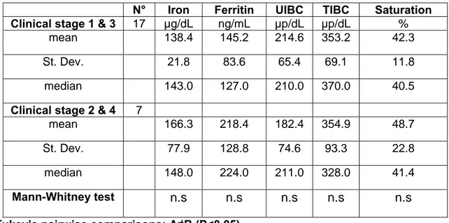
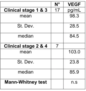
6.1 GROUPS DESCRIPTION...PAG 71 6.2 IRON SERUM STATUS, SERUM VEGF AND OXIDATIVE STRESS PATTERN BETWEEN GROUPS……….PAG 75
6.3 DIFFERENCES IN IRON SERUM STATUS, SERUM VEGF AND OXIDATIVE STRESS PATTERN WITHIN MCT PATIENT GROUPS..PAG 79
6.4 DIFFERENCES IN HAEMATOLOGICAL PROFILE BETWEEN GROUPS AND WITHIN MCT SUB GROUPS ...PAG 83 6.5 DIFFERENCES IN BIOCHEMICAL PROFILE BETWEEN GROUPS AND WITHIN MCT SUB GROUPS ...PAG 87 6.6 PROPORTION OF CANCER PATIENTS OUTSIDE THE REFERENCE RANGE (IRON PROFILE,VEGF,OXIDATIVE STRESS PATTERN)..PAG 94 6.7 RELATIONS BETWEEN VARIABLES...PAG 97
CHAPTER SEVEN
DISCUSSION...PAG 100 CHAPTER EIGHT
RESEARCH ACTIVITIES...PAG 104 REFERENCE...PAG 108
endothelial growth factor and oxidative stress pattern in canine cancer patients with particular attention on cutaneous mast cell tumour. Mast cell tumors (MCTs) are the most common cutaneous tumor in the dog, accounting for 16% to 21% of cutaneous tumors
(Bostock,1986; Finnie,1979; Rothwell, 1987; Brodey,1970). Wide variation is seen in the histologic pattern of canine MCT, and the histologic grade has been clearly established as a strong prognostic factor that is highly predictive of biologic behavior and clinical outcome. It has been demonstrated that mast cells and mast cells tumour can express, produce and release the Vascular endothelial growth factor (VEGF) has been implicated to contribute tissue edema through its effect on vascular permeability (Paturno, 2009; Mederle, 2010). Studies performed in vitro and in vivo (rats) on Mast cells, showed also how VEGF expression was regulated by hypoxiainducible factor-1a (HIF-1a) activation through the phosphatidylinositol 3-kinase (PI3K)–HIF-1a pathway (Lee, 2008). Systemic hypoxia produces an inflammatory response characterized by increases in reactive O2 species (ROS), venular leukocyte-endothelial adherence and emigration, and vascular permeability. The results showed that mast cells could play a key role in hypoxia-induced inflammation and suggested that alterations in the ROS-nitric oxide balance may be involved in mast cell activation during hypoxia (Steiner, 2003). Studies performed in vitro on mast cells have also demonstrated the role of the antioxidant barrier, especially of the vitamin E, in prevent degranulation of mast cells and proliferation of tumour cell lines (Gueck, 2002; Reiter, 2003). Most of these study have been performed in vitro and, except for the VEGF (which has been often investigated in the last years for his role in the anticancer target therapy in veterinary medicine), there are just few data on the role of oxidative stress pattern and iron profile in canine patients affected by mast cell tumour, as
neoplastic diseases as well as in the body’s response to the tumour. So the aim of this study was to investigate the role of this network in canine oncology patients through different tumour time and clinical substage focusing on mast cell tumour.
growths of metastatic disease. Although several different molecules and pathways are responsible of this complex event, the Vascular Endothelial Growth Factor (VEGF) is recognized as one of the main pro-angiogenetic factors (Folkman, 1992). In the early stage of tumour development, we observe a first a-vascular stage in which the tumour obtains oxygen and nutrients through the process of diffusion and simple diffusion (Grupta, 2003), and this stage the tumour is quiescent (Dvorak, 1986). A tumour is composed by a heterogenic population of cells and some of the them, due to molecular changes, increased characteristic of malignancy, inhibition of onco-suppressor genes (such as RAS, Raf and p53)(Chiarugi V., 1998), or due to any inflammatory or traumatic event acquiring an angiogenetic phenotype. The acquisition and expression of this phenotype allows the formation of an adequate vascular bed and the consequent exponential growth of the tumour. The angiogenesis is supported and promoted by the VEGF secreted by the tumor cells and also by stromal cells, platelets, the immune system (mast cells, macrophages, T lymphocytes) and vascular cells, particularly in hypoxic areas. Hypoxia leads to the production of HIF, hypoxia-inducible factor, a transcription factor that stimulates the transcription of the VEGF encoding gene. Angiogenesis, as well as be an essential process for the growth of primary tumor, has a special importance in enabling metastatic spread. In fact the new vessels, because of their thin wall with poor development of the basement membrane, are easily traversed by the neoplastic cells, representing a convenient access route to the bloodstream
resulting by physiological cells’ death is collected by macrophages, re-entering the life cycle. Body iron levels are then set only during absorption through the small intestine and the small amount of iron that is lost must be replenished daily to maintain balance (Finch, 1982). If not, either because of an excessive loss or
because the amount absorbed is not enough, we observe an iron deficiency that, over the time, will lead to the development of anemia. On the other hand, if the amount of iron entering the body exceeded the its requirements, this will be slowly accumulated in the body and particularly in the liver, leading to the development of iron overload. Due to the oxide-reducing capacity of the iron, this event will may lead to the formation of oxygen reactive species (ROS) that can damage cellular
(Gomme, 2005).
Vitamin E is a powerful antioxidant capable of blocking the lipid peroxidation of cell membranes, breaking the covalent bonds formed between ROS and fatty acids of the cell membrane. Iron deficiency increase HIF-1a and VEGF serum concentration and it is a promoter of angiogenesis, suggesting that systemic iron deficiency could play an important role in tumor progression (Eckard, 2010).
1.
Vascular Endothelial Growth Factor (VEGF)
1.1.
V
ASCULOGENESIS AND ANGIOGENESISThe formation of new blood vessels can develop from two different process, vasculogenesis and angiogenesis. Although the vasculogenesis is the process of de-novo formation of vessels from haemangioblast stem cells which occurs in the early stages of embryogenesis (Risau, 1995; Patan, 2004), endothelial cell precursors are also
found within the bone marrow of adults and are recruited due to trauma or ischemia
(Zammaretti, 2005). During angiogenesis, the new vessels are sprouting from endothelial
cells of preexisting vessels and despite angiogenesis is one of the main cause of solid tumor growth and progression it is also involved in inflammatory process (Asahara, 1997;
Djonov, 2000; Peichev, 2000; Djonov, 2003; Patan, 2004) showing also important physiological
function, such as wound healing (Ruiter,.1993; Paavonen, 2000).
In adults the vascular system is almost quiescent, except during physiological process like wound healing, hair growth and the menstruation (Salven, 2001, Ferrara, 2001). In
these circumstances the angiogenic mechanism is closely controlled and remains active for relatively short periods;
The imbalance in demand and supply of oxygen and nutrients, as for example chronic inflammatory conditions (rheumatoid arthritis, inflammatory bowel disease),
diabetes (diabetic retinopathy),
hyperproliferative skin diseases such as psoriasis, endometriosis and tumours (Salven, 2001) results in sprouting of new capillaries from pre-existing vessels through angiogenesis.
The study of the ability of a neoplastic tissue to induce formation of a new vascular network was one of the main objectives scientific researches over the past decade. Understanding this mechanism has allowed in human medicine to support the chemotherapy "standard" therapies, which aim to inhibition of tumor growth and treatment of both the primary tumor, both of possible metastatic spread. The tumor angiogenesis is a process formation of a vascular network within a tissue neoplastic and departing from it. Solid tumors are strictly dependent on the formation of new vessels, "a fabric cancer without a vascular bed can not grow larger than the diameter of 1-2 mm (Folkman, 1992). In pre-neoplastic vascular tissue seldom exceeds 2 mm in
size (eg, carcinoma in situ) and, at this stage the cells can get oxygen and nutrients needed for survival and growth, and eliminate catabolites produced through a process of simple passive diffusion (Gupta, 2003). Besides this extension, this mechanism is not
sufficient to trophic ensure the growth of tumors, also, the state of hypoxia that is established, leads via activation of p53, apoptosis (Cotran, 1999). At this stage, a tumour may remain quiescent for a long time, reaching a balance between the number replicating cells and those who are dying, until the time in which some of them switch to an angiogenic phenotype (List, 2001).
BioJobBLOGGER,2006
The angiogenic switch is a phenomenon caused by a rearrangement of gene expression, responsible of the balance between the factors stimulating and inhibiting angiogenesis. The acquisition of this phenotype by some tumour cells allows the formation of a proper vascular growth and the secondary tumour growth facilitating the metastatic process.
The local vessel growth is induced by the release of angiogenic soluble factors by the involved tissue activating endothelial cells. The interactions between angiogenic factors and their receptors provide signals for cell migration, proliferation and differentiation forming new capillaries. As previously mentioned, angiogenesis is a complex process, characterized by a cascade of
events such as an initial vasodilatation accompanied by an increase of vascular permeability and degradation of surrounding matrix, allowing activated endothelial cells in
proliferation and migration forming new vessels. The vessels derived from the sprouting of endothelial cells are supported by a network of differentiated endothelial cells and peri-matrix. Subsequently, at a stage of maturation and remodelling, these new vessels take place in the formation of a vascular network. The invasion of endothelial cells and their migration require the presence of various enzymatic activities such as plasminogen activator, metalloproteinase and various cysteine proteases (Mignatti, 1996). The expression of protease genes is induced by cytokines
and angiogenic factors such as of the basic fibroblast growth factor (FGFb) and vascular endothelial growth factor (VEGF), while proteolysis is stimulated by the activation of pro-proteases on the one hand, and in decreasing levels of endogenous inhibitors proteolytic enzymes. The activated endothelial cells express integrin-like αvβ3 e αvβ5 that allow the migration through the degraded matrix, followed by their
proliferation. Subsequently, the endothelial cells of new born capillaries synthesize a new basement membrane. In addition, the stabilization of new capillaries is accompanied by recruitment of pericytes and smooth muscle cells (Crocker, 1970). This
process is regulated by the platelet derived growth factor (PDGF) (Lindahl, 1997; Hellström, 2001). The process of angiogenesis is tightly controlled by a dynamic balance
between pro-angiogenic factors and anti-angiogenic factors (i.e. Thrombospondin-1). In physiological conditions the balance between pro angiogenic and anti angiogenic factors shifts in favour of the latter.
There is ample evidence for recognizing VEGF as the main pro-angiogenic factor
(Ferrara, 1999). The VEGF stimulates endothelial cells to degrade their basement
membrane, migrate and express integrins αvβ3 e αvβ5 . Furthermore, VEGF is able to
stimulate in vitro survival and proliferation of endothelial cells and the tube formation by endothelial cells themselves.
1.2. V
ASCULARE
NDOTHELIALG
ROWTHF
ACTOR(VEGF)
VEGF is a dimeric glycoprotein of about 40 kDa and is a potent mitogen stimulating the proliferation and migration of endothelial cells, expression of metalloproteinases
(Pufe, 2004) and the formation of endothelial fenestrations (Monaghan-Benson, 2009). VEGF
also induces the increase of vascular permeability causing extravasal accumulation of fibrin (the substrate for the activity of endothelial cells and tumor cells) and finally, interacts with the cells of the immune system (including natural killer) inducing the expression of adhesion molecules.
In mammals, the VEGF family comprises seven members: VEGF-A (usually the one considered as VEGF), VEGF-B, VEGF-C, VEGF-D, VEGF-E and PlGF (placental growth factor). Alternative splicing results in different variants of VEGF that include VEGF121, VEGF165, and VEGF189 VEGF206, and several other forms. The solubility of
these variants depends on the affinity for heparin. The VEGF206 and VEGF189 are
soluble forms that do not bind tightly to heparin and, therefore, remain sequestered in the extracellular matrix. The VEGF165 binds heparin with lower affinity, but may also
be associated with the matrix, while the VEGF121 lacks the ability to bind heparin and
is therefore the more soluble form of VEGF. The VEGF-A is involved in the increased vascular permeability and angiogenesis. The VEGF-B is currently being studied for its role in tumor progression and seems not to be linked to angiogenesis, whereas VEGF-C and VEGF-D are widely studied for their role in angiogenesis and linfoangiogenesis in cancer (Ferrara, 2003).
Ferrara , 2003
1.3. VEGF
RECEPTORSAngiogenesis is controlled by paracrine signals and most of them are produced by the binding between small molecules (i.e. VEGF) and transmembrane receptor tyrosine kinase (RTK). The human VEGF protein binds with high affinity with two classes of receptor tyrosine kinase: a) flt-1 (fms-like tyrosine-kinase) is expressed on vascular endothelial cells, confirming the primary action of VEGF on vascular endothelium
(Fong, 1995). b) KDR (kinase domain region) known also as flk-1 (fetal liver kinase-1) is
identified on endothelial cells, monocytes and tumor cells (Shalaby, 1995) showing affinity for VEGF165 but not VEGF121. As members of the RTK family, both the
seven extracellular region similar to immunoglobulins (Ig-like), a transmembrane and an intracellular domain responsible of the intracellular tyrosine kinase activity. Flt-4 is also a member of the RTK family and binds VEGF-C. The interaction with both receptors (flt-1 and KDR) is essential to induce the full spectrum of VEGF biological activity. The cascade of events involved in the transduction signal induced by VEGF (which begins with the autophosphorylation and dimerization of the receptors upon interaction with the ligand), is
followed by a succession of phosphorylations that leads to the activation of alternative pathways (i.e. Ras) (Grugel, 1995; Fong, 1995; Shalaby, 1995; Merenmies, 1997). VEGF
shows different biological effects depending on the binding receptor;
although flt-1 and KDR are highly homologous they activate distinct pathways sending different signals to the endothelial cells. Numerous experiments using cell lines transfected with flt-1 and KDR genes concluded that their expression is essential for most of the VEGF biological effects: while the interaction between VEGF and flk-1/KDR is followed by proliferation of endothelial cells, no mitogenic effects were observed after VEGF bound with flt-1. Other studies showed that the interaction of VEGF with flt-1 activates phosphatidylinositol pathway and, for this reason, it may be involved in monocytes migration. The expression of high affinity VEGF receptors during the early stages of embryonic development (hemangioblasts), suggests a role in vasculogenesis and angiogenesis and is one of the first events of the differentiation of endothelial cells. It has been also reported how conditions of chronic oxygen deficits, in which angiogenesis is a more appropriate response, VEGF receptors are
increased and thus potentially facilitate VEGF action. Indeed, hypoxia increases VEGF binding sites in cultured bovine retinal endothelial cells and increased mRNA levels of both KDR/Flk and Fit in the lung of rats exposed to hypoxia. (Takagi, 1996.)
1.4. R
EGULATION OF GENE EXPRESSION OFVEGF
Several mechanisms are involved in the regulation VEGF gene regulation. An important role is played by tissue O2 tension, as evidenced by reversible mRNA levels
of VEGF under hypoxic conditions in vivo and in vitro. The increased expression of VEGF mRNA is mainly mediated by the transcription factor HIF-1 (hipoxia-inducible factor 1), which binds to a recognition site in the promoter region of the VEGF gene.
(Harris,2002 ) The hypoxic induction of VEGF expression is mediated through adenosine
a2 receptors and consequent increase of intracellular cAMP through the activation of the translation related to c-src. Increased levels of VEGF mRNA in response to hypoxia is due to an increased stability of the mRNA by attachment of a protein induced by hypoxia at the 3 'non-coding mRNA. Is reported that the VEGF mRNA is regulated at the transcriptional and post-transcriptional level also by glucose deficit. Moreover, several cytokines are able to operate indirectly an up-regulation of VEGF mRNA, resulting in protein synthesis and secretion of vascular endothelial growth factor. Transcription Growth Factor-a (TGF-a), TGF-b, epidermal growth factor (EGF), the mediators of the inflammatory response such as interleukin (IL)-1b and prostaglandin E2 are able to induce the expression of VEGF, suggesting the latter's participation in inflammatory processes. It has been also reported that Insulin Growth Factor 1 (IGF-1), in addition to its proliferative effect, can induce the expression of VEGF by tumor cells. Activation of protein kinase C, protein kinase A, Ras oncogene
and overexpression of v-raf or v-src is also related to VEGF over expression (Rak, 1995). Moreover the cell cycle regulatory proteins such as p53 and suppressor gene
Von Hippel-Lindau (VHL) have been also described as VEGF up-regulatory factors.
(Mukhopadhyay, 1997). The increased level of concentration of VEGF also leads to
over-expression of the receptors Flt-1and KDR on endothelial cells. Later, after the vascular development the expression of VEGF is reduced gradually (Penn, 2008).
In the 1970s Judah Folkman stated that tumors were dipendent on blood vessels for their growth eh expansion (Folkman, 1971). This concept of angiogenesis is the formation of new blood vessels from pre-existing vessels. The 1980s saw the advent in several laboratories of a tumor-derived protein inducing angiogenesis, called vascular endothelial growth factor A (VEGFA) (Ferrara, 1989; Leung, 1989; Plouet, 1989)
,
and one vascular permeability factor (VPF), which increased permeability (Keck, 1989; Senger, 1986). It was later revealed that these two proteins were one and the same, and was upregulated in all solid tumors acting via its receptors found mainly on endothelial cells in blood vessels (Rennel, 2009)
1.5. VEGF,
O
XYGENR
EACTIVES
PECIES(ROS)
ANDMICROVASCULAR PERMEABILITY
VEGF greatly enhances microvascular permeability; however, the molecular mechanisms controlling VEGF-induced permeability remain unknown
.
Endothelial permeability is mediated by two pathways: the transcellular pathway and the paracellular pathway. In the transcellular pathway material passes through the cells, whereas in the paracellular pathway fluid and macromolecules pass between thecells. The paracellular pathway is regulated by the properties of endothelial cell-cell junctions (Dejana, 2004; Muller, 2003; Vestweber, 2008)
.
Treatment of microvascular endothelial cells with VEGF led to an increase in reactive oxygen species (ROS) production. Additionally, treatment with VEGF caused ROS-dependent tyrosine phosphorylation of both vascular-endothelial (VE)-cadherin and β-catenin (Monaghan-Benson, 2009). VE-cadherin is an endothelial cell-specific adhesion molecule that connects adjacent endothelial cells (Gotsch, 1997; Corada, 1999). Disruption
of VE-cadherin is sufficient to disrupt intercellular junctions (Corada, 1999; Carmeliet, 1999; May, 2005) Additionally, VE-cadherin is required to prevent disassembly of blood vessel walls (May, 2005; Hordijk, 1999; Crosby, 2005) and to coordinate the passage of
macromolecules through the endothelium (Fukuhara, 2005; Kooistra, 2005). Tyrosine
phosphorylation may provide the regulatory link, as increased phosphorylation of cadherins and potential dissociation of the cadherin/catenin complex results in decreased cell-cell adhesion and increased permeability (Ozawa, 1998; Potter, 2005).
Recent evidence has demonstrated that Rac1-induced reactive oxygen species (ROS) disrupt VE-cadherin based cell-cell adhesion (Van Wetering, 2002).
The mechanisms by which ROS affect endothelial permeability have not been fully characterized. VEGF has been reported to induce NADPH oxidase activity and induce the formation of ROS (Abid, 2000 & 2001).
In many cell types, including endothelial cells the major producer of ROS is NADPH oxidase (Babior, 2000).
These data indicate that VEGF treatment leads to the production of ROS and that these ROS are required for the increase in microvascular permeability.
Tyrosine phosphorylation of various adherens junction molecules in the endothelium is indicative of decreased junctional integrity (Volberg, 1992; Birchmeier, 1994). One of the
ROS production regulates the tyrosine phosphorylation of adherens junction proteins VE-cadherin, β-catenin, and p120-catenin and their association. VEGF has a well known and well studied role in angiogenesis. However, the molecular mechanisms that regulate VEGF-induced permeability in the microvasculature, the first described function of VEGF, remain incompletely understood (Senger, 1983; Senger, 1986). It was
shown thatVEGF regulates microvascular permeability through the activation of Rac1 and the production of ROS. These molecules, in turn, regulate the tyrosine phosphorylation of adherens junction proteins VEcadherin and β-catenin, ultimately regulating junctional integrity(Monaghan-Benson, 2009).
1.6. VEGF,
IRON DEFICIENCY AND ANGIOGENESISCellular iron deficiency increased HIF-1a, VEGF, and angiogenesis, suggesting that systemic iron deficiency might play an important part in the tumor angiogenesis. Iron is an essential metal for all living organisms participating in cellular processes, such as DNA synthesis, enzyme functions, and oxygen transport. Cellular iron metabolism is homologous among most cell types; cellular iron homeostasis is primarily mediated by transferrin (Tf), transferrin receptor-1 (TfR1), and ferritin (Pantopoulos, 2004; Rouault, 2006; Muckenthaler, 2008). Tf is an iron transport protein with two iron-binding sites and is
mainly found in the bloodstream, from which it circulates and delivers iron throughout the body. TfR1 is a ubiquitous membrane protein forming a complex with Tf, which initiates membrane endocytosis and serves as a major cellular iron uptake pathway. Excess iron within cells is stored in ferritin. When cells are iron deficient, iron regulatory proteins (IRPs) bind to iron responsive elements (IRE) in the 3′ untranslated region (UTR) of TfR1 mRNA as well as 5′ UTR of ferritin mRNA. The IRP
binding to 3′-UTR results in TfR1 upregulation and increased iron uptake, but its binding to 5′-UTR of ferritin leads to its downregulation and decreased iron storage
(Rouault, 2006; Muckenthaler, 2008; Recalcati, 2010).
HIF-1a is a critical transcription factor for the regulation of vascular endothelial growth factor (VEGF), a potent inducer of tumor angiogenesis and metastasis (Bertout, 2008). However, direct evidence of iron deficiency on VEGF is lacking and the effect of iron deficiency on tumor angiogenesis has not yet been studied.
To further associate iron deficiency with HIF-1a, VEGF, and angiogenesis, using iron supplementation causes a destabilization of HIF-1a , a result which supports the idea that iron deficiency contributes to HIF-1a induction and stabilization. Further confirmation, the effects of iron supplementation also extended beyond cellular HIF stabilization and into downstream HIF signaling, which lead to a lower level of VEGF and a decline in the in vitro angiogenesis .
1.7. F
UNCTION OFVEGF
IN TUMORSVEGF plays a crucial role in growth of most primary tumors and the subsequent process of metastasis. In many human tumors is observed an up-regulation of VEGF and its mRNA (mRNA and VEGF receptor), an increase in the levels of VEGF found both within the tumor into the circulation. Tumor growth requires vascularization that is mediated by VEGF. The VEGF released from the tumor acts at the level of VEGF receptors present on endothelial cells and activate angiogenesis. Thus, the VEGF stimulates the formation of a new and immature blood supply that has structural and functional abnormalities. The production of VEGF by the tumor creates a positive feedback by which the angiogenesis induced by VEGF allows further tumor growth.
As a result of this fast-growing cancer exceeds the limits of its blood supply and the contribution of oxygen and nutrients becomes inadequate, resulting in areas of hypoxia.
In a rapidly growing tumor, O2 demand increases and O2 delivery decreases, primarily because of: A) insufficient blood supply (at least to some tumor areas)
and B) increasing diffusion distances between the blood vessels and the O2-consuming cells (Vaupel, 1989; Giordano, 2001). This leads to hypoxia in the expanding
tumor mass, triggering events that stimulate angiogenesis in an effort to ameliorate the hypoxic condition. In tumor tissue, the ability to induce angiogenesis is associated with the development of an aggressive phenotype, as metastatic cells have more opportunity to enter the circulation in a well-vascularized tumor and thereby escape their hostile environment (Goonewardene, 2002; Hanahan, 1996). Production of VEGF is
driven by hypoxia via transcription activation of the VEGF gene by HIF-1 (Forsythe, 1996). The basic importance of HIF-1 in the angiogenic process has been
demonstrated in several experimental and clinical studies (Maxwell, 2002). Carmeliet et
al. reported a reduced hypoxic induction of VEGF in vitro in mouse embryonic stem cells with inactivated HIF-1α genes (Carmeliet, 1998).
In vivo studies, found that HIF-1α–/– embryonic stem-cell-derived tumors had fewer blood vessels and impaired hemodynamics within the tumor mass, and further, that HIF-1α knockout mice died in utero with a complete lack of cephalic vasculature as a consequence of disrupted angiogenesis (Ryan, 1998 & 2000). Additionally, VEGF has
been shown to stimulate migration of macrophages by activation of the VEGF receptor (Flt-1). Macrophages produce several angiogenic factors, including VEGF and tumor necrosis factor alpha (TNF-α) (Leibovich, 1987; Leek, 2000). At the clinical level, the results
of the majority of over a dozen studies comprising more than 3,500 patients generally speak in favor of an independent prognostic impact of VEGF expression regarding
relapse-free and overall survival. Additionally, VEGF expression may be predictive of the anatomical site of first recurrence (Goonewardene, 2002).
In addition to VEGF, other angiogenesis-related gene products and receptors are regulated by HIF-1, including PDGF-B, VEGFR-1, endothelin-1, inducible nitric oxide synthetase (iNOS), monocyte chemotactic protein, adrenomedullin, and EGF. Several of these, including iNOS, endothelin-1, heme oxygenase 1, and adrenomedullin, have been shown to play roles in the regulation of local blood flow by the modulation of vascular tone (Wenger, 2002). Thus, it appears that HIF-1 not only mediates angiogenesis by VEGF induction but also influences tumor blood flow by more complex mechanisms involving target genes playing a role in vessel tone.
Yet another mechanism for stimulation of tumor angiogenesis is induction of HIF-1 and VEGF subsequent to somatic mutation. One example of this is seen in the loss of p53 tumor-suppressor activity either by direct mutational inactivation or by overexpression of mouse double minute, a ubiquitin protein ligase involved in the degradation of p53. Loss of p53 activity results in decreased hypoxia-mediated apoptosis, possibly increased HIF-1α expression (Ravi, 2000), and a subsequent
increase in HIF-1-mediated transactivation of VEGF and other target genes, thereby facilitating tumor angiogenesis.
Angiogenesis, stimulated by VEGF, promotes the contact of tumor cells with blood vessels, providing a way for cells to invade the vessels themselves, Excessive production of VEGF leads to the formation of very permeable immature vessels, which will facilitate the invasion. VEGF is also involved in lymphangiogenesis. Stimulating the formation of giant lymphatic vessels, VEGF provides another possible route of metastatic spread. In humans, VEGF is overexpressed by a wide variety of different malignant tumours. Increased VEGF concentrations in the serum and plasma of patients indicate a poor prognosis for numerous tumor types and the magnitude of
VEGF concentration reflects tumour burden, response to therapy, and disease progression. Recently, VEGF has been shown to inhibit cells of dendritic cell lineage by impairing their antigen-presenting ability and limiting tumor cell detection by the immune system, thus enhancing metastasis.
1.8. VEGF
IN CANINE PATIENTSIn dogs were identified similar VEGF isoforms described in humans differing due to the absence of a residues at the N-terminal region. In dogs, as in most other mammals, it should then use the following terminology: VEGF164 and VEGF120 (soluble
isoforms) and VEGF205, VEGF144 and VEGF188 (soluble isoforms, linked to the matrix
extracellular). Between man and dog there is a complete homologue amino acid sequence involved in receptor development. This correspondence implies that the two proteins (human and canine) have the same binding properties and explains ability of canine VEGF164 to activate human endothelial cells in same degree of VEGF165;
VEGFR-1 overexpression (together with that of VEGF) has been highlighted in several canine cancers (Scheidegger, 1999)
.
Although VEGF expression has been documented in dogs with several solid tumors (Barbara kaser hotz) its role in canine malignancies is not well defined. In 2 recent studies (Wergin, 2004a & 2004b) concentrations of VEGF indogs with naturally occurring neoplasia were found to be highest with malignant melanoma. These investigators also identified a significant difference among types of tumors and plasma VEGF concentrations (Wergin 2004b). High concentrations of VEGF
were thought to be associated with the aggressiveness of the tumor but were not associated with tumor stage or tumor volume (Wergin 2004a). High concentrations of
VEGF were found in this study and were comparable to concentrations observed in the carcinoma category of tumors in the study (Wergin 2004a). VEGF serum
concentration has been also investigated in healty and noneoplastic dogs and were found to be detectable in less than 10% of these patients whereas more than 40% of tumor dogs had detectable VEGF serum concentration (Troy, 2006). A recent study
showed that canine patients with a serum VEGF level < 63 pg/mL had a doubled median overall survival if compared to those with a VEGF > 63 pg/mL (Marchetti, 2011).
1.8.1. VEGF
IN MAST CELL TUMOURS(MCT)
Mast cell tumors (MCT) are frequent neoplasms in dogs (Macy, 1985; London, 2003; Misdorp, 2004). These tumours are often detected in the skin, but can also develop in
and metastasize into other (visceral) organs (Macy, 1985; London and Seguin, 2003; Misdorp, 2004). The biological behaviour of canine mastocytomas varies considerably among
patients (Rogers, 1996; Govier, 2003; London, 2003; Cahalane, 2004; Misdorp, 2004). Metastatic
and progressive MCT usually exhibit a poor response to conventional antineoplastic drugs and thus have an unfavourable prognosis (Rogers, 1996; Govier, 2003; London, 2003; Cahalane, 2004; Misdorp, 2004). Aggressive MCT tumours usually exhibit a high grade
histology (Patnaik, 1984). The expression of VEGF and VEGF receptors in neoplastic canine MC is poorly understood.Thus, whereas it is well known that normal mast cells in various species can express and release VEGF under certain circumstances (cell activation) and that mast cell-derived VEGF is involved in the regulation of wound healing and angiogenesis (Boesiger, 1998; Artuc, 1999; Szukiewicz, 2005), little is known so
recently shown that neoplastic human MCs in patients with systemic mastocytosis express detectable amounts of VEGF (Wimazal, 2002). Several studies have shown that normal mast cells can express and release VEGF, and that mast cell-derived VEGF is involved in the regulation of angiogenesis (Boesiger, 1998; Artuc, 1999; Szukiewicz, 2005)
.
The expression of VEGF and of VEGF receptors in primary MCT in dogs and in canine mastocytoma cell line C2 has been investigated to determine whether VEGF acts as an autocrine growth regulator in neoplastic MCs (Rebuzzi, 2007) and although
neoplastic cells often utilize VEGF as an autocrine growth factor (Giles, 2001; Gerber, 2003; Shinkaruk, 2003), in contrast to other neoplasms, mastocytoma cells may
apparently do not utilize VEGF as an autocrine growth regulator (Rebuzzi, 2007). Only
one study investigated in deep the relationship between cytosol and circulating VEGF levels, microvascular density (MVD) and mast cell density (MCD) in regulating tumour angiogenesis and progression of canine MCT (Patruno, 2009). It was noticed that serum
VEGF may be an inaccurate indicator of circulating VEGF due to its release during sampling as no correlations between serum VEGF and MVD and between serum VEGF and malignancy grade of MCTs was found.
VEGF levels were found to be low in platelet poor plasma (P-PP) blood fraction suggesting that VEGF from serum samples is derived from platelets during the coagulation process as reported in human MCs (Verheul, 1997). Accordingly, VEGF
levels in the P-PP could reflect the excess of circulating VEGF to the steady state with platelets levels without any biological clinical significance. VEGF levels from both cytosol and P-APR were higher than in P-PP and showed a statistically significant increase in G3 tumours when compared to G1 and G2. This study showed also that MCs of canine MCTs were the main source of circulating VEGF protein, which may be released by degranulation and then stored in platelets. In fact, was demonstrated that MCs in G3 MCTs subgroup contained few or no cytoplasmatic granules and this
parameter correlated with high MVD, high VEGF level from cytosol and high VEGF level from P-APR. On the contrary, MCs in G1 or G2 CMCTs were less degranulated, showing more methacromatic granules in their cytoplasm and these data correlated with low angiogenesis biblio.
2. O
XIDATIVE STRESS AND ANTIOXIDANTS2.1. O
XIDATIVE STRESS AND ANTIOXIDANTSIn the last two decades there has been an explosive interest in the role of oxygen-free radicals, more generally known as “reactive oxygen species,” (ROS) and of “reactive nitrogen species” (RNS) in experimental and clinical medicine (Halliwell, 1999). ROS and RNS: (I) are generated during irradiation by UV light, by X-rays
and by gamma rays; (II) are products of metal-catalyzed reactions; (III) are present as pollutants in the atmosphere; (IV) are produced by neutrophils and macrophages during inflammation; (V) are by-products of mitochondria-catalyzed electron transport reactions and other mechanisms (Cadenas, 1989). ROS/RNS are known to play a dual role in biological systems, since they can be either harmful or beneficial to living systems (Valko, 2004). Beneficial effects of ROS involve
physiological roles in cellular responses to anoxia, as for example in defence against infectious agents and in the function of a number of cellular signalling systems. One further beneficial example of ROS at low concentrations is the induction of a mitogenic response. In contrast, at high concentrations, ROS can be important mediators of damage to cell structures, including lipids and membranes, proteins and nucleic acids (termed oxidative stress) (Poli, 2004). The harmful effects of ROS are balanced by the antioxidant action of non-enzymatic antioxidants in addition to antioxidant enzymes (Halliwell, 1996). Despite the presence of the cell’s antioxidant defence system to counteract oxidative damage from ROS, oxidative damage accumulates during the life cycle, and radical-related damage to DNA, to
proteins and to lipids has been proposed to play a key role in the development of age-dependent diseases such as cancer, arteriosclerosis, arthritis, neurodegenerative disorders and other conditions (Halliwell, 1999).
Free radicals can be defined as molecules or molecular fragments containing one or more unpaired electrons. The presence of unpaired electrons usually confers a considerable degree of reactivity upon a free radical. Those radicals derived from oxygen represent the most important class of such species generated in living systems (Valko, 2004). ROS can be produced from both endogenous and exogenous substances. Potential endogenous sources include mitochondria, cytochrome P450 metabolism, peroxisomes, and inflammatory cell activation
(Inoue, 2003). Mitochondria have long been known to generate significant quantities of hydrogen peroxide. The hydrogen peroxide molecule does not contain an unpaired electron and thus is not a radical species. Under physiological conditions,
the production of hydrogen peroxide is estimated to account for about 2% of the
total oxygen uptake by the organism. However, it is difficult to detect the occurrence of the superoxide radical in intact mitochondria, most probably in consequence of the presence of high SOD activity therein. Generation of the superoxide radical by mitochondria was first reported more than three decades ago (Loeschen, 1971). After the determination of the ratios of the mitochondrial
generation of superoxide to that of hydrogen peroxide, the former was considered as the stoichiometric precursor for the latter. Ubisemiquinone has been proposed
as the main reductant of oxygen in mitochondrial membranes (Inoue, 2003). Mitochondria generate approximately 2–3 nmol of superoxide/min per mg of protein, the ubiquitous presence of which indicates it to be the most important physiological source of this radical in living organisms (Inoue, 2003). Since
mitochondria are the major site of free radical generation, they are highly enriched with antioxidants including GSH and enzymes, such as superoxide dismutase (SOD) and glutathione peroxidase (GPx), which are present on both sides of their membranes in order to minimise oxidative stress in the organelle (Cadenas, 2000). Superoxide radicals formed on both sides of mitochondrial inner membranes are efficiently detoxified initially to hydrogen peroxide and then to water by Cu, Zn-SOD (Zn-SOD1, localised in the intermembrane space) and Mn-Zn-SOD (Zn-SOD2, localised in the matrix). Besides mitochondria, there are other cellular sources of superoxide radical, for example xanthine oxidase (XO), a highly versatile enzyme that is widely distributed among species (from bacteria to man) and within the various tissues of mammals (Li, 2002). Xanthine oxidase is an important source of
oxygen-free radicals. It is a member of a group of enzymes known as molybdenum iron–sulphur flavin hydroxylases and catalyzes the hydroxylation of purines. In particular, XO catalyzes the reaction of hypoxanthine to xanthine and xanthine to uric acid. In both steps, molecular oxygen is reduced, forming the superoxide anion in the first step and hydrogen peroxide in the second (Valko, 2004). Additional
endogenous sources of cellular reactive oxygen species are neutrophils, eosinophils and macrophages. Activated macrophages initiate an increase in oxygen uptake that gives rise to a variety of reactive oxygen species, including superoxide anion, nitric oxide and hydrogen peroxide (Conner, 1996). Cytochrome
the induction of cytochrome P450 enzymes, the possibility for the production of reactive oxygen species, in particular, superoxide anion and hydrogen peroxide, emerges following the breakdown or uncoupling of the P450 catalytic cycle. In addition, microsomes and peroxisomes are sources of ROS. Microsomes are responsible for the 80% H2O2 concentration produced in vivo at hyperoxia sites
(Gupta, 1997). Peroxisomes are known to produce H2O2, but not O2•−, under physiologic conditions. Although the liver is the primary organ where peroxisomal contribution to the overall H2O2 production is significant, other organs that contain peroxisomes are also exposed to these H2O2-generating mechanisms. Peroxisomal oxidation of fatty acids has recently been recognised as a potentially important source of H2O2 production as a result of prolonged starvation. The release of the biologically active molecules such as cytokines and others, from activated Kupffer cells (the resident macrophage of the liver) has been implicated in hepatotoxicological and hepatocarcinogenic events. Recent results indicate that there is a close link between products released form activated Kupffer cells and the tumour promotion stage of the carcinogenesis process (Klaunig, 2004). Reactive oxygen species can be produced by a host of exogenous processes. Environmental agents including non-genotoxic carcinogens can directly generate or indirectly induce reactive oxygen species in cells. The induction of oxidative stress and damage has been observed following exposure to various xenobiotics. These involve chlorinated compounds, metal (redox and non-redox) ions, radiation and barbiturates. For example 2-butoxyethanol is known to produce ROS indirectly, which causes cancer in mice (Klaunig, 1997).
2.2. O
XIDATIVE DAMAGE TO BIOMOLECULESReactive oxygen species are formed through a variety of events and pathways. It has been estimated that one human cell is exposed to approximately 1.5×10⁵ oxidative hits a day from hydroxyl radicals and other such reactive species
(Beckman, 1997). The hydroxyl radical is known to react with all components of the
DNA molecule: damaging both the purine and pyrimidine bases and also the deoxyribose backbone (Dizdaroglu, 2002). Permanent modification of genetic material resulting from these “oxidative damage” incidents represents the first step involved in mutagenesis, carcinogenesis and ageing. In fact, as is well established, in various cancer tissues free radical-mediated DNA damage has occurred. To date, more than 100 products have been identified from the oxidation of DNA. ROS-induced DNA damage involves single- or doublestranded DNA breaks, purine, pyrimidine, or deoxyribose modifications, and DNA cross-links. DNA damage can result either in arrest or induction of transcription, induction of signal transduction pathways, replication errors and genomic instability, all of which are associated with carcinogenesis (Marnett, 2000; Cooke, 2003). The hydroxyl
radical is able to add to double bonds of DNA bases at a second-order rate constant in the range of (3–10)×109M−1 s−1 and it abstracts an H-atom from the methyl group of thymine and each of the five carbon atoms of 2’ deoxyribose at a rate constant of approximately 2×10⁹M−1 s−1 (Dizdaroglu, 2002). While OH-adduct
radicals of DNA bases are generated via an addition reaction, the allylic radical derived from thymine and carbon-centred sugar radicals arise by abstraction reactions (Dizdaroglu, 2002). Further reactions of base and sugar radicals generate a
variety of modified bases and sugars, base-free sites, strand breaks and DNA– protein cross-links. The presence of 8-OH-G in human urine was first reported by Ames and co-workers (Shigenaga, 1989).
Shigenaga,1989
This oxidised DNA product is important because it is both relatively easily formed and is mutagenic and carcinogenic. It is a good biomarker of oxidative stress of an organism and a potential biomarker of carcinogenesis. We note that 8-hydroxyguanine undergoes keto-enol tautomerism and therefore 8-OH-G is often called 8-oxoguanine or 8-oxo-G, however, 8-oxo-G and 8-OH-G are equivalent. This base modification occurs in approximately one in 105 guanidine residues in a normal human cell. Ionising radiation, a carcinogenic and exogenous source of ROS, induced both urinary and leukocyte biomarkers of oxidative DNA damage
(Halliwell, 1999). Tobacco smoking, another carcinogenic source of ROS, increases
the oxidative DNA damage rate by 35–50%, as estimated from the urinary excretion of 8-oxo-G, and the level of 8-oxo-G in leukocytes by 20–50%. Measurements demonstrated that factors such as hard physical labour, day–night shift work, smoking and low meat intake significantly increased the 8-oxo-G level, while moderate physical exercise, such as sports reduced its level (Kasai, 2001).
The potential connection between 8-nitroguanine and the process of carcinogenesis is unknown. In addition to the extensive studies devoted to the role
of oxidative nuclear DNA damage in neoplasia, there exists evidence about the involvement of the mitochondrial oxidative DNA damage in the carcinogenesis process (Inoue, 2003). Mutations and altered expression in mitochondrial genes encoding for complexes I, III, IV and V, and in the hypervariable regions of mitochondrial DNA, have been identified in various human cancers. The following points account for the fact that mitochondrial DNA is more susceptible to oxidation than nuclear DNA (Inoue, 2003): (I) under physiological conditions, the mitochondria
convert 5% of oxygen consumed into superoxide anion and subsequently
hydrogen peroxide; (II) mitochondrial DNA repair capacity is limited, since they lack entirely the feature of nucleotide excision repair; (III) mitochondrial DNA is not protected by histones. Hydrogen peroxide and other reactive oxygen species have been implicated in the activation of nuclear genes that are involved in mitochondrial biogenesis, transcription, and replication of the mitochondrial genome. Although the region of tumour cells that possess mutated mitochondrial DNA and the extent to which mitochondrial DNA alterations participate in the cancer process have not been satisfactorily established, a significant amount of information supporting the involvement of the mitochondria in carcinogenesis exists (Penta, 2001). This connection supports the fact that fragments of
mitochondrial DNA have been found to be inserted into nuclear DNA, suggesting a possible mechanism for activation of oncogenes. In conclusion, as observed with oxidative genomic DNA modification, oxidative damage and the induction of
mutation in mitochondrial DNA may participate at multiple stages of the process of carcinogenesis, involving mitochondria-derived ROS, induction of mutations in mitochondrial genes, and possibly the insertion of mitochondrial genes into nuclear DNA (Penta, 2001). As described above, oxygen radicals may induce a number of
DNA base alterations that can lead to mutagenesis. However, there are specific and general repair mechanisms that can repair DNA base modifications (Evans, 2004; Kasai, 2002). Off interest is the fact, that the efficiency of repair mechanisms
may be enhanced following exposure to reactive oxygen species because expression of many DNA repair enzymes is upregulated following oxidative stress.
Since in nuclear DNA, 90% of oxidised bases are repaired by single nucleotide
repair mechanisms and the remaining 10% by long-patch base excision repair, the single nucleotide base excision repair is the primary pathway for repair of 8-OH-G. The first evidence of a repair mechanism for the 8-OH-G lesion was observed in irradiated mouse liver, where levels of this lesion were found to decrease with time
(Kasai, 2002).
2.3. O
XIDATIVE STRESS,
CELL SIGNALLING AND CANCERCells communicate with each other and respond to extracellular stimuli through biological mechanisms called cell signalling or signal transduction (Poli, 2004; Thannickal, 2000). Signal transduction is a process enabling information to be
transmitted from the outside of a cell to various functional elements inside the cell. Signal transduction is triggered by extracellular signals such as hormones, growth factors, cytokines and neurotransmitters (Hensley, 2000). Signals sent to the
transcription machinery responsible for expression of certain genes are normally transmitted to the cell nucleus by a class of proteins called transcription factors. By binding to specific DNA sequences, these factors regulate the activity of RNA polymerase II. These signal transduction processes can induce various biological activities, such as muscle contraction, gene expression, cell growth and nerve transmission (Sah, 2000). While ROS are predominantly implicated in causing cell damage, they also play a major physiological role in several aspects of intracellular signalling and regulation (Palmer, 1997). It has been clearly demonstrated that ROS interfere with the expression of a number of genes and signal transduction pathways (Thannickal, 2000). Because ROS are oxidants by
nature, they influence the redox status and may, according to their concentration, cause either a positive response (cell proliferation) or a negative cell response (growth arrest or cell death). As already mentioned above, while high concentrations of ROS cause cell death or even necrosis, the effects of ROS on cell proliferation occurred exclusively at low or transient concentrations of radicals. Low concentrations of superoxide radical and hydrogen peroxide in fact stimulate
proliferation and enhanced survival in a wide variety of cell types.ROS can thus play a very important physiological role as secondary messengers (Lowenstein, 1994). Other examples include regulation of the cytosolic calcium concentration (which itself regulates the above-mentioned biological activities), regulation of protein phosphorylation, and activation of certain transcription factors such as NF-kB and the AP-1 family factors (Storz, 2005).
Valko,2006
ROS and metal ions primarily inhibit phosphorserine/threonine-, phosphotyrosine- and phospholipidphosphatases, most probably by interacting with sulphydryl groups on their cystein residues, which are oxidised to form either intramolecular or intermolecular disulphide bonds (Valko, 2004; Thannickal, 2000). These structural
changes alter protein conformation which leads to the upregulation of several signalling cascades, most importantly growth factor kinase-, src/Abl kinase-, MAPK- and PI3-kinase-dependent signalling pathways. These signalling cascades
lead to the activation of several redox-regulated transcription factors (AP-1, NF-kB, p53, HIF-1, NFAT).
In veterinary oncology oxidant and antioxidant profile has not been evaluated so far, and few reports investigated mammary tumours (Szczubiał, 2004; Kumaraguruparan, 2005) and lymphomas (Vajdovich, 2005; Winter, 2009). In mammary
tumours was observed an enhanced lipid peroxidation, attributed to overproduction of ROS, followed by an increase of the antioxidant barrier. Authors suggested that an increased lipid peroxidation and host antioxidant defences associated with the development of canine mammary tumours may offer a selective growth advantage to tumours cells over their surrounding normal counterparts.
In canine lymphoma patients a decrease in vitamin E secondary to lymphoma development was observed, suggesting an imbalance between ROS production and antioxidant protection. Glutathione Peroxidase (GSHPx) was found to be increased in one study (Winter, 2009) according to the fact that Glutathione is a
principal cellular antioxidant and serves to regenerate oxidized vitamins C and E to their reduced and bioactive forms. On the other hand this is in contrast to the data reported by Vajdovich and collegues (Vajdovich, 2005) were GSHPx was found to be
reduced, however this conflict could be related to differences in WHO classification and substage (including inclusion of more dogs with advanced disease in the study of Winter and collegues) or different immunophenotypes.
2.3.1. CYTOKINES AND GROWTH FACTORS SIGNALLING
A variety of cytokines and growth factors that bind to receptors of different classes have been reported to generate ROS in nonphagocytic cells. Growth factor receptors are tyrosine kinases (RTKs) that play a key role in the transmission of information from outside the cell into the cytoplasm and the nucleus (Neufeld, 1999).
The information is transmitted via the activation of mitogen-activated protein kinases (MAPKs) signalling pathways (Hazzalin, 2002). ROS production as a result
of activated growth factor receptor signalling includes epidermal growth factor (EGF) receptor (Bae, 1997), platelet-derived growth factor (PDGF) receptor (Catarzi, 2002), vascular endothelial growth factor (VEGF) (Neufeld, 1999). Further examples
involve cytokine receptors (TNF-a and IFN-y) or interleukin receptors (IL-1b)
(Sundaresan, 1996). Cytokines receptors fall into a large and heterogenous group of receptors that lack intrinsic kinase activity and are most directly linked to ion channels or G proteins. Cytokines such as TNF-a, IL-1 and interferon (IFN-y) were among those first reported to generate ROS in nonphagocytic cells (Chapple, 1997).
It is generally accepted that ROS generated by these ligand/receptor-initiated pathways can function as true second messengers and mediate important cellular functions such as proliferation and programmed cell death. Abnormalities in growth factor receptor functioning are closely associated with the development of many cancers (Drevs, 2003). Several growth factor receptors (EGF, PDGF, VEGF)
are affected by carcinogenic metals such as nickel, arsenic, cobalt and beryllium
(Simeonova, 2002). The EGF receptor is associated with cell proliferation in normal cells. Nickel has been found to increase expression of the EGF receptors and overexpression of the EGF receptor has been observed in lung and urinary
cancers (Kim, 2001). Exogenous oxidative stress appears to stimulated secretion of heparin-binding EGF. VEGF is involved in proliferation and angiogenesis and also is induced by carcinogenic metals (Co, Ni and As) and hypoxia. Arsenic-induced VEGF expression appears to be associated with p38 (Leonard, 2004). Activation of
both EGF and VEGF results in increases in cellular Ca(II). A similar effect was observed in a various cell types following treatment with Ni(II), Cd(II) and Be(II) compounds. The VEGF is probably most strongly activated by hydrogen peroxide. The PDGF is found in endothelial cells, fibroblasts and mesenchymal cells; the overexpression of PDGF has been found in lung and prostate cancers.
2.4. A
NTIOXIDANT DEFENCE MECHANISMS IN CARCINOGENESISThe effect of reactive oxygen and nitrogen species is balanced by the antioxidant action of non-enzymatic antioxidants, as well as by antioxidant enzymes. Such antioxidant defences are extremely important as they represent the direct removal of free radicals (pro-oxidants), thus providing maximal protection for biological sites. A good antioxidant should: (I) specifically quench free radicals; (II) chelate redox metals; (III) interact with (regenerate) other antioxidants within the “antioxidant network”; (IV) have a positive effect on gene expression; (V) be readily absorbed; (VI) have a concentration in tissues and biofluids at a physiologically relevant level; (VII) work in both the aqueous and/or membrane domains. The most efficient enzymatic antioxidants involve superoxide dismutase, catalase and glutathione peroxidase (Mates, 1999). Non-enzymatic antioxidants
involve Vitamin C, Vitamin E, carotenoids, thiol antioxidants (glutathione, thioredoxin and lipoic acid), natural flavonoids, a hormonal product of the pineal gland, melatonin and other compounds (McCall, 1999). Some antioxidants act in a hydrophilic environment, others in a hydrophobic environment, and some act in both environments of the cell. For example, Vitamin C reacts with superoxide in the aqueous phase while Vitamin E does so in the lipophilic phase. In contrast, a-lipoic acid is both water and fat soluble and therefore can operate both in cellular membranes and in cytosol. Certain antioxidants are able to regenerate other antioxidants and thus restore their original function. This process is called an “antioxidant network” (Sies, 2005). The redox cycles of vitamins E and C form such
an antioxidant network. The capacity to regenerate one antioxidant by another is driven by the redox potentials of the [Red/Ox] couple. There is a link between increased levels of ROS and disturbed activities of enzymatic and non-enzymatic antioxidants in tumour cells.
2.4.1. VITAMIN E
Vitamin E is a fat-soluble vitamin that exists in eight different forms. α-Tocopherol is the most active form of vitamin E in humans and is a powerful biological antioxidant which is considered to be the major membranebound antioxidant employed by the cell (eight chemically distinct analogues are now known, consisting of alpha (α), beta (β), gamma (γ) and delta (δ)-tocopherols (TP) and alpha, beta, gamma and delta-tocotrienols (T3); all of themare referred to as
vitamin E. The tocopherols are saturated forms of vitamin E, whereas the tocotrienols are unsaturated and possess an isoprenoid side chain. (Burton, 1989).
Its main antioxidant function is protection against lipid peroxidation (ascorbic acid is regarded as the major aqueous phase antioxidant) (Prior, 2000). Recent evidence suggests that α-tocopherol and ascorbic acid function together in a cyclic-type of process. During the antioxidant reaction, α-tocopherol is converted to α-α-tocopherol radical by the donation of labile hydrogen to a lipid or lipid peroxyl radical. The α-tocopherol radical can thus be reduced to the original α-tocopherol form by ascorbic
acid (Kojo, 2004). Several epidemiological trials reported the effect of the intake of Vitamin E supplements. It has been demonstrated that the intake of Vitamin E [200 IU (international units)/day] reduced the incidence of coloretal cancer by triggered apoptosis of cancer cells by inducing p21wafi/cip1, a powerful cell cycle inhibitor
(White, 1997). Generally, the protective effect of Vitamin E is a result of the inhibition
of free radical formation and activation of endonucleases. Other study reported negative results for Vitamin E in combination with Vitamin C and betacarotene to prevent colorectal cancer adenoma over a period of 4 years (Greenberg, 1994). Since Vitamin C regenerates Vitamin E, it has been proposed that addition of Vitamin E hinders the protective effect of Vitamin C against oxidative damage (Dreher, 1996).
Of great surprise was a recent trial which revealed that daily Vitamin E doses of 400 IU or more can increase the risk of death and should be avoided (Miller, 2005).
According to the analysis, there is no increased risk of death with a dose of 200 IU per day or less, and there may even be some benefit.
2.4.1.1. VITAMIN E IN ANIMAL MODELS
In animals Tocotrienols exhibit activity in different models of both prevention and treatment of cancer. Perhaps the first report about the therapeutic potential of tocotrienols for cancer in animal models was by Kato et al., who in 1985 showed that tumor-bearing rats administered with tocotrienols had an extended life span
(Kato, 1985). Komiyama et al. observed antitumor activity when tocotrienols were
administered intraperitoneally to mice with established murine Meth A fibrosarcoma. They showed that tocotrienols were more effective than tocopherol, and among the tocotrienols, γ-tocotrienol was more effective than α-tocotrienol as an antitumor agent (Komiyama, 1989). They also showed that
tocotrienols are better antioxidants than tocopherols. The growth of highly metastatic B16 melanoma in female mice was inhibited by tocotrienols, and δ-tocotrienol was more active than γ-δ-tocotrienol in this setting (HE, 1997). In mice implanted with hepatoma, both γ-tocotrienol and δ-tocotrienol delayed tumor growth, and when examined for levels of tocotrienols, the tumors contained a specific accumulation of these analogues (Hiura, 2009). The antitumor effects of
angiogenesis (Nakagawa, 2007). Suppression of angiogenesis is mediated through reduction in serum levels of VEGF and inhibition of the PI3K–AKT pathway. The inhibition of HMG-CoA reductase and the consequent decrease in serum cholesterol level has been linked with the tumor-suppressive action of tocotrienols
(Elson, 1995). Tocotrienols have also been shown to enhance the antitumor effects
of other agents. In one study, δ-tocotrienol was reported to enhance the growth-suppressive effects of lovastatin in the B16 melanoma model in mice (McAnally, 2007). Γ-Tocotrienol preferentially sensitized human prostate cancer in nude mice to radiation (Kumar, 2006). Besides antitumor effects against established tumors,
tocotrienols have also been shown to be effective in cancer prevention models. Sundram et al. showed that palm oil, one of the richest dietary sources of tocotrienols, is effective in preventing 7,12-dimethylbenz[α]anthracene (DMBA)-induced mammary carcinogenesis in rats, but corn oil and soybean oil, which contain tocopherols but not tocotrienols, lack this activity (Sundram, 1989). Gould et
al. reported a statistically significant increase in tumor latency in the DMBA-induced rat mammary tumor model with tocotrienols but not with tocopherols
(Gould, 1995). Inhibition of tumor promotion by various palm-oil tocotrienols was also
reported by Goh et al. (Goh, 1994), in an in vitro assay utilizing the activation of Epstein–Barr virus (EBV) early antigen expression in EBV-genome-carrying human lymphoblastoid cells. They showed that gamma- and delta-tocotrienol derived from palm oil exhibit strong activity against tumor promotion by inhibiting EBV early antigen expression in Raji cells induced by phorbol ester. However, alpha- and tocopherol and dimers of tocotrienol or gamma-tocopherol lack this activity (Khanna, 2006). Iqbal et al. showed that feeding
mammary carcinogenesis, and this correlated with declines in serum cholesterol, low-density lipoprotein (LDL)-cholesterol, and HMG-CoA reductase protein (Parker, 1993). Wada et al. examined the effect of 0.05% oral tocotrienols on spontaneous liver carcinogenesis in male mice and on glycerol-induced lung tumor promotion in male mice initiated with 4-nitroquinolone 1-oxide (Wada, 2005). Incidence of liver
and lung tumors was almost 80% lower in treated animals than in untreated animals. Tocotrienols have been shown to prevent chemical-induced carcinogenesis of the liver (Iqbal, 2004) and found to suppress 2- acetylaminofluorene (AAF)-induced hepatocarcinogenesis (Ngah, 1991). In another
study, Rahmat et al. (Rahmat, 1993) examined the effect of long-term administration
of tocotrienols on hepatocarcinogenesis in rats. Liver carcinogenesis was induced by diethylnitrosamine and AAF in rats fed a diet containing 30mg/kg tocotrienols for 9 months. Expression ofbiomarkers of liver carcinogenesis suchas glutathione, alkaline phosphatase, and gamma-glutamyl transpeptidase was enhanced by the carcinogens but attenuated by tocotrienols, decreasing the impact of the carcinogens. A similar study by others confirmed these findings (Shamaan, 1993). All these studies suggest that tocotrienols have potential to both prevent and treat cancer.
2.4.1.2. VITAMIN E& MAST CELLS
Dietary vitamin E supplementation is reported to alleviate the symptoms of inflammatory diseases (Meydani, 1995). The pathogenesis of atopic dermatitis (AD),
an immunoglobulin (Ig)E-mediated hypersensitivity reaction, is characterized by the release of inflammatory mediators from mast cells. Effects of the preformed mast cell mediator histamine are mediated through histamine receptors (H1, H2). Stimulation of H1 on nerve cells causes pruritus. However, stimulation of H2 on neutrophils, lymphocytes, and monocytes inhibits the release of inflammatory mediators. The effect of histamine on blood vessels seems to be mediated via H1 and H2, whereby it influences the vascular tone and permeability (Scott, 1995). It was hypothesized that in AD mast cells have an enhanced releasability for histamine (Hill, 2001). Proteases such as chymase and tryptase are also preformed mast cell mediators. In AD they are involved in the generation of bioactive peptides and they take part in the destruction of the extracellular matrix. The expression of mast cell proteases seems to be dependent on the microenvironment. Ricarelli and colleagues (Ricarelli, 1999) showed an inhibition of
protease expression by vitamin E. Other mast cell mediators, such as prostaglandins, are produced and released immediately after stimulation. These
de novo synthesized prostaglandins, especially prostaglandin D2 (PGD2), are known as pro-inflammatory mediators. PGD2 leads to peripheral vasodilatation, inhibits platelet aggregation and functions as a chemo-attractant for neutrophils. The rate-limiting enzyme in prostaglandin production (prostaglandin H synthase, PGHS) is regulated by reactive oxygen species (ROS) (Chappel, 1997). The
dependent on membrane composition and redox state (Wolfreys, 1997). Vitamin E accumulates in biological membranes and stabilizes them. Furthermore, mast cells are able to generate ROS in response to different stimulants. It has been shown that ROS generation induced during mast cell activation is prevented by the antioxidant diphenyleneiodinium leading to reduced release of histamine (Matsui, 2000). Gueck and colleagues demonstrate that vitamin E as a natural antioxidant also is involved in modulation of histamine release, prostaglandin D production and possibly chymase activity in the well-characterized canine mastocytoma cell line (C2). The author observed that the percentage of spontaneous and mastoparan (mast cell-degranulating peptide)-stimulated histamine release was reduced significantly in RRR-α-tocopherol-treated cells, otherwise chymase activity tended to be decreased by vitamin E but not tryptase (Gueck, 2002).
The effects of tocopherols on the proliferation and signaling pathways in mast cells was evaluateded on the human mastocytoma cell line (HMC-1) by Kempna and colleagues (Kempna, 2004) . The tocopherols inhibited HMC-1 cell proliferation with
different potency (δ>α = γ>β). Growth inhibition correlated with the reduction of PKB (protein kinase B) phosphorylation by the different tocopherols. The reduction of PKB phosphorylation led to a decrease of its activity, as judged from a parallel reduction of GSK phosphorylation. The tocopherols did not significantly change oxidative stress in HMC-1 cells, suggesting that the observed effects were not the result of a general reduction of oxidative stress. Thus, the tocopherols interfered with PKB phosphorylation and reduced the proliferation of HMC-1 cells, possibly by modulating either phosphatidylinositol 3-kinase, a kinase phosphorylating PKB (PDK1/2), or a phosphatase that dephosphorylates it. Inhibition of proliferation and PKB signalling in HMC-1 cells by vitamin E suggested a role in preventing
diseases with mast cell involvement, such as allergies, atherosclerosis, and tumorigenesis (Reiter, 2007).