Candidate: Elena Tantillo
Supervisors: Prof. Matteo Caleo
Dr. Chiara Maria Mazzanti
Scuola Normale Superiore
CLASSE DI SCIENZE MATEMATICHE, FISICHE E NATURALI
PhD program in Neuroscience
PhD Thesis:
Bidirectional Neuron-Glioma Interactions:
Effects of Glioma Cells on Synaptic Activity and its
Impact on Tumor Growth
1
Index
Abstract ... 4 Introduction ... 6 1.1. Gliomas ... 6 1.1.1. Glioblastoma (GB) ... 8 1.2. Therapies ... 10 1.3. Tumor microenvironment ... 141.4. Neural activity shapes the tumor microenvironment ... 15
1.4.1. Neurotransmitters ... 17
1.4.1.1. Glutamate... 17
1.4.1.2. GABA ... 19
1.4.2. Neurotrophins ... 20
1.5. Neural alterations induced by glioma ... 22
1.5.1. Tumor-associated epilepsy ... 22
1.5.2. The hyper-activation of excitatory networks ... 24
1.5.3. The degradation of inhibitory networks ... 25
1.6. Direct neuron-glioma interaction ... 26
1.7. Glioma models ... 28
1.7.1. Transgenic murine models ... 29
1.7.2. Glioma cell transplant ... 33
1.7.2.1. GL261 mouse model ... 34
Aim of the thesis ... 40
Materials and Methods... 42
2.1. Animals and Rearing ... 42
2.2. GL261 cells ... 43
2.3. Glioma Induction ... 43
2.4. How glioma affects neural activity ... 44
2.4.1. Chronic electrode implantation ... 44
2.4.2. Visual evoked potentials (VEPs) recordings ... 44
2.4.3. Local field potential (LFP) recordings ... 46
2.4.4. Spectral analysis ... 47
2.4.5. Immunohistochemistry: NeuN fast protocol ... 48
2
2.4.7. Gene expression analysis: Customized Real-Time PCR panel ... 50
2.4.7.1. RNA extraction (Maxwell) ... 50
2.4.7.2. cDNA synthesis and Preamplification Reaction ... 51
2.4.7.3. Real-Time PCR and gene expression analysis ... 52
2.5. How neural activity influences glioma growth ... 53
2.5.1. Thy1-ChR2 optogenetic stimulation ... 53
2.5.2. AAV injection for ChR2 expression in Parvalbumin interneurons ... 55
2.5.3. PV-ChR2 optogenetic stimulation ... 55
2.5.4. Manipulation of visual afferent input ... 56
2.5.4.1. TMZ administration ... 57
2.5.5. Botulinum Neurotoxin A injection ... 58
2.5.6. Region-specific effect of dark rearing on glioma proliferation ... 58
2.5.7. Immunohistochemistry ... 59
2.5.8. Image acquisition and data analysis ... 60
2.5.8.1. Quantification of the density of cell proliferation ... 60
2.5.8.2. Proliferation index ... 61
Results ... 62
3.1. Impact of glioma progression on neural activity ... 62
3.1.1. Progressive decay of visual response during tumor progression ... 63
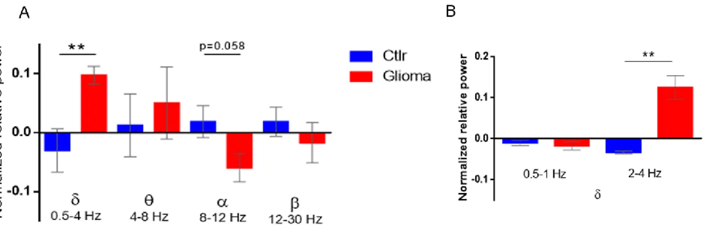
3.1.2. Spectral analysis of LFPs: enhancement of δ band and deterioration of α band during tumor growth ... 66
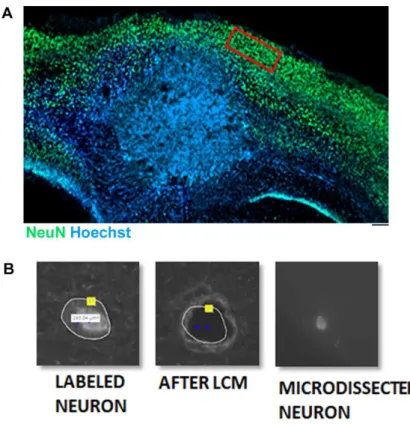
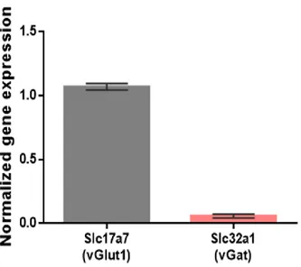
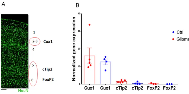
3.1.3. Molecular alterations in excitatory peritumoral neurons ... 68
3.1.4. LFP alterations in glioma-bearing mice ... 73
3.2. Impact of neural activity on glioma proliferation ... 77
3.2.1. Neuronal fibers infiltrate the glioma mass ... 78
3.2.2. Optogenetic stimulation of pyramidal, excitatory neurons promotes glioma growth 80 3.2.3. Optogenetic stimulation of Parvalbumin-positive, GABAergic interneurons restrains glioma proliferation... 81
3.2.4. Afferent sensory input bidirectionally regulates tumor proliferation ... 84
2. ... 87
2.6.13. Blockade of synaptic transmission via BoNT/A enhances glioma cell proliferation ... 87
2.6.14. The effects of sensory input on tumor proliferation are region-specific ... 89
2.6.15. Sensory stimulation combined with temozolomide treatment delays the deterioration of visual responses induced by glioma growth ... 91
3
Discussion ... 92
Effects of glioma progression on neural tissues ... 93
Effects of neural activity on glioma proliferation ... 99
Conclusions ... 105
Ongoing experiments ... 106
Publications ... 107
4
Abstract
Gliomas grow in a neuronal environment, but the interactions between glioma cells and peritumoral neurons remain poorly understood. Understanding this complex relationship could add useful information to develop more effective therapeutic approaches for the treatment of this deadly disease. In recent years, the interaction between cancer cells and tumor microenvironment has emerged as one important regulator of tumor progression. Thus, my thesis aimed to investigate the crosstalk between neural peritumoral tissue and glioma cells; in particular, I assessed i) functional impairments of peritumoral tissue occurring during tumor progression and ii) the impact of neural activity on glioma proliferation.
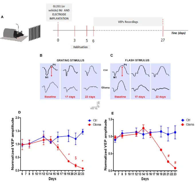
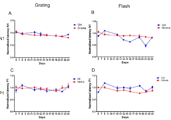
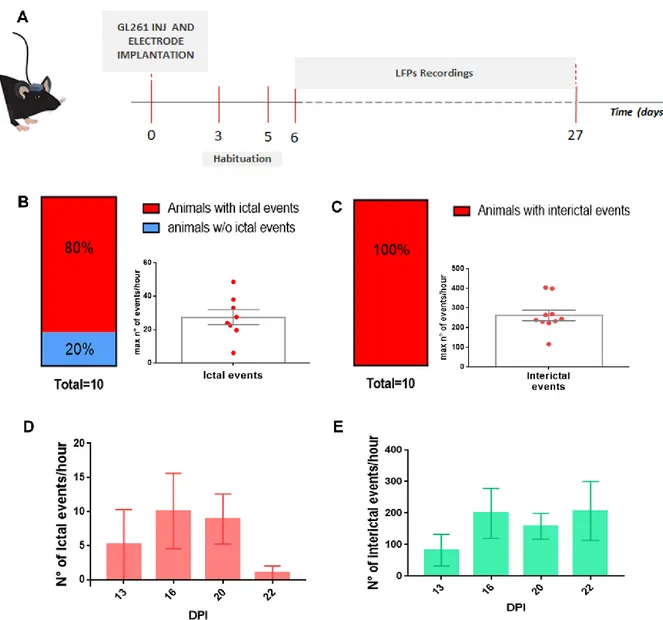
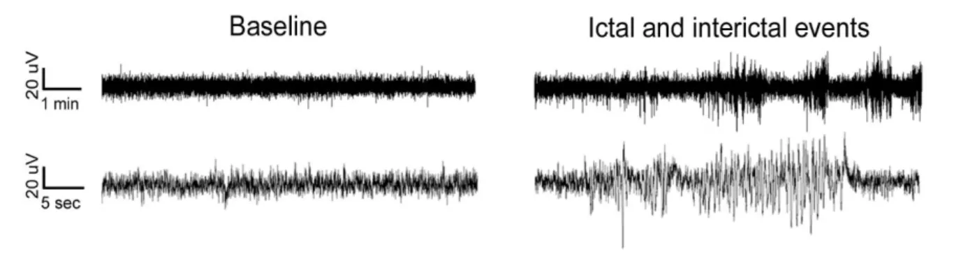
To monitor longitudinal changes in network activity, I recorded visual evoked potentials (VEP) and local field potentials (LFP) after transplant of GL261 glioma cells (or PBS) in mouse visual cortex. Gliomas were injected in visual cortex to allow a detailed investigation of peritumoral neurons using several physiological parameters. Thanks to this analysis, I detected a progressive deterioration of VEP amplitudes along with tumor progression and changes in the LFP power spectra typical of focal epilepsy, with an increase of the power of delta band and the deterioration of alpha rhythm in glioma-bearing mice. To understand the molecular alterations that underlie these perturbed patterns of neuronal activity, I analysed the gene expression profile of microdissected peritumoral pyramidal neurons in the cortical superficial layers (i.e., II-III). The data were clear in indicating that glioma induces alterations in both pre- and post-synaptic markers, demonstrating that its progression shapes the network activity of peritumoral areas towards hyperexcitability. Indeed, I recorded the occurrence of seizures in a subset of glioma-bearing animals, finding alterations in the LFP power spectra just before the onset of ictal events.
To investigate how levels of cortical activity affects tumor cell proliferation, I inoculated GL261 glioma cells into the mouse neocortex and modulated neuronal activity by different methods. Second, I dissected the role of inhibitory and excitatory circuitries on tumor proliferation and I found that while the activation of excitatory networks exacerbate glioma proliferation (confirming the data in literature), inhibitory circuits decrease GL261 cell proliferation. Based on these data, I investigated whether a sensory stimulation of the visual cortex may also impact on tumor growth. I found that a reduction of visual cortical activity via Dark Rearing enhanced the density of proliferating glioma cells, while a Visual
5 Stimulation had the opposite effect. Intriguingly the effect was region-specific, as visual deprivation had no significant effect on glioma proliferation in motor cortex. I found that local blockade of neurotransmission via administration of the synaptic blocker botulinum neurotoxin A (BoNT/A) enhances glioma cell proliferation, underlying the importance of neural activity in controlling glioma progression. In addition, the stimulation with visual patterns combined with temozolomide treatment delayed the deterioration of visual responses induced by glioma growth. Altogether, these data demonstrate complex effects of different neuronal subtypes and afferent sensory input in the control of glioma proliferation.
6
Introduction
1.1. GliomasThe treatment of gliomas represents one of the hardest challenge of our times for neuro-oncologists. Despite most neoplastic brain lesions derives from metastases of cancers outside the central nervous system (Gavrilovic and Posner, 2005), gliomas are among the most common primary brain tumors in adult and children (Ferlay et al., 2010; Ostrom et al., 2016), accounting for almost 30% of all primary brain tumors and 80% of all malignant ones (Weller et al., 2015). Although therapies are in continuous development, these tumors remain associated with high morbidity and mortality (Burnet et al., 2007). The name ‘glioma’ reflects the histomorphological resemblance of tumor cells with those of
glial lineages in the normal brain, such as astrocytes and oligodendroglia, which has been regarded, for a long time, as the only proliferating cells in the mature brain (Jones et al., 2012). For the past century, the classification of brain tumors has been based largely on their histogenesis including cellularity, mitotic activity, nuclear atypia, vascularity and necrosis; thus, tumors have been classified according to their microscopic similarities with different putative cells of origin and their presumed levels of differentiation (Louis et al., 2016). Nowadays, the most used and known classification is the international classification published by the World Health Organization (WHO) with a specific aim: to establish a classification and grading of human tumours that is accepted and used worldwide (Louis et al., 2007).
The classical WHO classification grades brain tumors on a WHO consensus-derived scale of I to IV, according to their degree of malignancy as
7 judged by various histological features accompanied by genetic alterations (Louis et al., 2007). Grade I tumors are biologically benign and can be surgically removed; grade II tumors are low-grade malignancies that may follow long clinical courses, but early diffuse infiltration of the surrounding brain renders them incurable by surgery; grade III tumors exhibit increased anaplasia and proliferation over grade II tumors and are more rapidly fatal; grade IV tumors exhibit more advanced features of malignancy including vascular proliferation and necrosis and are recalcitrant to radio/chemotherapy (Furnari et al., 2007).
The most common gliomas affecting the cerebral hemispheres are classified in the “diffuse” type due to the ability to infiltrate through the brain parenchyma (Wesseling et al., 2011). Individual tumor cells can spread along white-matter tracts of the cerebrum, sometimes crossing the corpus callosum, entrapping neurons, clustering around small vessels or accumulate in the subpial space (Louis et al., 2007). Based on histopathological analysis, these gliomas are diagnosed as diffuse astrocytomas, with glioblastoma (GB) as its most malignant form, oligodendrogliomas or oligoastrocytomas, classified as II, III or IV WHO grades(Louis et al., 2007, 2016; Perry and Wesseling, 2016). Across the years, the WHO classification has undergone a substantial evolution breaking with the century-old principle of diagnosis, integrating genetic and molecular parameters in the classification methods which, in some cases, override the classical histological features (Louis et al., 2016; Wesseling and Capper, 2018). For instance, the detection of genetic mutations, as in isocitrate dehydrogenase 1 and 2 (IDH1/IDH2) (Yan et al., 2009), in association with the 1p/19q codeletion, determine a new method of classification (Wesseling and Capper, 2018). So, even if a tumour has the histological appearance of an astrocytoma, detection of
8 complete 1p/19q codeletion leads to the diagnosis of oligodendroglioma, IDH-mutant and 1p/19q-codeleted (Perry et al., 2016; Wood et al., 2019).
1.1.1. Glioblastoma (GB)
The 60-70% of all gliomas is represented by GB, the most lethal tumor of the CNS, classified as a WHO grade IV tumor due to its histopathological features (Louis et al., 2016). Glioblastoma presents significant intratumoral heterogeneity at cytopathological, transcriptional and genomic levels and this complexity has conspired to make GB one of the most difficult cancer to understand and to treat (Furnari et al., 2007). Typical histological features of GB include regions of necrosis, microvascular proliferation, abundant mitosis and pleiomorphic cells (Wen and Kesari, 2008). According to recent discoveries, GBs are now subdivided on the basis of the mutational state of isocitrate dehydrogenase (IDH) genes in: IDH wild type, which most frequently corresponds with the clinically defined primary or de novo GB; IDH mutant, which corresponds to the secondary GB; those not otherwise specified (NOS), for which the IDH status could not be determined (Louis et al., 2016; Verhaak et al., 2010). The 90% of GB are primary IDH-wildtype tumors with a rapid clinical presentation, while the remainder (about 10%) are mostly IDH-mutant tumors, typically arising from lower grade infiltrating astrocytomas (Wood et al., 2019). Although the molecular features of IDH-wildtype glioblastoma have been studied extensively, the prognosis remains poor (Ceccarelli et al., 2016; Ostrom et al., 2016). Some common mutations that represent frequent gene signatures in human GB used for clinical classification are those in TP53, PTEN, NF1, ERBB2, RB1, PIK3R1 and PIK3CA genes (McLendon et al., 2008). The mentioned
9 cancer-associated genes represents a core set of pathways that are commonly deregulated in glioblastoma, including growth factor signalling (receptor tyrosine kinase (RTK)/phosphatidylinositide 3-kinase (PI3K)/Ras), p53, and Rb signalling pathways. Moreover, amplification of EGFR (epidermal growth factor receptor) gene is frequent in primary glioblastomas as well as the methylation of the O6-methylguanine-DNA methyltransferase (MGMT) promoter that occurs in about 50% of glioblastoma cases (Louis et al., 2016; Shinojima et al., 2003). Other cellular processes deregulated in GB could be exploited for diagnostic and prognostic applications as the homeostatic pathways (Franceschi et al., 2018). Altogether, these alterations lead to aberrant signalling in proliferation, cell cycle regulation, senescence and apoptosis, underscoring the importance of such pathways in tumorigenesis (Brennan et al., 2013; McLendon et al., 2008). Different clinically relevant GB subtypes (proneural, neural, classical, and mesenchymal), are identified on the basis of the gene expression profiles together with somatic alterations (Brennan et al., 2013; Franceschi et al., 2015; Verhaak et al., 2010).
Despite the growing experimental investigation in this field and the improved therapeutic strategies (see section 1.2), GB remains essentially incurable, with an overall survival time ranging from 12 to 18 months, and a five-year relative survival following diagnosis of 6.8% (Ostrom et al., 2016, 2019). An important factor that limits the identification of an efficient GB treatment is the high cellular and genetic heterogeneity because the tumor cells that form GB mass, although belonging to the same patient, are not genetic phenocopies (Patel et al., 2014). Moreover, the cellular origin of gliomas remains a topic to debate. Although glioblastomass from human patients contain different types of driver mutations that may represent diverse subtypes, the contribution of the cell
10 of origin remain unknown. The cell of origin can be an important determinant of tumour phenotype and genotype in GB, and thus plays an important role in its malignant behaviour (Llaguno and Parada, 2016). In the past years, one hypothesis was that gliomas arise from neoplastic transformation and dedifferentiation of mature glial cells, astrocytes, oligodendrocytes, ependymal cells or their precursors (Martin-Villalba et al., 2008). Nowadays, the most accredited hypothesis places the origin of high-grade gliomas, in adult neural stem and progenitor cells (Llaguno and Parada, 2016). In particular, the subventricular zone (SVZ) of the lateral ventricles is identified as the likely neurogenic region it may give rise to the cells of origin of GB (Alcantara Llaguno et al., 2009). It has been demonstrated that astrocyte-like neural stem cells (NSCs) of the SVZ could carry driver mutations that stimulate the development of glioma, migrating from the SVZ to distant sites of the brain (Lee et al., 2018). To support this idea, more differentiated neuronal cells showed reduced abilities in forming a tumor mass, demonstrating that increasing lineage restriction is an impediment to glioma formation (Alcantara Llaguno et al., 2019). Altogether, these advances attempt to clarify dynamic changes of tumor composition for a more accurate classification and the setting up of specific treatments (Alcantara Llaguno et al., 2019).
1.2. Therapies
The standard of care of glioblastoma consists in maximal safe surgical resection followed by radiotherapy (RT) with concurrent and adjuvant chemotherapy (Temozolomide, TMZ). However, none all the accepted treatments are of real efficacy for the patients, due to the high rate of
11 recurrence, overall resistance and devastating neurological deterioration provoked by GB (Bahadur et al., 2019; Kim et al., 2015; Lin et al., 2015). Surgery alleviates symptoms of mass compression and provides material for diagnosis and molecular analysis, improving overall and progression-free survival (Brown et al., 2016). In addition, surgical excision provides an opportunity to apply some therapies during the procedure as implantation of chemotherapeutic agents and photodynamic therapy which have a promising outcome (Jain, 2018). However, the tumor removal does not eliminate the microscopic foci of neoplastic cells that invade the surrounding normal brain substance beyond the main tumor mass, provoking the 90% of recurrences within a two centimetre margin of the primary tumor site (Corso et al., 2017). Novel fluorescence-based technologies have incremented the tumor visualization. For instance, molecular-imaging techniques have been combined with GB targeting agents as Chlorotoxin (CTX)-based bioconjugates to improve tumor diagnosis and imaging (Cohen et al., 2018).
Moreover, fluorescence-guided surgery using 5-aminolevulinic acid allows a more precise resection of glioma mass and a better identification of tumor border, thus prolonging patients’ survival(Munteanu et al., 2017; Stummer et al.,
2006).
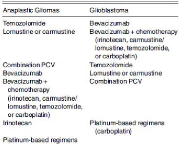
Unfortunately, the association with radiotherapy and chemotherapy cannot completely eradicate GB, since this would require unacceptably high radiation or chemotherapic doses that result in severe brain-neuron damage (Colman et al., 2006; Furnari et al., 2007). Thus, new therapeutic strategies are in continuous development, based on the combination of postoperative radiations with TMZ administration as a backbone on which to add new therapies summarized in the Table 1 below (Laub et al., 2018; Reitman et al., 2018):
12 In the last years there has been an explosion of research on glioblastoma, with thousands of new clinical trials (Bahadur et al., 2019; Ozdemir-Kaynak et al., 2018); however, no one resulted enough effective in counteracting GB growth, producing a significant increase of patients’ survival. Indeed, because of the
extreme heterogeneity of these tumors, finding effective therapies is still very challenging.
Table 1 Systemic therapy approved for recurrent high-grade Gliomas (Laub
et al., 2018)
13 The majority of clinical trials are focused on systemic therapies, as opposed to surgical or radiotherapeutic treatments (Cihoric et al., 2017). Whereas targeted drug therapies are focused on the abnormal molecular and cytogenic pathways that affect tumor growth, invasion, angiogenesis and cell death (Roy et al., 2015), other clinical strategies are focused on immunotherapies, such as switching check-point inhibitors (i.e., pembrolizumab and nivolumab) (Reardon et al., 2017). In addition, other clinical trials are examining the role of vaccines, such as dendritic cell or anti-EGFR (O’Rourke et al., 2017), or the use of electrical tumor
treating fields (TTF) to inhibit the formation of mitotic spindle (McClelland et al., 2018; Stupp et al., 2017; Wick et al., 2018).
Although the histological analysis remains essential in the diagnosis of gliomas, the methylation status of MGMT promoter has been recently identified as a potential determinant of chemotherapic treatment failure in glioblastoma (Wick et al., 2014). Indeed, while high levels of MGMT activity in cancer cells create a resistant phenotype by blunting the therapeutic effect of chemotherapy, the epigenetic silencing of the MGMT gene is associated with a diminished DNA-repair activity (Morandi et al., 2010; Wick et al., 2018).
Despite all studies lead on discovering new therapeutic targets or aimed at creating novel therapeutic approaches, the majority of the current proposed treatments have not reported significant improvements in the overall survival or in preventing recurrences for GB patients (Laub et al., 2018). This devastating scenario strongly indicates a lack of knowledge in the biology of gliomas, prompting scientists to go further the classical treatments, investigating peritumoral tissues as the tumoral invasion front into the neighbouring, healthy tissue (D’Alessio et al., 2019). Thus, scientists must now focus on elucidating the
14 optimizing surgical resection, but also to better defining the role of peritumoral tissue in gliomas progression, identifying new therapeutic targets (D’Alessio et al., 2019).
1.3. Tumor microenvironment
For long time, cancer research has focused mainly on understanding the biology of glioma cells, investigating the aberrant pathways that guide tumor onset and progression. Nowadays, the idea that the interaction between glioma cells and the local environment is crucial in driving tumor growth has strengthened (Hanahan and Coussens, 2012). Understanding microenvironmental determinants that contribute to the growth of glioma growth is therefore at the centre of current scientific research, with the aim of discovering new targets and strategies to prevent the high burden of morbidity and mortality by improving patients’ survival and quality of life (Johung and Monje, 2017).
The features of the tumor microenvironment (TME) are dynamically modified during glioma progression and the interaction of tumor cells with the main components of the TME (neurons, reactive astrocytes, microglia, oligodendrocytes, inflammatory cells, endothelial cells, pericytes and GB-associated stromal cells) seems to shape the progression of the disease (D’Alessio et al., 2019; Venkatesh et al., 2015). For instance, the enhancement of
cerebrovascular and lymphatic networks remodelling (Hahn et al., 2019; Hanahan and Coussens, 2012; Hoelzinger et al., 2007; Mancino et al., 2011), together with the metabolic and biochemical alterations in peritumoral tissues, are regulated by different communication routes which include secreted
15 molecules, gap junctions, tunnelling nanotubes and extracellular vesicles (D’Alessio et al., 2019; Jung et al., 2019; Lane et al., 2019; Matarredona and
Pastor, 2019; Osswald et al., 2015).
1.4. Neural activity shapes the tumor microenvironment
Increasing evidences show that neurons are active part of the tumor microenvironment, contributing, with their direct and indirect action, in shaping the interactions between different players of glioma development. However, complex interactions between various types of neurons make this mechanism difficult to understand (Deisseroth et al., 2004; Ge et al., 2007).
In a non-pathological condition, electrical activity influences the development of central and peripheral neural system (Gafarov, 2018). During the maturation of CNS, patterned waves of electrical activity influences neurodevelopment inducing calcium transients in all the parts of the nervous system (Corlew et al., 2004; Wong et al., 1995). Interestingly, other mechanisms are profoundly regulated by neurons also in the adult brain. For instance, excitatory neurons regulate proliferation and differentiation of stem cells in the subgranular zone of the dentate gyrus and neurogenesis in the subventricular zone (Deisseroth et al., 2004; Paez-Gonzalez et al., 2014). Moreover, neurons also regulate I) Schwann cell proliferation and survival in the peripheral nervous system (Maurel and Salzer, 2000) and II) glial precursor proliferation in myelin plasticity in the CNS, conditioning brain structure and function (Scholz et al., 2009; Takeuchi et al., 2010). Studies on oligodendrocyte precursor cells (OPC) demonstrate the suppression of cell proliferation by silencing the neural activity in the rat optic nerve, either via nerve transection or via tetrodotoxin (Barres and
16 Raff, 1993). Moreover, OPC, pre-OPC and neural precursor cells show a mitogenic response to optogenetically increased cortical activity and subsequent differentiation (Gibson et al., 2014). Altogether, these data strengthen the pivotal role of neurons in controlling glial precursor cells, a likely origin of gliomas, pointing out the possible influence of neural activity on tumor cells.
Actually, there are many robust evidences that neural activity affects glioma cell behaviour. The most relevant studies of the latest years have demonstrated that pyramidal cell activity promotes patient-derived high-grade glioma proliferation through activity-dependent secreted factors, in particular neuroligin-3 (NLGN3) (Venkatesh et al., 2015, 2017). The mitogenic effects are a consequence of the activation of the PI3K-mTor, SRC and RAS signalling pathways, leading to a potentially autocrine/paracrine loop of NLGN3 expression, that inversely correlates with overall survival of glioblastoma patients (Venkatesh et al., 2017). It has been also demonstrated that AMPA receptor activation promotes glioma growth, mainly through microenvironmental interactions such as neuron-to-glioma synaptic transmission (Venkatesh et al., 2019). Moreover, NLGN3 seems to be implicated in the formation of bona fide AMPA receptor-dependent synapses between glioma cells and peritumoral excitatory neurons, integrating high-grade gliomas in the neural network (Venkatesh et al., 2019). Several studies suggest also a probable pivotal role in regulation of glioma proliferation exerted by other neural secreted molecules, that are neurotransmitters and neurotrophins; however, the role of activity-dependent secretion has not been demonstrated yet, because of the difficulty in unravelling the source that secretes these fundamental factors, since both neural and glioma cells can produce them.
17 1.4.1. Neurotransmitters
Glioma growth is regulated by complex multifactorial interactions and it is affected by activity-dependent protein factors, as described above. Besides proteins as NLGN3, neurotransmitters are other possible candidates released by neurons to control tumor progression (Johung and Monje, 2017). Indeed, it is well known that glioma cells express functional neurotransmitter receptors, which could interfere with mitogenic pathways. For instance, adult GBs express the dopamine receptors DRD2 and DRD4, whose inhibition seems to be particularly effective in reducing glioma cell proliferation and survival (Caragher et al., 2019; Dolma et al., 2016). Moreover, human GB cells also express serotonin receptors, albeit it is not clear whether they have an impact on tumor cell proliferation (Mahé et al., 2004). In particular, a retrospective study made on GB patients showed that increasing serotonin levels through the administration of a selective serotonin reuptake inhibitor (SSRI) didn't produce a significant survival benefit (Caudill et al., 2011; Otto-Meyer et al., 2020).
1.4.1.1. Glutamate
One of the most characterized neurotransmitter is undoubtedly glutamate. Glutamate is one of the most important and abundant molecule of the CNS, but its extracellular levels must be maintained low to guarantee a normal brain function. Therefore, the scavenger role of glia and astrocytes is of paramount relevance to maintain this equilibrium (Bergles and Jahr, 1997; Robert and Sontheimer, 2014). Indeed, glial membranes are abundant of Na+-dependent transporters (i.e. EAAT1 or EAAT2), representing the 1% of total brain proteins and being necessary for the clearance of glutamate (Danbolt, 2001). In particular,
18 glioma cells have an almost complete absence of Na+-dependent glutamate uptake, due to the loss of EEAT2 protein production and to the mislocalization of EAAT1 to the nuclear membrane (Buckingham and Robel, 2013; Robert and Sontheimer, 2014; Ye et al., 1999). Furthermore, glioma cells extrude glutamate outside, predominantly through the overexpression of the cystine-glutamate antiporter (system xc-, SXC) that exchanges intracellular glutamate for extracellular cysteine and that is involved in the generation of antioxidant glutathione (de Groot and Sontheimer, 2011). The biochemical source of glutamate for glioma cells is not entirely known. Published data show that glutamate may be generated from glutamine via a glutaminase reaction or that it could be accumulated due to other metabolic impairments as the decreased conversion in α-ketoglutarate by GLUD2 enzyme (de Groot and Sontheimer, 2011; Franceschi et al., 2018). Glioma cells express ionotropic and metabotropic glutamate receptors (de Groot and Sontheimer, 2011) and the increased extracellular glutamate levels in proximity of the tumor mass (Behrens et al., 2000) promote survival, growth and migration through the activation of PI3K-Akt pathway via AMPA receptor activation (Ishiuchi et al., 2007). The high amount of glutamate acts in a paracrine/autocrine manner on tumor cells, facilitating their spread into the brain parenchyma and resulting to be excitotoxic for neurons in the tumor vicinity (de Groot and Sontheimer, 2011). Moreover, gliomas increase the neural excitability of peritumoral neurons, but the mechanism that links hyperactivity and a further cell proliferation is still lacking (Venkatesh and Monje, 2017).
19 1.4.1.2. GABA
The role of GABA neurotransmitter is far more complex. In a normal brain, GABA is not only the most important inhibitory neurotransmitter of the CNS, but it also controls stem cell proliferation in the subventricular zone, limiting the generation of new neuroblasts (Blanchart et al., 2017; Young and Bordey, 2009). It has been demonstrated that glioma cells express GABA receptors, but the effects of their activity is still under debate. Studies on patient-derived glioma cultures show that the expression of functional GABA A receptors is more correlated with low-grade gliomas and oligodendrogliomas than glioblastomas (Labrakakis et al., 1998), but other in vitro studies on human glioblastoma samples presume the opposite (D’Urso et al., 2012). In a p16 Arf -/- PDGFB
murine glioma model has been identified an endogenous ionotropic signalling within tumor cells, demonstrating that glioma cells are able not only to express GABA A receptor subunits and to respond to it, but they can also release GABA, especially in tumor bulk (Blanchart et al., 2017). Furthermore, studies demonstrated that the endogenous neurotransmitter limits cell proliferation of both murine and patient-derived glioblastoma cells, prolonging the survival of glioma-bearing mice (Blanchart et al., 2017). As a matter of fact, it is supposed that GABA could have a role not in the initial steps of transformation, but once the tumor is established, as proved by experiments with muscimol and bicuculline on animal models (Blanchart et al., 2017). Moreover, phosphorylation of H2AX by chloride influx within cells through GABA A receptor activation seems to be essential for the inhibition of glioma growth. It has to be noticed that in high-grade gliomas GABA A receptors are likely downregulated, reducing the inhibitory effect of the peptide on tumor proliferation (Jung et al., 2019). Intriguingly, the effect of
20 GABA is more robust in the quiescent stem-like population of glioma cells, suggesting a possible strategy to maintain tumor-initiating cells quiescent (Blanchart et al., 2017). This is sustained also by the downregulation of miRNA 155 in glioblastoma, which reduces the expression of GABA A receptors and, when the miRNA production is regained, GB cells recover the response to the GABA neurotransmitter (D’Urso et al., 2012). Taken altogether, these studies point out the interesting, but still not completely answered, question regarding the influence that GABAergic interneurons may exert on glioma.
1.4.2. Neurotrophins
Neurotrophins are a family of proteins that are historically implicated in several vital functions as growth, survival, development and plasticity in both central and peripheral nervous systems (Sofroniew et al., 2001); their role is much broader than their name might suggest (Vega et al., 2003). The classical molecules belonging to this group are nerve growth factor (NGF), brain-derived neurotrophic factos (BDNF), neurotrophin-3 (NT3) and neurotrophin-4 (NT4). They bind the p75NT receptor and one of the tyrosine kinase receptors (Trk; i.e. TrkA for NGF, TrkB for BDNF and NT4, TrkC for NT3) (Park and Poo, 2013), activating trophic or pro apoptotic pathways depending on molecular context (Meeker and Williams, 2015).
The role of neurotrophins in cancer and especially in gliomas is still under debate, because it is not clear whether they exert a pro-mitogenic effect on glioma cells or a protective role for the tumor microenvironment (Garofalo et al., 2015; Venkatesh et al., 2015). It is known that in paediatric tumors such as neuroblastoma or medulloblastoma tumor cells express neurotrophin receptors
21 and the clinical significance is suggested to be predictive of a favourable outcome (Donovan et al., 1993; Eberhart et al., 2001; Nakagawara et al., 1993). Also in adult low-grade gliomas TrkA and TrkB are highly expressed, while their presence in GB is weak, suggesting that those receptors might be involved only in the early stages of the disease (Wadhwa et al., 2003).
NGF was the first neurotrophin discovered to be responsible for sympathetic and sensory nerve growth and survival (Cohen et al., 1954). It has been demonstrated that in a rat model of glioma the activation of the TrkA receptor through NGF treatment has an anti-mitotic effect on C6 glioma cells, reducing their proliferation and inducing the differentiation, although tumor cells lack to synthetize neurotrophin (Kimura et al., 2002; Rabin et al., 1998). Also in humans, a phase II clinical trial with administration of NGF on patients with childhood optic gliomas led to a statistically significant improvement in electrophysiological parameters used to evaluate vision respect to placebo-treated patients (Falsini et al., 2016). Conversely, on human glioblastoma cell lines, NGF seems to improve tumor proliferation via Notch1 signalling (Park et al., 2018).
The role of BDNF in gliomas is more controversial. BDNF is a peptide secreted in an activity-dependent manner (Hong et al., 2008). In vitro studies demonstrate that neurotrophin stimulates high grade glioma cell proliferation, through the binding with its receptor TrkB (Xiong et al., 2013) and its release increases with neural activity (Venkatesh et al., 2015). On the other end, evidences suggests that BDNF overexpression in the hypothalamus has immune-augmenting properties, eliciting an increased anti-tumor immune response and reducing the activity of several proteins that would normally confer resistance to chemotherapeutic agents (Radin and Patel, 2017). Moreover,
22 glioma-bearing mice reared in an enriched environment (EE) showed increased level of BDNF together with significantly reduced tumor growth (Garofalo et al., 2015).
1.5. Neural alterations induced by glioma
Emerging researches suggest that glioma cells exert a powerful influence on neural tissues. Deficits in neurocognitive functioning frequently occur in glioma patients and heavily affect their quality of life (Aaronson et al., 2011; Seano et al., 2019). The impact of tumor mass on neurons could provoke mechanical compression on brain structures but also cell-death through tumor-released excitotoxins and disturbance in synaptic transmission, inducing a direct neuronal damage in the region of insurgence and disturbing the functional brain networks (van Kessel et al., 2017).
1.5.1. Tumor-associated epilepsy
Tumor-associated epilepsy (TAE) is among the most common comorbid condition in patients with gliomas, heavily affecting their quality of life (Beaumont and Whittle, 2000; Fisher et al., 2014; Taphoorn et al., 2010). Approximately 30-50% of patients with brain tumors manifest seizure as initial symptom of disease progression (van Breemen et al., 2007). Surprisingly, patients with low grade tumors are more prone to develop TAE with an incidence of the 80-90% (van Breemen et al., 2007; You et al., 2012a), whereas in high grade gliomas are less frequent (30-62%) but may be more difficult to control and occur predominantly during the onset of the disease (Armstrong et al., 2016). TAE often manifest as
23 focal seizures with secondary generalization and, despite its major clinical and social impact, the pathophysiological causes are poorly understood. Due to the complex heterogeneity of perturbed mechanisms, this kind of epilepsy is often refractory to antiepileptic treatments (Cowie and Cunningham, 2014; You et al., 2012a).
An important aspect that determines the insurgence of seizures is the glioma localization in the brain. As a matter of fact, the proximity to the cortical grey matter is a fundamental factor: tumors localized closely to the cortex or with limbic and perilimbic cortical localization are highly epileptogenic, whereas tumors located in the inner parts of the brain are less likely to manifest seizures (Berntsson et al., 2009; Cowie and Cunningham, 2014). In addition, epileptiform activity originates outside the tumor mass but within 1-2 mm from the tumor border for animal models (Köhling et al., 2006) and within 3 mm for human patients (Patt et al., 2000). Moreover, the tumor itself might generate action potentials through astrocytic tumor cell activity, which could also represent a source of epileptic activity (Bordey and Sontheimer, 1998).
The development of seizures is a complex phenomenon that involves multifactorial mechanisms in the peritumoral tissues as metabolic and pH changes, disruption of the BBB, immunological and inflammatory changes, ionic change (e.g. Mg2+ and Fe3+), hypoxia, acidosis and metabolic impairments that could affect neural morphology and function (Armstrong et al., 2016; Cowie and Cunningham, 2014; You et al., 2012a). Besides, the alterations in the balance between excitatory and inhibitory networks certainly play a pivotal role in the generation of seizures (MacKenzie et al., 2017; Nelson and Turrigiano, 1998; You et al., 2012a).
24 1.5.2. The hyper-activation of excitatory networks
As described in the section 1.4.1.1, glioma extrudes high amounts of glutamate in the extracellular space of the peritumoral area (Behrens et al., 2000; Buckingham et al., 2011), resulting in excitotoxicity, tumor invasion and hyperexcitability of neural tissues (Marcus et al., 2010; Robert and Sontheimer, 2014; Rzeski et al., 2001; Sontheimer, 2008a; Vanhoutte and Hermans, 2008). As a consequence, the elevated quantity of glutamate in peritumoral tissue can over-activativate NMDA and AMPA receptors (Savaskan et al., 2008). The prolonged activation of NMDA receptors provokes a sustained influx of Ca2+ into the cell, mediating excitotoxicity and affecting the inhibitory function of GABA B receptors (Terunuma et al., 2010). The reduction of neuronal cell density creates room for tumor expansion (Buckingham et al., 2011) and the over-activation of AMPA receptors boosts neural excitability (Savaskan et al., 2008). Taken altogether, all these studies highlight that the glutamate-driven NMDA and AMPS activation is now considered to be one of the main causes of peritumoral cortical hyperexcitability (Buckingham and Robel, 2013; Campbell et al., 2012). Indeed, targeting glutamate receptors with talampanel (antagonist of AMPA receptors) and memantine (antagonist NMDA receptors) are currently tested in clinical trials with encouraging results (Huberfeld and Vecht, 2016).
However, it is worth noticing that glutamate release per se, although necessary, is insufficient to drive peritumoral epilepsy and that other concomitant dysregulations are necessary in order to establish long-lasting hyperexcitation (Campbell et al., 2015).
For instance, the chloride (Cl-) homeostasis of pyramidal neurons is heavily affected, with the downregulation of KCC2 (K-Cl cotransporter 2) and the
25 upregulation of NKCC1 (Na-K-Cl cotransporter 1). The high amount of intracellular chloride caused by the defective extrusion through KCC2 and the increased influx by NKCC1, lead to GABA-mediated depolarization, reversing chloride potential and leading to an excitatory action of gamma-aminobutyric acid (GABA) receptor activation on pyramidal neurons (Campbell et al., 2015; Conti et al., 2011; Di Angelantonio et al., 2014; Huberfeld and Vecht, 2016; Jung et al., 2019; Pallud et al., 2014). The excitatory role of GABA in peritumoral tissues is also supported by some studies on temporal lobe epilepsy, where it was found that stimulating parvalbumin interneurons (PV) could produce differential effects with respect to their distance to the epileptic focus. Indeed, activating PV cells close to the epileptic focus fails to block ictal generation, enhancing synchrony and promoting ictal generation; on the contrary, the stimulation of PV cells distant from the epileptic focus lead to an anti-epileptic effect (Sessolo et al., 2015). In addition, the reduction of KCC2 by a prolonged glutamate-activation of NMDA receptors, causes the degradation of GABA receptor subunits. Furthermore, pyramidal cells show a reduction in inhibitory synapses on the soma and axon initial segment, indicating an incorrect regulation of the circuits (Marco et al., 1997). Therefore, the decreased inhibition, the GABA possible switch to excitation and an altered expression of its receptors, could concur in neural hyperexcitability.
1.5.3. The degradation of inhibitory networks
In the tumor microenvironment, also inhibitory networks appear perturbed but the role of GABA connections in glioma environment is not completely understood. In particular, it has been reported that the peritumoral regions show
26 a significant loss of approximately 35% of GABAergic interneurons, together with a reduction in their firing rates and in synapses with pyramidal neurons (Campbell et al., 2015; Tewari et al., 2018; Yu et al., 2020) that leads peritumoral tissue to be hyper-excitable and more prone to seizures (Buckingham et al., 2011; Venkatesh et al., 2019).
During the latest years, it has been developed the concept that seizures occurring in peritumoral tissues are not only the result of a specific alteration of excitatory and inhibitory neurons. Indeed, studies showed that in TME the perineuronal nets (PNNs), that surround fast spiking interneurons acting as ionic buffering, neuroprotecting and stabilizing their synaptic activity, are degraded by glioma-released proteases. This results in increased membrane capacitance and, in turn, reduces the firing rate of the remained peritumoral inhibitory interneurons within 400 μm from tumor edge. In particular, the pro-seizure effect of PNN degradation seems to be due to the lacking role of electrostatic insulator affecting the physiological properties of fast-spiking interneurons (Tewari et al., 2018). All these events contribute to establish an unbalanced network, that leads peritumoral tissue to be hyper-excitable and more prone to seizures (Buckingham et al., 2011; Venkatesh et al., 2019).
1.6. Direct neuron-glioma interaction
As previously described, glioma cells interact with the local environment modifying it for their own advantage, through the secretion of specific factors that facilitate tumor progression. But the invasiveness of brain tumors can follow complex patterns: many gliomas cross the corpus callosum to form butterfly lesions, some remain confined to the white matter stopping abruptly at the
gray-27 white-matter junction, whereas others grow in the gray matter around neurons and blood vessels (satellitosis) (Louis, 2006; Wesseling et al., 2011). The neural satellitosis is a perfect example of direct interaction between glioma cells and neurons and this phenomenon has pioneered the study of the tumor connectivity with the brain microenvironment. Recently it has been discovered that many astrocytomas are able to directly interact with the surrounding environment, extending ultra-long membrane protrusions called tumor microtubes (TMs) that represent a route for glioma proliferation to invade the brain (Osswald et al., 2015). The presence of these structures is correlated with the worst prognosis in human gliomas and confers a resistance against all the available therapies (Weil et al., 2017). Studies in drosophila demonstrate that glioblastoma cells protrude TMs to enwrap neurons, depleting in them the Wingless-related integration site (WNT) pathway and causing neurodegeneration (Portela et al., 2019). Moreover, through TMs, glioma cells interact with peritumoral excitatory neurons forming functional bona fide AMPA-mediated synapses (Venkataramani et al., 2019), in which pro-mitotic peptides as NLGN3 are involved (Venkatesh et al., 2019).
However, despite the latest evidences demonstrating a tumor-supportive activity of excitatory neurons, an anti-proliferative interaction between peritumoral neurons and glioma cells has also been well-described (Liu et al., 2013). Interestingly, peritumoral neurons are capable of restrain glioma progression through the pivotal role of program death-ligand1 (PD-L1) protein, and indeed neuronal PD-L1 signalling in brain cells is important for GB patient survival (Liu et al., 2013). In vitro studies demonstrated that cortical neurons (but not glia) significantly inhibit GL261 cell proliferation, as assessed by
5-Bromo-2′-deoxyuridine (BrdU) incorporation (Liu et al., 2013). Specifically, high expression of PD-L1 by peritumoral neurons positively associates with GB patient survival,
28 demonstrating the existence of a PD-L1-dependent interaction between peritumoral neurons and tumor cells also in vivo (Liu et al., 2013).
Moreover, evidences demonstrate that the interaction between glioma cell lines and neurons lead to an upregulation of functional GABA A receptor expression in glioma cells, both in vitro and in vivo (Synowitz et al., 2001).
All these studies depict a very complex scenario, underlining the need to clarify the contribution of distinct neuronal cell types in tumor development.
1.7. Glioma models
Animal models represent a suitable tool to study the biology of tumors and to investigate the role of candidate pathways that might be involved in the development of this pathology. To create animal models of brain tumors we can take advantage of three different strategies: i) the infusion of tumor cells, ii) the delivery of oncogenic transgenes, or iii) the genome manipulation (Robertson et al., 2019). However, for a reliable prediction and validation of the effects of different therapeutic modalities, glioma models need to comply with specific and more strict demands than other models of cancer (Lenting et al., 2017). These demands are directly related to the combination of I) genetic heterogeneity of cell population, II) driver mutations within canonical cell-growth and survival pathways, III) the infiltration into the brain, IV) complex microenvironmental interactions that change tumor features and composition during its progression (Lenting et al., 2017; Quail and Joyce, 2017). Therefore, an ideal animal model of glioma should be sustainable in vitro but also be able, when in vivo, to mimic the complexity of the human pathology with a predictable and reproducible pattern (Peterson et al., 1994; Robertson et al., 2019).
29 In particular, the laboratory mouse is a suitable system that shares extensive molecular and physiological similarities to humans, such as angiogenesis and metastasis processes; so, they represent a powerful tool for studying cancer (Chen et al., 2012). More importantly, murine glioma models provide temporally and genetically controlled systems for studying the tumorigenic process, as well as response to treatment (Reilly and Jacks, 2001). Many different murine models are actually available. These models are designed to recreate a tumor with increasingly similar characteristics to the real pathology in humans or to reproduce an altered pathway known to be important in glioma development. Thanks to modern technologies such as the combination of magnetic resonance and ultramicroscopy, a detailed analysis of different biological features of some murine models has been done, pointing out differences in angiogenesis, grow pattern and intratumoral heterogeneity, allowing the researchers to choose the most suitable model for their studies (Breckwoldt et al., 2019). The nowadays principal used murine models are transgenic or created through glioma cells transplant.
1.7.1. Transgenic murine models
Transgenic mouse models offer an opportunity to develop and utilize an easily replenished, reproducible, spontaneously manipulated and accurate pre-clinical model of human cancers, which we can use to enhance our molecular knowledge and to test promising therapies (Miyai et al., 2017). The availability of modern techniques of genome sequencing have allowed the identification of driven mutations of gliomas. Consequently, a wide variety of genetically engineered mouse models (GEMMs) that incorporate genetic alterations found in
30 human patients has been developed (Kersten et al., 2017). . Especially, they represent a very powerful tool to investigate early initiation events of glioma formation and also to be used as preclinical models for testing new therapies (Llaguno and Parada, 2016).
In particular, GEMMs have been created by introducing genetic alterations in the germline and using breeding strategies that generate compound mutants with alterations in both oncogenes and tumour suppressors. Inevitably, mutations in some of the relevant genes are early lethal and therefore must be engineered using conditional tools (e.g. Cre-loxP recombination strategies) (Robertson et al., 2019). The general approach for the development of GEMMs involves loss of function of principal driven tumor suppressor genes, such as Cdkn2a (Ink4A/Arf), Nf1, Trp53, Pten, and Rb1 and/or the expression of driver oncogenes, such as mutant epidermal growth factor receptor (e.g., EGFR VIII) and Ras (K-Ras, H-Ras), Akt, and platelet-derived growth factor (PDGF) ligands (Kegelman et al., 2014).
The strategies to generate transgenic animal models are varied and one of the most popular approach that has contributed to our understanding of the potential cells of origin of GB is the RCAS-TVA system (Holland et al., 2000). Cells producing TVA, the receptor for subgroup A avian leukosis viruses, are susceptible to infection with replication-competent avian sarcoma-leukosis virus long terminal repeat with splice acceptor (RCAS) viral vectors (Wang et al., 2016). Holland et al. have developed transgenic mouse lines expressing the TVA in Nes- or Gfap expressing cells, presumed to be respectively progenitor cells and differentiated astrocytes and bred these with Cdkn2a-knockout mice (Holland et al., 1998). In addition, they have demonstrated that staminal cells were more susceptible to tumor initiation than Gfap-TVA cells, but differentiated astrocyte
31 populations derived from neural stem cells were not discernible (Holland et al., 2000). To address this problem, the model has been subsequently implemented combining the RCAS with lineage-restricted promoters (Jiang et al., 2017). With this system, a significant impact of differentiation state on tumour aggressiveness has been confirmed, with more restricted progenitors being less malignant (Jiang et al., 2017). Recently, the RCAS-TVA system has been combined with powerful techniques of genetic engineer as CRISP/Cas9 to deliver oncogenes or induce specific mutations in tumor suppressors (Oldrini et al., 2018). The limitation of this approach is the need of specific TVA-expressing mouse strains and the viral cargo length.
The complexity and heterogeneity of glioma pathogenesis, has led to design plasmid-based techniques as CRISPR- and PiggyBac-based approaches, that do not require mouse breeding or virus production and enable the delivery of larger cargo sizes (Pathania et al., 2017). In fact, they can deliver combinations of oncogenes and tumour suppressors in multiplex, directly in vivo and with high enough efficiency for tumour formation (Robertson et al., 2019).
However, viral vectors have been extensively used as engineering system for the generation of mouse models of interest in the study of brain tumors (Robertson et al., 2019). Moreover, the direct injection of adenovirus, retrovirus and lentivirus in the site of interest, allows a great spatial control of tumor initiation and reduces the amount of time required to generate germline colonies(Miyai et al., 2017). Viral vectors represent a very versatile and efficient system to obtain different subtypes and grades of glioma changing the expression of specific genes (i.e. PDGFB, EGFRvIII, activated p21–RAS,
activated AKT), the lineage of the cell expressing specific receptors (GFAP, NES) and transgenic strains (null for Cdkn2a, Trp53 etc.) (Miyai et al., 2017). The
32 combination of genetic engineering tools together with viral techniques, conditional knock-out with cre-loop system and inducible promoters have produced a plethora of mouse models that develop gliomas with a range of histologic types, penetrance and latency and which recapitulate in varying degrees the pathological hallmarks of human gliomas (Llaguno and Parada, 2016). A glioma model that embraces diverse tools has been recently developed by Giachino et al. (Giachino et al., 2015) to study the tumor suppressor functions of Notch signalling. They have developed a mouse model expressing PDGF, thus activating receptor tyrosine kinase-PI3K-mTOR signalling in a Trp53-/- background, to identify tumor-initiating cells. A PDGF-IRES-Tomato retrovirus has been injected in the subventricular zone of mice carrying floxed Trp53, Hes5::CreERT2 allele and a Cre-reporter allele (Rosa-CAG::GFP) to lineage-trace cells. They also have exploited an inducible system to delete Trp53 in Hes5+ cells activating Cre recombinase by the administration of Tamoxifen. With this system, the formation of glioma has been induced in the subventricular zone and cells expressing both reporter genes have been identified as tumor initiating cells, marked in yellow (Giachino et al., 2015). Extensive evidences from this developing field suggest that formation of endogenous brain tumors, using viral vectors or plasmid systems to deliver oncogenes, is somewhat variable. The degree of penetrance, tumor latency and histopathological features are dependent not only on the species and age of animals, but also on the identity of specific genetic, on the vector system used to deliver them and on the anatomical location of genetic alterations (Candolfi et al., 2009).
33 1.7.2. Glioma cell transplant
The transplant of glioma cells is another strategy to induce gliomas in the brain of animal models, usually rats and mice. The unpredictable character of gliomas that develop in GEMMs has prompted researchers to create stable in
vitro cell cultures that can initiate a tumor when injected in an adult rodent brain.
Grafting glioma cells offers the advantage to spatially and temporally control tumor initiation (Miyai et al., 2017). The use of bioluminescence in vivo is possible to monitor the growth of transplanted cancer cells longitudinally; this experiment requires a stable expression of the luciferase cassette in the transplanted cells and it is now widely used because of its simplicity and low-cost (Robertson et al., 2019).
The graft can be classified in xenograft, where implanted cells come from a different species, or syngeneic graft, in which glioma cells are from the same species as the recipient (Robertson et al., 2019). The former is usually done with stable human glioma lines or with patient-derived glioblastoma (PDX) cells that maintain histopathological properties and genomic characteristics of parental tumor in situ (Wainwright et al., 2017; Zeng et al., 2020). It has been found that PDX xenograft models contain heterogeneous sub-clones derived from a single tumor (Soeda et al., 2015), so the similarities with human gliomas render this model the most suitable to evaluate cell- and patient- specific drug responses. Xenograft models, however, require immune-deficient mice i.e. SCID, with impaired T and B cell lymphocyte development NOD-SCID, with the addition of the deficiency in natural killer cells, or athymic nude mice (Miyai et al., 2017). Alternatively, immune-humanized mice may be used whether the murine Ig-locus is exchanged for the human one (Morton et al., 2016). However, the lack of a
34 complete immune system makes xenograft models unable to be used for the study of immune-mediated responses. Moreover, the surrounding tumor microenvironment is not of the same origin of grafted cells and this may interfere with drug response (Miyai et al., 2017).
Syngeneic mouse models could overcome most of the xenograft limitations, providing a tool to model immune interactions (Robertson et al., 2019).
The possibility to graft tumor cells into syngeneic hosts give an important contribution for studying immunotherapy and immunosuppression in GB. Indeed, an efficient immune system plays a pivotal role in determining the efficiency of approaches as dendritic cell vaccination and immune checkpoint inhibition (Li et al., 2016; Tsiatas et al., 2016), hampered by the immune suppressive milieu in patients with glioma (Gielen et al., 2015). But also syngeneic models cannot fully recapitulate the intratumoral heterogeneity of human gliomas, not completely representing the genetic features of human GB cells (Lenting et al., 2017).
1.7.2.1. GL261 mouse model
One of the most used syngeneic mouse model of glioma is obtained by the intracerebral graft of murine GL261 cells. The GL261 mouse model has been generated in 1939 for the first time by Seligman and Sear by intracranial injection of the alkylating agent 3-methylcholantrene (Seligman and Shear, 1939). After, Ausman et al. transplanted tumor fragments subcutaneously and intracranially into C57BL/6 mice, with the latter resulting in a median survival of 24–25 days
when implanted with 1x10⁵ tumor cells/10 μl (Ausman et al., 1970). Stable GL261
cell lines for transplantation has been then constituted in the mid-1990s (Szatmári et al., 2006).
35 For its histological features, GL261 tumor has been described like ependymoblastomas, but resembling GB phenotypes. They stain positively for the GB marker vimentin and harbour activating mutations of the K-ras oncogene as well as mutations of the p53 tumor suppressor gene, resulting in high expression of c-myc, as reported also in human gliomas (Candolfi et al., 2007; Gururaj et al., 2013; Sidransky et al., 1992; Trent et al., 1986). Furthermore, given the deficiency in PTEN, GL261 cells reproduce the dysregulation of PI3K pathway, as demonstrated by the detection of p-Akt in vitro and in vivo that is known to promote a tumor whose development is similar to human GB (Fresno Vara et al., 2004; Newcomb and Zagzag, 2009). Importantly, PTEN mutations up-regulate expression of PD-L1, a cell surface protein that can be expressed in GB tumors but not in normal physiologic states (Parsa et al., 2007) and that promotes immunosuppression by inducing T lymphocyte apoptosis (Dong et al., 2002).
In any event, the genetic features of GL261 are not completely accepted as a model of authentic GB, due to the presence of mutations not completely detected in the human pathology as those in K-ras (Miyai et al., 2017; Robertson et al., 2019). However, GL261 cells show interesting features of immunogenicity similar to the human counterpart, that make this murine model perhaps the most extensively used for preclinical testing of immunotherapeutic approaches for GB (Oh et al., 2014; Szatmári et al., 2006). These tumor cells are partially immunogenic, expressing high level of MHC I; however, the expression of molecules that activate the immune system as MHC II, B7-1, and B7-2, is low (Szatmári et al., 2006) as in GB cell lines, miming the escape from immune surveillance as in human GB (Anderson et al., 2007; Wu et al., 2007; Zagzag et al., 2005). Among the most relevant immunologic studies conducted in this model
36 there are i) the use of adoptive T cell transfer to restore and induce long-term immunity (Plautz et al., 1997) and ii) the use of antibodies to improve antitumor T cell activity via augmentation of costimulatory signalling (Kim et al., 2001). Gene therapy studies have also utilized the GL261 model, involving tumor modification for production of inflammatory cytokines (e.g. IL-2), to enhance tumor immunogenicity (Lichtor et al., 1995) as well as with IL-12-expressing DNA plasmids to slow tumor growth and stimulate a robust CTL response (Keke et al., 2004).
In summary, the structural characteristics of tumor development from GL261 cells recapitulate the main features of human GB. Hence, tumor formation
proceeds through five stages (Figure 1) over a four-week period after implantation: I) implantation; II) perivascular organization of tumor cells around blood vessels; III) proliferation near vasculature as mechanism of invasion; IV) hypoxia through blood vessel degeneration, which in turn promotes VEGF expression leading to angiogenesis; V) neovascularization towards necrotic regions to sustain tumor growth (Zagzag et al., 2000).
37 In the very early stages of neoplastic growth, just after implantation glioma cells are scattered in the neuropil, followed by early perivascular organization around existing cerebral vessels. This suggests an active homing of the tumor cells to the native cerebral vessels (Zagzag et al., 2000). In contrast, in the second phase there is formation of new blood vessels, mostly at the tumor periphery, which arise from existing vessels. This concept suggests that in richly
vascularized organs, as brain, tumors can obtain an efficient blood supply from a suitable native vascular bed (e.g., cerebral blood vessels) (Zagzag et al., 2000). Pre-existing vessels are able to temporarily provide essential blood supply for tumor progression, but failing to undergo angiogenesis, they involute. This leads in turn to tumor necrosis and, eventually, angiogenesis occurs (Zagzag et al., 2000).
The GL261 tumor mass presents also interesting histologic markers that recapitulate essentially all of the pathologic features of human glioblastomas, validating this model for preclinical studies. It presents areas of necrosis, initially focal zones around vasculatures and after, extensive regions with pseudopalisading cells surrounded by novel blood vessels. Tumor cell invasion
Figure 1 Five stages of GL261 growth in the brain.
Stage I: GL261 cell graft. This is the initial phase just after tumor cell inoculation. The tumor cells are dispersed within the neuropil. Stage II: Perivascular Organization (Week 1). The tumor cells are concentrated around nutrient-rich native blood vessels. Stage III: Proliferation (Week 2). The tumor cells proliferate around the viable blood vessels. Stage IV: Apoptosis and Involution (Week 3). Apoptosis in vascular cells occurs and degeneration of the blood vessels becomes evident. The involution of the host vessels is likely to lead to hypoxia, which in turn will induce VEGF release, leading to angiogenesis. Inset: Apoptic bodies are seen in endothelial cells. Stage V: Angiogenesis (Week 4). Neovascularization occurs as blood vessels grow toward and vascularize the now necrosing tumor (central gray stippled area), providing a new source of nutrition (taken from Zagzag et al., 2000).
38 occurred both around vascular channels and as individual infiltrating tumor cells but they are not able to metastasize (Szatmári et al., 2006; Zagzag et al., 2000).
As a matter of fact, GL261 cells create a solid tumor mass (Figure 2), defined nodular, that exert the so called “mass-effect” on surrounding tissues, inducing compression, tissue deformation and tension. Moreover, with intravital multiphoton microscopy, it has been demonstrated that solid-stress induces a distortion of the morphology of peritumoral cells that is associated with neurological dysfunctions and neural loss (Seano et al., 2019). A detailed characterization of neural dysfunctions in GL261 mouse mode has been described by Vannini et al. (Vannini et al., 2016a, 2017). The authors report alterations in the morphology of peritumoral neurons, showing a shrunken dendritic arbor and a reduction of spine density. Implanting the tumor in the visual cortex, they exploited the visual system to detect physiological alterations due to glioma growth including i) enhanced spontaneous firing, ii) reduced visual responsiveness over a range of stimulus contrasts, iii) impaired response reliability and iv) increased size of receptive fields (Vannini et al., 2016a). After this study, through a battery of sensitive motor tests, the longitudinal progression of the tumor has been monitored when GL261 cells were implanted in the motor cortex, assessing the time course and the extent of dysfunction induced by glioma growth (Vannini et al., 2017). This characterization has allowed the identification of the appearance of the first symptoms associated with neuronal impairments 9-12 days after GL261 cells injection, while at day 22 glioma-bearing mice displayed a severe damage affecting all features of motor function, in parallel with a dramatic extension of the tumoral mass (Vannini et al., 2017). Altogether, these results pave the way for the use of GL261 mouse model in
39 preclinical experimentation of therapies designed to ameliorate the physiological status of peritumoral neural tissues.
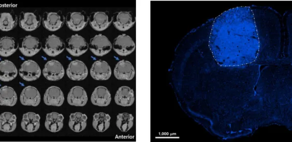
Figure 2 GL261 tumoral mass. On the left, 7 Tesla (7T) magnetic resonance
imaging (MRI) 14 days after GL261 inoculation; blue arrow indicates the tumor position. GL261 cells were implanted in the primary motor cortex. On the right, a representative image of the same tumour mass acquired from brain coronal section of GL261 glioma-bearing mouse stained with Hoechst (fluorescent stain of DNA, blue) 21 days after tumour induction. Image was captured with a microscope equipped with Apotome.2 using a 10× EC-PLAN-NEOFLUAR objective in ZENpro software. Scale bar is 1000 μm.
Based on the extensive characterization of the GL261 glioma model here reported, it can be said that this model accurately represents the relevant biology of human GBs as mandated by the NCI Brain Tumor Progress Review Group. All the studies reported here confirm the relevance of this glioma model in the preclinical setting with respect to its histopathology as shown in the brains of both immunosuppressed and immunocompetent mice (Newcomb and Zagzag, 2009; Oh et al., 2014).
40
Aim of the thesis
High-grade gliomas infiltrate surrounding brain parenchyma during their development, establishing new interactions with the environment that can be exploited to sustain their own progression. Traditionally, brain tumor research has been focused on the study of the biology of glioma cells. However, in the recent years, the interaction between cancer cells and tumor microenvironment has emerged as one important regulator of tumor progression. An ample amount of