Exploring Splicing-Switching Molecules For Seckel Syndrome Therapy
Daniela Scalet
a, Dario Balestra
a, Sara Rohban
b, Matteo Bovolenta
a, Daniela Perrone
c,
Francesco Bernardi
a, Stefano Campaner
b, Mirko Pinotti
a,⁎
a
Department of Life Sciences and Biotechnology, Section of Biochemistry and Molecular Biology, University of Ferrara, Via Fossato di Mortara 74, 44121 Ferrara, Italy
bCenter for Genomic Science, Fondazione Istituto Italiano di Tecnologia (IIT), Via Adamello 16, Milano 20139, Italy cDepartment of Chemical and Pharmaceutical Sciences, University of Ferrara, Via Luigi Borsari 46, Ferrara 44121, Italy
a b s t r a c t
a r t i c l e i n f o
Article history: Received 14 June 2016
Received in revised form 5 September 2016 Accepted 13 September 2016
Available online 14 September 2016
The c.2101 AN G synonymous change (p.G674G) in the gene for ATR, a key player in the DNA-damage response, has been thefirst identified genetic cause of Seckel Syndrome (SS), an orphan disease characterized by growth and mental retardation. This mutation mainly causes exon 9 skipping, through an ill-defined mechanism. Through ATR minigene expression studies, we demonstrated that the detrimental effect of this mutation (6 ± 1% of correct transcripts only) depends on the poor exon 9 definition (47 ± 4% in the ATRwtcontext), because the
change was ineffective when the weak 5′ or the 3′ splice sites (ss) were strengthened (scores from 0.54 to 1) by mutagenesis. Interestingly, the exonic c.2101 A nucleotide is conserved across species, and the SS-causing mutation is predicted to concurrently strengthen a Splicing Silencer (ESS) and weaken a Splicing Enhanc-er (ESE). Consistently, the artificial c.2101 A N C change, predicted to weaken the ESE only, modEnhanc-erately impaired exon inclusion (28 ± 7% of correct transcripts). The observation that an antisense oligonucleotide (AONATR)
targeting the c.2101 A position recovers exon inclusion in the mutated context supports a major role of the underlying ESS.
A U1snRNA variant (U1ATR) designed to perfectly base-pair the weak 5’ss, rescued exon inclusion (63 ± 3%) in
the ATRSS-allele. Most importantly, upon lentivirus-mediated delivery, the U1ATRpartially rescued ATR mRNA
splicing (from ~19% to ~54%) and protein (from negligible to ~6%) in embryonicfibroblasts derived from human-ized ATRSSmice.
Altogether these data elucidate the molecular mechanisms of the ATR c.2101 AN G mutation and identify two potential complementary RNA-based therapies for Seckel syndrome.
© 2016 Elsevier B.V. All rights reserved.
Keywords:
(up to 6) Seckel syndrome-1 Exonic splicing silencer modified U1snRNA Antisense oligonucleotide correction approaches
1. Introduction
Seckel syndrome (SS) is an extremely rare autosomal recessive disorder characterized by intrauterine grow retardation, dwarfism, microcephaly with mental retardation, and a “bird-headed” facial appearance[1]. So far, there is no cure and the molecular mechanisms are poorly known.
Thefirst genetic defect found to be associated to SS (SS-type 1; OMIM 210600) has been the c. 2101 AN G synonymous (p.G674G) mutation in the gene encoding for the ataxia-telangiectasia and RAD3-related protein (ATR)[2], a phosphatidylinositol 3-kinase like kinase playing a key role in the DNA damage response [3–5]. Studies in patients' cells and in vitro[2]have shown that this mutation mainly causes exon 9 skipping and synthesis of a shorter and frame-shifted mRNA form. Trace levels of correct transcripts are also present, which explains a very severe but not lethal phenotype that is expected from a null-condition[6].
The c.2101 AN G change has been used as model to investigate the role of ATR protein, and a mouse model harboring the humanized ATR gene, and recapitulating the SS-1 features, has been developed[7]. Notwithstanding, the molecular mechanisms through which this nucleo-tide change induces exon 9 skipping have not been elucidated. Despite this splicing mutation represents an ideal model to explore RNA-based correction approaches[8,9]that are attracting great attention for many other human genetic diseases, no attempts have been made so far. Indeed, depending on the regulatory elements involved, the skipped exon could be rescued by masking negative elements through antisense molecules (U7snRNA or oligonucleotides)[10–12]or by increasing exon definition through variants of the U1 small nuclear RNA (U1snRNA)[13–25], the component of the ribonucleoprotein U1snRNP driving the recognition of the 5′ splice site (5’ss) in the earliest splicing steps[26].
In the present study, through expression studies with ATR minigenes we demonstrated that the c.2101 AN G synonymous change promotes the skipping of the very weak exon 9 mainly by strengthening an Exonic Splicing Silencer (ESS). Thisfinding prompted us to develop two complementary correction approaches based on an antisense ⁎ Corresponding author.
E-mail address:[email protected](M. Pinotti).
http://dx.doi.org/10.1016/j.bbadis.2016.09.011 0925-4439/© 2016 Elsevier B.V. All rights reserved.
Contents lists available atScienceDirect
Biochimica et Biophysica Acta
j o u r n a l h o m e p a g e :w w w . e l s e v i e r . c o m / l o c a t e / b b a d i soligonucleotide masking the ESS or a modified U1snRNA with increased complementarity to the weak authentic exon 9 5’ss, which were able to remarkably rescue splicing. The U1snRNA was also challenged in Mouse Embryonic Fibroblasts (MEF) from the humanized SS-1 mouse, and resulted in partial rescue of ATR mRNA and protein levels.
2. Materials and Methods 2.1. Creation of vectors 2.1.1. ATR minigenes
To create the pATRwtconstruct, the 1561-bp genomic fragment
spanning ATR intron 8 (from position−434) trough intron 10 (until position +416)(Fig. 1a) was amplified from genomic DNA of a normal subject using high-fidelity PfuI DNA-Polymerase (Transgenomic, Glasgow, UK) with primers5’TGACCATATGTGCACATCTTCACCTCTATT CTG3’ (forward) and 5’TACGCATATGTGGAAAGTGGCCAAGAAGAT3’ (reverse), and cloned in the pTB expression vector by exploiting the NdeI restriction site included in the primers (underlined). The mutant constructs pATRSS, pATRSS5’ss+, pATRSS3’ss+ and pATR2101C were
generated by site-directed mutagenesis (QuickChange II XL Site direct-ed Mutagenesis Kit; Agilent Technologies) (primers: pATRSS,5’CTAGTT
GTGTTAGTGGGTTTTTTATCTTATTG3’and5’CAATAAGATAAAAAACCCAC TAACACAACTAG3’; pATRSS5’ss+,5’CCCAAGATT CTTAGGTATGTACTAA3’ and5’TTAGTACATACCTAAGAATCTTGGG3’; pATRSS3’ss+,5’CTTAATTTTT
TCAGGACCACAGGCACAATC3’and 5’GATTGTGCCTGTGGTCCTGAAAA AATTAAG3’; pATR2101C,5’CTAGTTGTGTTAGTGGCTTTTTTATCTTATTG3’
and5’CAATA AGATAAAAAAGCCACTAACACAACTAG3’. All minigenes were validated by sequencing.
To create the pU1ATRexpression vector, the BglII-BclI fragment of the
pU1wt was replaced using oligonucleotides5’GATCTGATACATACATAG CAGGGGAGATACCAT3’and5’GATCATGGT ATCTCCCCTGCTATGTATGT ATCA3’as previously described[14].
2.1.2. Lentiviral vector
For the creation of the U1ATR-expressing lentivirus vector, the coding
sequence for the U1ATRwas cloned in a lentiviral plasmid backbone
( PGK-EGFP, Cyagen Biosciences) using the BamHI site. The pLV-U1ATRlentiviral plasmid together with psPAX2 packaging and pMD2.G
envelope plasmids were combined at a ratio of 3:2:1 respectively, and then used to transfect 293T packaging cells using calcium phosphate method. Twenty-four hours post transfection the medium was replaced with a new medium. The virus supernatants were collected at 48 and 72 h post transfection andfiltered through a 0.45 μm filter.
2.2. Antisense oligonucleotide (AON)
The 5’UAAGAUAAAAAACCCACUAACACAA3’ antisense oligoribo nucleotide (AONATR) was designed to cover the c.2101 position and in the light of the secondary structures predicted by the Sfold program
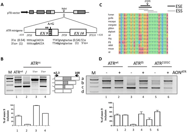
Fig. 1. Splicing features of the humanATR exon 9 context. A) Schematic representation of the ATR genomic sequence cloned as minigene in the pTB plasmid. Exonic and intronic sequences are represented by boxes and lines, respectively. Exons of the globin andfibronectin genes are indicated by grey and light grey boxes, respectively. The position of the c.2101 AN G (A N G) mutation and of cryptic 5’ss (asterics) are indicated above the ATR minigene. The black box represents the exonic splicing silencer (ESS) strengthened by the mutation. The intronic (lower cases) and exonic (upper cases) sequences and the scores (within parenthesis) of the 3’ss and 5’ss, either authentic or improved (+) by mutagenesis (nucleotides in bold), are reported below the scheme. B) Evaluation of ATR alternative splicing patterns in HEK293 cells transiently transfected with the indicated pATR constructs. The schematic representation of the normal (a) and aberrant (b-d) transcripts, and of primers used for the RT-PCR (arrows), is reported in the central panel. PCR amplified products were separated on 2% agarose gel. M, 100 bp molecular weight marker. Histograms indicate the relative percentage of correctly spliced transcripts upon densitometric analysis of bands, which is expressed as mean ± standard deviation (SD) from at least three independent experiments. C) Multiple sequence alignment of the region surrounding the c.2101 A position in various species. The position of the nucleotide under investigation, as well as the position of predicted ESE and ESS elements (dark and grey lines, respectively), are indicated above. D) Evaluation of ATR alternative splicing patterns in HEK293 cells transiently transfected with either the pATR minigenes alone (−) or in combination with 200 nM of AONATR
(+). The schematic representation of transcripts and of primers used for the RT-PCR as well as the electrophoretic conditions and the densitometric analysis is detailed as in panel B.
(http://sfold.wadsworth.org/cgi-bin/index.pl). The AONATR was
chemically synthesized to contain 2′-O-methyl modified RNA and a full-length phosphorothioate backbone[25].
2.3. Expression of ATR minigenes in mammalian cells and mRNA studies Human Embryonic Kidney (HEK293) cells were cultured and transiently transfected with Lipofectamine 2000 (ThermoFisher SCIENTIFIC, Carlsbad, CA, USA) in 12-well plate as previously described
[15]. pATR minigenes (1.5μg) were transfected either alone, with
AON-ATRor with 1.5× molar excess of pU1 vectors. Total RNA extraction was
performed 24 h post-transfection using TRIreagent (ThermoFisher SCIENTIFIC) and reverse transcribed using the M-MLV (ThermoFisher SCIENTIFIC). Theα-2,3 (5’CAACTTCAAGCTCCTAAGCCACTGC3’) and 10R
(5’GTCCACATGTCCGTGTTCAG3’) primers were used for the following PCR that was run for 40 cycles at the following conditions: 95 °C for 30 s, 56 °C for 30 s, 72 °C for 40 s. Densitometric analysis of bands upon electrophoresis on 2% agarose gel was performed using ImageJ software.
2.4. Infection of immortalized MEF cells and ATR mRNA/protein studies Seckel mouse embryonicfibroblast (MEF ATRS/S) cells were incubated
with thefiltered virus supernatant supplemented with polybrene (8μg/ml) at least for 3 h at 37 °C in a humidified incubator. Afterwards the medium was replaced by fresh medium. The RNA was isolated and reverse transcribed with Random hexamers as above mentioned and PCR amplified with primers 8F (5’CCATTCTGATGATGGCTGTTT3’) and
10R (see above) for 40 cycles at the following conditions: 95 °C for 30 s, 56 °C for 30 s, 72 °C for 40 s. Immortalized wild-type MEF, not harboring the human ATR cassette, were used as control to assess the impact on mouse ATR splicing though primers m8F (5’CTACTAAC CTTCTGCCATTAAGCCAG3’) and m10R (5’AACATGCCGTGAAGAGTACA GAC3’).
For the ATR protein analysis, cells were washed twice with ice-cold PBS and then directly lysed with 2× Laemmli buffer (120 mM Tris-HCl pH 6.8, 4% SDS and 20% Glycerol) supplemented with protease and phosphatase inhibitors. After sonication, the lysate was cleared by cen-trifugation and the supernatant was recovered. The protein concentra-tion was quantified using optical absorbance at 280 nm. 25 μg of protein was boiled withβ-mercaptoethanol (5% v/v) at 95 °C for 10 min. Samples were then separated on 4–15% gradient precast TGXTM polyacrylamide gel (Bio-Rad Laboratories, Hercules, CA, USA) and then were blotted onto nitrocellulose membrane using Trans-Blot® TurboTM transfer system (25 V, 1 A, 30 min). Following 1 h blocking in TBST containing 5% BSA, the blot was incubated with primary antibody against ATR (N-19, 1:1000 dilution, Santa Cruz Biotechnology, Dallas, TX, USA) for overnight at 4 °C. Secondary peroxidase-coupled antibody (rabbit anti-goat, Sigma-Aldrich, 1:5000 dilution) was incubated with the blot at room temperature for 1 h. ECL-based chemiluminescence (Bio-Rad Laboratories) was detected on BioRad ChemiDoc system and the image was processed using Image Lab 4.0 (Bio-Rad Laboratories).
2.5. Computational analysis
Bioinformatic prediction of splice sites and/or splicing regulatory elements was conducted by using thehttp://fruitfly.org/seqtools/ splice.html and the Human Splicing Finder (http://www.umd.be/ HSF3/) online softwares.
3. Results and Discussion
The knowledge of molecular mechanisms regulating the splicing process, and involving the interplay between positive and negative regulatory elements located within exons and/or introns, offers the
opportunity to design novel therapeutic approaches for splicing muta-tions, a relevant cause of human disease[27–29]. It is worth noting that RNA-based correction strategies have the advantage of maintaining the gene regulation only in the physiologic tissue and of exploiting small therapeutic cassettes that can be delivered by any viral vector suitable for gene therapy.
In this paper, to explore a correction approach for Seckel syn-drome, so far never attempted, we chose as model the Seckel Syndrome-1 causing ATR c.2101 AN G synonymous mutation that mainly promotes exon 9 skipping, and makes it an ideal target for splicing-switching molecules. The study took advantage of the expres-sion of ATR minigenes, which can be easily manipulated for mechanistic investigations, and of ex-vivo models represented by embryonic fibro-blasts from a humanized SS-1 mouse model, which recapitulates the SS-1 phenotype[7].
3.1. The c.2101 AN G mutation strengthens an Exonic Splicing Silencer in the intrinsically poorly defined exon 9
To dissect the molecular mechanism of the c.2101 AN G change we expressed the ATR minigene spanning exon 8 through 10 (Fig. 1a), either wild-type (pATRwt) or mutated (pATRSS), in the pTB backbone. The analysis of splicing patterns in transfected HEK293 cells recapitulated those previously described in the affected patients[2]. In particular, this mutation not only promoted exon 9 skipping (68 ± 2% of transcripts)(Fig. 1b) but also the usage of two exonic 5’ss cryptic sites at positions c.2093 and c.2075 (Fig. 1a, asterisks), in all cases producing frame-shifted transcripts. Trace levels (6 ± 1%) of the correctly spliced transcripts were also detected, which exclude a null ATR condition that would be lethal[6].
These data validated our experimental system and recapitulated the ATR processing in patients, thus providing us with a model for mechanistic studies. The occurrence of exon-skipping even in the wild-type context (29 ± 4% of transcripts) indicated the poor definition of exon 9, in part attributable to the remarkable divergence of the 5’ss (score 0.54) and 3’ss (0.54) sequences from the consensus ones. We therefore investigated the splicing patterns in an artificial context in which exon 9 definition was improved by increasing the 5’ss
(pATR-SS5’ss+) or the 3’ss (pATRSS3’ss+) scores (Fig. 1a). Noticeably, in both
cases the impact of the c.2101 AN G mutation was virtually abolished (Fig. 1b), with the ameliorated 5’ss context associated only with correct transcripts.
Comparative analysis showed that the c.2101 A nucleotide is con-served across species, and bio-informatic analysis of splicing regulatory elements predicted that the A to G substitution strengthens an Exonic Splicing Silencer (ESS) and weakens an Exonic Splicing Enhancer (ESE)(Fig. 1c). To verify this hypothesis, we investigated the artificial c.2101 AN C change (pATR2101C
) that is predicted to affect the ESE only. Interestingly, this change gave rise to remarkably higher amounts (28 ± 7%) of correct transcripts (Fig. 1d) as compared to the SS-causing mutation. To further corroborate ourfindings, we created an antisense oligoribonucleotide (AONATR) masking the c.2101 A-including region.
As shown inFig. 1d, co-transfection of the AONATRimproved exon 9
inclusion of both variants, particularly in the SS context (52 ± 10%), without showing a noticeable effect on the splicing pattern of the pATRwtminigene.
Taken together thesefindings demonstrate an interplay between ESS and ESE in governing the exon 9 definition, with a major role of the ESS that, once strengthened by the disease causing c.2101 AN G change, promotes exon skipping. It is worth noting that the mechanism involving an ESS in a weak exon does not appear to be uncommon, and has been described for disease-causing mutations in other genes
[30–35].
Interestingly, the effect of the ESS vanished in the presence of strong 5’ss and 3’ss, thus confirming the crucial contribution of the splice site junction in driving exon 9 definition by the spliceosome. These
observations further highlight thefine balance between positive and negative regulatory elements governing the complex exon definition
[36]that makes RNA processing very susceptible to derangements, as indicated by the growing number of disease-causing mutations through aberrant splicing[27–29]. On the other hand, our data with the AONATRprovide an additional example of the potential of
cor-rection approaches based on antisense molecules, which have been successfully exploited by us and others in different disease models
[25,37]and also in humans[38].
3.2. Strengthening recognition of the exon 9 5’ss by an engineered U1snRNA rescues ATR expression in cellular models
In the earliest splicing step, the 5’ss is recognized by the spliceosomal U1 small nuclear ribonucleoprotein (U1snRNP) by com-plementarity with the 5′ tail of its RNA component, the U1snRNA[26]. The low score of the authentic 5’ss of exon 9 is expected to weaken the U1snRNA binding, and accordingly its amelioration by mutagenesis restores exon inclusion. This observation prompted us to act in trans by
exploiting a U1snRNA variant (U1ATR) having the 5′ tail perfectly
matching by complementarity the authentic exon 9 5’ss (Fig. 2a). This approach has been exploited by us and others in several human disease models, both in vitro and in vivo, to rescue splicing in the presence of mutations at the 5’ss, 3’ss or within exons
[13–25].
Co-transfection of HEK293 cells expressing the ATR minigenes with the pU1ATRremarkably rescued splicing in the wild-type (from 47 ±
4% to 96 ± 2% of correct transcripts) and, most importantly, in the presence of the SS-causing mutation (from 6 ± 1 to 62 ± 3%)(Fig. 2b). On the other hand, co-transfection of the pU1wt, mimick-ing overexpression of the endogenous one, had negligible effects in both conditions, thus indicating the functional role of the modified U1snRNA 5′ tail.
Altogether, these findings indicate the ability of the modified U1snRNA to efficiently rescue the SS mutation.
3.3. The lentiviral-mediated U1ATRdelivery rescues ATR expression in
humanized MEFSScells
To further assess its correction efficacy, the U1ATR
was challenged in embryonicfibroblasts from mice harboring the mutated human ATR cassette (MEF ATRS/S), which recapitulates the ATR splicing profile
ob-served in SS patients and the SS phenotype[7]. To overcome the very low transfection efficiency of these cells, which precluded us the direct exploitation of the AONATR, we packaged the U1ATRcassette into a
lentiviral vector (LV-U1ATR,Fig. 3a, left). As shown inFig. 3a (right
panel), the infection resulted in the expression of the U1ATRand, most
importantly, in a partial rescue of ATR splicing, with correctly spliced transcripts raising from 19 ± 1 to 54 ± 3% (Fig. 3b). On the other hand, transduction with LV-U1ATRdid not change the splicing pro
file of mouse ATR in MEF cells from normal mice, thus indicating the U1 specificity toward the human 5’ss (Fig. 3b).
The U1ATR-mediated rescue of ATR expression was also assessed at the intracellular protein level by Western Blotting. Noticeably, the ATR protein was observed in MEF ATRS/Scells only upon transduction with
the LV-U1ATR, and its levels raised to approximately 6% of the mouse ATR in control cells (Fig. 3c).
4. Conclusions
Taken together thesefindings elucidate the molecular mechanism underlying the defective ATR expression in the presence of the synony-mous c.2101 AN G change, which causes Seckel Syndrome type 1. This led us to design two alternative approaches to modulate ATR splicing by AON or modified U1snRNA, both resulting in the remarkable rescue of the correct ATR transcripts, which could be exploited for other similar disease-causing mechanisms involving an ESS in a poorly defined exon
[30–35].
It is worth noting that the AONs and the engineered U1snRNA, successfully explored for therapeutic purposes for other neurologic disorders[24], possess different features in an in vivo application perspective. AONs are easy to produce and deliver but have limited half-life and cellular specificity and permeability[39]. On the other hand, the U1snRNA have to be delivered by viral vectors such as adeno-associated virus[22,24], which can mediate cell target and guarantee long-lasting expression but can trigger an immune response. We are conscious that several issues have to be addressed (i.e. target tissues, time of treatment, clinical phenotype reversibility, etc) to evaluate the feasibility of these approaches for Seckel Syndrome thera-py. Notwithstanding, the results obtained in this pioneer study lay the foundation for further studies in the available SS humanized mouse, a particularly complex model to develop RNA-based strategy for severe neuro-developmental disorders.
Fig. 2. Rescue of ATR splicing patterns by the modified U1ATR
. A) Sequences of the authentic ATR exon 9 5’ss and of the U1ATR 5′ tail, with full complementarity.
B) Evaluation of ATR alternative splicing patterns in HEK293 cells transfected with pATR minigenes alone (−), or in combination (+) with plasmids expressing the pU1 constructs. The normal and aberrant transcripts are indicated by letters (a-d) as inFig. 1. Amplified products were separated on 2% agarose gel. M, 100 bp molecular weight marker. Histograms indicate the relative percentage of correctly spliced transcripts upon densitometric analysis of bands, which is expressed as mean ± standard deviation (SD) from at least three independent experiments.
Conflicts of interest
D.S, D.B, S.R and S.C have no competing interests to declare. M.P. and F.B. are founders of the start-up company Raresplice.
Contributions
D.S. created the ATR minigenes, characterized the mechanism and performed the experiments with the AONATR; D.B. created the
modified U1snRNA and assessed the rescue; S.R. created the LV vec-tor, transduced MEF cells and evaluated ATR protein expression; M.B. designed the AON and revised the manuscript; D.P. synthe-sized the AONATR; F.B., S.C. and M.P. conceived the study, designed the experiments, analyzed and interpreted data and wrote the manuscript.
Transparency document
TheTransparency documentassociated with this article can be found, in online version.
Acknowledgements
This work was supported by Telethon-Italy (GGP14190 to D.B., D.S., and M.P.) and the University of Ferrara, and by the Italian Association for Cancer Research (AIRC, IG grant 13135 to S.C.).
The authors thank Oskar Fernandez Capetillo for the MEFS/Scells.
References
[1] A. Shanske, D.G. Caride, L. Menasse-Palmer, A. Bogdanow, R.W. Marion, Central nervous system anomalies in Seckel syndrome: report of a new family and review of the literature, Am. J. Med. Genet. 70 (2) (1997) 155–158.
[2] M. O'Driscoll, V.L. Ruiz-Perez, C.G. Woods, P.A. Jeggo, J.A. Goodship, A splicing mutation affecting expression of ataxia-telangiectasia and Rad3-related protein (ATR) results in Seckel syndrome, Nat. Genet. 33 (4) (2003) 497–501.
[3] B.B. Zhou, S.J. Elledge, The DNA damage response: putting checkpoints in perspective, Nature 408 (6811) (2000) 433–439.
[4] D. Durocher, S.P. Jackson, DNA-PK, ATM and ATR as sensors of DNA damage: variations on a theme? Curr. Opin. Cell Biol. 13 (2) (2001) 225–231.
[5] Y. Shiloh, ATM and ATR: networking cellular responses to DNA damage, Curr. Opin. Genet. Dev. 11 (1) (2001 Feb) 71–77.
[6] A. de Klein, M. Muijtjens, R. van Os, Y. Verhoeven, B. Smit, A.M. Carr, A.R. Lehmann, J.H. Hoeijmakers, Targeted disruption of the cell-cycle checkpoint gene ATR leads to early embryonic lethality in mice, Curr. Biol. 10 (8) (2000) 479–482.
[7] M. Murga, S. Bunting, M.F. Montaña, R. Soria, F. Mulero, M. Cañamero, Y. Lee, P.J. McKinnon, A. Nussenzweig, O. Fernandez-Capetillo, A mouse model of ATR-Seckel Fig. 3. Rescue of ATR expression in MEFS/S
cells transduced with the LV-U1ATR
. A) Schematic representation of plasmid used to create the lentivirus vector and harboring the U1ATR
coding cassette (left panel), and RT-PCR based detection of the U1ATR
expression in MEF ATRS/Scells alone (−) or transduced (+) with the LV-U1ATR
(right panel). B) Evaluation of ATR alternative splicing patterns in MEF ATRS/Sor MEF cells before (−) or after transduction (+) with LV-U1ATR. The schematic representation of the normal and aberrant transcripts,
and of primers used for the RT-PCR (arrows), is reported in the central panel. Amplified products were separated on 2% agarose gel. M, 100 bp molecular weight marker. The relative percentage of correctly spliced transcripts upon densitometric analysis of bands, expressed below as mean ± standard deviation (SD), is reported below. C) Evaluation of ATR expression by Western blotting analysis in MEF and MEF ATRS/Scells before (−) or after transduction (+) with LV-U1ATR
shows embryonic replicative stress and accelerated aging, Nat. Genet. 41 (8) (2009) 891–898,http://dx.doi.org/10.1038/ng.420.
[8] M. Pinotti, F. Bernardi, A. Dal Mas, F. Pagani, RNA-based therapeutic approaches for coagulation factor deficiencies, J. Thromb. Haemost. 9 (11) (2011) 2143–2152, http://dx.doi.org/10.1111/j.1538-7836.2011.04481.x.
[9] P. Spitali, A. Aartsma-Rus, Splice modulating therapies for human disease, Cell 148 (6) (2012) 1085–1088,http://dx.doi.org/10.1016/j.cell.2012.02.014(16). [10] K. Meyer, J. Marquis, J. Trüb, R. Nlend Nlend, S. Verp, M.D. Ruepp, H. Imboden, I.
Barde, D. Trono, D. Schümperli, Rescue of a severe mouse model for spinal muscular atrophy by U7 snRNA-mediated splicing modulation, Hum. Mol. Genet. 18 (3) (2009) 546–555,http://dx.doi.org/10.1093/hmg/ddn382.
[11] R.N. Nlend, D. Schümperli, Antisense genes to induce exon inclusion, Methods Mol. Biol. 867 (2012) 325–347,http://dx.doi.org/10.1007/978-1-61779-767-5_21. [12] F. Rigo, Y. Hua, A.R. Krainer, C.F. Bennett, Antisense-based therapy for the treatment
of spinal muscular atrophy, J. Cell Biol. 199 (1) (2012) 21–25,http://dx.doi.org/10. 1083/jcb.201207087.
[13] M. Baralle, D. Baralle, L. De Conti, C. Mattocks, J. Whittaker, A. Knezevich, C. Ffrench-Constant, F.E. Baralle, Identification of a mutation that perturbs NF1a gene splicing using genomic DNA samples and a minigene assay, J. Med. Genet. 40 (3) (2003) 220–222.
[14] L. Susani, A. Pangrazio, C. Sobacchi, A. Taranta, G. Mortier, R. Savarirayan, A. Villa, P. Orchard, P. Vezzoni, A. Albertini, A. Frattini, F. Pagani, TCIRG1-dependent recessive osteopetrosis: mutation analysis, functional identification of the splicing defects, and in vitro rescue by U1 snRNA, Hum. Mutat. 24 (3) (2004) 225–235.
[15]M. Pinotti, L. Rizzotto, D. Balestra, M.A. Lewandowska, N. Cavallari, G. Marchetti, F. Bernardi, F. Pagani, U1-snRNA-mediated rescue of mRNA processing in severe factor VII deficiency, Blood 111 (5) (2008) 2681–2684.
[16] M. Pinotti, D. Balestra, L. Rizzotto, I. Maestri, F. Pagani, F. Bernardi, Rescue of coagu-lation factor VII function by the U1 + 5 A snRNA, Blood 113 (25) (2009) 6461–6464, http://dx.doi.org/10.1182/blood-2009-03-207613.
[17] G. Tanner, E. Glaus, D. Barthelmes, M. Ader, J. Fleischhauer, F. Pagani, W. Berger, J. Neidhardt, Therapeutic strategy to rescue mutation-induced exon skipping in rhodopsin by adaptation of U1 snRNA, Hum. Mutat. 30 (2) (2009) 255–263, http://dx.doi.org/10.1002/humu.20861.
[18] L. Hartmann, K. Neveling, S. Borkens, H. Schneider, M. Freund, E. Grassman, S. Theiss, A. Wawer, S. Burdach, A.D. Auerbach, D. Schindler, H. Hanenberg, H. Schaal, Correct mRNA processing at a mutant TT splice donor in FANCC ameliorates the clinical phenotype in patients and is enhanced by delivery of suppressor U1 snRNAs, Am. J. Hum. Genet. 87 (4) (2010) 480–493,http://dx.doi.org/10.1016/j.ajhg. 2010.08.016.
[19] E. Glaus, F. Schmid, R. Da Costa, W. Berger, J. Neidhardt, Gene therapeutic approach using mutation-adapted U1 snRNA to correct a RPGR splice defect in patient-derived cells, Mol. Ther. 19 (5) (2011) 936–941,http://dx.doi.org/10.1038/mt. 2011.7.
[20] F. Schmid, E. Glaus, D. Barthelmes, M. Fliegauf, H. Gaspar, G. Nürnberg, P. Nürnberg, H. Omran, W. Berger, J. Neidhardt, U1 snRNA-mediated gene therapeutic correction of splice defects caused by an exceptionally mild BBS mutation, Hum. Mutat. 32 (7) (2011) 815–824,http://dx.doi.org/10.1002/humu.21509.
[21] E. Fernandez Alanis, M. Pinotti, A. Dal Mas, D. Balestra, N. Cavallari, M.E. Rogalska, F. Bernardi, F. Pagani, An exon-specific U1 small nuclear RNA (snRNA) strategy to cor-rect splicing defects, Hum. Mol. Genet. 21 (11) (2012) 2389–2398,http://dx.doi.org/ 10.1093/hmg/dds045.
[22] D. Balestra, A. Faella, P. Margaritis, N. Cavallari, F. Pagani, F. Bernardi, V.R. Arruda, M. Pinotti, An engineered U1 small nuclear RNA rescues splicing defective coagulation F7 gene expression in mice, J. Thromb. Haemost. 12 (2) (2014) 177–185. [23] A. Dal Mas, P. Fortugno, I. Donadon, L. Levati, D. Castiglia, F. Pagani, Exon-Specific
U1 s Correct SPINK5 Exon 11 Skipping Caused by a Synonymous Substitution that Affects a Bifunctional Splicing Regulatory Element, Hum. Mutat. 36 (5) (2015) 504–512,http://dx.doi.org/10.1002/humu.22762.
[24] A. Dal Mas, M.E. Rogalska, E. Bussani, F. Pagani, Improvement of SMN2 pre-mRNA processing mediated by exon-specific U1 small nuclear RNA, Am. J. Hum. Genet. 96 (1) (2015) 93–103,http://dx.doi.org/10.1016/j.ajhg.2014.12.009.
[25] D. Balestra, E. Barbon, D. Scalet, N. Cavallari, D. Perrone, S. Zanibellato, F. Bernardi, M. Pinotti, Regulation of a strong F9 cryptic 5'ss by intrinsic elements and by combina-tion of tailored U1snRNAs with antisense oligonucleotides, Hum. Mol. Genet. 24 (17) (2015) 4809–4816,http://dx.doi.org/10.1093/hmg/ddv205.
[26] X. Roca, A.R. Krainer, I.C. Eperon, Pick one, but be quick: 5' splice sites and the prob-lems of too many choices, Genes Dev. 27 (2) (2013) 129–144,http://dx.doi.org/10. 1101/gad.209759.112.
[27] F. Pagani, F.E. Baralle, Genomic variants in exons and introns: identifying the splicing spoilers, Nat. Rev. Genet. 5 (5) (2004) 389–396.
[28] T. Sterne-Weiler, J. Howard, M. Mort, D.N. Cooper, J.R. Sanford, Loss of exon identity is a common mechanism of human inherited disease, Genome Res. 21 (10) (2011) 1563–1571,http://dx.doi.org/10.1101/gr.118638.110.
[29] J. Tazi, N. Bakkour, S. Stamm, Alternative splicing and disease, Biochim. Biophys. Acta 1792 (1) (2009 Jan) 14–26,http://dx.doi.org/10.1016/j.bbadis.2008.09.017. [30]D. Matern, M. He, S.A. Berry, P. Rinaldo, C.B. Whitley, P.P. Madsen, S.C. van Calcar,
R.C. Lussky, B.S. Andresen, J.A. Wolff, J. Vockley, Prospective diagnosis of 2-methylbutyryl-CoA dehydrogenase deficiency in the Hmong population by new-born screening using tandem mass spectrometry, Pediatrics 112 (1 Pt 1) (2003 Jul) 74–78.
[31]M. Santoro, A. Modoni, M. Sabatelli, F. Madia, F. Piemonte, G. Tozzi, E. Ricci, P.A. Tonali, G. Silvestri, Chronic GM2 gangliosidosis type Sandhoff associated with a novel missense HEXB gene mutation causing a double pathogenic effect, Mol. Genet. Metab. 91 (1) (2007 May) 111–114.
[32] B.D. Theophilus, M.S. Enayat, M.D. Williams, F.G. Hill, Site and type of mutations in the factor VIII gene in patients and carriers of haemophilia A, Haemophilia 7 (4) (2001 Jul) 381–391.
[33] A. Masuda, X.M. Shen, M. Ito, T. Matsuura, A.G. Engel, K. Ohno, hnRNP H enhances skipping of a nonfunctional exon P3A in CHRNA1 and a mutation disrupting its binding causes congenital myasthenic syndrome, Hum. Mol. Genet. 17 (24) (2008 Dec 15) 4022–4035,http://dx.doi.org/10.1093/hmg/ddn305.
[34] S.F. Dobrowolski, H.S. Andersen, T.K. Doktor, B.S. Andresen, The phenylalanine hydroxylase c.30CN G synonymous variation (p.G10G) creates a common exonic splicing silencer, Mol. Genet. Metab. 100 (4) (2010 Aug) 316–323,http://dx.doi. org/10.1016/j.ymgme.2010.04.002.
[35] I. Talukdar, S. Sen, R. Urbano, J. Thompson, J.R. Yates III, N.J. Webster, hnRNP A1 and hnRNP F modulate the alternative splicing of exon 11 of the insulin receptor gene, PLoS One 6 (11) (2011) e27869,http://dx.doi.org/10.1371/journal.pone.0027869. [36]J. Kralovicova, I. Vorechovsky, Global control of aberrant splice-site activation by
auxiliary splicing sequences: evidence for a gradient in exon and intron definition, Nucleic Acids Res. 35 (2007) 6399–6413.
[37] Y. Hua, T.A. Vickers, H.L. Okunola, C.F. Bennett, A.R. Krainer, Antisense masking of an hnRNP A1/A2 intronic splicing silencer corrects SMN2 splicing in transgenic mice, Am. J. Hum. Genet. 82 (4) (2008 Apr) 834–848,http://dx.doi.org/10.1016/j.ajhg. 2008.01.014.
[38] M. Kinali, V. Arechavala-Gomeza, L. Feng, S. Cirak, D. Hunt, C. Adkin, M. Guglieri, E. Ashton, S. Abbs, P. Nihoyannopoulos, M.E. Garralda, M. Rutherford, C. McCulley, L. Popplewell, I.R. Graham, G. Dickson, M.J. Wood, D.J. Wells, S.D. Wilton, R. Kole, V. Straub, K. Bushby, C. Sewry, J.E. Morgan, F. Muntoni, Local restoration of dystrophin expression with the morpholino oligomer AVI-4658 in Duchenne muscular dystro-phy: a single-blind, placebo-controlled, dose-escalation, proof-of-concept study, Lancet Neurol. 8 (10) (2009) 918–928, http://dx.doi.org/10.1016/S1474– 4422(09)70211-X.
[39] K.S. Frazier, Antisense oligonucleotide therapies: the promise and the challenges from a toxicologic pathologist's perspective, Toxicol. Pathol. 43 (1) (2015 Jan) 78–89,http://dx.doi.org/10.1177/0192623314551840.