. . . .
. . . .
. . . .
Transplantation of adipose tissue mesenchymal cells
conjugated with VEGF-releasing microcarriers
promotes repair in murine myocardial infarction
Rosalinda Madonna
1, Lyubomir Petrov
2, Maria Anna Teberino
3, Lamberto Manzoli
4,
Jean-Pierre Karam
5, Francesca Vera Renna
3, Peter Ferdinandy
6,
Claudia N. Montero-Menei
5, Seppo Yla¨-Herttuala
2, and Raffaele De Caterina
1,3*
1
Center of Excellence on Aging, ‘G. d’Annunzio’ University, Chieti, Italy;2
Biocenter Kuopio, A. I. Virtanen Institute for Molecular Sciences, Kuopio, Finland;3
Institute of Cardiology, Department of Neurosciences, Imaging, and Clinical Sciences, ‘G. d’Annunzio’ University, Chieti, Italy;4
Department of Medicine and Aging Sciences, ‘G. d’Annunzio’ University, Chieti, Italy;5
INSERM U 1066, Micro et nanome´decine biomime´tiques, LUNAM, Universite´ d’Angers, Angers, France; and6
Department of Pharmacology and Pharmacotherapy, Semmelweis University, Budapest, Hungary
Received 18 March 2015; revised 15 June 2015; accepted 30 June 2015; online publish-ahead-of-print 17 July 2015
Time for primary review: 34 days
This manuscript was handled by a Consulting Editor.
Rationale Engraftment and survival of transplanted stem or stromal cells in the microenvironment of host tissues may be im-proved by combining such cells with scaffolds to delay apoptosis and enhance regenerative properties.
Aims We examined whether poly(lactic-co-glycolic acid) pharmacologically active microcarriers (PAMs) releasing vascular endothelial growth factor (VEGF) enhance survival, differentiation, and angiogenesis of adipose tissue-mesenchymal stromal cells (AT-MSCs). We analysed the efficacy of transplanted AT-MSCs conjugated with PAMs in a murine model of acute myocardial infarction (AMI).
Methods and results
We used fibronectin-coated (empty) PAMs or VEGF-releasing PAMs covered with murine AT-MSCs. Twelve-month-old C57 mice underwent coronary artery ligation to induce AMI, and were randomized into five treatment groups: AMI control (saline 20 mL, n ¼ 7), AMI followed by intramyocardial injection with AT-MSCs (2.5× 10⁵cells/20 mL, n ¼ 5), or concentrated medium (CM) from AT-MSCs (20 mL, n ¼ 8), or AT-MSCs (2.5× 10⁵cells/20 mL) conjugated with empty PAMs (n ¼ 7), or VEGF-releasing PAMs (n ¼ 8). Sham-operated mice (n ¼ 7) were used as controls. VEGF-re-leasing PAMs increased proliferation and angiogenic potential of AT-MSCs, but did not impact their osteogenic or adi-pogenic differentiation. AT-MSCs conjugated with VEGF-releasing PAMs inhibited apoptosis, decreased fibrosis, increased arteriogenesis and the number of cardiac-resident Ki-67 positive cells, and improved myocardial fractional shortening compared with AT-MSCs alone when transplanted into the infarcted hearts of C57 mice. With the excep-tion of fracexcep-tional shortening, all such effects of AT-MSCs conjugated with VEGF-PAMs were paralleled by the injecexcep-tion of CM.
Conclusions AT-MSCs conjugated with VEGF-releasing PAMs exert paracrine effects that may have therapeutic applications.
-Keywords Adipose tissue-derived mesenchymal stromal cells † Microspheres † Myocardial infarction
1. Introduction
Coronary artery disease and peripheral arterial disease are the major causes of morbidity and mortality.1While collateral vessel formation as an alternative pathway for blood supply occurs in some of these pa-tients, vascular networks adequate to compensate for the loss of the original blood supply are formed infrequently and insufficiently. Such
patients might benefit from therapies that accelerate natural processes of postnatal collateral vessel formation, an approach referred to as therapeutic angiogenesis. On the other hand, recent reports have indi-cated that the adult heart is self-healing and self-renewing since con-taining a pool of resident cardiac stem/progenitor cells that are capable of differentiating into new blood vessels or into new myo-cytes.2This suggests the opportunity to complement exogenous
*Corresponding author. Tel:+39 0871 41512; fax: +39 0871 402817, Email: [email protected]
Published on behalf of the European Society of Cardiology. All rights reserved.&The Author 2015. For permissions please email: [email protected].
stem cell therapy by inducing endogenous cardiac cells to migrate, dif-ferentiate, and proliferate in situ, thereby replacing lost endothelial cells and cardiomyocytes. However, major obstacles in such approach are the difficulty in engraftment and survival of transplanted stem cells in the harmful microenvironment of the host tissue, and the difficulty in stimulating recruitment of endogenous stem cells into the ischaemic tissue.
To address these issues, there has been increasing focus on develop-ment of novel biomaterials, including pharmacologically active micro-spheres (PAMs) that allow in situ prolonged/controlled growth factor delivery and cell adhesion to their surface functionalized with extracel-lular matrix molecules.3,4The combination of stem cells, PAMs and, in further addition, growth factors may enhance the efficacy of cell ther-apy in several ways: by mobilizing endogenous stem/progenitor cells in vivo; by promoting cell proliferation and differentiation; and by aug-menting cell engraftment and survival in the injured myocardium. Mes-enchymal stromal cells (MSCs) are a population of regenerative cells derived from the bone marrow or the adipose tissue (AT), which con-tain a population of adult multipotent mesenchymal stem cells that can regenerate damaged cardiovascular tissues.5AT-MSCs are attractive for regenerative medicine since they have strong paracrine properties, as they are capable of secreting angiogenic factors such as vascular endothelial growth factor (VEGF), which is a master regulator of angio-genesis,6hepatocyte growth factor (HGF), and fibroblast growth factor (FGF); and are therefore expected to synergize with PAMs in modulat-ing the arterial response to ischaemia or in inducmodulat-ing mobilization and commitment of resident cardiac stem cells.
On this basis, we examined whether PAMs functionalized with fibro-nectin and releasing VEGF enhance survival, growth, differentiation, and angiogenesis of AT-MSCs compared with AT-MSCs alone. We also analysed their therapeutic efficacy in a murine model of acute myocar-dial infarction (AMI).
2. Methods
2.1 Animal care
All procedures were approved by the Chieti Institutional Ethics Committee for animal research. All studies complied with the guidelines from Directive 2010/63/EU of the European Parliament on the protection of animals used for scientific purposes or the NIH guidelines.
2.2 Isolation of mesenchymal stromal cells, cell
culture, and cell conjugation with PAMs
For our in vivo studies, we used a total number of 45 recipients animals (12-month-old male C57/BL6 (C57) mice) and 12-month-old donors male transgenic mice expressing green fluorescent protein (GFP) (Charles River Laboratories, Calco, Lecco, Italy). For the isolation of MSCs, mice were anaesthetized by inhalation of 2 – 5% isofluorane in oxygen, and eutha-nized. Adequate level of anaesthesia was monitored by the lack of palpebral and toe pinch reflexes. AT-MSCs were isolated from the peri-epididymal visceral adipose tissue of C57 and GFP-expressing mice7selected on the basis of their adherence to plastic. AT-MSCs were cultured for up to three passages and characterized for the expression of markers for mesenchymal stem cells, endothelial cells, pericytes, fibroblasts, smooth muscle cells, and monocyte/macrophages. Passage 3 cells were used for transplantation in the in vivo experiments. Before injection, the GFP+AT-MSCs were incu-bated with 4′,6-diamidino-2-phenylindole (DAPI) (50 mmol/L) at 378C for 15 min and re-suspended in phosphate-buffered saline (PBS). Poly(lactic-co-glycolic acid) (PLGA)-microspheres of an average diameter of 60 mm containing or not VEGF were prepared using an emulsion solvent
extraction – evaporation process previously described.8,9Microspheres were functionalized with fibronectin to obtain empty PAMs, as previously described.8For cell attachment to PAMs, 0.5 mg of empty or VEGF165
-releasing PAMs were incubated with 1× 10⁵AT-MSCs. The percentage
of PAMs bound to the cells was evaluated by counting under optic micros-copy. Murine AT-MSCs alone or conjugated with empty or VEGF-releasing PAMs were characterized for their resistance to H2O2-induced apoptosis.
Detailed methods are described in the Supplementary material online.
2.3 Cell proliferation assays
Murine AT-MSCs (1× 10³cells/cm2) from 12-month-old male C57 (n ¼ 4 mice per group), alone or conjugated with empty or VEGF-releasing PAMs were plated onto a 96-well plate and counted daily from day 0 to day 21. At each time point, the population doubling time was calculated by using the following equation: t ¼ (log10[N/N0]× 3.33), where N is the total number
of cells and N0is the number of seeded cells.
2.4 Osteogenic, adipogenic, and myogenic
differentiation assays
For the osteogenic, adipogenic, and myogenic differentiation analyses, we examined Alizarin Red S staining, Oil-Red-O staining, or expression of the myogenic markers cardiac actin and/or smooth muscle alpha-actin, re-spectively, in AT-MSCs from 12-month-old male C57 (n ¼ 4 mice per group), alone or conjugated with empty or VEGF-releasing PAMs, cultured in specific differentiation media. Detailed methods are described in the Supplementary material online.
2.5 Tube formation assays
For tube formation assays, AT-MSCs (alone or conjugated with empty or VEGF-releasing PAMs) were plated on Matrigel for 24 h, then analysed for tube areas, tube length, and tube number. Detailed methods are described in the Supplementary material online.
2.6 Kinetics of VEGF and HGF release
At different time points after the change of fresh medium (from day 0 to day 21), supernatants were collected from AT-MSCs from 12-month-old male C57, alone or conjugated with empty or VEGF-releasing PAMs. We ana-lysed the concentration of murine VEGF and HGF in the supernatants by using an enzyme-linked immunosorbent assay (ELISA; Quantikine Murine VEGF and HGF Immunoassay, R&D Systems, Minneapolis, MN, USA). Detailed methods are described in the Supplementary material online.
2.7 Induction of acute myocardial infarction,
treatments, and functional evaluation
AMIs were produced in adult C57 mice (12 months old, weighing 25 – 30 g) irrespective of sex, by permanently ligating the left anterior descending (LAD) coronary artery, as described in detail in the Supplementary material online. Mice were randomly assigned to receive a single dose of one of the following treatments 5 min after LAD ligation: (a) allogeneic DAPI+/GFP+ AT-MSCs (from 12-month-old GFP male mice) (2.5× 10⁵cells/20 mL, n ¼ 5); (b) concentrated medium (CM) from AT-MSCs (20 mL, n ¼ 8); or (c) allogeneic DAPI+/GFP+AT-MSCs (2.5× 10⁵cells/20 mL) conjugated with empty PAMs (n ¼ 7); or (d) allogeneic DAPI+/GFP+ AT-MSCs (2.5× 10⁵cells/20 mL) conjugated with VEGF-releasing PAMs (n ¼ 8); (e) PBS (20 mL), as a non-cellular control (n ¼ 7). Each treatment was ad-ministered via five intramyocardial injections in the anterior and posterior infarct border zones of the ischaemic myocardium. Seven control mice underwent a sham operation followed by PBS injections. In preliminary ex-periments, we tested escalating doses of AT-MSCs by injecting mice in the peri-infarct area with a single dose (20 mL volume divided into five injec-tions of 4 mL each) of one of the following cell concentrainjec-tions: 1× 10⁵, 1.5× 10⁵, 2.0× 10⁵, 2.5× 10⁵, 5.0× 10⁵, or 7.5× 10⁵. In those experi-ments, we found that the maximum cell dosage compatible with mouse
survival was 2.5× 10⁵cells. After the surgical procedure, animals were monitored every 24 h for 1 week, for the absence or presence of sign of pain, distress, or discomfort. Animals received standard postoperative care and returned to the laboratory 3 weeks later for echocardiography analyses.
To assess the functional effects of each cell therapy, investigators blinded to the treatment groups measured left ventricular end-systolic diameter (LVESD), left ventricular end-diastolic diameter (LVEDD), and left ventricu-lar fractional shortening (FS) 1 week before coronary artery ligation and at 7 and 21 days after the procedure by using a portable ultrasound apparatus (Esaote, Genova, Italy) equipped with a 21-MHz linear probe. After the last assessment of myocardial function, the mice were euthanized, and their hearts were excised for histologic examination. Detailed methods are de-scribed in the Supplementary material online.
2.8 Histologic evaluation of fibrosis, arteriolar
density, cell proliferation, and cell engraftment
rates
The excised hearts were transversely cut to obtain 15 sections (5 mm thick each): 5 from the base of the ventricles, 5 from the middle of the ventricles, and 5 from the apex. For each set of five sections, two were embedded in paraffin and three were embedded in Tissue-Tekw
OCT Compound me-dium. All histologic sections were analysed in a blinded fashion. Fibrotic areas were detected in the paraffin-embedded sections with haematoxylin and eosin staining. Arteriolar density and cellular proliferation were deter-mined by immunofluorescenly staining the OCT-embedded tissue sections with cyanine 3- or phycoerythrin-conjugated rabbit IgG against alpha-smooth muscle actin or Ki-67 (Sigma-Aldrich, St Louis, MO, USA), res-pectively. The number and distribution of AT-MSCs alone or AT-MSCs conjugated with empty PAMs or with VEGF-releasing PAMs were
determined by counting the GFP+/DAPI+cells. The number and location of smooth muscle alpha-actin-positive cells were determined by counting cells positive for Cy3. Detailed methods are described in the Supplemen-tary material online.
2.9 Enzyme-linked immunosorbent assay for
VEGF
Concentrations of VEGF in frozen heart tissues in each experimental group were determined by ELISA (quantikine murine VEGF Immunoassay, R&D System, Minneapolis, MN, USA). A standard curve was prepared from nine VEGF dilutions. The lower limit of sensitivity was 8.4 pg/mL. The over-all inter-assay and intra-assay coefficients of variation were ,10%. In each well, total cellular proteins were measured with a bicinchoninic acid assay (Pierce, Rockford, IL, USA). The VEGF content was normalized to cell protein values.
2.10 Western blots
Total proteins from AT-MSCs or from heart tissues in each experimental group were isolated and analysed for the expression of the following mar-kers: (i) cardiac actin, (ii) collagen type III, (iii) caspase-3, or (iv) cleaved caspase-3. Detailed methods are described in the Supplementary material online.
2.11 Statistical analysis
Within each of the six treatment groups, the differences between baseline and week 1 or 3 in FS, L-VEDD, and L-VESD were evaluated using a paired t-test. To evaluate the differences in FS, L-VEDD, and L-LESD change across groups, we used one-way ANOVA with Scheffe´’s multiple contrasts, con-firmed using ANCOVA adjusting for baseline values, to compare the mean differences between baseline and week 1 or 3. Statistical significance
Figure 1 Left ventricular FS 1 week and 3 weeks after AMI and intramyocardial injection of AT-MSCs with or without conjugation with PLGA micro-spheres. Changes in FS in C57BL/6 mice that underwent a sham operation plus PBS injections (sham+ PBS) or coronary artery ligation to induce AMI plus injections of PBS (n ¼ 7 mice), or CM (n ¼ 8 mice) from AT-MSCs, or AT-MSCs only (n ¼ 5 mice), or AT-MSCs conjugated with empty micro-spheres (n ¼ 7 mice) or VEGF-releasing PAMs (n ¼ 8 mice). Box-plot graph showing the median FS for each study group at baseline (1 week before coronary artery ligation) and at 1 week and 3 weeks after intervention; for each study group at each time point, the graph shows the median, 25 and 75% percentiles, minimum, and maximum values. *P , 0.05 vs. baseline;aP , 0.05 vs. sham+ PBS;bP , 0.05 vs. AMI+ PBS;cP , 0.05 vs. AMI+ CM;
d
P , 0.05 vs. AMI+ MSCs;eP , 0.05 vs. AMI+ MSCs + ePAM (one-way ANOVA with Scheffe correction, confirmed with ANCOVA adjusting for baseline values). ePAM, empty PAMs; vPAM, VEGF-releasing PAMs; AT-MSCs, adipose tissue-derived mesenchymal stromal cells.
was defined as a two-sided P-value ,0.05 for all analyses, which were per-formed using Stata 13.1 (Stata Corp., College Station, TX, 2013).
3. Results
3.1 In vivo transplantation of AT-MSCs
conjugated with VEGF-releasing
microspheres improved cardiac function in a
murine model of AMI
Given that AT-MSCs conjugated with VEGF-releasing PAMs showed in vitro evidence of decreased apoptosis (see Supplementary material on-line, Figure S1), increased angiogenic potential (see Supplementary ma-terial online, Figure S2), and VEGF production (see Supplementary material online, Figure S3), we hypothesized that the delivery of AT-MSCs in conjunction with such scaffolds would augment the en-graftment and survival of AT-MSCs transplanted after AMI, and there-fore enhance their regenerative properties. To examine the in vivo effect of this novel combinatorial approach for myocardial repair, AT-MSCs only, AT-MSCs conjugated with empty PAMs or with VEGF-releasing PAMs, or PBS was injected into the infarcted hearts
of C57 mice by means of multiple intramuscular injections. Since para-crine activity likely accounts for a substantial portion of the in vivo bene-ficial effects produced by this cell type,10we also included injections of conditioned media from AT-MSCs as a non-cellular control. Left ven-tricular FS (Figures1and2) and diameters (Table1) were measured be-fore coronary ligation, 1 week and 3 weeks after ligation and treatments. All mice with AMI, except those treated with AT-MSCs conjugated with VEGF-releasing PAMs, showed a significant decrease in FS at 1 and 3 weeks after coronary artery ligation, when compared with baseline measurements and those from sham-operated mice. FS was greater in post-AMI hearts at 1 and 3 weeks if treated with AT-MSCs conjugated with VEGF-releasing PAMs than in post-AMI hearts treated with AT-MSCs, AT-MSCs conjugated with empty PAMs or PBS (Figures1and2). Interestingly, inter-group and intra-group comparisons of hearts before and after 1 week and 3 weeks post-AMI+ treatment revealed that CM did not exert similar positive effects on LV function. Mice with AMI that were treated with AT-MSCs, AT-MSCs conjugated with empty PAMs or PBS showed a significant in-crease in LVEDD at 3 weeks after ligation, reflecting left ventricular dila-tion (Table1). However, transplantation of AT-MSCs conjugated with VEGF-releasing PAMs and injection of CM significantly limited the
Figure 2 Left ventricular FS 1 week and 3 weeks after AMI and intramyocardial injection of AT-MSCs with or without conjugation with PLGA micro-spheres. Individual changes in FS in C57BL/6 mice that underwent a sham operation plus PBS injections (sham+ PBS) or coronary artery ligation to induce AMI plus injections of PBS (n ¼ 7 mice), or CM (n ¼ 8 mice) from AT-MSCs, or AT-MSCs only (n ¼ 5 mice), or AT-MSCs conjugated with empty microspheres (n ¼ 7 mice) or VEGF-releasing PAMs (n ¼ 8 mice). Values shown for baseline (1 week before coronary artery ligation) and for 1 week and 3 weeks after intervention. Results are presented as the mean + SD. *P , 0.05 vs. baseline (paired t-test).
increase in LVEDD induced by AMI (Table1). Similar results were found for LVESD.
No evidence of inflammation was observed at the injection site in mice among the six groups, as indicated by the absence of cell infiltrates on haematoxylin/eosin-stained slides of the infarcted hearts (Figure4A – F ). These results show that AT-MSCs reduce the post-AMI deterior-ation of cardiac function and remodelling, and this cardioprotective effect is mostly attributable to a paracrine effect of grafted cells.
3.2 In vivo transplantation of AT-MSCs
conjugated with VEGF-releasing
microspheres reduced post-ischaemic
fibrosis and improved arteriogenesis in a
murine model of AMI
Because neovascularization is believed to be essential for the functional recovery of the heart after ischaemia, we examined arteriogenesis in the infarcted hearts of C57 mice after cell therapy. To identify arter-ioles, we used antibodies against smooth muscle cell alpha-actin to im-munostain serial tissue sections from the mid-ventricle to the apex of left ventricle, 21 days after treatment. Arteriolar density was markedly
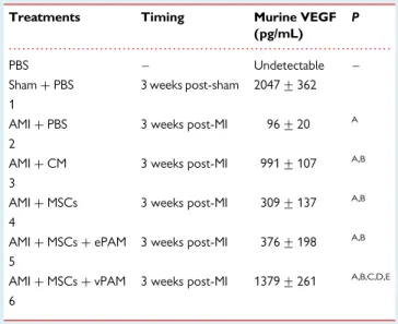
increased in mice receiving either AT-MSCs conjugated with VEGF-releasing PAMs or injection of CM when compared with those that received PBS or AT-MSCs alone or AT-MSCs conjugated with empty PAMs (Figure3). Such results were paralleled by increased ex-pression of VEGF in vivo. Indeed, in normal hearts (sham-operated mice), the abundance of VEGF protein was much higher than that of infarcted hearts treated with PBS. At 21 days after transplantation, VEGF levels in the ischaemic transplanted area were much higher in hearts injected with AT-MSCs conjugated with VEGF-releasing PAMs or CM compared with those that received AT-MSCs alone or AT-MSCs conjugated with empty PAMs (Table2).
We also evaluated the effects of cell transplantation or CM injection on cardiac remodelling. Formation of a collagen-rich scar was signifi-cantly increased after AMI in mice treated with PBS or AT-MSCs alone or AT-MSCs conjugated with empty PAMs (Figure4B, D, E and G). These effects were paralleled by increased expression of collagen type III and decreased content of cardiac actin (Figure4H – L). Trans-plantation of AT-MSCs conjugated with VEGF-releasing PAMs or injec-tion of CM decreased the extent of the area of fibrosis (Figure4C and F ) and the expression of collagen type III (Figure4G and I ), while increasing the expression of cardiac actin (Figure4H and L).
. . . . Table 1 Effects of treatments on remodelling after the experimental AMI
Treatments Timing FS (%) P LVEDD (mm) P LVESD (mm) P
Sham+ PBS Baseline (before sham) 50.6 + 3.9 3.1 + 0.5 1.9 + 0.5 1 1 week post-sham 50.0 + 5.2 3.2 + 0.8 1.7 + 0.7 3 weeks post-sham 48.7 + 4.4 3.0 + 0.5 1.6 + 0.5 D 1 week – baseline 20.6 + 2.6 0.1 + 0.3 20.1 + 0.3 D 3 weeks – baseline 21.9 + 3.1 20.1 + 0.1 20.2 + 0.8 AMI+ PBS Baseline (before AMI) 50.0 + 2.2 3.7 + 0.4 2.4 + 0.6 2 1 week post-MI 19.0 + 7.2 4.3 + 0.7 3.4 + 0.9 3 weeks post-MI 17.1 + 4.2 5.3 + 1.1 4.5 + 1.1
D 1 week – baseline 231.0 + 6.6 *,A 0.6 + 0.9 1.1 + 0.4 *,A D 3 weeks – baseline 232.8 + 4.0 *,A 1.5 + 1.4 *,A 2.0 + 1.6 *,A,D AMI+ CM Baseline (before AMI) 48.2 + 5.4 3.4 + 0.6 2.1 + 0.4
3 1 week post-MI 32.0 + 7.7 3.7 + 0.7 2.4 + 0.7 3 weeks post-MI 33.2 + 6.2 4.2 + 0.7 2.4 + 0.6 D 1 week – baseline 216.3 + 3.7 *,A B 0.4 + 0.9 0.4 + 1.1 D 3 weeks – baseline 215.0 + 10.0 *,A,B 0.7 + 1.1 0.3 + 0.7 AMI+ MSCs Baseline (before AMI) 51.2 + 2.9 2.9 + 0.6 1.4 + 0.4 4 1 week post-MI 43.4 + 3.8 3.5 + 0.6 2.1 + 0.6 3 weeks post-MI 29.4 + 6.3 4.0 + 0.7 3.3 + 0.6 D 1 week – baseline 27.8 + 6.1 *,B 0.7 + 1.1 0.7 + 0.6
D 3 weeks – baseline 221.8 + 8.0 *,A 1.0 + 1.0 A 2.0 + 0.4 *,A,D AMI+ MSCs + ePAM Baseline (before AMI) 48.4 + 2.4 2.9 + 0.2 1.9 + 0.3
5 1 week post-MI 41.0 + 9.1 3.7 + 0.7 2.5 + 0.5 3 weeks post-MI 32.8 + 6.6 4.1 + 0.6 3.3 + 0.6 D 1 week – baseline 27.4 + 9.1 B 0.8 + 0.6* 0.5 + 0.6
D 3 weeks – baseline 215.6 + 7.0 *,A,B 1.1 + 0.7 *,A 1.3 + 0.6 *,A AMI+ MSCs + vPAM Baseline (before AMI) 48.7 + 2.1 2.3 + 0.6 1.9 + 0.7
6 1 week post-MI 54.1 + 1.6 3.6 + 0.5 2.6 + 0.5 3 weeks post-MI 52.0 + 3.6 3.3 + 0.5 2.2 + 0.6 D 1 week – baseline 5.3 + 2.3 *,B,C,D,E 1.2 + 0.3 *,A 0.5 + 0.8
D 3 weeks – baseline 3.3 + 3.6 *,B,C,D,E 0.9 + 0.7 * 0.3 + 0.5 B,C
All values are reported as means + SDs.
All P-values have been computed using one-way ANOVA with Scheffe´’s correction, and confirmed using ANCOVA, adjusting for baseline values.
Sham, toracotomy+ PBS injection; LVEDD, left ventricular end-diastolic diameter; LVESD, left ventricular end-systolic diameter; AT-MSCs, adipose tissue-mesenchymal stromal cells; CM, conditioned medium; AMI, acute myocardial infarction; PAMs, pharmacologically active microspheres; VEGF, vascular endothelial growth factor.
3.3 In vivo transplantation of GFP
1AT-MSCs conjugated with VEGF-releasing
microspheres resulted in cell engraftment
into infarcted cardiac tissue and cellular
differentiation into vascular structures
To examine the engraftment and incorporation of transplanted GFP+/ DAPI+AT-MSCs into vascular structures, we examined the long-term engraftment of cells on transverse histologic sections of the infarcted hearts. Cell retention 21 days after the transplantation of AT-MSCs alone or of AT-MSCs conjugated with empty PAMs or VEGF-releasing PAMs is shown in Figure5. GFP+/DAPI+cells were found in infarcted hearts in the areas of GFP+/DAPI+AT-MSC injections, whereas no GFP+/DAPI+cells were found in infarcted hearts that did not receive cell delivery (Figure5). At 21 days after transplantation, the number of GFP+/DAPI+AT-MSCs was much higher in hearts injected with AT-MSCs conjugated with VEGF-releasing PAMs compared with those that received AT-MSCs alone or AT-MSCs conjugated with empty PAMs, indicating an increase in cell engraftment in the presence of PAMs.
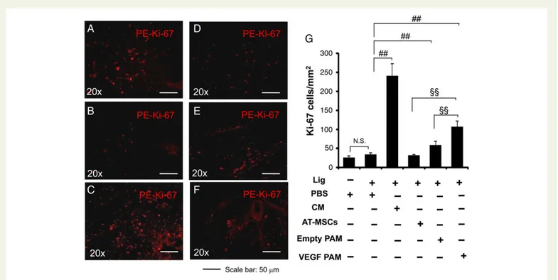
To further determine whether VEGF-releasing PAM conjugation af-fects cell proliferation of the parenchyma surrounding the injection site, we analysed the percentage of Ki-67-positive cells in the infarcted hearts of cell-treated C57 mice. At 21 days after transplantation, C57 mice that received GFP+/DAPI+ AT-MSCs conjugated with VEGF-releasing PAMs or injection of CM had a significantly higher per-centage of Ki-67-positive cells than did those that received GFP+/
DAPI+AT-MSCs alone or GFP+/DAPI+AT-MSCs conjugated with empty PAMs (Figure6), indicating increased cell proliferation in the par-enchyma surrounding the application site of GFP+/DAPI+AT-MSCs conjugated with VEGF-releasing PAMs or the injection of CM.
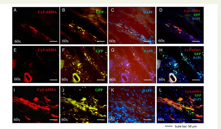
To determine whether transplanted cells integrate into the vascula-ture directly or have a more indirect perivascular effect, we performed morphometric analyses of infarcted hearts in cell-treated C57 mice 21 days after transplantation. We quantified the co-localization of GFP expression with nuclei (DAPI) and smooth muscle alpha-actin. Im-munofluorescence imaging of transverse heart sections immunostained for smooth muscle alpha-actin revealed that GFP expression co-localized with smooth muscle alpha-actin and DAPI staining in vascular structures (Figure7), suggesting the incorporation of GFP+/DAPI+ AT-MSCs into arterioles of the infarcted hearts. Co-localization of GFP with DAPI and smooth muscle alpha-actin was much higher in the infarcted hearts of C57 mice transplanted with AT-MSCs conju-gated with VEGF-releasing PAMs than in those transplanted with AT-MSCs alone or AT-MSCs conjugated with empty PAMs.
4. Discussion
This is the first demonstration that AT-MSCs conjugated with VEGF-releasing PAMs results in increased proliferation, angiogenic dif-ferentiation, and VEGF production, as well as decreased cell death when compared with AT-MSCs alone or conjugated with empty PAMs in vitro. Furthermore, in a murine model of AMI transplantation of AT-MSCs conjugated with VEGF-releasing PAMs into the ischaemic Figure 3 Arteriogenesis in hearts from C57 mice with AMI after the intramyocardial injection of AT-MSCs with or without conjugation with PLGA microspheres. Histologic analysis of arteriogenesis is shown in the hearts of C57 mice that received injections of PBS (B, n ¼ 7 mice), or CM (n ¼ 8 mice) from AT-MSCs (C ), or AT-MSCs only (n ¼ 5 mice, D), or AT-MSCs conjugated with empty microspheres (n ¼ 7 mice, E) or VEGF-releasing PAMs (n ¼ 8 mice, F ). Control heart that received only PBS without coronary ligation (sham, n ¼ 7 mice) is shown in (A). The extent of arteriogenesis is shown in (G). 8P , 0.05 AMI+ PBS vs. sham + PBS,#P , 0.05 AMI+ PBS vs. AMI + CM or AMI + AT-MSCs + VEGF-PAMs,§P , 0.05 AMI+ AT-MSCs + VEGF-PAMs vs. AMI+ AT-MSCs or AMI + AT-MSCs + empty PAMs. N.S. not significant AMI + PBS vs. AMI + MSCs or AMI + CM vs. AMI + AT-MSCs+ VEGF PAM. Magnification¼60×. Scale bar: 50 mm.
region improved post-AMI cardiac function and increased arteriogesis. In these infarcted hearts in mice we provided evidence of cell en-graftment and differentiation into vascular structures. Our findings suggest that AT-MSCs conjugated with VEGF-releasing PAMs may have therapeutic applications for use in treating patients with vascular disease, particularly patients with a previous AMI.
Although encouraging results have been reported in cardiac cell therapy studies, all reports concur that only few of the transplanted cells survive in the hostile environment of the host tissue, such as that occurring after an infarction, and integrate into the host myocar-dium/myocardial scar.11Transplanted cells quickly disappear from the site of injection because they are removed by the blood flow and degraded by specific enzymes in the extracellular microenvironment.11 Strategies using cells overexpressing pro-survival genes such as Akt,12 or co-transplantation with other cells such as endothelial cells,13have been investigated to improve cell survival in ischaemic models. All these approaches have exploited the anti-apoptotic and proliferative proper-ties of the VEGF signalling pathway, along with its capability of inducing neovascularization in ischaemic conditions. Irrespective of the results
obtained, the amount of VEGF released by the cells in the ischaemic/in-farcted area is not enough to promote sufficient protection of trans-planted cells. On the other hand, strategies that employ viral vectors aimed at conveying and releasing high amounts of VEGF at the site of injection suffer from the several drawbacks of viral gene therapy, in-cluding the potent induced immune response to the virus that limits the duration of their effect and decrease cell engraftment.14
In the present study, we tried to overcome these limitations by em-ploying PAMs releasing VEGF. Polymeric scaffolds that have been tested previously for their ability to support transplanted cells have been only marginally effective (reviewed in Ref.4). In our study, we used PAMs, which in general are capable of combining in situ controlled drug delivery with implantation of cells adhered onto a biomaterial-based microcarrier functionalized with extracellular matrix molecules.8 As a specific strategy, we used a microcarrier with a fibronectin-coated surface developed to gradually release the encapsulated VEGF into the injection area.8The PAMs used in the present study are biocompatible and biodegradable microspheres made of PLGA, a polymer approved by the U.S. Food and Drug Administration and the European Medicinal Figure 4 Structural analysis and myogenesis in hearts from C57 mice with AMI after the intramyocardial injection of AT-MSCs with or without con-jugation with PLGA microspheres. Representative haematoxylin/eosin-stained cross-sections (A – G) or western analysis of cardiac actin and collagen type III expression (H – I) of hearts of C57 mice that received injections of PBS (n ¼ 7 mice, B), or CM (n ¼ 8 mice, C ) from AT-MSCs, or AT-MSCs only (n ¼ 5 mice, D), or AT-MSCs conjugated with empty microspheres (n ¼ 7 mice, E) or VEGF-releasing PAMs (n ¼ 8 mice, F ). Control heart that received only PBS without coronary ligation (sham, n ¼ 7 mice) is shown in (A). The extent of fibrosis area is shown in (G). 88P , 0.01 sham+ PBS vs. AMI+ PBS;##P , 0.01 AMI+ PBS vs. AMI + CM or AMI + AT-MSCs + VEGF-PAMs;§§P , 0.01 AMI+ AT-MSCs + VEGF-PAMs vs. AMI + PBS or AMI+ AT-MSCs or AMI + AT-MSCs + empty PAMs or AMI + CM;§§P , 0.05 AMI+ AT-MSCs + VEGF-PAMs vs. AMI + AT-MSCs or AMI + AT-MSCs+ empty PAMs; N.S. not significant AMI + PBS vs. AMI + MSCs or AMI + AT-MSCs + empty PAM. (J) Densitometric analysis of the protein bands shown in (H) and (I ). Results are representative of three different experiments, and data are presented as the mean + SD of n mice per group (AMI+ PBS, n ¼ 7 mice; AMI + CM, n ¼ 8 mice; AMI + AT-MSCs, n ¼ 5 mice; AMI + AT-MSCs + empty PAMs, n ¼ 7 mice; AMI + AT-MSCs + VEGF-PAMs, n ¼ 8 mice). 88P , 0.01 sham+ PBS vs. AMI + PBS;##P , 0.01 AMI+ PBS vs. AMI + CM or AMI + AT-MSCs + VEGF-PAMs; N.S. not significant AMI+ PBS vs. AMI + MSCs or AMI + AT-MSCs + empty PAM. Magnification¼40×. Scale bar: 50 mm. GAPDH levels were assessed as a loading control.
Agency for the formulation of implantable devices. These PAMs can mi-mic, prolong, and amplify specific functions of adhered stem cells by promoting their retention within the damaged area and enhancing their survival, proliferation, and differentiation into cardiac and vascular cells, all functions otherwise critically impaired in the delivery of cells alone. This in vivo outcome further confirms previous in vitro results obtained with PAMs releasing VEGF.9
In our approach, biomimetic PAMs have been used in conjunction with AT-MSCs, which have per se strong paracrine properties, since capable of secreting growth factors with pro-survival and angiogenic properties, such as VEGF, HGF, and FGF.10Our data thus reinforce the concept that an additive or perhaps synergistic effect between the release of VEGF, from the PAMs, and the release of other growth factors, secreted by AT-MSCs, may exist, with a resulting improved modulation of the angiogenic responses of the infarcted tissues. AT-MSCs conjugated with VEGF-releasing PAMs indeed featured, after transplantation into the infarcted tissue, a better formation of arterioles than AT-MSCs alone or than AT-MSCs conjugated with empty PAMs. This augmented pro-angiogenic property of AT-MSCs conjugated with VEGF-releasing PAMs may reflect a decrease in the loss of vascular cells due to the anti-apoptotic/anti-necrotic properties of VEGF. Alterna-tively, the increased vascular density observed in infarcted tissues trea-ted with AT-MSCs conjugatrea-ted with VEGF-releasing PAMs may reflect a response to the release of pro-angiogenic growth factors by AT-MSCs, which was shown in our study to be potentiated by their conjugation with VEGF-releasing PAMs. Our observations are in line with recent
Figure 5 Engraftments of AT-MSCs with or without conjugation with PLGA microspheres in the infarcted hearts. Representative fluorescence mi-croscopy images of tissue sections showing the retention of DAPI/GFP+AT-MSCs (n ¼ 5 mice) (D) and (E and F ) DAPI/GFP+AT-MSCs conjugated with empty PAMs (n ¼ 7 mice) (E) or VEGF-releasing PAMs (n ¼ 8 mice) (F ) at the injection site 21 days after transplantation. Control hearts without cell transplantation are shown in (A) (sham, n ¼ 7 mice), (B) (injection of PBS, n ¼ 7 mice) and (C) (injection of CM, n ¼ 8 mice). Quantitative data showing the retention of DAPI/GFP+AT-MSCs are shown in (G). Graph represents data combined from three independent experiments; results are presented as the mean + SD. 8P , 0.05 AMI+ MSCs vs. AMI + MSCs + empty PAMs; §§P , 0.01 AMI+ MSCs + empty PAMs vs. AMI + AT-MSCs+ VEGF-PAMs. Magnification¼60×. Scale bar: 50 mm.
. . . . Table 2 Expression of murine VEGF as measured by ELISA in various experimental conditions
Treatments Timing Murine VEGF (pg/mL)
P
PBS – Undetectable – Sham+ PBS 3 weeks post-sham 2047 + 362 1
AMI+ PBS 3 weeks post-MI 96 + 20 A 2
AMI+ CM 3 weeks post-MI 991 + 107 A,B
3
AMI+ MSCs 3 weeks post-MI 309 + 137 A,B
4
AMI+ MSCs + ePAM 3 weeks post-MI 376 + 198 A,B 5
AMI+ MSCs + vPAM 3 weeks post-MI 1379 + 261 A,B,C,D,E
6
All values are reported as means + SDs.
Sham, toracotomy+ PBS injection; AT-MSCs, adipose tissue-mesenchymal stromal cells; CM, conditioned medium; AMI, acute myocardial infarction; PAMs, pharmacologically active microspheres; VEGF, vascular endothelial growth factor.
A
P , 0.05 vs. sham;BP , 0.05 vs. AMI+ PBS;CP , 0.05 vs. AMI+ MSCs;DP , 0.05 vs. AMI+ CM;E
data in the literature showing that VEGF-releasing PAMs enhance AT-MSCs survival, most likely through either the activation of pro-survival kinases, such as the ERK 1/2 pathway, and Bcl-2-dependent me-chanisms.8Further studies are needed to assess the role of all of the angiogenic factors secreted by AT-MSCs and assess their individual and/or combined effects on endothelial cell proliferation and differen-tiation into microvessels.
We also observed that the transplantation of AT-MSCs conjugated with VEGF-releasing PAMs resulted in cell engraftment into infarcted tissues and into cellular differentiation or integration into vascular structures, as shown by the proliferation of these cells and their co-localization with nuclei and smooth muscle cells. In vitro studies have shown that PAMs releasing VEGF stimulate ADSCs differentiation to-wards a cardiomyogenic lineage.9However, it is not clear whether this co-localization and concomitant increase in arteriogenesis resulted from the fusion of transplanted cells with native vasculature (and pos-sibly a vascular rescue due to decreased cell death), the directed differ-entiation of transplanted cells, or the paracrine secretion of pro-angiogenic growth factors by the transplanted cells in vivo. Distin-guishing among these possibilities requires further investigation and may lead to further refinement of our strategies to favour stem cell im-plantation in the future.
The complexity of angiogenesis by itself suggests the combination of cells and growth factors as a better strategy than using cells alone. In our study, we demonstrate that conditioned medium (CM) is suf-ficient to reproduce the effects of AT-MSCs conjugated with VEGF-releasing PAMs on most endpoints here investigated.
However, AT-MSCs conjugated with VEGF-releasing PAM trans-plantation exerted a superior therapeutic potential compared with CM injection in terms of improvement of post-infarction cardiac function. This superiority of AT-MSCs conjugated with VEGF-releasing PAMs can be attributed at least in part to the ability of AT-MSCs and PAMs to stabilize and support the formation of vas-cular networks. The enhanced reconstruction of supporting micro-vasculature that is functional to the improvement of cardiac performance, as obtained with the use of transplanted AT-MSCs conjugated with VEGF-releasing PAMs rather than of growth factors only, may represent one important reason for continuing to pursue cell-based therapies rather than therapies with cell-derived products (CM). Observations from our study are in line with recent data show-ing that CM derived from cardiospheres only modestly augmented the percentage of Ki67+cardiomyocytes, but did not improve pump function in a murine model of AMI.15
Provided that our observations in a rodent model can be translated into humans, our results suggest that the delivery of AT-MSCs and VEGF-releasing PAMs is a potentially valuable novel strategy for reducing stem cell apoptosis and loss in the transplanted area and for enhancing the angiogenic response in the infarcted tissue. PLGA micro-spheres are made-up of a biodegradable polymer, fully metabolized by the organism. Such material has been already used in clinical trials for treating gliomas,16PLGA-based implantable devices are already ap-proved by regulatory agencies, and PLGA sutures are widely used in surgery. Thus, the use of such material has practical applicability already at present for further testing.
Figure 6 Evaluation of proliferation by using the Ki-67 marker after myocardial infarction and cell transplantation. (A – F) Representative Ki-67-stained cross-sections of heart of C57 mice that received injections of PBS (n ¼ 7 mice, B), or CM (n ¼ 8 mice, C ) from AT-MSCs, or AT-MSCs only (n ¼ 5 mice, D), or AT-MSCs conjugated with empty microspheres (n ¼ 7 mice, E) or VEGF-releasing PAMs (n ¼ 8 mice, F ). A section from a control heart without coronary ligation is shown in (A) (sham, n ¼ 7 mice). (G) Quantitative data showing the percentage of red-fluorescent Ki-67-positive cells in relation to the total number of nuclei. Graph represents data combined from three independent experiments; results are presented as the mean + SD.##P , 0.01 AMI+ PBS vs. AMI + CM or AMI + AT-MSCs + empty PAMs or AMI + AT-MSCs + VEGF-PAMs;§§P , 0.01 AMI+ AT-MSCs + VEGF-PAMs vs. AMI+ CM or AMI + AT-MSCs + empty PAMs. N.S. not significant sham + PBS vs. AMI + PBS. Magnification¼20×. Scale bar: 50 mm.
Strengths of our study include the presence of appropriate multiple controls—here provided by the injection of empty PAMs, AT-MSCs alone, and CM—and the use of various analyses exploring mechanisms of myocardial regeneration—i.e. arteriogenesis and the relevance of paracrine effects—as well as the efficacy of cell transplantation in terms of myocardial function. Such a complete demonstration was needed because of the paucity of experimental data and appropriate controls in the in vivo setting of previous studies.
In summary, VEGF-releasing PAMs may prolong and improve regen-erative activities of stem cells, whether they are resident cardiac stem cells (potentially—not investigated by us) or transplanted stem cells from ex-ogenous sources (such as in our cases), in the healing processes of in-farcted tissues. This system appears to maximize the biological effects of stem cells through specifically targeting the injured area, and therefore appears to be suited for potential clinical use and industrial applicability.
Supplementary material
Supplementary material is available at Cardiovascular Research online.
Acknowledgements
We thank Alfonso D’Orazio for the assistance with immunofluores-cence and immunohistochemistry analyses.
Conflict of interest: none declared.
Funding
This study was supported by grants from the Italian Istituto Nazionale Ri-cerche Cardiovascolari (I.N.R.C.), the Italian Ministry of the University and Research (PRIN projects), and the CARIPLO Foundation (to R.M. and R.D.C.) ‘Angers Loire Me´tropole’ and ‘Institut National de le Recherche et de la Sante Medical (INSERM)’, France (for J.P.K. and C.M.M.).
References
1. MERIT-HF Study Group. Effect of metoprolol CR/XL in chronic heart failure: Metopro-lol CR/XL Randomised Intervention Trial in Congestive Heart Failure (MERIT-HF). Lancet 1999;353:2001 – 2007.
2. Marban E. Big cells, little cells, stem cells: agents of cardiac plasticity. Circ Res 2007;100: 445 – 446.
3. Delcroix GJ, Garbayo E, Sindji L, Thomas O, Vanpouille-Box C, Schiller PC, Montero-Menei CN. The therapeutic potential of human multipotent mesenchymal stromal cells combined with pharmacologically active microcarriers transplanted in hemi-parkinsonian rats. Biomaterials 2011;32:1560 – 1573.
4. Madonna R, De Caterina R. Stem cells and growth factor delivery systems for cardio-vascular disease. J Biotechnol 2011;154:291 – 297.
5. Madonna R, Taylor DA, Geng YJ, De Caterina R, Shelat H, Perin EC, Willerson JT. Transplantation of mesenchymal cells rejuvenated by the overexpression of telomer-ase and myocardin promotes revascularization and tissue repair in a murine model of hindlimb ischemia. Circ Res 2013;113:902 – 914.
6. Ferrara N. Vascular endothelial growth factor: basic science and clinical progress. Endocr Rev 2004;25:581 – 611.
7. Zuk PA, Zhu M, Mizuno H, Huang J, Futrell JW, Katz AJ, Benhaim P, Lorenz HP, Hedrick MH. Multilineage cells from human adipose tissue: implications for cell-based therapies. Tissue Eng 2001;7:211 – 228.
8. Penna C, Perrelli MG, Karam JP, Angotti C, Muscari C, Montero-Menei CN, Pagliaro P. Pharmacologically active microcarriers influence VEGF-A effects on mesenchymal stem cell survival. J Cell Mol Med 2013;17:192 – 204.
Figure 7 Engraftment and vascular differentiation of AT-MSCs with or without conjugation with PLGA microspheres in the infarcted hearts. Repre-sentative micrographs showing GFP+cells co-localized with DAPI in the infarcted hearts of C57 mice at 21 days after treatment with injections of AT-MSCs (A – D), or AT-MSCs conjugated with empty microspheres (E – H ), or AT-MSCs conjugated with VEGF-releasing PAMs (I – L ). Cy3-conjugated anti-smooth muscle cell a-actin antibody (ASMA) was used to identify smooth muscle cells in cross-sections of heart tissue. GFP+cells co-localized with ASMA, indicating vasculature formation from transplanted cells. Results are representative of those obtained for the n ¼ 5 C57 mice in each treatment group. Magnification¼60×. Scale bar: 50 mm.
9. Karam JP, Muscari C, Sindji L, Bastiat G, Bonafe F, Venier-Julienne MC, Montero-Menei NC. Pharmacologically active microcarriers associated with thermo-sensitive hydrogel as a growth factor releasing biomimetic 3D scaffold for cardiac tissue-engineering. J Control Release 2014;192:82 – 94.
10. Madonna R, Delli Pizzi S, Di Donato L, Mariotti A, Di Carlo L, D’Ugo E, Teberino MA, Merla A, Tartaro A, De Caterina R. Non-invasive in vivo detection of peripheral limb ischemia improvement in the rat after adipose tissue-derived stromal cell transplant-ation. Circ J 2013;76:1517 – 1525.
11. Wu KH, Mo XM, Han ZC, Zhou B. Stem cell engraftment and survival in the ischemic heart. Ann Thorac Surg 2011;92:1917 – 1925.
12. Gnecchi M, He H, Liang OD, Melo LG, Morello F, Mu H, Noiseux N, Zhang L, Pratt RE, Ingwall JS, Dzau VJ. Paracrine action accounts for marked protection of ischemic heart by Akt-modified mesenchymal stem cells. Nat Med 2005;11:367 – 368.
13. Lee WY, Wei HJ, Wang JJ, Lin KJ, Lin WW, Chen DY, Huang CC, Lee TY, Ma HY, Hwang SM, Chang Y, Sung HW. Vascularization and restoration of heart function in rat myocardial infarction using transplantation of human cbMSC/HUVEC core-shell bodies. Biomaterials 2012;33:2127 – 2136.
14. Madonna R, Rokosh G, De Caterina R, Bolli R. Hepatocyte growth factor/Met gene transfer in cardiac stem cells--potential for cardiac repair. Basic Res Cardiol 2010;105: 443 – 452.
15. Xie Y, Ibrahim A, Cheng K, Wu Z, Liang W, Malliaras K, Sun B, Liu W, Shen D, Cho HC, Li T, Lu L, Lu G, Marban E. Importance of cell-cell contact in the therapeutic benefits of cardiosphere-derived cells. Stem Cells 2014;32:2397 – 2406.
16. Menei P, Montero-Menei C, Venier MC, Benoit JP. Drug delivery into the brain using poly(lactide-co-glycolide) microspheres. Expert Opin Drug Deliv 2005;2: 363 – 376.